EM · Obstetric, gynaecological and sexual-health emergencies
Antepartum haemorrhage
Also known as APH · Antepartum bleed · Third-trimester bleeding · Placenta praevia bleed · Placental abruption
Antepartum haemorrhage (APH) is bleeding from the genital tract from 24 weeks of gestation until the onset of labour or birth. The four major causes are placenta praevia (painless, bright-red, recurrent bleeding from a low-lying placenta), placental abruption (painful, dark bleeding with a rigid woody tender uterus and concealed or revealed retroplacental haemorrhage), vasa praevia (fetal bleeding at rupture of membranes with fetal distress and a stable mother), and uterine rupture. The physiological show and cervical or vaginal lesions are the benign mimics. Management is simultaneous maternal and fetal resuscitation — ABCDE, two large-bore cannulae, crossmatch 4 units, coagulation and fibrinogen, continuous CTG, Kleihauer-Betke with anti-D for the Rh-negative mother, speculum (never digital) examination until praevia is excluded, early obstetric referral, and Caesarean section for praevia covering the os. ACEM-primary, globally tagged.
On this page & tools
Your progress
Saved locally on this device.
Target exams
Red flags
Antepartum haemorrhage is bleeding from the genital tract from 24 weeks of gestation until the onset of labour or birth, and it is one of the few emergencies in which the emergency physician is simultaneously resuscitating two patients — the mother and the fetus. The Fellowship candidate must recognise the four major causes from their fingerprint at the bedside (painless bright-red bleeding for placenta praevia, painful rigid-uterus dark bleeding for abruption, bleeding at rupture of membranes with fetal distress for vasa praevia, sudden cessation of contractions with shock for uterine rupture), run simultaneous maternal and fetal resuscitation, and refuse to perform a digital vaginal examination until praevia is excluded. The two non-negotiables in every APH are the early obstetric call and the anti-D immunoglobulin for the Rh-negative mother, dosed by the Kleihauer-Betke test.[1][3]
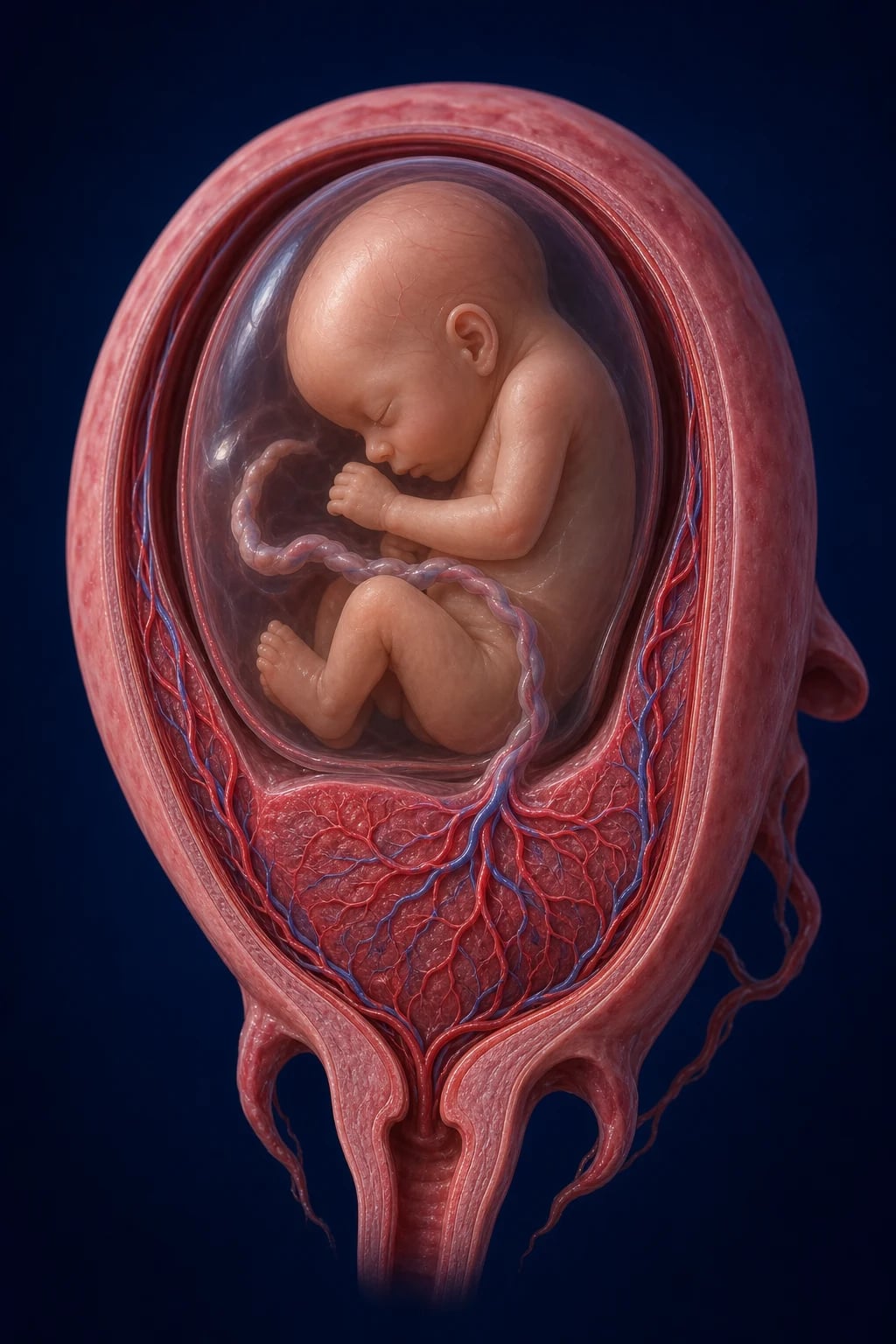
Definition and classification
Antepartum haemorrhage is defined as bleeding from the genital tract occurring at or after 24 weeks of gestation and before the birth of the baby. The 24-week threshold is the RCOG and ANZ convention, anchored to the limit of viability; some North American texts define APH from 20 weeks, but the practical cut-off for the emergency physician is viability, and the management from 24 weeks is obstetric. Bleeding before 24 weeks belongs to the early-pregnancy pathway (miscarriage, ectopic, molar, cervical) and is managed differently.[1]
APH is classified by its severity into minor and major, and by its cause into the big four and the mimics. Severity governs disposition; cause governs the definitive operation. [1]
[1]By cause, the four serious pathologies are placenta praevia, placental abruption, vasa praevia and uterine rupture. The benign mimics are the physiological show (blood-tinged mucus near term) and the cervical or vaginal lesion (ectropion, polyp, cancer, varix, trauma). The clinical task is to separate the serious four from the benign mimics without provoking a catastrophe — most importantly, without performing a digital vaginal examination in a woman with an undiagnosed praevia. [1]
Epidemiology and risk factors
APH complicates approximately 3 to 5 per cent of pregnancies. It remains a leading cause of maternal morbidity and a major driver of perinatal mortality, through prematurity, exsanguination and, in abruption, pathological disseminated intravascular coagulation. The incidence of placenta praevia is rising in parallel with the Caesarean rate, advanced maternal age and assisted reproduction; the incidence of placental abruption is approximately 0.5 to 1 per cent; vasa praevia is rare, between 1 in 1200 and 1 in 5000 pregnancies, but it is catastrophic for the fetus when it occurs.[1][4]
The risk factors are cause-specific and the examiner wants them by cause. For placenta praevia, the dominant risks are a previous Caesarean section (the risk rises with each successive scar, and praevia plus previous scar is the setting for placenta accreta spectrum), previous placenta praevia, multiparity, advanced maternal age, multiple gestation, assisted reproduction, and smoking. For placental abruption, the dominant risk is hypertension in pregnancy — chronic hypertension or pre-eclampsia is the single strongest risk factor — followed by previous abruption, abdominal trauma (including motor-vehicle collision and intimate-partner violence), smoking, cocaine use, advanced maternal age, multiparity, thrombophilias, polyhydramnios with rapid decompression at rupture of membranes, and chorioamnionitis.[3] For vasa praevia, the risks are a velamentous cord insertion, a succenturiate or bilobed placenta, a low-lying placenta, a second-trimester praevia, in vitro fertilisation pregnancy, and multiple gestation.[4][5] For uterine rupture, the dominant risk is a previous Caesarean or other uterine scar, followed by grand multiparity, oxytocin augmentation, previous rupture, and blunt abdominal trauma.
Pathophysiology — the four causes
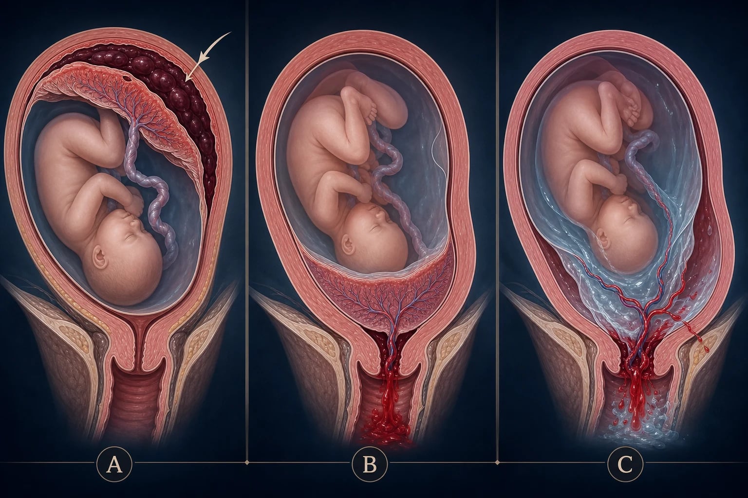
The four serious causes are distinguished at the bedside because each has a distinct mechanism, and the mechanism dictates both the clinical fingerprint and the operation. [1]
Placenta praevia is the implantation of the placenta wholly or partly in the lower uterine segment, covering or encroaching on the internal cervical os. As the lower segment forms and thins during the third trimester, and as Braxton-Hicks contractions tug at the lower segment, the placental edge shears away from the decidua and a spiral artery bleeds into the vagina. The bleeding is painless because there is no myometrial injury, it is bright red because it is maternal arterial blood that exits quickly, and it tends to recur because the mechanical problem persists. The praevia is graded by its relationship to the internal os: a praevia (covering the os) and a low-lying placenta (within 20 mm of the os) are the two clinically actionable categories; the older four-grade system is now simplified.[1]
Placental abruption is the premature separation of a normally situated placenta before delivery. A retroplacental haemorrhage forms behind the placenta and dissects it off the uterine wall. The blood may track down between the membranes and escape through the cervix (revealed abruption) — producing dark, often old-looking blood with clots — or it may be trapped behind the placenta and never appear externally (concealed abruption), in which case the visible bleeding is minimal but the maternal shock and the uterine pain are severe. Blood extravasates into the myometrium, producing the rigid, woody, tender Couvelaire uterus, and the damaged decidua releases thromboplastin into the maternal circulation, driving disseminated intravascular coagulation — abruption is the commonest cause of pathological DIC in pregnancy.[3]
Vasa praevia is the presence of fetal blood vessels running unprotected through the fetal membranes, over or in close proximity to the internal cervical os — either from a velamentous cord insertion (the cord inserts into the membranes rather than the placenta) or from a succenturiate placental lobe with connecting vessels crossing the os. The vessels are unsupported by placental tissue or Wharton's jelly, so they are vulnerable to tearing. Rupture of the membranes, whether spontaneous or artificial, tears a vessel and the fetus bleeds. Because the fetal circulating blood volume is only about 80 to 100 mL per kilogram (around 250 mL at term), a loss that would be trivial to the mother is lethal to the fetus; the mother is completely haemodynamically unaffected.[4][5]
Uterine rupture is a full-thickness tear of the uterine wall, usually through a previous Caesarean or myomectomy scar. The tear allows the fetus, placenta or both to extrude into the peritoneal cavity, producing the sudden severe constant abdominal pain, the cessation of previously painful uterine contractions, the loss of the fetal presenting part from the pelvis, and the rapid progression to maternal shock. [1]
THE 4 CAUSES
Clinical presentation
The clinical presentation is the discriminator, and the examiner builds every APH question around the fingerprint of one cause. [1]
Placenta praevia presents with painless, bright-red vaginal bleeding, typically in the third trimester, often first as a small "warning" bleed that settles and then recurs with larger volume. The abdomen is soft, relaxed and non-tender, the uterus is of normal tone, the fetal parts are easily palpable, and the presenting part is often high, breech or transverse because the praevia blocks the inlet. There is no uterine activity between bleeds. The mother is often haemodynamically stable at the first bleed, which can lull the unwary — the next bleed can be torrential. [1]
Placental abruption presents with sudden severe constant abdominal pain and dark, often old, vaginal bleeding — or, in the concealed variant, with shock out of proportion to the visible loss. The uterus is rigid, woody and tender to palpation, the fetal parts are difficult to feel through the hypertonic uterus, and uterine contractions are frequent and painful, often with a raised baseline tone on the CTG. Fetal distress is common and may progress to fetal death. The hypertensive woman, the smoker, the cocaine user and the woman with abdominal trauma are the high-risk hosts. [1]
Vasa praevia presents with vaginal bleeding at or immediately after rupture of the membranes, accompanied by acute fetal distress — a profound bradycardia, a sinusoidal CTG pattern, or a flat trace — while the mother is completely haemodynamically normal. The split between a haemodynamically normal mother and a dying fetus is the signature of vasa praevia. When the diagnosis is made antenatally by ultrasound, the presentation is planned elective Caesarean; the emergency physician meets the undiagnosed case presenting at term with rupture of membranes.[5]
Uterine rupture presents with sudden severe constant abdominal pain, the cessation of previously painful contractions, loss of the fetal presenting part from the pelvis, palpable fetal parts just under the abdominal wall, vaginal bleeding, and a rapid progression to maternal shock. The history of a previous Caesarean is almost always present. [1]
Bloody show is blood-tinged mucus, small in volume, near term, with the onset of labour. The mucoid character and the small volume are the keys, but show is a diagnosis of exclusion in any woman with a significant bleed. [1]
Differential diagnosis
The differential of third-trimester bleeding is the four serious causes plus the benign mimics, and the candidate must distinguish them at the bedside from the history, the abdominal and speculum examination, and the fetal monitoring. [1]
Placenta praevia
- Painless, bright-red, recurrent bleeding; soft relaxed non-tender uterus; high or malpresenting fetus
- Previous Caesarean, multiparity, IVF, advanced age; the warning bleed precedes a torrential one
- NEVER digital vaginal examination; speculum and transvaginal ultrasound to localise the placenta
- Caesarean section if the placenta covers the os or is low-lying with bleeding
Placental abruption
- Sudden severe constant pain; dark or concealed bleeding; rigid woody tender uterus; fetal distress
- Hypertension or pre-eclampsia is the dominant risk; also trauma, smoking, cocaine, thrombophilia
- Send coagulation and fibrinogen early and repeat — abruption is the commonest cause of pathological DIC in pregnancy
- Emergency Caesarean if the fetus is alive and viable; correct coagulopathy before surgery
Vasa praevia
- Bleeding at rupture of membranes; fetal bradycardia or sinusoidal CTG; mother haemodynamically normal
- Velamentous cord insertion, succenturiate lobe, low-lying placenta, IVF pregnancy
- Fetal blood loss is catastrophic for the fetus and trivial to the mother; Apt test or Kleihauer-Betke confirms fetal red cells
- Immediate Caesarean section to save the fetus
Uterine rupture
- Sudden severe constant pain; cessation of contractions; loss of fetal station; maternal shock
- Almost always through a previous Caesarean or myomectomy scar; also grand multiparity, oxytocin, trauma
- Do not delay for imaging — the diagnosis is clinical and the operation is immediate
- Immediate laparotomy with repair or hysterectomy
Bloody show / show
- Blood-tinged mucus, small volume, near term, with the onset of labour
- The mucoid character distinguishes it from the watery or liquid blood of praevia and abruption
- A diagnosis of exclusion — confirm a stable mother, normal CTG, small volume, and no praevia before reassurance
- Reassurance and ongoing labour management
Cervical or vaginal lesion
- Ectropion, cervical polyp, cervical cancer, vaginal varix, or trauma; bleeding is local and the uterus is quiet
- Visualised on speculum examination of the cervix and vagina once praevia is excluded
- Consider cervical cancer in any pregnant woman with postcoital bleeding — speculum is diagnostic
- Treat the lesion; cervical biopsy or cautery as indicated in the postpartum period
Investigations
The investigations run in parallel with the resuscitation and never delay it. Two large-bore cannulae are sited and blood is drawn as the resuscitation begins. [1]
Bloods. A full blood count gives the haemoglobin and the platelet count. A group and save is the minimum for every APH; a crossmatch of 4 units is sent for any major APH or any praevia, with O-negative blood used if the crossmatch is not ready within 15 minutes. Coagulation — the INR, the APTT and, critically, the fibrinogen — is sent early and repeated, because abruption drives DIC and the fibrinogen is the most sensitive marker of consumption in obstetric bleeding; a fibrinogen under 2 g per litre predicts severe ongoing bleeding and triggers cryoprecipitate. Urea and electrolytes and liver function tests complete the panel, and a venous gas quantifies the lactate and the base deficit that mark occult maternal shock. [1]
Fetomaternal tests. The Kleihauer-Betke test (or the more accurate flow cytometry) is sent on every APH. It has two jobs: it quantifies the fetomaternal haemorrhage so that anti-D can be dosed correctly in the Rh-negative mother, and it detects fetal red cells in the maternal circulation, which supports vasa praevia when the bleed is fetal in origin. The Apt test (the Singer alkali denaturation test) is the bedside test that distinguishes fetal from adult haemoglobin: fetal haoglobin resists alkali denaturation and remains pink, while adult haemoglobin denatures to brown; a positive Apt test on vaginal blood confirms that the bleeding is fetal — the signature of vasa praevia. [1]
Imaging. Transabdominal ultrasound is performed first to localise the placenta and assess the fetus; if the placenta is low-lying, transvaginal ultrasound is both safe and more accurate for defining its relationship to the internal os — transvaginal ultrasound does not provoke bleeding in praevia and is the standard. Ultrasound also identifies velamentous cord insertion and succenturiate lobes that mark vasa praevia. The placenta is "ruled up" — confirmed as not praevia — before a digital vaginal examination is even contemplated.[1][5]
Fetal monitoring. A continuous cardiotocograph (CTG) is applied to every viable APH. A bradycardia, a sinusoidal pattern or a flat trace after rupture of membranes is vasa praevia until proven otherwise; late decelerations and a raised baseline tone suggest abruption; an acute prolonged deceleration with cessation of contractions suggests rupture. [1]
[1]Immediate management and resuscitation
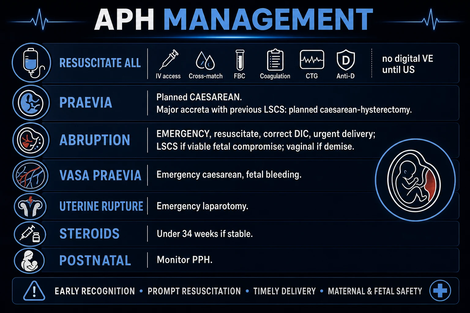
APH is a simultaneous maternal and fetal resuscitation, run by a team with a team leader, and the early call to obstetrics, anaesthetics, haematology and neonatology is the single most important action. The massive transfusion protocol is activated for any major APH. [1]
-
ABCDE. Secure the airway and give high-flow oxygen to the shocked or hypoxic mother. Place the mother in a left lateral tilt to relieve aortocaval compression by the gravid uterus — supine hypotension compounds the shock. Establish two large-bore cannulae (14 or 16 gauge) and draw the bloods. Give balanced crystalloid in 500 mL boluses titrated to the response, with all fluids warmed, and crossmatch 4 units — O-negative blood if the crossmatch is not ready within 15 minutes. Give tranexamic acid 1 g intravenously early in any major bleed. Activate the massive transfusion protocol and aim for a balanced ratio of red cells, plasma and platelets. [1]
-
Fetal resuscitation runs with the maternal. Apply the continuous CTG, treat the maternal hypotension (which compromises the uteroplacental perfusion), and position the mother in the left lateral tilt. The fetus is resuscitated by resuscitating the mother until the obstetric team takes the decision on delivery. [1]
-
Never digital, speculum only. Until praevia is excluded by ultrasound, perform a speculum examination only — visualise the cervix, assess the volume and character of the bleeding, and look for a cervical lesion. Once the placenta is confirmed as "ruled up" (not praevia), a digital vaginal examination is safe and may proceed under obstetric guidance. [1]
-
Anti-D for every Rh-negative mother. Give anti-D immunoglobulin 500 IU intramuscularly to every Rh-negative woman with an APH, within 72 hours, and dose the additional requirement from the Kleihauer-Betke result — a significant fetomaternal haemorrhage needs a larger calculated dose. Omitting anti-D after a sensitising APH is a recurring and avoidable cause of haemolytic disease of the fetus and newborn in the next pregnancy. [1]
-
Corticosteroids and magnesium where time permits. If the gestation is under 34 weeks and the mother is stable enough to allow 48 hours, give betamethasone 12 mg intramuscularly, two doses 24 hours apart, for fetal lung maturation. If the gestation is under 32 weeks and delivery is likely within 24 hours, give magnesium sulfate for fetal neuroprotection (a 4 g intravenous loading dose over 20 minutes, then 1 g per hour maintenance), coordinated with obstetrics. [1]
The first hour of a major APH in one breath
Recognise the cause from the fingerprint — painless bright-red bleed with a soft uterus (praevia), painful rigid-woody uterus with dark or concealed blood (abruption), bleed at rupture of membranes with fetal distress and a stable mother (vasa praevia), sudden cessation of contractions with shock (rupture). Call obstetrics, anaesthetics, haematology and neonatology now. Run the ABCDE: high-flow oxygen, left lateral tilt, two large-bore cannulae, bloods including coagulation and fibrinogen, crossmatch 4 units (O-negative if delayed), balanced crystalloid boluses warmed, tranexamic acid 1 g IV, activate the massive transfusion protocol. Apply the continuous CTG. Perform a speculum examination only — never digital until praevia is excluded — and a transvaginal ultrasound to localise the placenta. Send the Kleihauer-Betke and give anti-D 500 IU IM to every Rh-negative mother. Give betamethasone 12 mg IM if under 34 weeks and stable, and magnesium sulfate if under 32 weeks and delivery is likely. Hand to obstetrics for the delivery decision — Caesarean for praevia covering the os, abruption with a live viable fetus, vasa praevia, or rupture. [1]
Definitive management — the cause-specific operations
Beyond the universal resuscitation, each cause has a definitive operation, and the obstetric team leads each. [1]
Placenta praevia. A praevia that covers the internal os is delivered by Caesarean section — there is no role for vaginal delivery, because the placenta would be sheared and bled from before the fetus delivers. A low-lying placenta (within 20 mm of the os) with significant bleeding is also delivered by Caesarean; a low-lying placenta without bleeding may be considered for a trial of vaginal delivery under obstetric protocol. The timing: an asymptomatic praevia is delivered by elective Caesarean at 37 weeks; a praevia with a major bleed is delivered by emergency Caesarean now, after resuscitation and correction of coagulopathy. In the woman with praevia and a previous Caesarean, the placenta accreta spectrum must be anticipated — the senior obstetrician, the interventional radiology, the cell salvage and the consent for hysterectomy are pre-alerted, because attempted separation of an accreta causes catastrophic haemorrhage.[1][2]
Placental abruption. If the fetus is alive and viable, the definitive management is emergency Caesarean section, after rapid resuscitation and correction of coagulopathy. If the fetus has died and the mother is stable, vaginal delivery may be attempted to minimise maternal surgical risk, with amniotomy and oxytocin — but any maternal deterioration mandates Caesarean. The coagulopathy of abruption is corrected with fresh frozen plasma, cryoprecipitate (for a fibrinogen under 2 g per litre) and platelets before and during surgery. The Couvelaire uterus that will not contract after delivery is a cause of postpartum haemorrhage and may require the full obstetric haemorrhage ladder.[3]
Vasa praevia. When vasa praevia is diagnosed antenatally, the planned management is elective Caesarean section at 34 to 36 weeks, before labour and before rupture of membranes. When it presents acutely with rupture of membranes and fetal distress, the management is immediate emergency Caesarean section — every minute of delay is fetal blood loss, and the fetus has very little to lose. There is no role for resuscitating the fetus medically; only delivery saves it.[4][5]
Uterine rupture. The diagnosis is clinical, and the management is immediate laparotomy — do not delay for imaging. The tear is repaired if possible; if the bleeding cannot be controlled, a hysterectomy is performed. The fetus is delivered, the paediatric and neonatal team resuscitate, and the mother receives the full massive transfusion support. [1]
The definitive operations at a glance
Subtypes and scenarios
The concealed abruption is the trap: there is little or no visible bleeding, but the retroplacental clot is large, the uterus is rigid and woody, the pain is severe, and the mother is shocked out of proportion to the apparent loss. The candidate who waits for visible blood misses it. The fibrinogen and the clinical picture — not the drip count — make the diagnosis. [1]
The placenta accreta spectrum is the catastrophe that complicates praevia with a previous scar. The placenta invades the myometrium (accreta), the myometrium deeply (increta), or through the serosa into the bladder (percreta), and it cannot separate normally at delivery. Attempted forced separation causes torrential haemorrhage. The diagnosis is made antenatally on ultrasound (loss of the retroplacental clear space, placental lacunae, bladder wall disruption) and increasingly on MRI; the undiagnosed accreta presenting to the emergency department with a praevia bleed is a highest-risk scenario that needs the most senior obstetric and anaesthetic response, interventional radiology on standby, and the consent for hysterectomy.[2]
The minor APH with a normal placenta — a small bleed, a stable mother, a normal CTG, ultrasound confirming the placenta is clear of the os, and a benign speculum finding — is observed, investigated for the cause, and followed up as an outpatient. The show and the cervical ectropion fall here once praevia and abruption are excluded. [1]
The trauma-related abruption can present immediately or be delayed by hours to days after a motor-vehicle collision, a fall or intimate-partner violence. Any pregnant woman over 20 weeks presenting after significant abdominal trauma is monitored for a minimum of 4 to 6 hours with a CTG even in the absence of bleeding, because the abruption and the fetomaternal haemorrhage can be occult. (See trauma-in-pregnancy for the full pathway.) [1]
Complications and pitfalls
The maternal complications are the consequences of haemorrhage and DIC: haemorrhagic shock with multi-organ failure, disseminated intravascular coagulation (most characteristic of abruption), acute kidney injury and the rare acute cortical necrosis from prolonged hypotension, Sheehan syndrome (anterior pituitary infarction from profound hypovolaemia, presenting postpartum with failure to lactate and hypopituitarism), postpartum haemorrhage after delivery (abruption and accreta are the highest-risk settings), and hysterectomy. The fetal complications are hypoxia, intrauterine death, prematurity (from early delivery) and intrauterine growth restriction. [1]
The recurring pitfalls are the inverse of the protocol. The first is the digital vaginal examination in an undiagnosed praevia, which shears the placental edge and converts a warning bleed into a catastrophe — speculum only until the placenta is ruled up. The second is missing the concealed abruption because there is no visible bleeding, attributing the shock to "something else" while the rigid woody uterus and the falling fibrinogen are ignored. The third is attributing fetal distress to maternal hypotension when the cause is vasa praevia — a stable mother with a dying fetus is vasa praevia until proven otherwise, and the Apt test and Kleihauer-Betke confirm it. The fourth is forgetting the anti-D in the Rh-negative mother, or dosing it without the Kleihauer-Betke for a significant fetomaternal haemorrhage. The fifth is under-transfusing because the visible loss looked small, while the concealed retroplacental clot was a litre. The sixth is delaying the obstetric call while "stabilising" the patient in the emergency department — obstetrics and theatre run in parallel with the resuscitation, not after it. [1]
[1]Prognosis and disposition
The prognosis depends on the cause and the speed of the response. Placenta praevia managed electively has an excellent maternal prognosis; the major praevia bleed that presents acutely carries the risk of exsanguination and hysterectomy. Placental abruption carries the highest perinatal mortality of the four — fetal death occurs in 10 to 30 per cent of cases, and the maternal risk is driven by DIC and postpartum haemorrhage. Vasa praevia has a high fetal mortality when undiagnosed (over 50 per cent) but an excellent fetal prognosis when diagnosed antenatally and delivered by elective Caesarean — the screening of high-risk pregnancies (IVF, low-lying placenta, velamentous insertion) has transformed the outcome. Uterine rupture carries maternal and fetal mortality proportional to the delay to laparotomy.[3][4]
The disposition is labour ward or theatre for any major APH, with obstetrics, anaesthetics, haematology and neonatology mobilised; high-dependency or intensive care for the mother who has been shocked, transfused massively, or had a hysterectomy. A minor APH with a stable mother, a normal CTG, a clear placenta on ultrasound and a benign speculum finding is observed and investigated on the ward, with outpatient follow-up. The preterm gestation that is stable enough to wait receives corticosteroids and magnesium on the labour ward under obstetric care. [1]
Special populations
The woman with a previous Caesarean and a praevia is the highest-risk patient — praevia plus previous scar carries an accreta risk that rises with each successive scar (from a few per cent after one scar to over 60 per cent after four). The accreta must be anticipated, the senior team and interventional radiology pre-alerted, and the consent for hysterectomy obtained. The emergency physician receiving the undiagnosed accreta calls the most senior obstetric help immediately.[2]
The Rh-negative mother needs anti-D after every sensitising APH, dosed by the Kleihauer-Betke. Omission is a recurring and avoidable error; the anti-D prevents the haemolytic disease of the fetus and newborn in the next pregnancy. [1]
The woman on anticoagulants — low-molecular-weight heparin for thromboprophylaxis, or a direct oral anticoagulant or warfarin for a prosthetic valve — is at additional bleeding risk, and the anticoagulant reversal is part of the immediate workup of any major APH. [1]
The preterm gestation under 34 weeks adds the timing decisions of corticosteroids (under 34 weeks) and magnesium sulfate for neuroprotection (under 32 weeks), balanced against the urgency of delivery — a stable mother buys the 48 hours for the steroids; an unstable mother is delivered now. [1]
The woman presenting after trauma is monitored for the occult, possibly delayed abruption and fetomaternal haemorrhage; the Kleihauer-Betke is part of the trauma-in-pregnancy workup, and intimate-partner violence is considered in any unexplained abdominal injury in pregnancy. [1]
Evidence and regional guidelines
The contemporary framework rests on the RCOG Green-top Guideline No. 27a (Jauniaux and colleagues, 2026) for placenta praevia and placenta accreta spectrum, which sets the diagnostic criteria, the transvaginal ultrasound as the safe and accurate modality, the timing of delivery, and the anticipatory management of accreta.[1] The RCOG Green-top Guideline No. 63 for antepartum haemorrhage sets the severity classification (major and minor), the resuscitation, and the obstetric referral. The Jauniaux 2026 PLoS Medicine review challenges the older dogma around the placenta accreta spectrum and redefines the diagnostic and management approach.[2] The Wright 2026 American Journal of Perinatology study documents the temporal trends, the risk factors and the adverse maternal and neonatal outcomes of placental abruption.[3] The Ruiter 2016 BJOG systematic review of vasa praevia defines the incidence and the risk indicators (velamentous cord insertion, low-lying placenta, IVF, multiple gestation), and the Ruiter 2015 Ultrasound in Obstetrics and Gynecology systematic review confirms the high accuracy of ultrasound in the antenatal diagnosis that transforms the fetal prognosis.[4][5]
ANZ practice note. Antepartum haemorrhage is managed by simultaneous maternal and fetal resuscitation — ABCDE, left lateral tilt, two large-bore cannulae, crossmatch 4 units, coagulation and fibrinogen, continuous CTG, tranexamic acid 1 g IV, speculum examination only (never digital until praevia is excluded), and transvaginal ultrasound to localise the placenta. The Kleihauer-Betke is sent on every APH and anti-D 500 IU IM is given to every Rh-negative mother within 72 hours, dosed additionally by the Kleihauer. The obstetric, anaesthetic, haematology and neonatology teams are called at the moment the diagnosis is recognised, and the massive transfusion protocol is activated for any major APH. The definitive operation is obstetric — Caesarean for praevia covering the os, abruption with a live viable fetus, and vasa praevia; laparotomy for rupture. RANZCOG guidance mirrors the RCOG green-top framework. [1]
Exam pearls
- Painless, bright-red, recurrent third-trimester bleeding is placenta praevia until imaging proves otherwise; the uterus is soft and non-tender.
- Painful, dark bleeding (or concealed bleeding) with a rigid woody tender uterus is placental abruption; hypertension is the dominant risk and DIC the dominant complication — check the fibrinogen.
- Bleeding at rupture of membranes with fetal distress and a haemodynamically normal mother is vasa praevia — the Apt test and the Kleihauer-Betke confirm fetal blood; deliver by immediate Caesarean.
- Never perform a digital vaginal examination until praevia is excluded — speculum and transvaginal ultrasound only. Transvaginal ultrasound is safe and more accurate.
- Every Rh-negative mother gets anti-D 500 IU IM within 72 hours, dosed additionally by the Kleihauer-Betke; omitting it is a recurring and avoidable error.
- Abruption is the commonest cause of pathological DIC in pregnancy — send coagulation and fibrinogen early and repeat; a fibrinogen under 2 g per litre triggers cryoprecipitate.
- Crossmatch 4 units, two large-bore cannulae, tranexamic acid 1 g IV, massive transfusion protocol for any major APH; O-negative blood if the crossmatch is delayed.
- Cessation of contractions with sudden severe pain, loss of fetal station and shock is uterine rupture — immediate laparotomy, do not delay for imaging.
- Praevia plus previous Caesarean is the placenta accreta spectrum until proven otherwise — senior obstetric, anaesthetic, interventional radiology and consent for hysterectomy.
- The estimated blood loss underestimates major APH by up to half — manage on the physiological derangement, not the drip count. [1]
Exam practice
SAQ — Major placenta praevia haemorrhage at 34 weeks with two previous Caesarean sections
10 minutes · 10 marks
A 34-year-old woman (G3P2) at 34 weeks gestation with two previous lower-segment Caesarean sections is brought to the emergency department by ambulance after a sudden, painless, bright-red vaginal bleed that has soaked through her clothes and the ambulance stretcher. On arrival she is pale, anxious and cold: BP 86/52, HR 124, RR 24, SpO2 96 per cent on room air. The abdomen is soft, relaxed and non-tender, the fundal height is consistent with dates, and the presenting part is high and mobile. The continuous cardiotocograph shows a baseline fetal heart rate of 145 with normal variability and no decelerations. Her haemoglobin is 82 g/L and the crossmatch is in progress.
SAQ — Placental abruption with disseminated intravascular coagulation at 38 weeks
10 minutes · 10 marks
A 29-year-old woman at 38 weeks gestation with chronic hypertension on labetalol presents with a sudden severe constant abdominal pain that woke her from sleep, followed by dark vaginal bleeding. On arrival the uterus is rigid, woody and exquisitely tender, with a baseline tone that does not relax between the contractions. BP 92/58, HR 118, RR 26, SpO2 94 per cent on room air, with cold peripheries and a capillary refill of 4 seconds. The cardiotocograph shows late decelerations with a reduced variability and a rising baseline. Bloods return: haemoglobin 78 g/L, platelets 72 x 10^9/L, INR 2.1, APTT 48 seconds, fibrinogen 1.3 g/L, D-dimer markedly elevated.
Red flags
[1]References
- [1]Jauniaux E, Alfirevic Z, Bhide AG, et al. Placenta Praevia and Placenta Accreta Spectrum: Diagnosis and Management: Green-Top Guideline No. 27a BJOG, 2026.PMID 42374711
- [2]Jauniaux E, Alfirevic Z, Silver RM, et al. Placenta accreta spectrum in the 21st century: Challenging dogma and redefining disorder PLoS Med, 2026.PMID 42284360
- [3]Wright GL, Fritsch KL, Kruska SP, et al. Placental Abruption: Temporal Trends, Risk Factors, and Associated Adverse Maternal Outcomes Am J Perinatol, 2026.PMID 40940025
- [4]Ruiter L, Kok N, Lopriore E, et al. Incidence of and risk indicators for vasa praevia: a systematic review BJOG, 2016.PMID 26694639
- [5]Ruiter L, Burgers M, Oepkes D, et al. Systematic review of accuracy of ultrasound in the diagnosis of vasa previa Ultrasound Obstet Gynecol, 2015.PMID 25491755