EM · Shock
Shock states — the recognition and the approach
Also known as Circulatory shock · Acute circulatory failure · Tissue hypoperfusion
Shock — the definition (the inadequate tissue perfusion and the cellular hypoxia); the four patterns by the haemodynamics (the hypovolaemic, the distributive, the cardiogenic, the obstructive); the compensated vs the decompensated phases; the clinical signs (the compensatory tachycardia, the vasoconstriction, the tachypnoea that precede the hypotension); the lactate as the occult-shock marker; the bedside ultrasound (the IVC, the cardiac); and the management framework (the treat-the-cause, the fluid, the vasopressor, the transfusion, the targets). ACEM-primary, globally tagged.
On this page & tools
Your progress
Saved locally on this device.
Target exams
Red flags
Shock is the emergency physician's bread and butter — the recognition of the tissue hypoperfusion before the blood pressure drops, the classification by the haemodynamic pattern, and the rapid correction with the fluid, the vasopressor and the treat-the-cause approach. The Fellowship candidate must know the four patterns, the compensated vs the decompensated phases, the role of the lactate, and the targets for the resuscitation, because the shock is the final common pathway of every acute presentation from the trauma to the sepsis to the MI.[1][2]
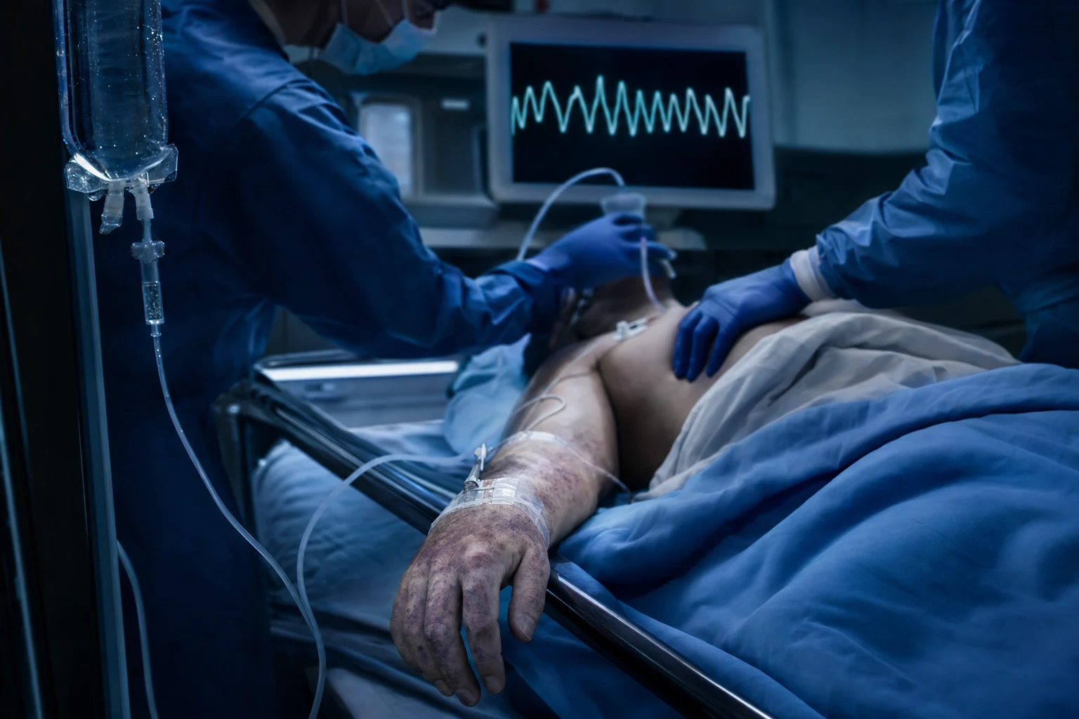
Definition
Shock is the state of inadequate cellular oxygen delivery — the oxygen supply to the tissues falls below the oxygen demand, producing the anaerobic metabolism, the lactate generation and, if untreated, the multi-organ failure and the death. The definition is haemodynamic: the cardiac output (the stroke volume times the heart rate) is insufficient to meet the tissue demand, whether because of the low preload (the hypovolaemic), the low contractility (the cardiogenic), the low systemic vascular resistance (the distributive), or the external obstruction to the flow (the obstructive).[1]
The four patterns of shock
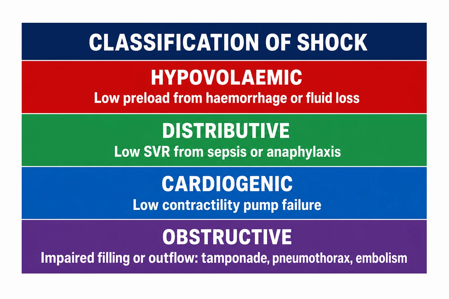
The classification is by the haemodynamic mechanism, and each pattern has a characteristic clinical profile. [1]
Hypovolaemic
- Low preload from the volume loss (the haemorrhage, the dehydration, the burns)
- Cold, clammy, tachycardic; narrow pulse pressure
- Responds to the fluid (the crystalloid, the blood)
- The commonest pattern in the trauma
Cardiogenic
- Low contractility from the pump failure (the MI, the myocarditis, the cardiomyopathy)
- Cold, clammy, tachycardic; the pulmonary oedema, the raised JVP
- Does NOT respond to the fluid (may worsen)
- The inotrope (the dobutamine, the adrenaline) and the cause treatment
Distributive
- Low SVR from the vasodilation (the sepsis, the anaphylaxis, the neurogenic)
- WARM and DRY initially; the wide pulse pressure
- Partially responds to the fluid; needs the vasopressor
- The noradrenaline is the first-line pressor
Obstructive
- The external obstruction to the flow (the tamponade, the tension pneumothorax, the massive PE)
- Cold, clammy; the raised JVP (except the tension PTX), the muffled sounds
- Needs the immediate cause treatment (the needle decompression, the pericardiocentesis)
- Time-critical — the delay kills
The compensated vs the decompensated phase
The compensated phase is the period before the blood pressure drops — the sympathetic nervous system and the renin-angiotensin-aldosterone system maintain the blood pressure through the tachycardia, the vasoconstriction and the tachypnoea. The blood pressure is a LATE sign — the compensatory mechanisms maintain it until approximately 30 per cent of the circulating volume is lost. The decompensated phase is when the compensation fails: the blood pressure drops, the perfusion collapses, and the patient deteriorates rapidly. The recognition of the compensated shock (the tachycardia, the narrow pulse pressure, the delayed capillary refill, the cool peripheries, the tachypnoea, the altered mental state) is the critical skill — the intervention in the compensated phase saves the life; the intervention in the decompensated phase is the salvage. [1]
Differential diagnosis — the cause of the shock
The identification of the cause drives the treatment. Each pattern of the shock has its common causes:
- The hypovolaemic shock — the haemorrhage (the trauma, the GI bleed, the ruptured AAA, the ectopic pregnancy), or the dehydration (the DKA, the gastroenteritis, the heat stroke), or the burns.
- The cardiogenic shock — the MI (the STEMI, the post-cardiac-arrest stunning), or the myocarditis, or the decompensated heart failure, or the arrhythmia (the VT, the fast AF), or the valvular catastrophe.
- The distributive shock — the sepsis (the commonest in the ED), or the anaphylaxis, or the neurogenic (the spinal cord injury above T6), or the adrenal insufficiency (the Addisonian crisis).
- The obstructive shock — the cardiac tamponade, or the tension pneumothorax, or the massive pulmonary embolism, or the severe pulmonary hypertension.
- The mixed pattern — the septic cardiomyopathy, or the trauma with the tension pneumothorax and the haemorrhage, or the mixed overdose. [1]
The clinical assessment
The ABCDE is the framework. The airway (patent? the stridor?), the breathing (the rate, the saturation, the auscultation — the bilateral air entry? the crackles? the wheeze?), the circulation (the heart rate, the blood pressure, the pulse character, the capillary refill, the skin temperature, the JVP), the disability (the conscious level, the pupils — the altered mental state is the sign of the poor cerebral perfusion), the exposure (the rash of the anaphylaxis, the fever of the sepsis, the wounds of the trauma). The key signs: the narrow pulse pressure (under 25 per cent of the systolic) signals the low stroke volume; the wide pulse pressure signals the low SVR; the raised JVP with the cold shock signals the cardiogenic or the obstructive; the flat JVP with the cold shock signals the hypovolaemic; the warm shock with the low diastolic signals the distributive. [1]
The investigations
The venous lactate is the single most important test — a lactate over 2 mmol/L (or the arterial base deficit over minus 2) is the marker of the tissue hypoperfusion, even with a normal blood pressure. The lactate clearance (the rate of the fall over the first hours) is the prognostic marker — a clearance under 10 per cent in the first 2 hours is associated with the higher mortality. The venous blood gas also gives the ScVO2 (the central venous oxygen saturation) — a low ScVO2 (under 70 per cent) signals the inadequate oxygen delivery. The bedside ultrasound (the RUSH protocol or the extended FAST) evaluates the IVC (the collapsible IVC signals the hypovolaemia; the plethoric IVC signals the cardiogenic or the obstructive), the cardiac contractility (the hyperdynamic small heart signals the hypovolaemia; the dilated poorly contracting heart signals the cardiogenic), the pericardial effusion (the tamponade), and the pleural space (the pneumothorax, the effusion). The ECG (the MI, the arrhythmia), the chest X-ray (the pulmonary oedema, the pneumothorax, the widened mediastinum), and the blood cultures (before the antibiotics in the suspected sepsis) complete the workup.[2]
Management — the approach and the drug doses
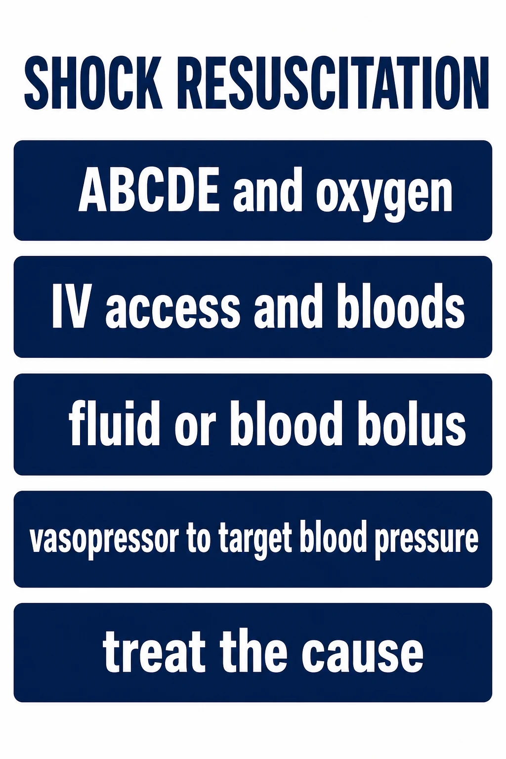
The management is the simultaneous treat-the-cause and the haemodynamic support. [1]
[1]The shock resuscitation targets
The vasopressor and the inotrope selection
The noradrenaline is the first-line vasopressor for the distributive shock (the sepsis, the anaphylaxis) — the alpha-1 vasoconstriction restores the SVR and the MAP. The vasopressin (the 0.03 to 0.04 units per minute, fixed dose) is the second-line for the refractory septic shock — it acts on the V1 receptor and is catecholamine-sparing. The adrenaline is the third-line for the septic shock and the first-line for the anaphylaxis (the IM route) — it provides both the alpha and the beta effect. The dobutamine is the first-line inotrope for the cardiogenic shock — the beta-1 effect increases the contractility; the beta-2 effect causes some vasodilation (the SVR may need the noradrenaline support). The milrinone (the phosphodiesterase inhibitor) is the alternative inotrope, particularly in the pulmonary hypertension. The dopamine is no longer recommended (the arrhythmia risk is higher than the noradrenaline). [1]
Complications and pitfalls
The complications are the multi-organ failure (the AKI, the ARDS, the hepatic dysfunction, the DIC), the ischaemia (the mesenteric, the limb, the myocardial — from the high-dose vasopressor), the fluid overload (the pulmonary oedema — the crystalloid is not harmless), and the arrhythmia (from the catecholamine and the electrolyte derangement). The pitfalls are: not recognising the compensated shock (the normal blood pressure does not exclude it); under-resuscitating (the single small fluid bolus in the shocked patient); over-resuscitating with the crystalloid (the 5 L of saline in the septic patient — the CLARITY trial and the CLOVERS trial caution against the excessive fluid); delaying the vasopressor (the noradrenaline should be started when the MAP is under 65 after the initial fluid challenge, not after 3 L of fluid); not treating the cause (the septic shock needs the antibiotic within 1 hour, not the vasopressor alone). [1]
Prognosis and disposition
The mortality depends on the cause and the timeliness: the septic shock 25 to 30 per cent, the cardiogenic shock 40 to 50 per cent, the hypovolaemic shock from the trauma 10 to 30 per cent (the early transfusion and the damage-control resuscitation have improved this), the obstructive shock near 100 per cent if untreated (the tension PTX, the tamponade) but salvageable if the cause is treated immediately. The patient with the shock is admitted to the HDU or the ICU; the patient with the resolved shock after the resuscitation is observed in the ED for the recurrence. [1]
Special populations
The elderly patient has the blunted sympathetic response (the beta-blocker, the calcium channel blocker, the age-related) and may not mount the tachycardia — the normocardiac shock is a danger sign. The pregnant patient is managed with the left-lateral tilt (the aortocaval compression) and the early fluid (the increased volume requirement). The child has the excellent physiological reserve and the late, sudden collapse — the bradycardia in the child is the pre-terminal sign (the hypoxia is the cause until proven otherwise). The anticoagulated patient with the shock of unknown cause has the retroperitoneal bleed until proven otherwise. [1]
Evidence and regional guidelines
The contemporary framework is the Surviving Sepsis Campaign for the septic shock, the European Society of Cardiology for the cardiogenic shock, and the ATLS for the haemorrhagic shock.[1][2] The noradrenaline first-line, the balanced crystalloid, the early vasopressor, the lactate-guided resuscitation, and the treat-the-cause approach are the global standards.
ANZ practice note. The shock resuscitation follows the local ED resuscitation protocol; the noradrenaline is the first-line vasopressor for the distributive shock; the balanced crystalloid (the Plasmalyte or the Hartmann's) is the fluid of choice; the early vasopressor and the lactate-guided resuscitation are the standard; and the ICU admission for the persistent shock is the expectation. [1]
The compensatory mechanisms — the sequence of the failure
The body defends the perfusion of the brain and the heart in a predictable order, and the recognition of this sequence is the single most examined concept in the shock viva. The sympathetic nervous system and the renin-angiotensin-aldosterone system (the RAAS) are activated within seconds of the falling cardiac output, and they restore the blood pressure by mechanisms operating in this fixed order. [1]
- The tachycardia — the beta-1 effect on the sinus node increases the heart rate to maintain the cardiac output (the cardiac output equals the stroke volume times the heart rate; the falling stroke volume is offset by the rising rate). This is the FIRST detectable sign.
- The vasoconstriction — the alpha-1 effect on the arterioles and the angiotensin II effect increase the systemic vascular resistance, shunting the blood from the skin, the splanchnic bed and the muscles to the heart, the brain and the kidneys. This produces the cool, clammy skin and the delayed capillary refill.
- The narrow pulse pressure — as the stroke volume falls, the systolic pressure falls (it depends on the stroke volume) while the diastolic pressure is maintained or rises (it depends on the SVR). The pulse pressure therefore narrows BEFORE the systolic drops; a pulse pressure under 25 per cent of the systolic is a sensitive marker of the falling stroke volume.
- The tachypnoea — the compensatory respiratory alkalosis (the blow-off of the CO2 from the anaerobic metabolism and the compensation for the metabolic acidosis) and the increased work of breathing.
- The hypotension — the LAST sign. The blood pressure is maintained until approximately 30 per cent of the circulating volume is lost (around 1500 mL in the 70 kg adult), because the vasoconstriction and the tachycardia together preserve the MAP. By the time the blood pressure falls, the patient is already deep in the decompensated phase and the cellular injury is underway. [1]
The compensatory cascade and the failure sequence
The stimulus — the falling cardiac output (the low preload, the low contractility, or the low SVR) is detected by the arterial baroreceptors and the cardiopulmonary receptors.
The tachycardia — the vagal withdrawal and the sympathetic beta-1 drive increase the heart rate. This is the FIRST sign; the heart rate over 100 (or over 110 in the sepsis criteria) is the trigger to assess the perfusion.
The vasoconstriction — the alpha-1 and the angiotensin II constrict the cutaneous, the splanchnic and the muscular arterioles. The skin becomes cool, pale, clammy and the capillary refill delays (over 3 seconds).
The narrow pulse pressure — the systolic falls (the low stroke volume) while the diastolic is maintained; the pulse pressure narrows to under 25 per cent of the systolic.
The tachypnoea and the lactate rise — the anaerobic metabolism generates the lactate, the metabolic acidosis drives the compensatory tachypnoea, and the venous lactate exceeds 2 mmol/L.
The hypotension — the LAST sign; the MAP falls below 65 once 30 per cent of the volume is lost and the compensation is exhausted. By now the cellular injury (the AKI, the hepatic dysfunction, the myocardial depression) is established.
Shock classification — the clinical profile of each pattern (the deep dive)
The four haemodynamic patterns each produce a distinct clinical and biochemical fingerprint. The Fellowship candidate must be able to predict the haemodynamics from the bedside findings and vice versa, because the bedside pattern directs the first intervention. [1]
Hypovolaemic
- The problem: the low preload from the volume loss. The cardiac output falls, the SVR rises.
- Haemodynamics: the low CVP, the low PCWP, the low cardiac output, the high SVR, the low ScVO2.
- Bedside: cold, clammy, tachycardic; the flat JVP, the narrow pulse pressure, the collapsed IVC on the ultrasound, the hyperdynamic small heart.
- Response: the rapid and the complete response to the fluid (the crystalloid, the blood).
- Examples: the haemorrhage, the dehydration, the burns, the GI losses.
Cardiogenic
- The problem: the low contractility from the pump failure. The cardiac output falls, the SVR rises, the preload is high.
- Haemodynamics: the high CVP, the high PCWP, the low cardiac output, the high SVR, the low ScVO2.
- Bedside: cold, clammy, tachycardic; the RAISED JVP, the pulmonary oedema (the bilateral crackles), the gallop, the dilated poorly contracting heart on the ultrasound.
- Response: does NOT respond to the fluid (may worsen the pulmonary oedema); needs the inotrope and the cause treatment.
- Examples: the STEMI, the myocarditis, the decompensated heart failure, the VT, the valvular catastrophe.
Distributive
- The problem: the low SVR from the vasodilation. The cardiac output is high (initially), the SVR is low.
- Haemodynamics: the low CVP, the low PCWP, the HIGH cardiac output (initially), the LOW SVR, the high ScVO2.
- Bedside: WARM and DRY initially; the wide pulse pressure, the low diastolic, the bounding pulse, the flushed skin, the fever (the sepsis) or the urticaria (the anaphylaxis).
- Response: partially responds to the fluid; needs the vasopressor (the noradrenaline) early.
- Examples: the sepsis, the anaphylaxis, the neurogenic (the spinal cord injury above T6), the adrenal insufficiency.
Obstructive
- The problem: the external obstruction to the flow. The cardiac output falls, the SVR rises, the preload is variable.
- Haemodynamics: the high CVP (except the tension PTX where the high intrathoracic pressure may compress the venous return), the low cardiac output, the high SVR.
- Bedside: cold, clammy; the RAISED JVP (the tamponade, the massive PE), the muffled heart sounds (the tamponade), the tracheal deviation and the absent breath sounds (the tension PTX), the right heart strain on the ultrasound.
- Response: needs the immediate cause treatment — the needle decompression (the tension PTX), the pericardiocentesis (the tamponade), the thrombolysis or the embolectomy (the massive PE).
- Examples: the cardiac tamponade, the tension pneumothorax, the massive PE, the severe pulmonary hypertension.
The bedside assessment of the perfusion — the clinical markers
The perfusion is assessed at the bedside by four clinical windows: the skin, the capillary refill, the urine output and the mental state. These are more sensitive than the blood pressure and they are the Fellowship viva staples — the candidate who cites the blood pressure as the first marker fails the viva. [1]
The skin
- The cool, pale, clammy, mottled skin signals the vasoconstriction of the cold shock (the hypovolaemic, the cardiogenic, the obstructive).
- The warm, dry, flushed skin signals the vasodilation of the distributive shock.
- The mottling (the reticular pattern over the knees and the extensor surfaces) is the marker of the severe peripheral vasoconstriction and the poor prognosis in the septic shock.
The capillary refill
- The capillary refill over 3 seconds (on the sternum or the distal phalanx) signals the poor peripheral perfusion.
- The mottling score (the ASTRINGENT 0 to 5 score) correlates with the mortality in the septic shock.
- Beware the cold environment — warm the finger before the assessment; the ambient cold causes the false positive; the pressor on the finger causes the false positive too.
The urine output
- The urine output under 0.5 mL/kg/h signals the poor renal perfusion; it is the sensitive and the continuous marker of the perfusion once the catheter is in.
- The oliguria precedes the creatinine rise by hours — the urine output is the real-time marker.
- Target the urine output over 0.5 mL/kg/h as one of the resuscitation end-points; the falling urine output during the resuscitation is the alarm.
The mental state
- The altered mental state (the agitation, the confusion, the lethargy) signals the poor cerebral perfusion — it is the sign of the advanced shock.
- The GCS drop, especially the new confusion in the elderly, is the sepsis and the shock red flag.
- The coma is the pre-terminal sign — the brain is the last organ to fail but the most unforgiving of the hypoperfusion.
The 60-second bedside perfusion assessment
The hands — feel the temperature (the warm vs the cold), the moisture (the clammy vs the dry), the pulse character (the thready vs the bounding) and the capillary refill (use the sternum if the hands are cold).
The pulse — the rate, the rhythm and the character. The narrow pulse pressure (the systolic minus the diastolic under 25 per cent of the systolic) signals the low stroke volume; the wide pulse pressure signals the low SVR.
The JVP — the flat JVP with the cold shock is the hypovolaemic; the raised JVP with the cold shock is the cardiogenic or the obstructive; the warm shock with the low diastolic is the distributive.
The chest — the bilateral crackles (the cardiogenic), the absent breath sounds and the tracheal deviation (the tension PTX), the muffled heart sounds and the pulsus paradoxus (the tamponade).
The abdomen — the distension and the tenderness (the intra-abdominal bleed, the ruptured AAA, the peritonitis), the pregnant uterus in the woman of the reproductive age.
The mental state and the urine output — the confusion and the oliguria are the perfusion markers that confirm the shock even with a normal blood pressure.
The lactate — the perfusion marker and the prognostic tool
The venous lactate is the single most important investigation in the shocked patient. It reflects the anaerobic metabolism that results when the oxygen delivery falls below the oxygen demand, and it rises BEFORE the blood pressure drops — it is the marker of the OCCULT shock and the reason the normotensive patient with the raised lactate is treated as the shocked patient. [1]
[1]The lactate thresholds and the prognostic markers
The early goal-directed therapy (EGDT) and the trilogy of the trials
The early goal-directed therapy (EGDT) was introduced by Rivers and colleagues in the 2001 NEJM trial, a single-centre randomised study from the Henry Ford Hospital in Detroit. The EGDT protocol mandated a 6-hour resuscitation in the ED with the central venous line, the CVP target of 8 to 12 mmHg, the MAP target over 65 mmHg, and the central venous oxygen saturation (ScVO2) target over 70 per cent, with the fluid, the vasopressor, the dobutamine and the transfusion (to the haematocrit over 30 per cent) used to achieve the targets. The EGDT reduced the in-hospital mortality from 46.5 per cent to 30.5 per cent (an absolute reduction of 16 per cent). This trial transformed the sepsis management and embedded the EGDT in the Surviving Sepsis Campaign guidelines for over a decade.[3]
Three large multicentre trials — the ProCESS, the ARISE and the ProMISe — tested whether the protocolised EGDT was superior to the usual care in the modern era (where the early recognition, the antibiotics and the fluid are the standard). All three found NO difference.[4][5][6]
Rivers — the original EGDT (NEJM 2001)
PMID 11794169
Key finding
The single-centre randomised trial; 263 patients with the severe sepsis or the septic shock. The EGDT (the CVP, the MAP and the ScVO2 targets over 6 hours) reduced the in-hospital mortality from 46.5 per cent to 30.5 per cent (P = 0.009) and the organ dysfunction.
Practice change
The landmark trial that established the EGDT and the early, aggressive, protocolised resuscitation as the standard for over a decade.
ProCESS (NEJM 2014) — the protocolised care vs the usual care
PMID 24635773
Key finding
The multicentre randomised trial; 1341 patients with the early septic shock across 31 US EDs. Three arms: the protocolised EGDT (21.0 per cent 60-day mortality), the protocolised standard therapy (18.2 per cent) and the usual care (18.9 per cent). NO significant difference.
Practice change
The invasive EGDT (the central line, the ScVO2, the routine transfusion and the dobutamine) is NOT superior to the good usual care. The era of the mandatory central venous oxygenation monitoring ended.
ARISE (NEJM 2014) — the ANZ EGDT vs the usual care
PMID 25272316
Key finding
The multicentre randomised trial; 1600 patients across 51 centres (mostly Australia and New Zealand). The EGDT (18.6 per cent 90-day mortality) vs the usual care (18.8 per cent); NO difference (P = 0.90). The EGDT group received more fluid, more vasopressor, more transfusion and more dobutamine — for no benefit.
Practice change
The EGDT does not improve the outcome when the usual care is good. The message: the early recognition, the antibiotics, the judicious fluid and the early vasopressor — NOT the protocolised central venous targets — are what matter.
ProMISe (NEJM 2015) — the UK EGDT vs the usual care
PMID 25776532
Key finding
The multicentre randomised trial; 1260 patients across 56 UK hospitals. The EGDT (29.5 per cent 90-day mortality) vs the usual care (29.2 per cent); NO difference (RR 1.01, P = 0.90). The EGDT was more expensive and had no benefit.
Practice change
The trilogy (the ProCESS, the ARISE, the ProMISe) is complete — the protocolised EGDT is equivalent to the good usual care. The Surviving Sepsis Campaign removed the mandatory ScVO2 and the central line from the 2016 update.
The point-of-care ultrasound (POCUS) of the shock — the RUSH protocol
The point-of-care ultrasound is the modern stethoscope of the shocked patient. The RUSH protocol (the Rapid Ultrasound in SHock) is the structured, the bedside and the reproducible assessment that categorises the shock by the "Pump, Tank and Pipes" framework and identifies the reversible causes within minutes. It is now the expected first-line adjunct in the undifferentiated shock in every ACEM, FRCEM and ABEM curriculum.[7]
The Pump (the heart)
- The parasternal long-axis and the subxiphoid views: the contractility (the hyperdynamic small heart in the hypovolaemic; the dilated poorly contracting heart in the cardiogenic), the pericardial effusion (the tamponade — the RA and the RV diastolic collapse), the right heart strain (the D-shaped septum and the dilated RV in the massive PE).
- The hyperdynamic squeeze (the EF over 70 per cent with the small, kissing LV) is the volume depletion; the sluggish, dilated LV is the pump failure.
The Tank (the volume)
- The IVC: the small, collapsible (over 50 per cent) IVC signals the hypovolaemia; the plethoric, fixed (under 20 per cent collapsibility) IVC signals the cardiogenic or the obstructive shock.
- The FAST views (the Morison pouch, the splenorenal, the suprapubic, the subxiphoid): the free fluid signals the haemorrhage (the trauma, the ruptured AAA, the ectopic).
- The pleural views: the bilateral B-lines (the pulmonary oedema of the cardiogenic); the absent lung sliding with the A-lines (the pneumothorax).
The Pipes (the vessels)
- The abdominal aorta: the diameter over 3 cm signals the aneurysm (the ruptured AAA in the shocked patient).
- The femoral and the popliteal veins: the non-compressible vein signals the DVT (the source of the massive PE in the obstructive shock).
- The aortic root on the parasternal long view: the root over 3 cm with the intimal flap signals the aortic dissection (the obstructive or the cardiogenic shock from the acute aortic regurgitation or the tamponade).
The RUSH protocol — the 2-minute shock ultrasound
The Pump — the parasternal long and the subxiphoid cardiac views: assess the contractility, the pericardial effusion and the right heart strain. The hyperdynamic small heart = the hypovolaemic; the dilated poor LV = the cardiogenic; the dilated RV with the septal bowing = the massive PE; the effusion with the RA collapse = the tamponade.
The Tank (the IVC) — the subxiphoid longitudinal view: measure the IVC diameter and the collapsibility with the sniff. The small collapsible IVC (under 1.5 cm, over 50 per cent) = the hypovolaemic; the plethoric fixed IVC (over 2 cm, under 20 per cent) = the cardiogenic or the obstructive.
The Tank (the FAST) — the Morison pouch, the splenorenal, the suprapubic and the subxiphoid views: the free fluid = the haemorrhage (the trauma, the ruptured AAA, the ectopic, the GI bleed).
The Tank (the pleura) — the anterior chest bilaterally: the B-lines = the pulmonary oedema (the cardiogenic); the absent lung sliding with the A-lines = the pneumothorax.
The Pipes — the abdominal aorta (the aneurysm) and the femoral veins (the DVT); the aortic root on the parasternal long view (the dissection).
The synthesis — combine the Pump, the Tank and the Pipes to classify the shock pattern and to direct the immediate intervention (the fluid, the vasopressor, the inotrope, the transfusion, the needle decompression, the pericardiocentesis).
The exam-exhaustive pearls — the high-yield viva facts
[1] [1] [1] [1] [1] [1] [1] [1] [1] [1]The resuscitation end-points — the macro, the micro and the cellular
The resuscitation is guided by the three levels of the end-point — the macro-circulation, the micro-circulation and the cellular. The macro-circulation (the MAP over 65, the urine output over 0.5 mL/kg/h) is the minimum; the micro-circulation (the lactate clearance over 10 per cent per hour, the capillary refill under 3 seconds, the mottling resolution) is the target; the cellular (the ScVO2 over 70 per cent, the base deficit resolution, the venous-arterial CO2 gap under 6) is the confirmation. The MAP over 65 alone is NOT the end-point — the patient with the MAP of 70 and the lactate of 6 is still in the shock. [1]
The three levels of the resuscitation end-points
SAQ — The undifferentiated shock and the RUSH ultrasound
10 minutes · 10 marks
A 65-year-old man is brought to the resuscitation bay with the breathlessness and the presyncope. He is cold and clammy, the heart rate is 128, the blood pressure is 76 over 50 with a narrow pulse pressure, the jugular venous pressure is visible at the ear lobes, and the chest has the bilateral crackles to the mid-zones. The venous lactate is 4.8 mmol per litre. The bedside ultrasound shows a dilated poorly contracting left ventricle and a plethoric fixed inferior vena cava.
SAQ — The obstructive shock and the warm-versus-cold distinction
10 minutes · 10 marks
A 38-year-old man is brought in 20 minutes after a fall while hiking, with the rib pain and the breathlessness. He is cold and clammy, the heart rate is 132, the blood pressure is 70 over 50, the jugular venous pressure is distended, the trachea is central, and the right chest is hyperresonant with the absent breath sounds. The venous lactate is 5.2 mmol per litre.
Exam pearls
- The four patterns: hypovolaemic (cold, low preload), cardiogenic (cold, low contractility), distributive (warm, low SVR), obstructive (cold, external block).
- The hypotension is LATE — the tachycardia, the narrow pulse pressure, the delayed capillary refill precede it by 30 per cent of the volume.
- The lactate over 2 mmol/L is the marker of the occult shock — the lactate clearance under 10 per cent predicts the mortality.
- The noradrenaline 0.05-0.5 mcg/kg/min is the first-line pressor for the distributive shock.
- The dobutamine 2.5-20 mcg/kg/min is the first-line inotrope for the cardiogenic shock.
- The balanced crystalloid 250-500 mL aliquots — the 30 mL/kg is the sepsis starting dose, not a mandate for all shock.
- The RUSH/eFAST protocol: the IVC, the cardiac, the pericardium, the pleura — the bedside diagnosis of the shock pattern.
- The treat-the-cause is simultaneous, not sequential — the antibiotic, the adrenaline, the needle, the pericardiocentesis. [1]
Red flags
[1]References
- [1]Standl T, Annecke T, Cascorbi I, et al. The Nomenclature, Definition and Distinction of Types of Shock Dtsch Arztebl Int, 2018.PMID 30573009
- [2]Chioncel O, Parissis J, Mebazaa A, et al. Epidemiology, pathophysiology and contemporary management of cardiogenic shock - a position statement from the Heart Failure Association of the European Society of Cardiology Eur J Heart Fail, 2020.PMID 32469155
- [3]Rivers E, Nguyen B, Havstad S, et al. Early goal-directed therapy in the treatment of severe sepsis and septic shock N Engl J Med, 2001.PMID 11794169
- [4]The ProCESS Investigators, Yealy DM, Kellum JA, Huang DT, et al. A randomized trial of protocol-based care for early septic shock N Engl J Med, 2014.PMID 24635773
- [5]ARISE Investigators, ANZICS Clinical Trials Group, Peake SL, et al. Goal-directed resuscitation for patients with early septic shock N Engl J Med, 2014.PMID 25272316
- [6]Mouncey PR, Osborn TM, Power GS, et al., for the ProMISe Trial Investigators. Trial of early, goal-directed resuscitation for septic shock N Engl J Med, 2015.PMID 25776532
- [7]Perera P, Mailhot T, Riley D, Mandavia D. The RUSH exam: Rapid Ultrasound in SHock in the evaluation of the critically lll Emerg Med Clin North Am, 2010.PMID 19945597