EM · Acute severe asthma
Acute severe asthma
Also known as Severe asthma exacerbation · Status asthmaticus · Near-fatal asthma
Acute severe asthma — the bronchoconstriction and airway-inflammation pathophysiology, the severity classification (acute severe, life-threatening, near-fatal with a rising CO2), the escalating therapy ladder with doses (oxygen, salbutamol, ipratropium, a systemic corticosteroid, intravenous magnesium), the danger of a normal or rising PaCO2, the hazards of intubating the asthmatic, and the tension-pneumothorax risk in the ventilated patient. ACEM-primary, globally tagged.
On this page & tools
Your progress
Saved locally on this device.
Target exams
Red flags
Acute severe asthma is an exacerbation of airway obstruction — from bronchospasm, mucosal oedema and mucus plugging — that threatens the patient's life by fatigue, hypercapnia and arrest. The Fellowship candidate must grade the severity at the bedside, run the escalating therapy ladder with the right drugs at the right doses, and recognise the single most dangerous sign — a normal or rising PaCO₂ — as the signal that the tiring patient can no longer compensate and needs critical care.[1][2]
Definition and severity classification
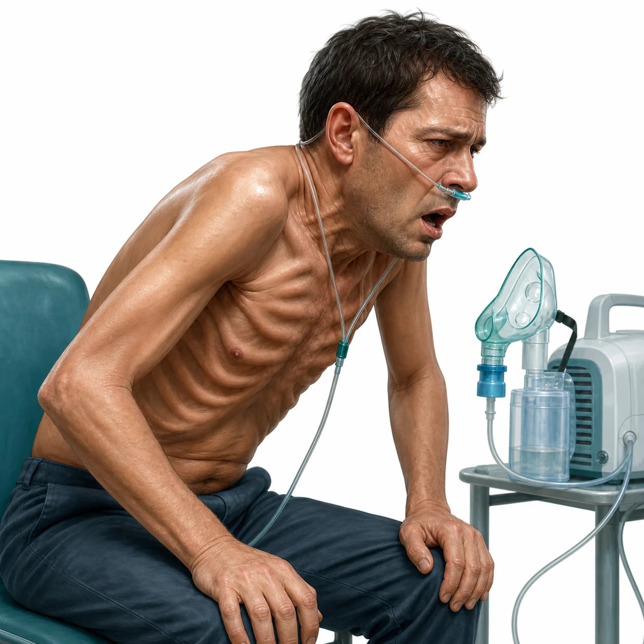
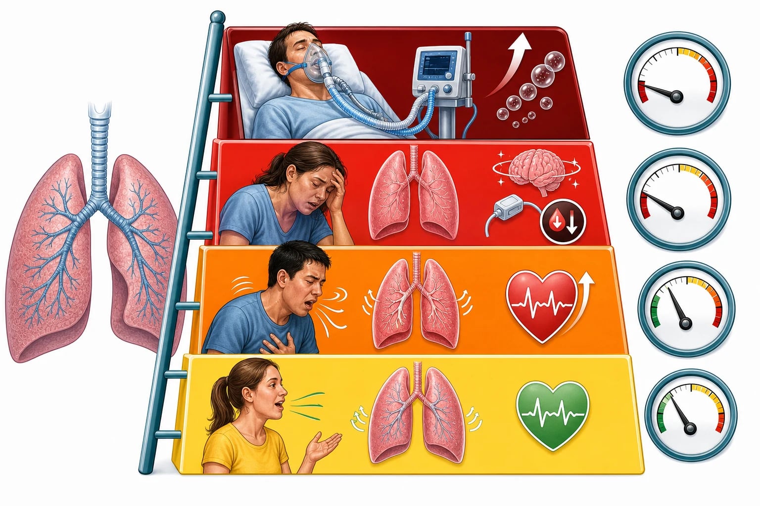
An acute severe asthma attack is graded at the bedside by the peak flow, the observations and the ability to speak. Acute severe asthma has a peak flow of 33 to 50 per cent of the predicted or best value, a respiratory rate of 25 or more, a heart rate of 110 or more, and an inability to complete sentences in one breath. Life-threatening asthma has any one of a peak flow under 33 per cent, an oxygen saturation under 92 per cent, a silent chest, cyanosis, exhaustion, a low or slow respiratory effort, hypotension, a bradycardia, or an altered conscious level. Near-fatal asthma is defined by a normal or rising arterial carbon dioxide — the single most reliable sign that the patient is tiring and near arrest. These thresholds are reproduced because they drive the escalation. [1]
Pathophysiology — why a rising CO₂ is the alarm
The airway obstruction of an asthma attack — bronchospasm, inflamed oedematous mucosa, and tenacious mucus plugging the small airways — produces air trapping and dynamic hyperinflation. The trapped air creates ventilation–perfusion mismatch and shunt, so the patient hypoxaemias; and the obstruction raises the work of breathing, so the patient hyperventilates and, in the early phase, blows off carbon dioxide. As the attack worsens and the respiratory muscles fatigue, the patient can no longer maintain that compensatory hyperventilation, and the carbon dioxide, which had been low, climbs back to normal and then rises — a normal or rising PaCO₂ in asthma is therefore a sign of a failing, fatiguing ventilator, not an improving one. The hyperinflation also raises the intrathoracic pressure, reducing the venous return, and the hypoxia and the beta-agonist drive raise the arrhythmia risk. This is why a rising carbon dioxide mandates critical care without delay. [1]
Clinical presentation
The patient is breathless, wheezy and distressed, using the accessory muscles, unable to speak in full sentences, tachypnoeic and tachycardic. The life-threatening features are the silent chest (no wheeze because there is no airflow — worse, not better), cyanosis, exhaustion, a slow or feeble respiratory effort, hypotension, a bradycardia, and drowsiness or confusion. A pulse that paradoxically varies markedly with respiration (a pulsus paradoxus) is a sign of severity. The trigger — a viral infection, an allergen, an NSAID or a beta-blocker, cold air or smoke, or non-adherence with preventer therapy — is sought in the history. [1]
Differential diagnosis
The wheezy, breathless patient has a differential, and the history, the chest and the response to therapy distinguish them. [1]
Acute severe asthma
- Known asthma, young, atopic; reversible obstruction
- PEF 33–50% severe, <33% life-threatening
- Wheeze, accessory-muscle use; responds to bronchodilator
- Rising CO₂ = near-fatal
COPD exacerbation
- Older, smoker, chronic airflow obstruction
- Baseline hypercapnia/hypoxia; less reversible
- Longer history; pursed-lip breathing
- Steroids, bronchodilators, ± NIV for type-2 failure
Anaphylaxis
- Rapid onset, urticaria, angiooedema, hypotension
- Trigger obvious; multi-system
- Adrenaline IM is first-line, not just bronchodilator
- Airway swelling may coexist
Cardiac asthma / pneumothorax
- Cardiac: raised JVP, crackles, ischaemic cause
- Pneumothorax: sudden, reduced air entry, hyperresonance
- BNP, CXR, echo distinguish
- Distinct pathways
Bedside assessment
Assess the severity at once: a peak expiratory flow if the patient can manage it, the respiratory rate, the heart rate, the oxygen saturation, the ability to speak, the work of breathing, and the life-threatening signs (silent chest, exhaustion, hypotension, bradycardia, cyanosis, conscious level). Take an arterial blood gas in any severe or life-threatening attack, specifically to look for the rising carbon dioxide. Examine for the trigger and the complications (a pneumothorax, a pneumomediastinum, pneumonia). [1]
Investigations and the targets
The peak expiratory flow grades the severity and tracks the response to therapy. The arterial blood gas is the key investigation in the severe attack: a low or normal-range carbon dioxide is expected in the early compensatory phase, and a normal or rising PaCO₂ is the danger sign of a near-fatal attack. The blood gas also shows the hypoxaemia and, in the late phase, a respiratory (and then a mixed) acidosis. The chest radiograph is not needed to diagnose asthma but excludes a pneumothorax, a pneumomediastinum and a pneumonia. The serum potassium falls with beta-agonist therapy and is monitored and corrected. [1]
Immediate management — the escalating therapy ladder
Sit the patient up and give high-flow oxygen to a target saturation of 94 to 98 per cent, then run the ladder, escalating as the severity demands. [1]
[1]Severity thresholds and the ladder
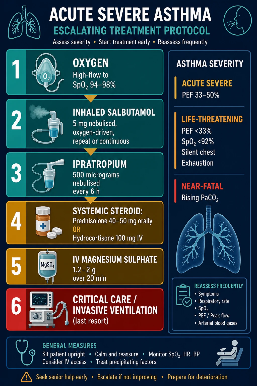
The magnesium evidence — that intravenous or nebulised magnesium modestly improves lung function in the severe attack — comes from the 3Mg trial and a systematic review.[1][2] For the refractory attack the second-line intravenous agents are added in critical care: intravenous salbutamol (a loading dose of about 250 micrograms, then an infusion of 5 to 20 micrograms per minute, with close cardiac monitoring for arrhythmia and hypokalaemia) and an aminophylline infusion (a loading dose of 5 mg per kilogram over 20 minutes, then 0.5 to 0.7 mg per kilogram per hour, with the level monitored for toxicity — nausea, tachycardia, seizure). Ketamine serves double duty as the bronchodilating induction agent, a helium–oxygen mixture (heliox) lowers the work of breathing through narrowed airways, and an inhaled volatile anaesthetic (isoflurane, sevoflurane) is a potent bronchodilator of last resort in the anaesthetised patient. Invasive ventilation is a last resort in the failing asthmatic: the dynamic hyperinflation risks hypotension and a tension pneumothorax, and the induction itself is hazardous. Where intubation is unavoidable, ketamine is the induction agent of choice (it bronchodilates), the ventilator is set to a slow rate with a long expiratory time and permissive hypercapnia, and the team watches constantly for a tension pneumothorax.
[1]Subtypes and special scenarios
The near-fatal asthmatic with a rising carbon dioxide needs critical care and, if failing, ventilatory support. The ventilated asthmatic is ventilated slowly with a long expiratory time and permissive hypercapnia, with a low threshold to decompress a tension pneumothorax at any sudden deterioration. Thunderstorm asthma is a mass-casualty phenomenon of allergen-driven exacerbations. Pregnancy modifies the approach only slightly — magnesium and steroids are safe, and the goal remains oxygenation of the mother and the fetus. [1]
Complications and pitfalls
The complications are respiratory failure and arrest, a tension pneumothorax (especially in the ventilated patient — a sudden rise in airway pressure with hypotension), an arrhythmia from hypokalaemia and beta-agonist drive, and hypokalaemia itself. The pitfalls are the dangerous inverse of the management: under-treating the attack as "anxiety"; missing a rising carbon dioxide; misreading a silent chest as improvement; relying on intravenous over oral steroid when the gut works; delaying critical care; sedating the tiring patient; and not watching for a tension pneumothorax in the ventilated asthmatic. [1]
Prognosis and disposition
A severe or life-threatening attack is admitted to a high-dependency or intensive-care bed; a near-fatal attack goes to intensive care. The trigger is treated, the preventer therapy is optimised, the patient is given a written asthma action plan, and a follow-up is arranged — because a near-fatal attack marks a high risk of a fatal one, and the preventable cause (often non-adherence with an inhaled corticosteroid) is addressed before discharge. For the ventilated patient, a slow weaning strategy with ongoing deep sedation prevents re-triggering the bronchospasm, and the arterial carbon dioxide is allowed to normalise gradually rather than rapidly to avoid a rebound arrhythmia. [1]
Special populations
Pregnancy uses the same ladder, with magnesium and steroids both safe and the oxygenation goal protecting both the mother and the fetus; salbutamol may relax the uterus but the maternal benefit outweighs this. Children use weight-based doses and a low threshold for critical care. The elderly and those with comorbid cardiopulmonary disease decompensate faster and are ventilated earlier. The non-adherent patient is identified and supported, because non-adherence with preventer inhaled-corticosteroid therapy is the commonest and most preventable driver of the near-fatal attack, and it is addressed with education and a written action plan before discharge. [1]
Severity grading — the three tiers in detail
Severity is graded at the bedside from the peak expiratory flow, the observations and the ability to speak, and the single most important investigation — the arterial blood gas — is taken in any severe attack to detect the rising carbon dioxide. [1]
Moderate
- PEF >50–75% predicted/best
- SpO₂ ≥92%, speaking in full sentences
- RR <25, HR <110
- No life-threatening features
- Manage in ED; recheck PEF 15–30 min after bronchodilator
Acute severe
- Any ONE: PEF 33–50% predicted/best
- RR ≥25, HR ≥110
- Inability to complete sentences in one breath
- Use of accessory muscles / respiratory distress
- Admit; start the full ladder; recheck PEF and ABG
Life-threatening
- Any ONE in a severe patient: PEF <33%
- SpO₂ <92% (or cyanosis)
- Silent chest, feeble respiratory effort, exhaustion
- Hypotension (SBP <100) or bradycardia
- Arrhythmia, altered conscious level, exhaustion
- ABG: normal/high PaCO₂, acidosis, or hypoxia
Near-fatal
- Rising PaCO₂ AND/OR requiring mechanical ventilation
- Defined by the ABG, not the bedside signs
- Critical care NOW — prepare for intubation
- Permissive hypercapnia if ventilated
- Mortality concentrated here
The thresholds are deliberately inclusive: any single life-threatening feature in a severe patient escalates the attack to the life-threatening tier, and a normal or rising PaCO₂ moves the patient to near-fatal and mandates critical-care involvement regardless of how well the patient looks. [1]
The severity thresholds, verbatim
Objective severity scores
For the intubated, paediatric, or hard-to-assess patient the modified pulmonary index score (MPIS) quantifies severity objectively across ten domains (oxygen saturation, accessory-muscle use, inspiratory:expiratory ratio, degree of wheeze, and mental status, each scored 0–3). An MPIS above 8 is severe and tracks the response to therapy when a peak flow cannot be measured. The pulmonary score and the Asthma Severity Score are alternatives for children. The objective scores are adjuncts, not substitutes, for the bedside classification — but they rescue grading in the patient who cannot perform a peak flow. [1]
[1]The first fifteen minutes — a resuscitation timeline
The first 15 minutes of the acute severe asthma attack
0 min — assess and call for help
ABCDE. Grade severity at the bedside: PEF (if the patient can manage it), RR, HR, SpO₂, BP, work of breathing, ability to speak, conscious level. Identify the life-threatening features (silent chest, exhaustion, hypotension, bradycardia, cyanosis, SpO₂ <92%, PEF <33%). Call the senior and critical care early for any life-threatening or near-fatal feature. Sit the patient upright.
0–5 min — oxygen and the first bronchodilator
High-flow oxygen 15 L/min via a non-rebreather mask (or oxygen-driven nebuliser), targeting SpO₂ 94–98% (88–92% only if the patient retains CO₂ — rare in pure asthma). Start **salbutamol 5 mg nebulised, oxygen-driven, immediately** — do not wait for the doctor. This is the single highest-impact intervention. Attach cardiac monitoring; take IV access.
5–10 min — steroid and ipratropium
Give a **systemic corticosteroid early** — prednisolone 40–50 mg orally if the patient can swallow, or hydrocortisone 100–200 mg IV if not (IV is not superior to oral when the gut works, but the oral route is unsafe in a tiring, breathless patient). Add **ipratropium bromide 500 micrograms nebulised** (combined with the salbutamol as a "duo-neb") for any severe or life-threatening attack. Steroids take 4–6 hours to work — give them early.
10–15 min — reassess and the ABG
Recheck PEF, SpO₂, RR, HR and the work of breathing after the first bronchodilator. Take an **arterial blood gas** in any severe or life-threatening attack to detect the rising PaCO₂ and the acidosis. If the patient is severe and not improving, or is life-threatening, give **IV magnesium sulphate 2 g over 20 minutes** and repeat the nebulisers back-to-back. Escalate to critical care.
15 min onward — escalate or admit
Reassess every 15 minutes. Continue back-to-back (or continuous) nebulised salbutamol ± ipratropium. If deteriorating or near-fatal (rising CO₂), move to second-line IV therapy (IV salbutamol, aminophylline), NIV as a bridge, and intubation as a last resort. If improving, transition to the admitting pathway and a discharge bundle.
Pharmacology of the bronchodilators
The bronchodilators reverse the bronchospasm; the corticosteroid addresses the inflammation that maintains it. Knowing the mechanism, the dose and the hazard of each agent is core Fellowship material. [1]
Salbutamol (β₂-agonist)
- Short-acting β₂-agonist — the first-line bronchodilator
- Dose: 5 mg nebulised, oxygen-driven; repeat or continuous
- IV: 250 µg load then 5–20 µg/min infusion
- Hazards: hypokalaemia, tachycardia, tremor, lactic acidosis
- β₂ selectivity incomplete at high dose — arrhythmia risk
Ipratropium (antimuscarinic)
- Short-acting antimuscarinic — added in severe attacks
- Dose: 500 µg nebulised, 6-hourly (combined with salbutamol)
- Mechanism: blocks M3 muscarinic receptors → bronchodilate
- Onset slower than salbutamol; synergistic with β₂-agonist
- Few systemic effects (poorly absorbed); dry mouth, urinary retention
Magnesium (IV)
- Smooth-muscle relaxant — inhibits Ca²⁺ influx in bronchial muscle
- Dose: 2 g (1.2–2 g) IV over 20 min in the severe attack
- Onset minutes; given once, may repeat
- Hazards: hypotension, flushing; monitor BP and reflexes
- Evidence: modest PEF improvement; 3Mg trial did not show outcome benefit
Aminophylline (methylxanthine)
- Phosphodiesterase inhibitor — raises cAMP → bronchodilate
- Dose: 5 mg/kg load over 20 min, then 0.5–0.7 mg/kg/h infusion
- Narrow therapeutic window — monitor the level
- Toxicity: nausea, vomiting, tachyarrhythmia, seizures
- Second-line; add in ICU for the refractory attack
Intravenous magnesium sulphate
Intravenous magnesium is a smooth-muscle relaxant that inhibits calcium influx into bronchial smooth muscle and modestly improves lung function in the severe attack not responding to standard therapy. It is given as 2 g (1.2 to 2 g) intravenously over 20 minutes, with the blood pressure and reflexes monitored for hypotension and areflexia. The nebulised route has weaker evidence. [1]
3Mg trial — Goodacre 2013 (Lancet Respir Med)
Design
Multicentre, randomised, double-blind, placebo-controlled factorial trial; 1109 adults with severe acute asthma across 34 UK EDs
Intervention
Single IV magnesium sulphate 2 g vs nebulised magnesium 3 × 2.5 g vs placebo (saline), in addition to standard therapy (salbutamol, ipratropium, steroid)
Primary outcome
Need for hospital admission; secondary: breathlessness, PEF, FEV₁, quality of life
Key result
Neither IV nor nebulised magnesium reduced admission vs placebo in this pragmatic trial. Subgroup with more severe airflow obstruction showed a PEF/FEV₁ improvement, but no outcome benefit. Magnesium remains in guidelines for the refractory severe attack despite a negative primary endpoint.
Kew 2014 — Cochrane review: intravenous magnesium for acute asthma in adults
Design
Cochrane systematic review and meta-analysis of randomised trials of IV magnesium sulphate in adults with acute asthma in the ED
Comparison
IV magnesium vs placebo, as an adjunct to standard therapy
Key result
IV magnesium improved lung function (PEF, FEV₁) in the severe attack and reduced hospital admission in the subgroup not responding to initial therapy. No serious adverse effects; transient hypotension and flushing reported.
Bottom line
IV magnesium 2 g is a reasonable adjunct in the severe attack not responding to standard bronchodilator/steroid therapy; the benefit is modest and concentrated in the most obstructed patients.
Second-line intravenous agents — salbutamol and aminophylline
For the attack refractory to nebulised bronchodilators, steroids and magnesium, the second-line intravenous agents are added in critical care. Intravenous salbutamol (a loading dose of about 250 micrograms, then an infusion of 5 to 20 micrograms per minute, titrated to effect and the tachycardia) bronchodilates but carries a real arrhythmia, hypokalaemia and lactic-acidosis risk and demands continuous cardiac monitoring. Aminophylline (a loading dose of 5 mg per kilogram over 20 minutes, then 0.5 to 0.7 mg per kilogram per hour) bronchodilates by phosphodiesterase inhibition but has a narrow therapeutic window — monitor the level for nausea, vomiting, tachyarrhythmia and seizures. Neither is routine first-line; both are rescue therapy in the deteriorating patient. [1]
Travers 2012 — Cochrane review: addition of IV β₂-agonists to inhaled β₂-agonists
Design
Cochrane systematic review and meta-analysis of randomised trials of IV β₂-agonists added to inhaled β₂-agonists for acute asthma
Comparison
IV β₂-agonist + inhaled β₂-agonist vs inhaled β₂-agonist alone (or vs IV methylxanthine)
Key result
No clear evidence that IV β₂-agonists add benefit to inhaled β₂-agonists for most patients. In the subgroup with life-threatening asthma not responding to inhaled therapy, IV β₂-agonists may improve lung function, but at the cost of more tremor, tachycardia and arrhythmia. IV salbutamol is not superior to IV aminophylline.
Bottom line
IV β₂-agonists are a second-line rescue, not a routine escalation; the inhaled route is superior for most patients. Use IV salbutamol with cardiac monitoring only in the refractory life-threatening attack.
Corticosteroids — dose, route, timing
A systemic corticosteroid is given early to every severe attack; it reduces admission, relapse and mortality, but takes 4 to 6 hours to work. Prednisolone 40 to 50 mg orally is standard; hydrocortisone 100 to 200 mg intravenously is used when the gut is unsafe (the tiring, breathless patient). Intravenous is not superior to oral when the gut works — the equivalence is a classic exam point. High-dose inhaled corticosteroid in the ED (e.g. budesonide or fluticasone) reduces admission in some trials but is not a substitute for the systemic steroid. [1]
Edmonds 2012 — Cochrane review: early inhaled corticosteroids in the ED treatment of acute asthma
Design
Cochrane systematic review and meta-analysis of randomised trials of inhaled corticosteroids given early in the ED for acute asthma
Comparison
Inhaled corticosteroid (alone or with systemic steroid) vs placebo or systemic steroid alone
Key result
High-dose inhaled corticosteroid in the ED modestly reduced hospital admission, particularly in patients not receiving systemic steroids. When systemic steroids were given, the additional benefit of inhaled steroids was marginal. No increase in adverse effects.
Bottom line
Inhaled corticosteroid in the ED is an adjunct, not a replacement, for the systemic corticosteroid — which remains the standard of care in the severe attack.
Ipratropium bromide
Ipratropium bromide, a short-acting antimuscarinic that blocks the M3 receptor on bronchial smooth muscle, is added to salbutamol as a "duo-neb" in any severe or life-threatening attack. The combination is synergistic and more effective than salbutamol alone in the severe attack — the evidence is the Cochrane review of combined anticholinergic and β₂-agonist therapy. The dose is 500 micrograms nebulised every 6 hours, continued until the patient improves. Systemic absorption is poor, so dry mouth, urinary retention and blurred vision are uncommon but occur in the elderly. [1]
Griffiths 2013 — Cochrane review: combined inhaled anticholinergics and short-acting β₂-agonists
Design
Cochrane systematic review and meta-analysis of randomised trials in children; combined anticholinergic + SABA vs SABA alone for acute asthma
Comparison
Single or multiple doses of ipratropium + SABA vs SABA alone
Key result
Adding ipratropium to SABA reduced hospital admission and improved lung function (FEV₁) in the moderate-to-severe exacerbation, with the benefit greatest in the most severe and over multiple doses. Minor side effects (tremor, dry mouth, nausea) only.
Bottom line
A single ipratropium dose is standard in any severe attack; multiple doses (every 6 hours) are used in the life-threatening or poorly responding attack.
Rescue and ventilatory therapies — heliox, volatiles, NIV
For the patient failing maximal medical therapy, rescue options bridge to — or sometimes avoid — intubation. Heliox (a helium–oxygen mixture, typically 70:30 or 80:20) is less dense than air and lowers the turbulent-flow work of breathing through narrowed airways; it is a temporising measure, not a cure, and the evidence is mixed. An inhaled volatile anaesthetic (isoflurane or sevoflurane), a potent bronchodilator, is an anaesthetist-delivered therapy of last resort in the anaesthetised, ventilated, refractory patient. Non-invasive ventilation (NIV or BiPAP) is occasionally used as a bridge in the fatiguing, hypercapnic asthmatic to reduce the work of breathing, but the evidence is limited and the risk of gastric insufflation and delayed intubation is real — NIV is a temporising measure in a monitored setting, never a substitute for intubation in the failing patient. Extracorporeal membrane oxygenation (ECMO) is the final salvage for the refractory, unventilatable asthmatic in a tertiary centre. [1]
Deciding to intubate the severe asthmatic — when to secure the airway
The trigger — deterioration despite maximal therapy
Intubate for any of: a rising PaCO₂ with exhaustion, a falling conscious level, respiratory arrest, refractory hypoxaemia, or cardiac instability. The decision is clinical and made early — the asthmatic who arrests is the one intubated too late.
The dangerous induction
The dynamic hyperinflation reduces venous return, and the induction agent drops the vascular tone — anticipate **profound hypotension on induction**. Pre-oxygenase fully, have noradrenaline drawn up, and use a reduced dose of induction agent. Ketamine is the agent of choice because it bronchodilates and supports the blood pressure.
Ketamine for induction
**Ketamine 1–2 mg/kg IV** (or a dissociative dose) is the bronchodilating induction agent; it releases catecholamines and supports the blood pressure where propofol would cause cardiovascular collapse. A benzodiazepine pre-treatment blunts the emergence phenomenon. Rocuronium or suxamethonium for paralysis; the largest tube that fits (to reduce resistance and allow bronchial toilet).
Ventilate slowly — the permissive-hypercapnia strategy
Set a **slow respiratory rate (6–10 breaths/min) with a long expiratory time (I:E ratio 1:4 or greater)** and a low tidal volume, accepting permissive hypercapnia (PaCO₂ up to 10–12 kPa with a compensating pH). The goal is to empty the lungs between breaths and avoid breath-stacking. Watch the expiratory flow waveform return to baseline.
Watch for the tension pneumothorax
A sudden rise in airway pressure with hypotension, hypoxaemia and asymmetric chest movement in the ventilated asthmatic is a **tension pneumothorax until proven otherwise** — needle-decompress immediately (2nd intercostal space, mid-clavicular line) and do not wait for a chest X-ray.
The ventilated asthmatic — settings and monitoring
Once intubated, the asthmatic is ventilated to minimise dynamic hyperinflation, not to normalise the blood gas. The settings: a low tidal volume (6–8 mL/kg ideal body weight), a slow rate (6–10 breaths/min), a long expiratory time (I:E 1:4–1:6), and permissive hypercapnia (allow the PaCO₂ to rise to 10–12 kPa provided the pH stays above ~7.15). The plateau pressure is kept under 30 cmH₂O. Deep sedation (and often a ketamine or cisatracurium infusion) prevents patient-ventilator dyssynchrony and re-triggering bronchospasm. The expiratory flow waveform is the key monitor: if it does not return to baseline before the next breath, there is gas trapping — slow the rate or shorten inspiration. [1]
Thunderstorm asthma — the mass-casualty phenomenon
Thunderstorm asthma is a sudden, large cluster of allergen-driven exacerbations during a thunderstorm in spring or early summer, classically from rye-grass pollen that is ruptured and dispersed by the storm, delivering allergen-laden particles deep into the airways of a sensitised population. The Melbourne 2016 event killed ten and overwhelmed the ambulance and ED systems within hours. The Fellowship lesson is systems-level: recognise the phenomenon early, surge the inhaler and oxygen stocks, and triage the at-risk (those with allergic rhinitis, undiagnosed asthma, or non-adherent preventer therapy) — because the standard per-patient therapy ladder does not scale to a mass-casualty presentation. [1]
The life-threatening features — any one escalates
Pitfalls and the dangerous inverse
The dangerous inverse of correct management is the recurring source of the preventable death: under-treating a severe attack as "anxiety"; missing the rising carbon dioxide on the blood gas; misreading a silent chest as improvement (no wheeze = no airflow, not a resolved attack); relying on intravenous over oral steroid when the gut works; delaying critical care for the near-fatal patient; sedating the tiring asthmatic; and not watching for the tension pneumothorax in the ventilated patient. A peak flow that improves but a gas that worsens, or a patient who "looks better" but is becoming bradycardic and drowsy, is deteriorating, not recovering. [1]
Evidence and regional guidelines
The contemporary framework is the BTS/SIGN British asthma guideline and the GINA global report; the bronchodilator and steroid choices are global. The intravenous-magnesium evidence is the 3Mg trial[1] and the systematic review.[2] The local respiratory pathway governs the escalation thresholds and the critical-care referral.
ANZ practice note. The severity grading and the therapy ladder follow the BTS/SIGN framework via the Thoracic Society of Australia and New Zealand and local respiratory pathways; intravenous magnesium 1.2 to 2 g is used for the severe attack not responding to standard therapy, and a near-fatal attack (a rising CO₂) triggers an immediate critical-care referral. [1]
The discharge bundle — closing the loop on a near-fatal attack
A near-fatal attack marks a high risk of a future fatal one, so discharge is not the end of the encounter but the start of prevention. The bundle has five elements, all delivered before the patient leaves. [1]
The asthma discharge bundle — five elements
1 — Optimise the preventer (inhaled corticosteroid)
Start or step up an inhaled corticosteroid (ICS) — the preventer — as a combination ICS/formoterol or ICS + SABA. Confirm the inhaler technique with a teach-back. The ICS is the single most effective intervention to prevent relapse and fatal recurrence; the LABA is never used alone (SMART trial).
2 — Give a written asthma action plan
A written, personalised plan that tells the patient what to do if symptoms worsen or the peak flow falls: increase the reliever, start or double the oral prednisolone, and when to seek urgent help. A written action plan reduces relapse and is a discharge standard.
3 — A course of oral prednisolone
A 5-day course of prednisolone 40 mg daily (or up to 10–14 days for a severe attack) is given to consolidate the recovery and reduce the early relapse rate. Review the response at 48 hours.
4 — Arrange follow-up
Primary-care or respiratory follow-up within 2–4 weeks (sooner for a near-fatal attack). Review the trigger, the adherence, the technique, and step up the preventer if needed. A near-fatal attack warrants specialist referral and assessment of adherence and comorbidities (rhinitis, reflux, obesity, anxiety).
5 — Address the trigger and the adherence
Identify and address the trigger (a viral infection, an allergen, an NSAID or beta-blocker, non-adherence with the ICS). Smoking-cessation advice. Vaccinate (influenza, pneumococcal). Non-adherence with preventer ICS therapy is the commonest and most preventable driver of the near-fatal attack — name it and fix it.
The drug doses — the verbatim ladder
Prognosis, relapse and disposition
The relapse rate after an ED visit for acute asthma is 10 to 20 per cent within two weeks; the predictors of relapse are a low peak flow on presentation, a poor response to the first bronchodilator, a recent ED visit or admission, and non-adherence with preventer therapy. A severe or life-threatening attack is admitted to a high-dependency or intensive-care bed; a near-fatal attack goes to intensive care. The peak flow response to therapy (the percentage improvement after the first hour) is a better predictor of safe discharge than any single admission value. The ventilated patient is weaned slowly with deep sedation to prevent re-triggering bronchospasm, and the carbon dioxide is allowed to normalise gradually to avoid a rebound arrhythmia. [1]
SAQs — exam practice
SAQ — Acute severe asthma with a silent chest
10 minutes · 10 marks
A 24-year-old woman with poorly controlled asthma (salbutamol reliever only, no inhaled corticosteroid) is brought to the ED by ambulance with worsening wheeze and breathlessness over six hours despite repeated salbutamol inhaler puffs at home. On arrival she is unable to speak, sitting upright and using accessory muscles; respiratory rate 32, heart rate 132, SpO2 88% on room air, blood pressure 104/68 with a palpable pulsus paradoxus. The chest is silent on auscultation — no wheeze is audible. PEF cannot be performed. She is drowsy but responsive.
SAQ — Near-fatal asthma post-intubation: ventilator strategy and the deteriorating ventilated patient
10 minutes · 10 marks
A 28-year-old man with near-fatal asthma (presenting PaCO2 9.2 kPa, pH 7.18, exhaustion) has just been intubated by the retrieval team using ketamine 1.5 mg/kg and rocuronium 1.2 mg/kg after pre-oxygenation. On arrival to the ICU ten minutes later he is ventilated at a rate of 14, I:E ratio 1:2, tidal volume 8 mL/kg ideal body weight, PEEP 5 cmH2O, FiO2 0.6. He abruptly develops a peak inspiratory pressure of 48 cmH2O with blood pressure 72/40 (was 110/65 on arrival), SpO2 84%, and asymmetric chest expansion.
Exam pearls
- Severity: PEF 33 to 50 per cent severe, under 33 per cent life-threatening; an SpO₂ under 92 per cent, a silent chest, exhaustion, hypotension or bradycardia are life-threatening.
- A normal or rising PaCO₂ is fatigue and a near-fatal attack → critical care.
- The ladder: oxygen, salbutamol 5 mg nebulised, ipratropium 500 micrograms, a steroid (prednisolone 40 to 50 mg orally, or hydrocortisone 100 mg intravenously), intravenous magnesium 1.2 to 2 g.
- Intravenous steroid is not superior to oral when the gut works.
- A silent chest is no airflow — deterioration, not improvement.
- Intubating the asthmatic is hazardous: ketamine, slow rate, long expiratory time, permissive hypercapnia, watch for tension pneumothorax. [1]
Red flags
[1]References
- [1]Goodacre S, Cohen J, Bradburn M, et al. Intravenous or nebulised magnesium sulphate versus standard therapy for severe acute asthma (3Mg trial): a double-blind, randomised controlled trial Lancet Respir Med, 2013.PMID 24429154
- [2]Mohammed S, Goodacre S. Intravenous and nebulised magnesium sulphate for acute asthma: systematic review and meta-analysis Emerg Med J, 2007.PMID 18029512
- [3]Kew KM, Kirtchuk L, Michell C, et al. Intravenous magnesium sulfate for treating adults with acute asthma in the emergency department Cochrane Database Syst Rev, 2014.PMID 24865567
- [4]Travers AH, Milan SJ, Jones AP, et al. Addition of intravenous beta(2)-agonists to inhaled beta(2)-agonists for acute asthma Cochrane Database Syst Rev, 2012.PMID 23235685
- [5]Griffiths B, Ducharme FM. Combined inhaled anticholinergics and short-acting beta2-agonists for initial treatment of acute asthma in children Cochrane Database Syst Rev, 2013.PMID 23966133
- [6]Nelson HS, Weiss ST, Bleecker ER, et al. The Salmeterol Multicenter Asthma Research Trial: a comparison of usual pharmacotherapy for asthma or usual pharmacotherapy plus salmeterol Chest, 2006.PMID 16424409
- [7]Edmonds ML, Milan SJ, Camargo CA Jr, et al. Early use of inhaled corticosteroids in the emergency department treatment of acute asthma Cochrane Database Syst Rev, 2012.PMID 23235589
- [8]Powell C, Dwan K, Milan SJ, et al. Inhaled magnesium sulfate in the treatment of acute asthma Cochrane Database Syst Rev, 2012.PMID 23235599