EM · Acute COPD exacerbation
Acute exacerbation of chronic obstructive pulmonary disease
Also known as COPD exacerbation · Acute COPD · COAD exacerbation
The acute COPD exacerbation — the Anthonisen criteria for the clinical definition, the controlled-oxygen principle (88 to 92 per cent, not 94 to 98 per cent), the four-pillar management (controlled oxygen, nebulised bronchodilators with doses, a systemic corticosteroid, antibiotics for the purulent exacerbation), the early non-invasive ventilation (BiPAP) for the respiratory acidosis (the strongest evidence in emergency respiratory medicine), and the CO2-retainer dangers. ACEM-primary, globally tagged.
On this page & tools
Your progress
Saved locally on this device.
Target exams
Red flags
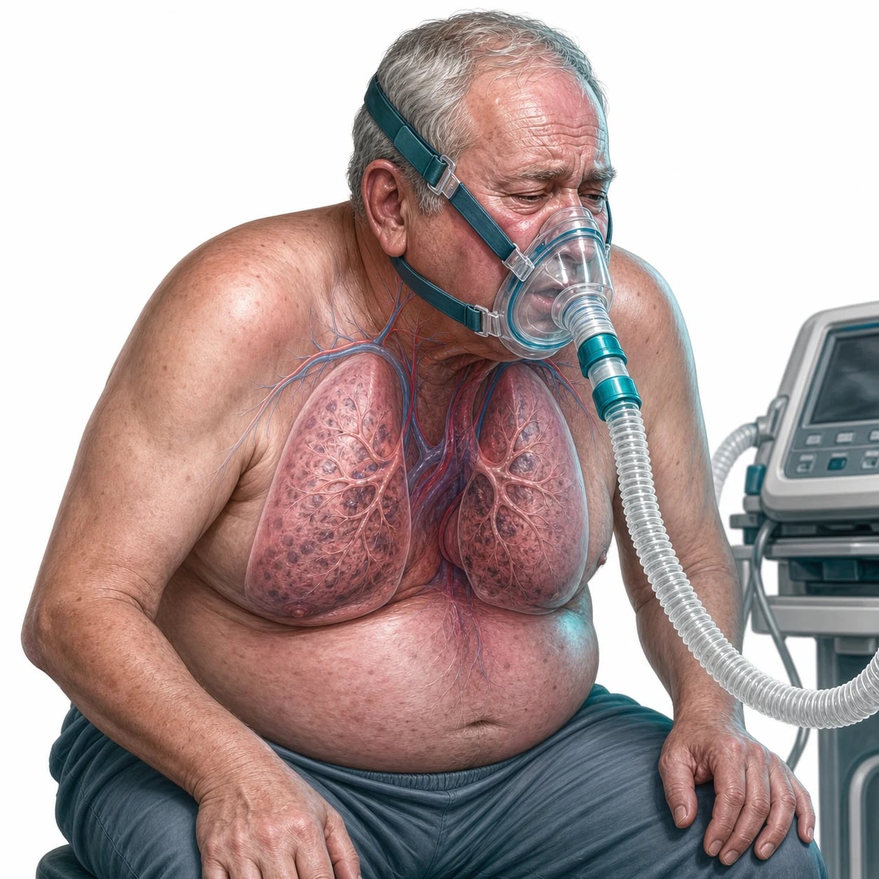
An acute exacerbation of chronic obstructive pulmonary disease is among the commonest and most dangerous presentations in the emergency department, and it is the condition for which non-invasive ventilation has the strongest evidence in all of emergency medicine. The Fellowship candidate must control the oxygen to 88 to 92 per cent, deliver the four pillars of the pharmacotherapy, and start the BiPAP early for the acidotic exacerbation, because every minute of a worsening acidosis is a minute closer to the intubation and the intensive care.[1][2]
Definition and the Anthonisen criteria
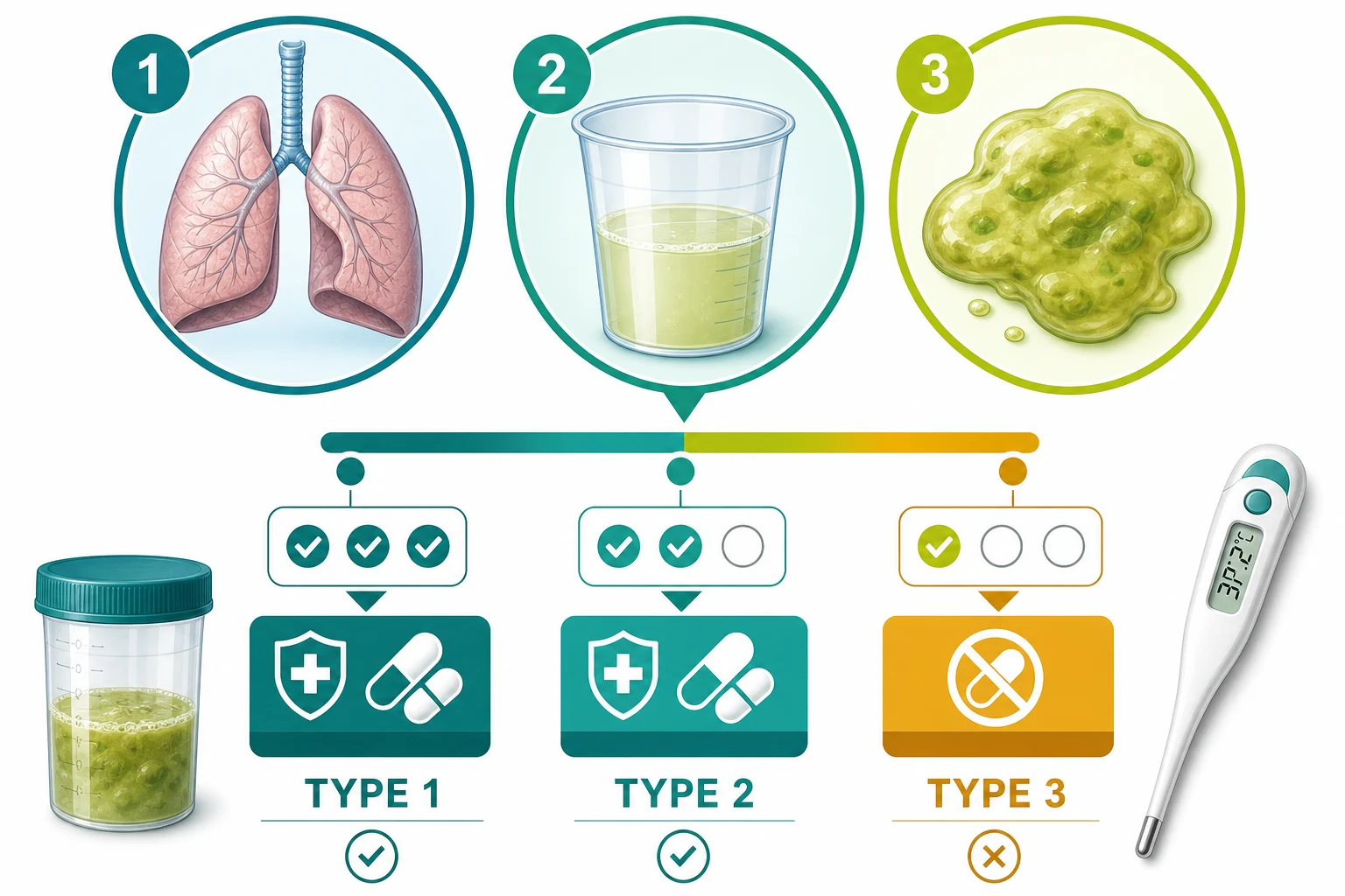
An acute exacerbation of COPD is a sustained worsening of the respiratory symptoms — the breathlessness, the sputum volume and the sputum purulence — that is beyond the day-to-day variability and acute in onset. The Anthonisen criteria classify the exacerbation by the clinical presentation: a type 1 exacerbation has all three features (the increased dyspnoea, the increased sputum volume and the increased purulence); a type 2 has two of the three; and a type 3 has one of the three plus at least one additional clinical criterion (an upper-airway infection, an unexplained fever, or an increased respiratory rate or heart rate). The Anthonisen type matters because it guides the antibiotic decision: the type 1 and the purulent type 2 warrant the antibiotic, while the non-purulent type 2 and the type 3 may not. [1]
Pathophysiology — the retainer and the oxygen
The chronic obstructive pulmonary disease produces a fixed and a partially reversible airflow obstruction, with a loss of the elastic recoil, a hyperinflation and a ventilation–perfusion mismatch. An exacerbation — usually triggered by a viral or a bacterial airway infection — worsens the bronchoconstriction, the mucosal oedema and the mucus plugging, which increases the airway resistance and the work of breathing. The hypoxia worsens, and if the patient is a chronic CO₂ retainer, the hypercapnia and the acidosis worsen. The critical mechanism — which the Fellowship candidate must explain at a viva — is the reason the retainer is targeted to 88 to 92 per cent, not 94 to 98 per cent: the excess oxygen raises the PaCO₂ by the Haldane effect (the oxygen displaces the CO₂ from the haemoglobin) and by the loss of the hypoxic pulmonary vasoconstriction (the blood is redirected to underventilated alveoli). The result is a CO₂ narcosis — a drowsy, hypoventilating patient heading for a respiratory arrest. [1]
Causes and precipitants
Approximately 70 per cent of acute exacerbations are infective — split roughly between the viral and the bacterial — and the remainder are environmental, the medication non-adherence, or the cardiovascular overlap. The identification of the trigger matters because it directs the antibiotic decision and the active search for the alternative diagnosis. The sputum inflammatory markers (the interleukin-6, the interleukin-8, the tumour necrosis factor) rise with the exacerbation and track the symptom severity and the lung-function decline.[9]
[1]Viral (~50%)
- Rhinovirus is #1 (the common cold)
- Also RSV, influenza A/B, parainfluenza, coronavirus, adenovirus
- Purulent-looking sputum may be inflammation, not bacteria
- Antibiotic NOT routinely needed unless the Anthonisen type 1
Bacterial (~30–50%)
- Haemophilus influenzae (the commonest)
- Streptococcus pneumoniae
- Moraxella catarrhalis (often a β-lactamase producer)
- Pseudomonas aeruginosa in severe/bronchiectatic disease
Environmental
- Air pollution, particulate matter (PM2.5)
- Cold weather and the temperature drops
- Cigarette smoke, the biomass fuel
- Occupational dusts and fumes
Other precipitants
- Pulmonary embolism (up to 25% of hospitalised "exacerbations")
- Pneumothorax, pneumonia, the LV failure
- Medication non-adherence (the inhaler ran out)
- Arrhythmia — the atrial fibrillation is common
The non-infective precipitants must be actively excluded at every presentation. A pulmonary embolism is found in up to a quarter of the COPD patients hospitalised for an "exacerbation," and the D-dimer and the imaging are considered when the dyspnoea is disproportionate to the infection, the effusion, or the hypercapnia. The pneumothorax in the hyperinflated, bullous lung may be small and easily missed on the mobile chest film; the bedside ultrasound (the absent lung sliding) is the rapid screen. The left-ventricular failure overlap (the "cardiac asthma") raises the BNP and shows the B-lines on the ultrasound. The relapse risk is real: the patient who has had one exacerbation is more likely to have another, and the frequency and the severity of the exacerbations increase over time.[8]
Severity assessment
The severity of the exacerbation is judged at the bedside from the respiratory effort, the gas exchange, the mental state and the haemodynamic stability, and it determines the disposition — the discharge, the ward, the high-dependency, the intensive care, or the intubation. The arterial blood gas is the objective severity anchor. [1]
Mild
- Dyspnoea increased, coping at rest
- SpO₂ 88–92% on no or low-flow O₂
- No respiratory acidosis (pH ≥7.35)
- RR <25; managing oral intake — consider discharge
Moderate
- Increased work of breathing, accessory-muscle use
- SpO₂ <88% needing controlled O₂
- pH 7.30–7.35 — borderline, watch closely
- Admit to the ward; start the four pillars
Severe
- pH 7.25–7.30, PaCO₂ high — a respiratory acidosis
- RR >30; using accessory muscles
- BiPAP indicated (within 60 min of the ABG)
- High-dependency or ICU level care
Life-threatening
- pH <7.25 or GCS <8, drowsy, exhausted
- Slow, falling respiratory rate (fatigue)
- NIV failing or contraindicated
- Intubate and ventilate; ICU
The severity-driven escalation in the ED
1 — Assess
The airway, the breathing, the circulation; the SpO₂, the RR, the accessory-muscle use, the GCS, the temperature.
2 — Controlled oxygen
The Venturi 24–28 per cent, target SpO₂ 88–92 per cent; recheck the ABG at 30 to 60 minutes.
3 — Mild/moderate (pH ≥7.35)
The four pillars on the ward; repeat the ABG if any deterioration.
4 — Severe (pH 7.25–7.35, PaCO₂ >6.5)
Start the BiPAP within 60 minutes; IPAP 12–20 / EPAP 4–6.
5 — Life-threatening (pH <7.25, GCS <8, or NIV failing)
Rapid-sequence intubation and ICU.
Clinical presentation
The patient presents with a worsening dyspnoea, an increased sputum volume and purulence, a wheeze, and a reduced exercise tolerance over days. The signs include a tachypnoea, a prolonged expiration, a pursed-lip breathing, a hyperinflated chest, an accessory-muscle use, a wheeze and a cyanosis. The severe exacerbation shows a drowsiness (the hypercapnia), a confusion, a coarse tremor (the asterixis of the CO₂ retention) and, in the worst case, a bradycardia and a hypotension that signal the impending arrest. The Anthonisen type is assigned at the bedside. [1]
Differential diagnosis
The COPD patient can have any of the common respiratory emergencies, and the assumption that "it is just the COPD" is a dangerous one. [1]
COPD exacerbation
- Worsening dyspnoea + sputum + purulence (Anthonisen)
- Known COPD; hyperinflated chest, pursed-lip breathing
- Controlled O2 88-92%; BiPAP for pH<7.35
- Four pillars: O2, bronchodilators, steroid, antibiotics
Pneumonia complicating COPD
- Fever, consolidation, sepsis on top of COPD
- CURB-65 for the pneumonia; broader antibiotic
- CXR: consolidation; blood cultures
- May still need BiPAP if acidotic
Pneumothorax in COPD
- Sudden onset; reduced air entry, hyperresonance
- CXR: pleural line; USS: absent sliding
- Drain; do not assume "just the COPD"
- High incidence in the hyperinflated lung
LV failure overlap
- "Cardiac asthma" — crackles, raised JVP, S3
- BNP raised; echo: LV dysfunction
- Treat both the heart and the lung
- Diuretic + nitrate + bronchodilator
Investigations and the acidosis decision
The arterial blood gas is the investigation that decides the ventilatory strategy. It provides the pH, the PaCO₂, the PaO₂ and the bicarbonate, and it is the pH that triggers the NIV: a pH below 7.35 with a PaCO₂ above 6.5 kPa is a respiratory acidosis that warrants the BiPAP. The earlier the NIV is started (within 60 minutes of the acidotic ABG), the better the outcome. The chest radiograph shows the hyperinflation and seeks a pneumonia, a pneumothorax or a pulmonary oedema — any of which may be the true precipitant rather than "just the COPD." The ECG screens for an arrhythmia (the atrial fibrillation is common in the exacerbation) and an ischaemia. The bloods (the full blood count, the CRP, the urea and electrolytes) and the sputum culture are sent. [1]
Immediate management — the four pillars and the controlled oxygen
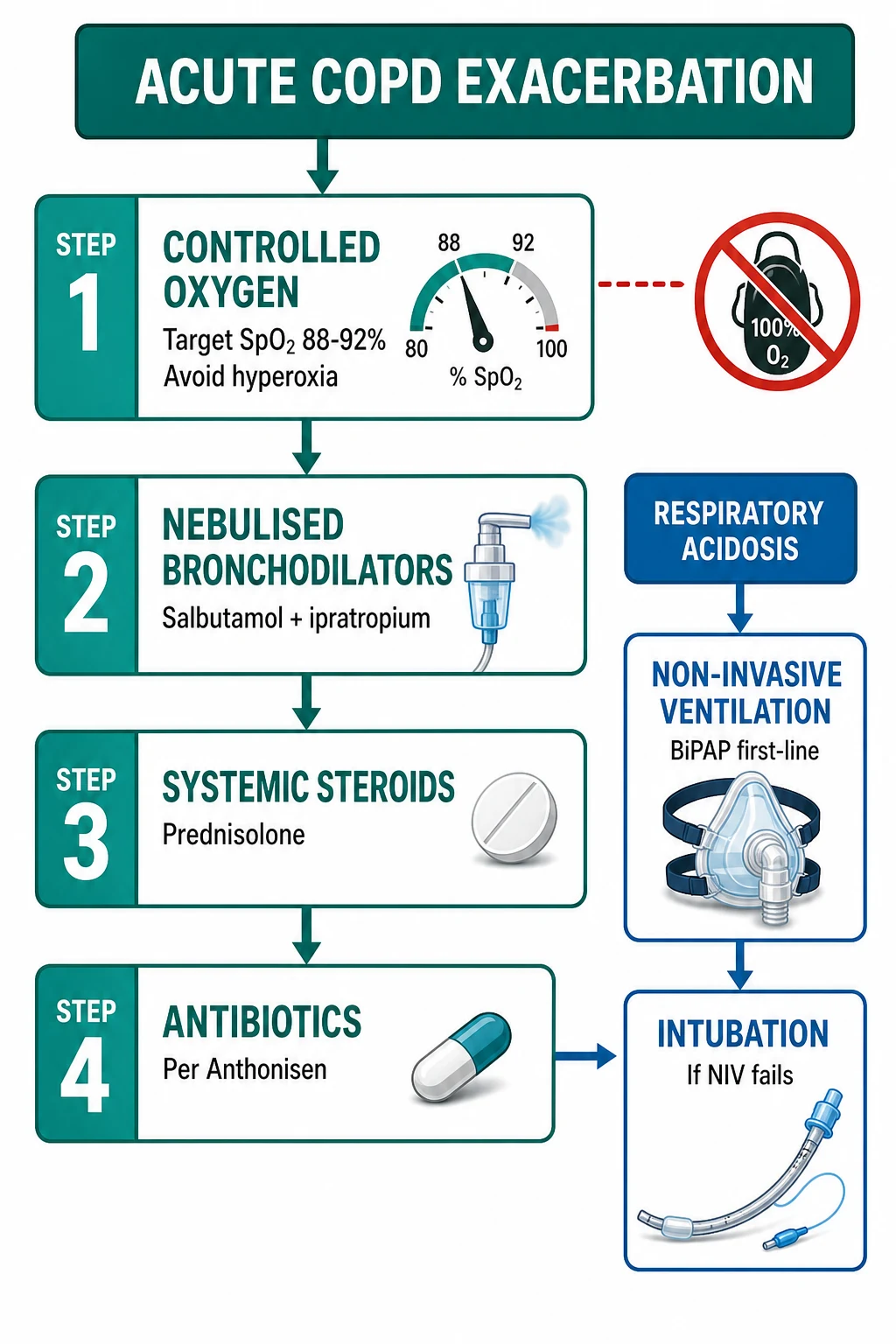
Stabilise the airway and the breathing. Give the controlled oxygen via a Venturi mask (24 to 28 per cent, titrated to a target of 88 to 92 per cent), and never the uncontrolled high-flow oxygen of the non-rebreather. Then deliver the four pillars. [1]
[1]The COPD exacerbation targets
The non-invasive ventilation — the strongest evidence
The non-invasive ventilation (the BiPAP — a bilevel positive airway pressure with an inspiratory and an expiratory pressure) is the single most evidence-supported intervention in the emergency respiratory management of the COPD exacerbation. The landmark trial of Plant and colleagues showed that an early BiPAP on the general respiratory ward reduced the need for the intubation, the mortality and the length of stay in the acidotic exacerbation.[1] The indication is a pH below 7.35 with a PaCO₂ above 6.5 kPa (a respiratory acidosis). The settings start at an IPAP of 12 to 20 and an EPAP of 4 to 6, titrated to the CO₂ clearance and the comfort. The patient who fails the NIV — the persisting or worsening acidosis, the drowsiness, the exhaustion — is intubated and ventilated. The patient who is already obtunded (a GCS under 8), who cannot protect the airway, or who refuses the mask goes directly to the invasive ventilation.
The Cochrane meta-analysis of fourteen randomised trials confirmed and quantified the benefit: the NIV reduced the mortality, the intubation, the treatment failure and the length of the stay.[6]
Antibiotics — when and which
The antibiotic is given for the Anthonisen type 1 (the increased dyspnoea, the increased sputum volume and the increased purulence) or the type 2 with a purulent sputum. The first-line agent is amoxicillin-clavulanate (it covers the Haemophilus influenzae, the pneumococcus and the Moraxella catarrhalis, many of which produce a beta-lactamase), with doxycycline as an alternative. The intravenous route is chosen for the severe or the ward-non-responsive patient. The course is 5 to 7 days.[2] The antibiotic is not needed for the non-purulent exacerbation without the Anthonisen criteria, because the cause is often viral.
Evidence in focus — the landmark trials
REDUCE — Leuppi 2013 (JAMA)
JAMA
PMID 23695200
Key finding
A multicentre randomised non-inferiority trial of 314 patients with the acute COPD exacerbation, comparing a 5-day course of the prednisone 40 mg daily against a 14-day course. The short course was non-inferior for the time to the next exacerbation and the recovery, with fewer corticosteroid-related adverse effects.
Practice change
The 5-day course of the prednisolone 30–40 mg is the evidence-based default; the taper is unnecessary for a short course, and the longer course adds steroid harm without the benefit.
Anthonisen 1987 (Ann Intern Med)
Annals of Internal Medicine
PMID 3492164
Key finding
A randomised trial of the antibiotic against the placebo in the exacerbations, stratified by the Anthonisen type. The antibiotic benefited the type 1 (all three criteria) and the purulent type 2 exacerbations; there was no benefit for the non-purulent exacerbations.
Practice change
The antibiotic is for the Anthonisen type 1 and the purulent type 2 exacerbation — not for every wheeze. This trial defined both the Anthonisen criteria and the antibiotic indication in one study.
Plant 2000 (Lancet)
Lancet
PMID 10859037
Key finding
A multicentre randomised trial of 236 patients with the acidotic COPD exacerbation (pH 7.25–7.35), comparing the early NIV on the general respiratory ward against the standard care. The NIV reduced the need for the intubation, the in-hospital mortality and the length of stay.
Practice change
The BiPAP within 60 minutes of the acidotic ABG is the standard of care — the single strongest evidence in the emergency respiratory medicine.
Ram 2004 — Cochrane (NIV in COPD)
Cochrane Database of Systematic Reviews
PMID 15266518
Key finding
A meta-analysis of 14 randomised trials of the NIV for the acidotic COPD exacerbation. The NIV reduced the mortality (number-needed-to-treat around 10), the need for the intubation, the treatment failure and the length of the hospital stay.
Practice change
The NIV is the most evidence-supported respiratory intervention in the emergency department for the acidotic COPD exacerbation.
Austin 2010 (BMJ)
BMJ
PMID 20959284
Key finding
A randomised controlled trial of 405 patients with the suspected COPD in the prehospital setting, comparing the titrated oxygen against the high-flow oxygen. The titrated oxygen reduced the mortality (a relative risk reduction of around 78 per cent).
Practice change
Do NOT give the high-flow oxygen to the suspected COPD patient in the prehospital or the ED setting — titrate from the first contact. This is the trial behind the controlled-oxygen principle.
Walters 2018 — Cochrane (corticosteroid duration)
Cochrane Database of Systematic Reviews
PMID 29553157
Key finding
A meta-analysis of the corticosteroid durations for the COPD exacerbation. The shorter courses (7 days or less) were as effective as the longer courses, with fewer adverse effects (the hyperglycaemia, the insomnia, the infection risk).
Practice change
The 5-day course of the prednisolone is the evidence-based default; the longer course is reserved for the non-resolving or the severe exacerbation.
The first 60 minutes of the acidotic COPD exacerbation
0 min — recognise and the controlled oxygen
The ABCDE. The Venturi 24–28 per cent to the 88–92 per cent target — NEVER the high-flow. The full monitoring (the SpO₂, the ECG, the NIBP every 3 min). Two large-bore cannulae.
0–10 min — the duo-neb and the steroid
The salbutamol 5 mg + the ipratropium 500 mcg, oxygen-driven, immediately. The prednisolone 30–40 mg orally (or the hydrocortisone 100 mg IV if the patient cannot swallow). Send the ABG now.
10–30 min — the ABG and the antibiotic decision
Read the pH and the PaCO₂. The pH <7.35 with the PaCO₂ >6.5 kPa → the BiPAP. The Anthonisen type 1 or the purulent type 2 → the amoxicillin-clavulanate 500/125 mg orally TDS (or the doxycycline/azithromycin if penicillin-allergic).
30–60 min — the BiPAP setup and the reassessment
Start the BiPAP at the IPAP 12–20 and the EPAP 4–6, titrated to the comfort and the CO₂ clearance. Recheck the ABG at 1 hour on the support. A worsening acidosis, a drowsiness or a loss of the effort → the intubation.
Complications and pitfalls
The complications are the CO₂ narcosis from the excess oxygen, the respiratory arrest, the arrhythmia (the atrial fibrillation), the pneumothorax, the NIV failure and the need for the intubation, and the hospital-acquired infection. The pitfalls are the inverse of the management: giving 94 to 98 per cent oxygen to the retainer; not escalating to the NIV when the acidosis persists; using an air-driven nebuliser (an unknown FiO₂); missing the alternative diagnosis (the pneumonia, the pneumothorax, the pulmonary embolism, the left-ventricular failure); not treating with the steroid; and delaying the antibiotic for the purulent exacerbation. [1]
Prognosis and disposition
The COPD exacerbation treated with the NIV has an in-hospital mortality of about 10 per cent; the intubated exacerbation carries a 20 to 30 per cent mortality, and a patient who has been intubated once for a COPD exacerbation has a significantly shorter subsequent life expectancy. The patient is admitted — the NIV-dependent to the high-dependency or the intensive care, the improving to the ward. The patient is switched to the oral therapy when the respiratory rate normalises and the sputum eases. The long-term management — the inhaled dual or triple therapy, the pulmonary rehabilitation, the smoking cessation, the vaccination, and the long-term oxygen therapy if indicated — is addressed before the discharge. [1]
Discharge and follow-up
The discharge decision rests on the resolution of the four-pillar-reversible features — the respiratory rate and the work of breathing normalising, the sputum easing, the SpO₂ back at the patient's baseline, and the patient mobile and eating — not on a single normalised blood gas. The structured discharge bundle reduces the re-admission, and it is examined. [1]
The COPD discharge bundle
1 — Inhaler technique and optimisation
Checked and corrected; optimise to the dual (LAMA + LABA) or the triple (plus ICS) therapy per the GOLD group.
2 — Smoking cessation
Offer the pharmacotherapy (the varenicline, the nicotine replacement) and the referral.
3 — Vaccination
The influenza annually, the pneumococcal (the PCV13/PPSV23), the COVID-19, and consider the pertussis and the zoster.
4 — Pulmonary rehabilitation referral
Reduces the re-admission, the dyspnoea and the anxiety, and improves the exercise capacity and the quality of life.
5 — Oxygen assessment
Check the ABG at least 4 weeks after the discharge for the LTOT eligibility (the PaO₂ <7.3 kPa on the air).
6 — Written action plan
Recognising and self-managing the early exacerbation, with the standby rescue medication (the oral steroid and the antibiotic).
7 — Follow-up
An early review within 4 weeks and a specialist review if the recurrent exacerbations.
The patient who has needed the BiPAP is not sent home directly; the weaning to the nasal cannula or the low-flow oxygen, the repeat ABG off the support, and the inpatient pulmonary rehabilitation are completed first. The patient on the long-term oxygen therapy at baseline is discharged back to the prescribed flow rate, with the oxygen assessment repeated at the four-week clinic review. The pulmonary rehabilitation, started in the hospital and continued in the community, is among the most cost-effective interventions in the chronic COPD care. [1]
The discharge readiness markers
Mild exacerbation
- Discharged on the oral steroid 5 days
- The duo-neb inhaler and the action plan
- The early follow-up within 4 weeks
- The pulmonary rehabilitation referral
Moderate exacerbation
- Admitted to the ward for the 48–72 h
- The four pillars + the physiotherapy
- The wean to the oral therapy
- The discharge bundle before the home
Severe (BiPAP)
- The HDU or the ICU for the NIV
- The repeat ABG at 1 h and on the wean
- The wean to the standard oxygen over 24–48 h
- The inpatient pulmonary rehabilitation
Intubated
- The ICU, the lung-protective ventilation
- The daily sedation holds and the weaning plan
- The tracheostomy if the prolonged ventilation
- The 20–30% in-hospital mortality — counsel the family
Special populations
The elderly have a higher mortality and more comorbidity. The chronic oxygen-dependent patient (the LTOT patient) has a baseline hypoxaemia and a higher risk from the intercurrent illness. The alpha-1 antitrypsin deficiency patient is younger and may be offered the replacement therapy and the transplantation assessment. The overlap with the heart failure (the "failing heart and the failing lung") is common and is distinguished by the BNP, the echocardiogram and the response to the therapy. [1]
Evidence and regional guidelines
The contemporary framework is the NICE COPD guideline and the GOLD global report, aligned with the BTS/TSANZ respiratory pathway. The NIV evidence is the Plant trial[1] and its confirmations. The pharmacotherapy evidence (the steroid, the bronchodilator, the antibiotic) is summarised in the recent reviews.[2] The four-pillar approach and the oxygen targets are global; the exact antibiotic and the NIV settings follow the local protocol.
ANZ practice note. The controlled-oxygen target (88 to 92 per cent), the four-pillar pharmacotherapy and the early BiPAP for the pH under 7.35 follow the NICE/GOLD framework via the TSANZ and the local respiratory pathway. The nebuliser is oxygen-driven, and the BiPAP is started within 60 minutes of the acidotic ABG, in the emergency department if the high-dependency bed is not immediately available. [1]
Exam practice
SAQ — COPD exacerbation with type 2 respiratory failure commenced on non-invasive ventilation
10 minutes · 10 marks
A 68-year-old man with known severe COPD (FEV₁ 38 per cent predicted, on triple inhaler therapy and home oxygen 2 L) presents with three days of worsening dyspnoea, increased sputum volume and purulence, and a productive cough. On arrival he is cyanosed, respiratory rate 32, using accessory muscles and pursed-lip breathing, SpO₂ 84 per cent on room air, BP 152/88, HR 108 in sinus tachycardia, GCS 14, temperature 37.8°C. The chest is hyperinflated with a diffuse expiratory wheeze. The initial arterial blood gas on a Venturi 28 per cent mask shows pH 7.28, PaCO₂ 9.2 kPa, PaO₂ 6.9 kPa, HCO₃⁻ 32 mmol/L, base excess +6, lactate 1.4. The chest radiograph shows hyperinflation and increased markings at the right base but no consolidation or pneumothorax.
SAQ — Acidotic COPD patient failing non-invasive ventilation: intubation and invasive ventilation
10 minutes · 10 marks
A 72-year-old woman with severe COPD (FEV₁ 30 per cent predicted, two hospital admissions in the last year, on triple therapy and long-term oxygen) was commenced on BiPAP one hour ago for an acidotic exacerbation (initial pH 7.24, PaCO₂ 10.5 kPa). She has received the duo-neb, the prednisolone 40 mg, and the amoxicillin-clavulanate. Despite an IPAP of 16 and an EPAP of 5, with a good mask seal and a conscious cooperative start, she has become progressively drowsy (GCS now 12), her respiratory rate has fallen from 28 to 12, she is unable to clear her secretions, and the repeat ABG shows pH 7.18, PaCO₂ 11.8 kPa, PaO₂ 7.1 kPa. The blood pressure is 96/58. She is no longer triggering the BiPAP reliably.
Exam pearls
- Controlled oxygen 88 to 92 per cent — never 94 to 98 per cent in the CO₂ retainer.
- The four pillars: O₂ (Venturi 24-28%), bronchodilators (salbutamol 5 mg + ipratropium 500 mcg nebulised, O₂-driven), steroid (prednisolone 30-40 mg for 5-7 days), antibiotics (amoxicillin-clavulanate if purulent).
- BiPAP for the pH under 7.35 — the strongest evidence in emergency respiratory medicine (Plant 2000).
- Oxygen-driven nebuliser, NOT air-driven — the air-driven delivers an unknown FiO₂.
- Anthonisen: increased dyspnoea + volume + purulence → type 1 → antibiotics.
- The COPD patient with a sudden deterioration may have a pneumothorax, a PE or an LV failure — do not assume "just the COPD."
- A patient on BiPAP who becomes drowsy is failing → prepare for the intubation.
- Prednisolone 30–40 mg for 5 days — the REDUCE trial proved 5 days is enough (non-inferior to 14 days); the taper is NOT needed for a short course.[4]
- Antibiotic only for the purulent / the Anthonisen type 1 — the amoxicillin-clavulanate (covers the β-lactamase-producing H. influenzae and M. catarrhalis); the doxycycline or the azithromycin if penicillin-allergic.[3]
- The duo-neb: salbutamol 5 mg + ipratropium 500 mcg, oxygen-driven, repeated as needed — the nebulised magnesium has NO role in the COPD (unlike the asthma).
- The methylxanthines (theophylline, aminophylline) are NOT recommended — the narrow therapeutic window, the arrhythmia, the no outcome benefit.
- Check the D-dimer / consider the PE in a COPD "exacerbation" with the disproportionate dyspnoea — the PE is found in up to 25%.
- Beware the falling respiratory rate — the fatiguing CO₂ retainer is arresting, not improving; escalate to the intubation.
- Repeat the ABG at 30–60 minutes after starting the controlled oxygen, and at 1 hour on the BiPAP, to confirm the CO₂ is falling.
- The Pseudomonas aeruginosa in the severe COPD with the bronchiectasis or the frequent antibiotics → the anti-pseudomonal agent (the piperacillin-tazobactam).
- No routine chest physiotherapy in the acute exacerbation (no outcome benefit); encourage the sputum clearance as tolerated.
- The atrial fibrillation is the commonest arrhythmia in the COPD exacerbation — the rate-control with a beta-blocker is acceptable once the bronchospasm is treated.
- The Heliox and the inhaled corticosteroids have no role in the acute rescue; the ICS is for the chronic maintenance (the triple therapy) only.
- The asthma–COPD overlap (ACO) — more reversibility and the eosinophilia; lean towards the earlier ICS in the maintenance, but the acute management is identical.
- The prehospital oxygen matters — titrate from the first contact; the high-flow oxygen in the suspected COPD increases the mortality (Austin 2010).[7]
- The corticosteroid duration is 5 days — the longer course adds the hyperglycaemia, the insomnia and the infection risk without the benefit (Walters 2018).[5]
Red flags
[1]References
- [1]Plant PK, Owen JL, Elliott MW. Early use of non-invasive ventilation for acute exacerbations of chronic obstructive pulmonary disease on general respiratory wards: a multicentre randomised controlled trial Lancet, 2000.PMID 10859037
- [2]Calvello Hlap E, Sakles JC, Patanwala AE, et al. Acute Exacerbation of Chronic Obstructive Pulmonary Disease: Pharmacological Treatment of AECOPD New Perspectives Semin Respir Crit Care Med, 2026.PMID 41844237
- [3]Anthonisen NR, Manfreda J, Warren CPW, Hershfield ES, Harding GK, Nelson NA. Antibiotic therapy in exacerbations of chronic obstructive pulmonary disease Ann Intern Med, 1987.PMID 3492164
- [4]Leuppi JD, Schuetz P, Bingisser R, et al. Short-term vs conventional glucocorticoid therapy in acute exacerbations of chronic obstructive pulmonary disease: the REDUCE randomized clinical trial JAMA, 2013.PMID 23695200
- [5]Walters JAE, Tan DJ, White CJ, Gibson PG, Wood-Baker R, Walters EH. Different durations of corticosteroid therapy for exacerbations of chronic obstructive pulmonary disease Cochrane Database Syst Rev, 2018.PMID 29553157
- [6]Ram FSF, Picot J, Lightowler J, Wedzicha JA. Non-invasive positive pressure ventilation for treatment of respiratory failure due to exacerbations of chronic obstructive pulmonary disease Cochrane Database Syst Rev, 2004.PMID 15266518
- [7]Austin MA, Wills KE, Blizzard L, Walters EH, Wood-Baker R. Effect of high flow oxygen on mortality in chronic obstructive pulmonary disease patients in prehospital setting: randomised controlled trial BMJ, 2010.PMID 20959284
- [8]Donaldson GC, Seemungal TAR, Bhowmik A, Wedzicha JA. Longitudinal changes in the nature, severity and frequency of COPD exacerbations Eur Respir J, 2003.PMID 14680081
- [9]Bhowmik A, Seemungal TAR, Sapsford RJ, Wedzicha JA. Relation of sputum inflammatory markers to symptoms and lung function changes in COPD exacerbations Thorax, 2000.PMID 10639527