EM · Aspiration pneumonia & lung abscess
Aspiration pneumonia and lung abscess
Also known as Aspiration pneumonitis · Anaerobic pneumonia · Lung abscess · Aspiration syndrome
Aspiration pneumonia and lung abscess — the two-hit mechanism (the chemical pneumonitis from the acid and the bacterial infection from the oropharyngeal flora), the risk factors (the reduced consciousness, the dysphagia, the alcohol, the seizure, the stroke), the organisms (the anaerobes and the mixed aerobes), the presentation (the fever, the foul-smelling sputum, the cavitation), the management (the clindamycin or the co-amoxiclav covering the anaerobes, the 2-to-4-week course, the drainage for the large abscess), and the treatment of the underlying cause. ACEM-primary, globally tagged.
On this page & tools
Your progress
Saved locally on this device.
Target exams
Red flags
Aspiration pneumonia is the infection of the lung parenchyma by the oropharyngeal and the gastric contents aspirated into the lower airway, and the lung abscess is its cavitating complication — a localised collection of pus within the lung that may follow the inadequately treated aspiration pneumonia. The Fellowship candidate must distinguish the aspiration from the community-acquired pneumonia (the risk factors, the organisms, the antibiotic cover — the anaerobic cover is the key difference), and must recognise the lung abscess on the imaging.[1][2]
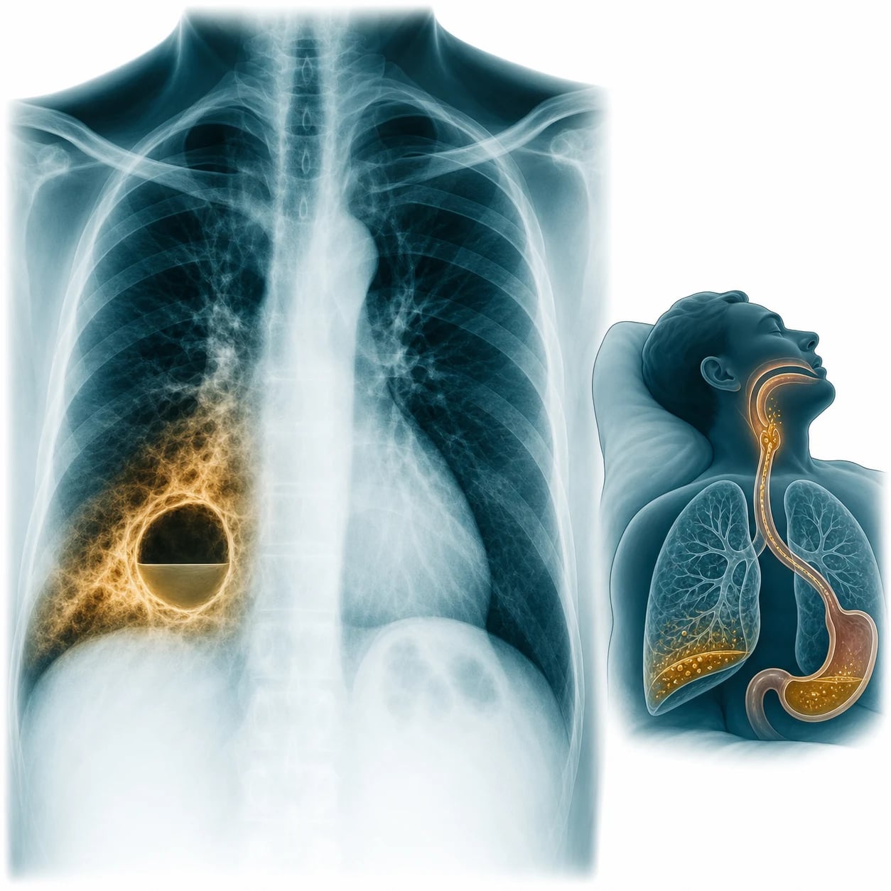
Pathophysiology — the two-hit mechanism
The aspiration pneumonia follows a two-hit mechanism. The first hit is the chemical pneumonitis — the aspirated gastric acid (pH below 2.5) injures the alveolar-capillary membrane directly, producing a rapid-onset inflammatory exudate, a hypoxaemia and a radiographic opacity that may be indistinguishable from the acute respiratory distress syndrome. The chemical pneumonitis is not initially infected (the antibiotics are not needed in the first 24 to 48 hours if the aspiration was witnessed and there is no sign of the infection), but the injured lung is highly susceptible to the bacterial colonisation. The second hit is the bacterial infection — the aspirated oropharyngeal secretions contain a rich anaerobic and aerobic flora (the Peptostreptococcus, the Bacteroides, the Fusobacterium, the Prevotella, and the aerobic S. aureus, the E. coli, the Klebsiella, the H. influenzae), which seed the chemically injured lung and produce the pneumonia. If the pneumonia is untreated or partially treated, the necrosis and the cavitation produce the lung abscess — a localised collection of the pus within the lung, with an air-fluid level on the imaging. [1]
Risk factors — who aspirates
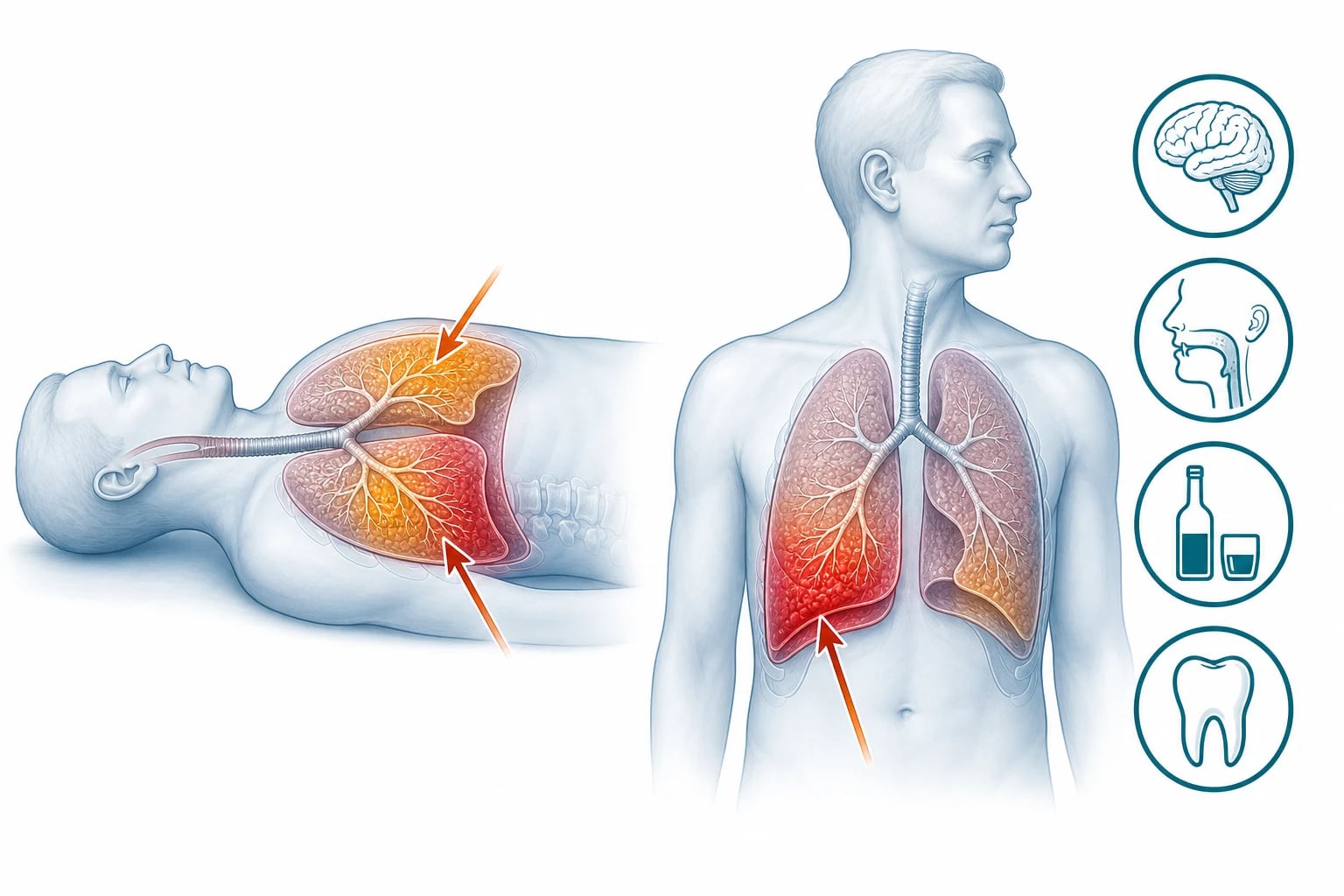
The aspiration requires the failure of the airway protection — the swallow mechanism and the cough reflex. The risk factors include the reduced consciousness (the stroke, the seizure, the alcohol intoxication, the drug overdose, the general anaesthesia, the dementia), the dysphagia (the neurological: the stroke, the Parkinson's, the motor neurone disease; the mechanical: the oesophageal stricture, the achalasia, the nasogastric tube, the tracheostomy), the gastro-oesophageal reflux disease, the poor dentition (the increased bacterial load in the oropharynx), the prolonged intubation, and the vomiting (the bulimia, the bowel obstruction, the post-operative). The identification of the risk factor is the first step to the prevention of the recurrence. [1]
Differential diagnosis — the cavitating lung lesion
The aspiration pneumonia and the lung abscess have a differential, and the history and the imaging resolve it. [1]
Aspiration pneumonia
- Risk factor: reduced consciousness, dysphagia
- Dependent lobe: posterior upper / lower
- Anaerobic organisms; foul-smelling sputum
- Clindamycin or co-amoxiclav; 2-4 weeks
Community-acquired pneumonia
- No aspiration risk factor
- Any lobe; the pneumococcus is the commonest
- No anaerobic cover needed (usually)
- 5-7 days; amoxicillin + macrolide
Tuberculosis
- Chronic; weight loss, night sweats, cough
- Upper-lobe predominant; cavitation
- Sputum AFB positive; the interferon-gamma
- Anti-TB therapy; isolate
Cavitating malignancy
- Chronic; weight loss, smoker
- A squamous cell carcinoma is the classic cavitator
- CT-guided biopsy for the diagnosis
- Oncology; not antibiotics
Investigations — the imaging and the cultures
The chest radiograph shows the consolidation (often in the dependent segments — the posterior segments of the upper lobes or the superior segments of the lower lobes, where the aspirated material settles by gravity), and, if the abscess has formed, a cavity with an air-fluid level. The CT scan of the chest is more sensitive and shows the cavity wall (the thick, irregular wall of the abscess, vs the thin wall of the bulla), the air-fluid level, the surrounding consolidation and any pleural involvement (the empyema). The sputum culture is sent for the Gram stain, the culture and the sensitivity — the anaerobic culture requires the special handling (the rapid transport, the anaerobic media) and may not grow in the routine culture. The blood cultures are taken before the antibiotics. The full blood count may show a leukocytosis; the CRP is raised; the albumin may be low (the chronic abscess, the poor nutrition). If the TB is suspected, the sputum AFB and the interferon-gamma release assay are sent. [1]
Management — the antibiotics covering the anaerobes
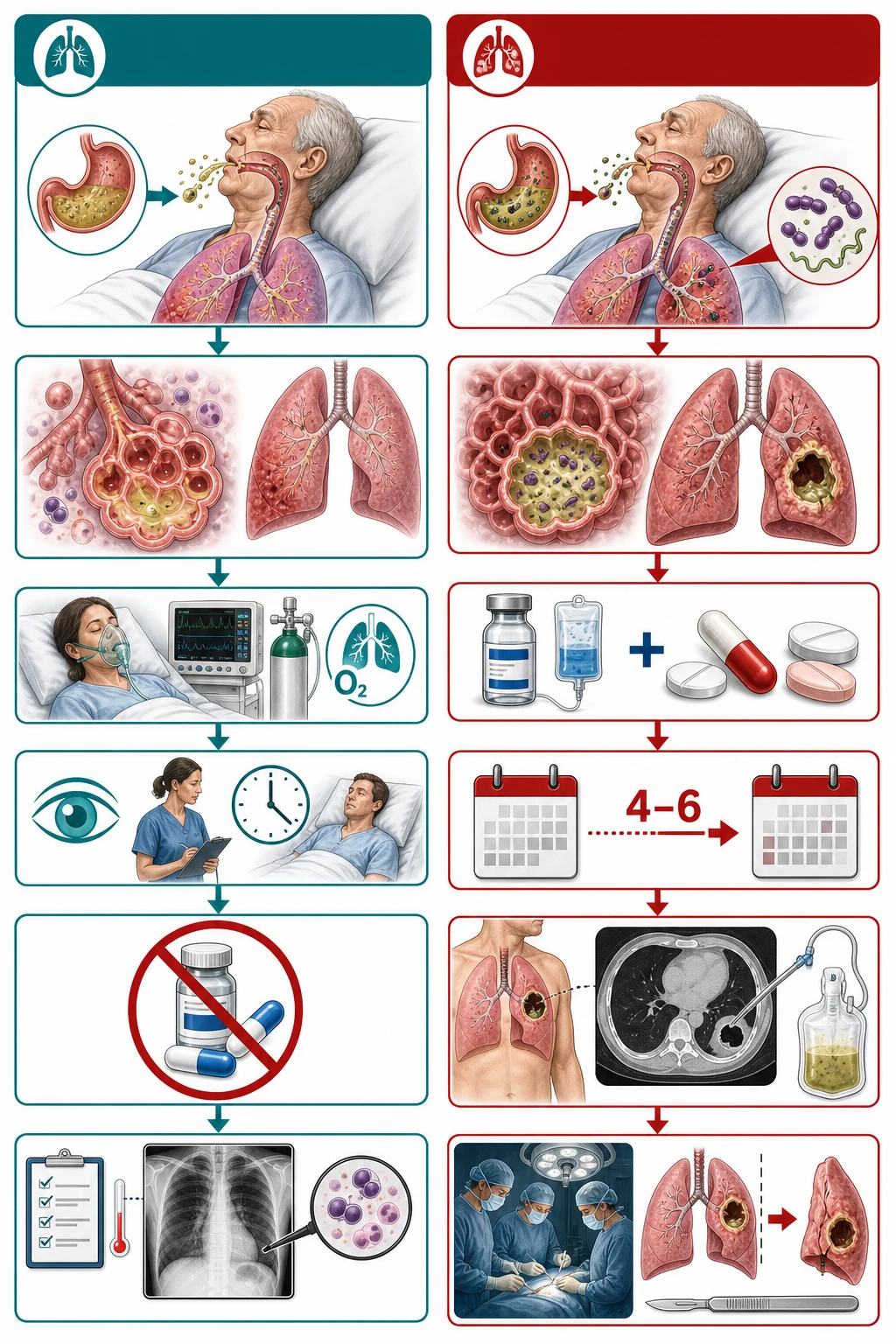
The antibiotic regimen for the aspiration pneumonia MUST cover the anaerobes — the standard community-acquired-pneumonia regimen (the amoxicillin and the macrolide) does NOT cover the anaerobes adequately. [1]
[1]The aspiration targets and the doses
The large lung abscess (over 6 cm) or the abscess that fails to respond to the antibiotics within 2 weeks may need the percutaneous drainage (the CT-guided catheter) or the surgical drainage (the VATS or the open decortication). The bronchoscopic drainage is an option for the central abscess. The underlying cause is treated throughout — the dysphagia is assessed by the speech and language therapy, the GERD is treated with the proton-pump inhibitor, the poor dentition is addressed, and the aspiration prevention (the head elevation, the swallowing technique, the NG feeding if needed) is implemented before the discharge.[2]
Complications and pitfalls
The complications are the empyema (the pleural-space extension — needs the chest drain), the bronchopleural fistula (the persistent air leak from the necrotic cavity into the pleural space — needs the surgical repair), the sepsis and the multi-organ failure, the haemorrhage (from the erosion of a vessel in the abscess wall — may be massive), the ARDS (from the chemical pneumonitis), and the recurrence (if the underlying cause is not addressed). The pitfalls are: not covering the anaerobes (using the standard CAP regimen without the clindamycin or the co-amoxiclav); treating for too short a duration (the standard 5-day CAP course is insufficient for the aspiration); not recognising the lung abscess on the chest radiograph (the air-fluid level may be subtle); not investigating and treating the underlying cause (the dysphagia, the alcohol, the seizure); and missing the TB or the cavitating malignancy behind the "abscess."[1]
Prognosis and disposition
The prognosis depends on the underlying cause and the severity — the uncomplicated aspiration pneumonia resolves with the 2-to-4-week antibiotic course; the lung abscess has a mortality of 5 to 10 per cent (higher in the immunocompromised and the elderly). The patient is admitted for the intravenous antibiotics; the improvement is monitored by the falling fever, the falling CRP and the shrinking cavity on the serial imaging. The switch to the oral therapy is made when the clinical response is confirmed. The follow-up includes the repeat CT at 4 to 6 weeks to confirm the resolution of the abscess, the speech-and-language assessment for the dysphagia, and the dental review. [1]
Special populations
The stroke patient with the dysphagia is the classic aspiration-risk patient — the swallow assessment and the modified diet reduce the recurrence. The alcoholic has the reduced consciousness, the poor dentition and the immunosuppression — the triple risk. The post-surgical patient aspirates during the recovery from the anaesthesia. The tracheostomised patient aspirates silently around the cuff. The dementia patient aspirates chronically and may present with the recurrent pneumonias. [1]
Aspiration pneumonitis vs aspiration pneumonia — the critical distinction
This is the highest-yield distinction in the aspiration syndrome, and the one Fellowship candidates most often blur. The two are not the same disease, they do not share a management, and conflating them produces two opposite errors: giving the antibiotics to a chemical pneumonitis that does not need them, and withholding the anaerobic cover from a genuine aspiration pneumonia that does.[3][4]
Aspiration pneumonitis — Mendelson syndrome
Aspiration pneumonitis is the acute chemical lung injury produced by the inhalation of the sterile, acidic gastric contents. Curtis Mendelson described it in 1946 in the obstetric patients (the original paper was an autopsy review of 66 cases of the aspiration under the anaesthesia), and the syndrome that bears his name is the archetype. The pathophysiology is a direct chemical burn of the alveolar-capillary membrane by the gastric acid. There are three determinants of the severity of the chemical injury: [1]
- The pH below 2.5 — the acid must be sufficiently caustic; the aspiration of the material with a pH above 2.5 produces little or no injury.
- The volume — the aspiration of more than 0.3 mL per kilogram of the gastric contents is sufficient to produce the syndrome.
- The particulate matter — the food particles and the bile worsen the injury and amplify the secondary inflammatory cascade. [1]
The clinical picture is the acute onset (within the hours) of the dyspnoea, the hypoxaemia, the tachypnoea, the diffuse bilateral radiographic infiltrates (in the dependent zones), the low-grade fever (the inflammatory, not the infectious, response) and the leukocytosis — in a patient with a witnessed or strongly suspected aspiration and a reduced consciousness. The syndrome peaks at 24 to 36 hours and then improves over 3 to 5 days if no bacterial infection supervenes.[3]
Aspiration pneumonia — the bacterial infection
Aspiration pneumonia is the bacterial infection of the lung parenchyma by the aspirated oropharyngeal flora. It may follow the chemical pneumonitis (the injured lung is the substrate for the colonisation) or it may occur without any preceding chemical injury — the small, unwitnessed, recurrent aspiration of the colonised oropharyngeal secretions in the patient with the dysphagia or the reduced consciousness. The clinical picture is the subacute onset (over the days) of the fever, the productive cough, the purulent and often foul-smelling sputum, the focal consolidation in the dependent segments, and the raised inflammatory markers — in a patient with a risk factor for the aspiration.[4][5]
The distinction in a table is the examination centrepiece: [1]
Pneumonitis (Mendelson)
- Witnessed aspiration of acidic gastric contents
- Acute onset over hours; diffuse bilateral infiltrates
- Sterile chemical injury; low-grade fever
- Antibiotics NOT routine; observe 24-48 h
- Corticosteroids NOT recommended
Pneumonia
- Often unwitnessed, recurrent aspiration
- Subacute onset over days; focal dependent consolidation
- Bacterial infection; foul-smelling sputum
- Antibiotics required; cover the anaerobes
- Duration 5-7 d (modern) up to 2-4 wk (severe)
The antibiotic and corticosteroid evidence — two classic traps
The two most-tested therapeutic questions are: do the prophylactic antibiotics help the witnessed aspiration, and do the corticosteroids attenuate the chemical injury? The answer to both is no, not routinely, and the routine use of either is the examination trap. [1]
DiBardino 2015 — the modern-trends review of the aspiration pneumonia
Journal of Critical Care
PMID 25129577
Key finding
A narrative review that re-examined the dogma of the aspiration pneumonia. It argues that the aspiration pneumonia is, microbiologically, a community-acquired-pneumonia equivalent, that the routine anaerobic cover is not needed for the hospital-acquired aspiration unless the risk factors for the anaerobic infection (the poor dentition, the chronic aspiration, the abscess, the foul sputum) are present, and that the antibiotic duration can mirror the CAP course (5 to 7 days) for the uncomplicated infection.
Practice change
The modern challenge to the reflexive 2-to-4-week clindamycin course — the uncomplicated aspiration pneumonia with the good response can be treated for 5 to 7 days; the necrotising or the cavitating disease still needs the prolonged course. The duration is now a clinical-response decision, not a fixed number.
The organisms in depth — the polymicrobial oropharyngeal flora
The aspiration pneumonia is polymicrobial — the oropharyngeal secretions carry a mixed anaerobic and aerobic load, and the infection reflects that mix. The anaerobes dominate, and they are the principal reason that the standard community-acquired-pneumonia regimen is inadequate.[3][5]
The organisms by the group, with the antibiotic implications: [1]
| Group | The organisms | The comment |
|---|---|---|
| Anaerobes (the majority) | Peptostreptococcus, Prevotella, Bacteroides, Fusobacterium, Porphyromonas | The mouth anaerobes; the foul-smelling sputum; the target of the clindamycin and the metronidazole |
| Gram-positive aerobes | Streptococcus pneumoniae, Staphylococcus aureus, Haemophilus influenzae | The usual CAP organisms, often co-aspirated |
| Gram-negative aerobes | Klebsiella, Escherichia coli, Pseudomonas | More in the nosocomial, the healthcare-associated and the alcoholic aspiration |
| The periodontal pathogens | Actinomyces, Eikenella, Capnocytophaga | The patient with the poor dentition and the gingival disease; the lung abscess with the actinomycosis |
The dependent lobes — where the aspirate settles
The aspirated material settles by the gravity in the dependent segments of the lung, and the position of the patient at the moment of the aspiration determines the location of the infiltrate: [1]
- The recumbent patient (asleep, intoxicated, post-ictal, post-anaesthetic) — the posterior segments of the upper lobes and the superior segments of the lower lobes are the most dependent. This is the classic aspiration distribution, and the one the Fellowship viva probes.
- The upright patient (the stroke with the swallow impairment while eating) — the basal segments of the lower lobes are the most dependent.
- The right lung is more often affected than the left — the right main bronchus is wider, shorter and more vertical, and the aspirate follows the path of the least resistance into the right lung. [1]
The ED workup of the suspected aspiration syndrome
Step 1 — Recognise the syndrome
The reduced consciousness, the dysphagia, the witnessed aspiration, the foul sputum, the dependent infiltrate. The recognition of the aspiration risk factor is the first and the most important step — without it, the anaerobic cover is missed.
Step 2 — The ABCDE assessment
The airway (the protective reflexes, the suction), the breathing (the hypoxaemia, the work of breathing, the need for the oxygen or the non-invasive ventilation), the circulation (the sepsis, the lactate, the need for the fluids and the vasopressors), the disability (the GCS and the cause of the reduced consciousness), the exposure (the full examination including the dentition).
Step 3 — The imaging
The chest radiograph for the infiltrate and the air-fluid level; the CT of the chest for the cavity, the wall, the effusion and the empyema. The CT is the definitive test for the abscess and the necrotising pneumonia.
Step 4 — The cultures and the bloods
The blood cultures before the antibiotics, the sputum for the Gram stain, the culture and the sensitivity (request the anaerobic handling — the rapid transport and the anaerobic media), the arterial blood gas, the full blood count, the CRP, the electrolytes, the lactate and the albumin.
Step 5 — The antibiotics
The clindamycin 600 mg IV q8h or the co-amoxiclav 1.2 g IV q8h; add the Gram-negative cover (the piperacillin-tazobactam or the meropenem) for the nosocomial or the healthcare-associated aspiration, or the known or suspected Pseudomonas.
Step 6 — The source control
For the large or the unresponsive abscess, the percutaneous catheter drainage (the CT-guided), the bronchoscopic drainage (the central abscess) or the surgical drainage (the failed percutaneous, the bronchopleural fistula, the massive haemoptysis).
Step 7 — The disposition
The ward for the stable patient, the HDU or the ICU for the septic or the respiratory-failure patient, and the early involvement of the respiratory and the infectious-diseases teams for the prolonged course and the drainage.
Step 8 — The underlying cause
The swallow assessment by the speech and language therapy, the dental review, the alcohol and the seizure management, the head elevation and the swallowing technique — the prevention of the recurrence is the real long-term treatment.
Lung abscess — the necrotic cavity
The lung abscess is the localised collection of the pus within the lung parenchyma, produced by the necrosis of the infected tissue and its central breakdown. It is the cavitating complication of the inadequately treated or the particularly virulent pneumonia, and the aspiration is its single commonest cause.[6][7]
The pathogenesis of the cavitation — the four stages
The abscess forms in the stages: [1]
- The consolidation — the pneumonia fills the alveoli with the inflammatory exudate.
- The necrosis — the intense inflammation and the bacterial toxins (the Staphylococcus aureus, the Klebsiella and the anaerobes produce the tissue-destructive enzymes, the leukocidins and the proteases) cause the coagulative necrosis of the lung tissue.
- The liquefaction and the bronchial communication — the necrotic centre liquefies; when the cavity communicates with a bronchus (the check-valve or the direct erosion), the air enters and the liquefied pus is partially expectorated, leaving the air-fluid level that is the radiographic signature of the abscess.
- The wall formation — the surrounding fibrosis and the granulation tissue form the thick, irregular wall that distinguishes the abscess from the thin-walled bulla or the cyst. [1]
The size thresholds and the drainage decision
The size of the abscess is the principal determinant of the need for the drainage in addition to the antibiotics. The thresholds and the rationale: [1]
The lung abscess thresholds
The drainage modalities — the ladder
The drainage options are chosen in the ascending order of the invasiveness, with the percutaneous catheter drainage as the contemporary first-line for the large or the unresponsive abscess.[8]
Percutaneous catheter
- CT-guided, the least invasive; the first-line drainage
- High clinical resolution over 80 per cent
- Risks: the pneumothorax, the bleeding, the bronchopleural fistula
- Lee 2022 meta-analysis supports the efficacy and the safety
Bronchoscopic drainage
- For the central abscess or the endobronchial obstruction
- Drains via the communicating bronchus
- Lower success for the peripheral abscess
- Allows the biopsy and the lavage for the diagnosis
Surgical drainage or resection
- VATS or the open decortication and the lobectomy
- For the failed percutaneous drainage
- For the bronchopleural fistula or the massive haemoptysis
- The highest morbidity; the last resort
Lee 2022 — percutaneous catheter drainage for the lung abscess
European Radiology
PMID 34327579
Key finding
A systematic review and meta-analysis of the percutaneous transthoracic catheter drainage for the lung abscess. The pooled clinical resolution rate was high (over 80 per cent), with the acceptable complication profile — the pneumothorax, the bleeding and the bronchopleural fistula being the principal events. The catheter drainage was effective for the large abscess (over 6 cm) and for the abscess unresponsive to the antibiotics.
Practice change
The percutaneous catheter drainage is the evidence-supported, first-line drainage modality for the large or the unresponsive lung abscess — it avoids the surgery in the majority and achieves the resolution with the acceptable risk. The threshold for the drainage is the abscess over 6 cm or the failure to respond to the antibiotics within 2 weeks.
Antibiotic duration — the modern evidence reconciled
The duration of the antibiotic therapy for the aspiration pneumonia has been contested, and the Fellow must hold both positions. The traditional teaching — still reflected in many guidelines and in much of the bedside practice — has been the 2-to-4-week course, on the rationale that the necrotising, the anaerobic infection is slower to sterilise than the pneumococcal pneumonia, and that the risk of the cavitation warrants the prolonged cover.[1]
The modern evidence and the contemporary reviews argue for a shorter course — 5 to 7 days — in the uncomplicated aspiration pneumonia with the good clinical response, on the principle that the aspiration pneumonia is, microbiologically, a CAP equivalent, and that the prolonged course does not improve the outcome but does increase the Clostridioides difficile risk and the antibiotic resistance.[5][3]
[1]Necrotising pneumonia and pulmonary gangrene — the severe end of the spectrum
The lung abscess sits at one end of the spectrum of the necrotising lung infection; the other end is the necrotising pneumonia and the rare, catastrophic pulmonary gangrene. The Fellow must recognise the spectrum, because the management escalates across it. [1]
- Necrotising pneumonia — the multifocal necrosis within a consolidated lobe, produced by the particularly virulent organisms (the community-acquired MRSA with the Panton-Valentine leukocidin, the Klebsiella pneumoniae, the Streptococcus pyogenes, the type-3 pneumococcus). It may progress to the multiple small abscesses, the pneumatoceles, and the bronchopleural fistula. The imaging shows the multifocal lucencies within the consolidation, without the discrete single cavity of the abscess.
- Pulmonary gangrene — the total necrosis of a lobe or a lung, with the sloughing of the necrotic tissue into the airway (the lung-within-a-lung appearance on the CT). It is rare, carries a very high mortality, and is managed by the surgical resection. [1]
The differential of the cavitating lung lesion — expanded
The cavitating lung lesion has a wide differential, and the history, the risk factors and the imaging resolve it. The expanded table covers the entities the Fellowship viva most often contrasts: [1]
Lung abscess
- Aspiration risk factor; acute-to-subacute onset
- Thick, irregular wall; the air-fluid level
- Surrounding consolidation; the foul sputum
- Anaerobic and the mixed cultures
- 4 to 6 weeks antibiotics; drain if over 6 cm or unresponsive
Cavitating tuberculosis
- Chronic; the weight loss, the night sweats, the cough
- Upper-lobe predominant; the thick wall
- Sputum AFB positive; the interferon-gamma release assay
- Anti-TB therapy; isolate the patient
- Risk factors: the TB exposure, the immunocompromise, the endemic area
Squamous cell carcinoma
- Chronic; the smoker, the weight loss
- The mural nodule; the eccentric wall
- No surrounding consolidation
- CT-guided biopsy for the diagnosis
- Resection or the oncology referral
Pneumatocele
- Post-infectious (PJP, staph) or the traumatic
- Thin, smooth wall
- No air-fluid level usually
- Conservative; resolves over the weeks to months
- May become secondarily infected
Infected bulla
- Underlying emphysema or the COPD
- Thin wall with the new air-fluid level
- Surrounding infiltrate if infected
- Antibiotics; drain if large
- May need the bullectomy if recurrent
Septic embolus
- Right-sided endocarditis; the IV drug use
- Multiple, peripheral, wedge-shaped lesions
- Cavitate over the days
- Blood cultures; the echocardiography
- Treat the source (the endocarditis)
Pitfalls — the exam and the bedside traps
[1] [1] [1] [1] [1]Complications — the full list
The complications of the aspiration syndrome and the lung abscess, from the common to the catastrophic: [1]
- The empyema — the pleural-space extension of the abscess; needs the chest drain and, often, the surgical decortication. The empyema complicating the abscess carries a higher mortality than the uncomplicated abscess.
- The bronchopleural fistula — the persistent air leak from the necrotic cavity into the pleural space; a surgical problem, often after the drainage or the necrotising pneumonia. The management is the chest drain, the bronchial-artery or the surgical closure, and the prolonged recovery.
- The massive haemoptysis — from the erosion of a vessel in the abscess wall; the airway protection (the bleeding-side-down positioning, the large-bore endotracheal tube, the selective lung isolation), the bronchial-artery embolisation and the surgical resection are the options.
- The ARDS — from the severe chemical pneumonitis or the necrotising pneumonia; the lung-protective ventilation and the supportive ICU care.
- The septic shock and the multi-organ failure — the Surviving Sepsis bundle (the antibiotics within 1 hour, the 30 mL per kg crystalloid, the early vasopressor for the MAP 65 mmHg or above).
- The recurrence — if the underlying cause is not addressed; the highest-risk patient is the chronic aspirator with the uncorrected dysphagia.
- The antibiotic-related complications — the Clostridioides difficile colitis from the prolonged clindamycin, the antibiotic resistance, and the line infection.
- The post-obstructive abscess — the abscess distal to a bronchial obstruction (the foreign body, the tumour, the bronchial stenosis); it will not resolve until the obstruction is relieved, and the bronchoscopy is both the diagnostic and the therapeutic. [1]
Prognosis — the determinants
The prognosis depends on the underlying cause, the severity and the host. The uncomplicated aspiration pneumonia resolves with the 5-to-7-day (modern) or the 2-to-4-week (traditional) antibiotic course. The lung abscess has a mortality of 5 to 10 per cent, higher in the immunocompromised, the elderly, the very large abscess, the bronchopleural fistula and the ARDS. The necrotising pneumonia and the pulmonary gangrene carry a substantially higher mortality and the longer recovery. The patient is admitted for the intravenous antibiotics; the improvement is monitored by the falling fever, the falling CRP and the shrinking cavity on the serial imaging. The follow-up includes the repeat CT at 4 to 6 weeks to confirm the resolution of the abscess, the speech-and-language assessment for the dysphagia, and the dental review. [1]
Evidence and regional guidelines
The contemporary framework is the BTS CAP guideline (the aspiration is a subset of the CAP with the anaerobic cover)[1] and the lung-abscess evidence (the prolonged antibiotics, the drainage).[2] The antibiotic choice (the clindamycin or the co-amoxiclav), the duration (2 to 4 weeks for the pneumonia, 4 to 6 weeks for the abscess) and the drainage thresholds follow the local respiratory and the infectious-diseases pathway.
ANZ practice note. The aspiration pneumonia and the lung abscess follow the BTS/eTG framework via the local respiratory pathway; the clindamycin 600 mg IV q8h or the co-amoxiclav 1.2 g IV q8h covers the anaerobes, the duration is 2 to 4 weeks for the pneumonia and 4 to 6 weeks for the abscess, and the large or the unresponsive abscess is drained percutaneously or surgically. [1]
Exam practice
SAQ — Witnessed aspiration in the post-ictal patient: pneumonitis versus pneumonia
10 minutes · 10 marks
A 72-year-old man with a history of ischaemic stroke and residual bulbar dysphagia is brought to the emergency department 90 minutes after a witnessed generalised tonic-clonic seizure at home. During the post-ictal period he vomited and was noted to aspirate gastric contents. He is now rousable (GCS 14), temperature 37.4 degrees C, RR 26, SpO2 90 per cent on room air improving to 97 per cent on 6 L via a simple face mask, BP 142/86, HR 104. He is not producing foul-smelling sputum. The white-cell count is 11.2, CRP 28. The chest radiograph shows a new infiltrate in the right lower lobe.
SAQ — The cavitating lesion in the alcoholic: lung abscess management and the drainage decision
10 minutes · 10 marks
A 58-year-old man with a 30-year history of heavy alcohol use, poor dentition and chronic dysphagia presents with a three-week history of fevers, drenching night sweats, an 8-kg weight loss and copious foul-smelling purulent sputum. On arrival he is cachectic, febrile 38.9 degrees C, RR 24, SpO2 92 per cent on room air, HR 110, BP 108/68. The white-cell count is 21.4 with a neutrophilia, CRP 210, albumin 26. The contrast-enhanced CT of the chest shows a 5.5 cm cavity with a thick wall and an air-fluid level in the posterior segment of the right upper lobe, with surrounding consolidation.
Exam pearls
- The aspiration pneumonia MUST cover the anaerobes — clindamycin 600 mg q8h or co-amoxiclav 1.2 g q8h.
- The two-hit mechanism: the chemical pneumonitis (the acid) + the bacterial infection (the anaerobes).
- The dependent segments: the posterior upper lobe or the superior lower lobe (the gravity).
- The foul-smelling sputum = the anaerobic infection.
- The lung abscess: an air-fluid level on the CT; 4-6 weeks of antibiotics; drain if large or unresponsive.
- The standard 5-day CAP course is insufficient — the aspiration needs 2-4 weeks.
- Treat the underlying cause (the dysphagia, the alcohol, the seizure, the poor dentition).
- Pneumonitis vs pneumonia: the witnessed sterile aspiration is the pneumonitis (observe, no antibiotics); the unwitnessed recurrent aspiration is the pneumonia (cover the anaerobes).
- The pH below 2.5 and the volume above 0.3 mL per kg are the thresholds for the Mendelson chemical injury.
- The right lung is more often affected — the right main bronchus is wider, shorter and more vertical.
- The prophylactic antibiotics and the corticosteroids are NOT routine after the witnessed aspiration — both are examination traps.
- The modern duration for the uncomplicated aspiration pneumonia is 5 to 7 days; the abscess still needs 4 to 6 weeks.
- The percutaneous catheter drainage is the first-line drainage for the abscess over 6 cm or unresponsive at 2 weeks.
- The wall of the squamous-cell-carcinoma cavity is nodular and eccentric with a mural nodule and no surrounding consolidation — biopsy, do not just treat.
- The community-acquired MRSA (the Panton-Valentine leukocidin) and the Klebsiella are the necrotising organisms — cover them when the course is severe or rapidly progressive.
- The BAL amylase is the silent-aspiration biomarker in the ventilated patient.
- The IV-to-oral switch: afebrile 48 to 72 hours, clinically improved, haemodynamically stable, tolerating the oral intake — do not wait for the radiographic resolution. [1]
Red flags
[1]References
- [1]Lim WS, Baudouin SV, George RC, et al. BTS guidelines for the management of community acquired pneumonia in adults: update 2009 Thorax, 2009.PMID 19783532
- [2]Marra A, Haverich A, Wiebe K. [Management of Lung Abscess] Zentralbl Chir, 2015.PMID 26351764
- [3]Marik PE Aspiration pneumonitis and aspiration pneumonia N Engl J Med, 2001.PMID 11228282
- [4]Marik PE Aspiration pneumonia: mixing apples with oranges and tangerines Crit Care Med, 2004.PMID 15190985
- [5]DiBardino DM, Wunderink RG Aspiration pneumonia: a review of modern trends J Crit Care, 2015.PMID 25129577
- [6]Kuhajda I, Zarogoulidis K, Tsirgogianni K, Tsavlis D, et al. Lung abscess-etiology, diagnostic and treatment options Ann Transl Med, 2015.PMID 26366400
- [7]Sperling S, Dahl VN, Floe A Lung abscess: an update on the current knowledge and call for future investigations Curr Opin Pulm Med, 2024.PMID 38411181
- [8]Lee JH, Hong H, Tamburrini M, Park CM Percutaneous transthoracic catheter drainage for lung abscess: a systematic review and meta-analysis Eur Radiol, 2022.PMID 34327579