EM · Pleural effusion
Pleural effusion (the emergency department workup and the Light criteria)
Also known as Pleural fluid · Water on the lung · Parapneumonic effusion · Empyema
Pleural effusion — the transudate-versus-exudate distinction by the Light criteria (the pleural fluid to serum protein ratio over 0.5, the LDH ratio over 0.6, the pleural fluid LDH over two-thirds the upper limit of normal), the causes (the heart failure, the pneumonia, the malignancy, the PE), the diagnostic approach (the chest radiograph, the ultrasound, the pleural fluid analysis), the management (the treat-the-cause, the therapeutic drainage, the chest drain for the empyema), and the parapneumonic effusion and the empyema. ACEM-primary, globally tagged.
On this page & tools
Your progress
Saved locally on this device.
Target exams
Red flags
A pleural effusion is an accumulation of fluid in the pleural space, and it is a common finding in the emergency department — the dyspnoea, the dullness to the percussion and the blunted costophrenic angle on the chest radiograph. The Fellowship candidate's task is to classify the fluid as a transudate or an exudate (using the Light criteria), because the classification narrows the differential and guides the management: a transudate is a systemic problem (the heart failure, the cirrhosis, the nephrotic) managed by the systemic treatment; an exudate is a local problem (the infection, the malignancy, the PE) that may need the drainage and the specific investigation.[1][2]
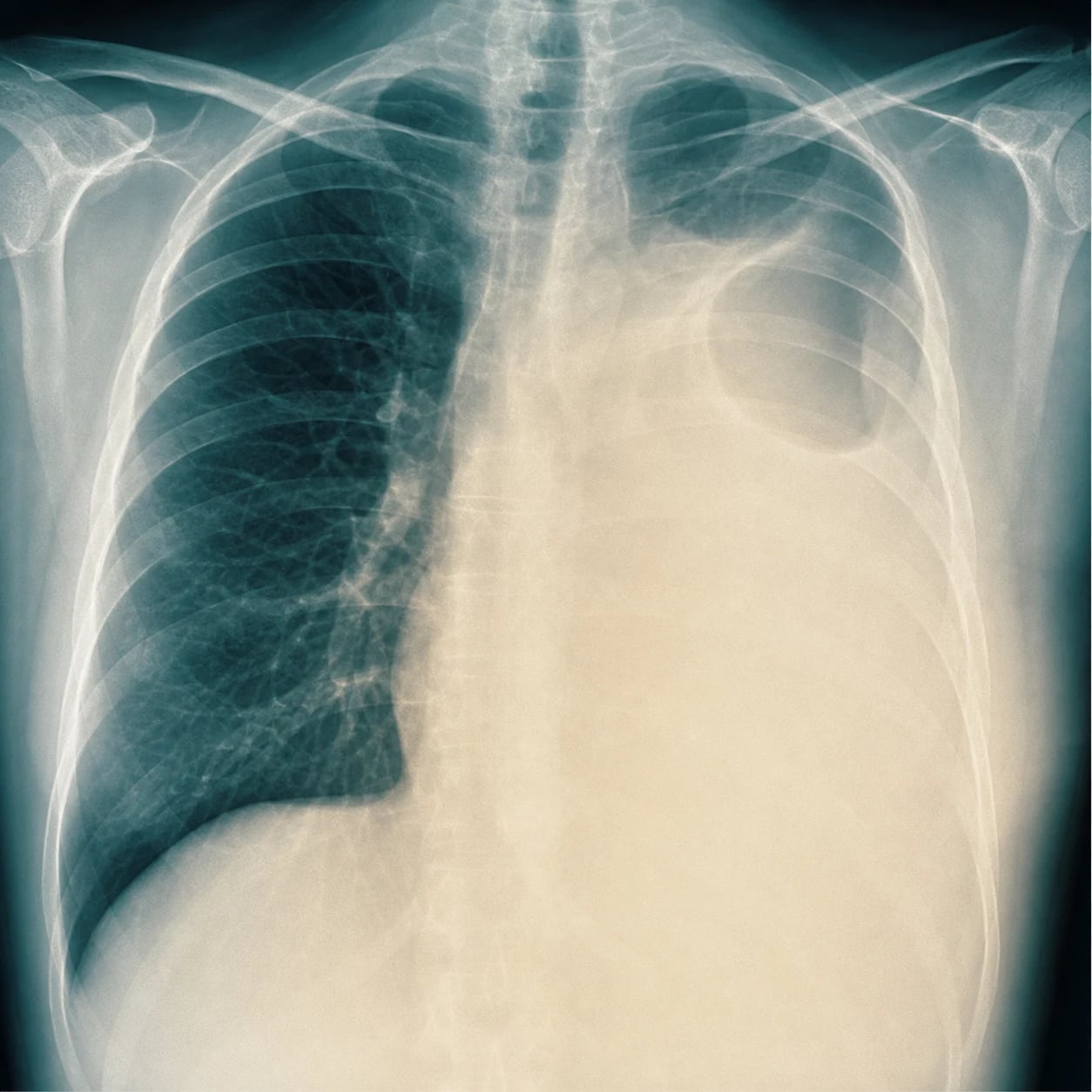
Pathophysiology — the fluid balance
The pleural space normally contains a small volume of fluid (5 to 10 mL) that lubricates the pleural surfaces. The fluid is produced by the parietal pleura and absorbed by the lymphatics, and the balance is maintained. The effusion forms when the production exceeds the absorption — from an increased hydrostatic pressure (the heart failure), a reduced oncotic pressure (the hypoalbuminaemia, the nephrotic), an increased capillary permeability (the infection, the inflammation, the malignancy) or a lymphatic obstruction (the malignancy). The transudate is a "water leak" from the systemic problem; the exudate is a "local inflammation." [1]
The Light criteria — the transudate and the exudate
The Light criteria separate the transudate from the exudate using the simultaneous measurement of the pleural fluid and the serum protein and the LDH (the lactate dehydrogenase). The three criteria, of which any one classifies the fluid as an exudate: [1]
- The pleural fluid-to-serum protein ratio over 0.5 (the fluid/serum protein ratio)
- The pleural fluid-to-serum LDH ratio over 0.6
- The pleural fluid LDH over two-thirds (2/3) of the upper limit of the normal serum LDH [1]
If none of the three is met, the fluid is a transudate. The Light criteria have a sensitivity of about 98 per cent and a specificity of about 80 per cent — they are highly sensitive (they rarely miss an exudate) but may misclassify about 20 per cent of the transudates as exudates (the "pseudoexudate" — the heart failure patient on the diuretics whose serum protein is lowered by the diuresis, making the pleural fluid-to-serum ratio falsely high).[3] The serum-to-pleural-fluid albumin gradient (the serum albumin minus the pleural-fluid albumin) resolves the pseudoexudate: a gradient over 1.2 g per dL reclassifies an apparent exudate as a transudate. The NT-proBNP on the pleural fluid (over 1,500 pg per mL) is another sensitive marker of a heart-failure effusion. The Light criteria were designed for the binocular classification; the modern approach pairs them with the clinical context — a bilateral, symmetric effusion in a patient with the heart failure is almost always a transudate regardless of the borderline ratios, and a unilateral effusion in a smoker with the weight loss is a malignancy until proven otherwise.
The alternate criteria and the bedside shortcuts
When the serum values are unavailable, the single-test Heffner criteria (pleural fluid protein over 2.9 g per dL, LDH over two-thirds ULN, or cholesterol over 45 mg per dL) classify most effusions with a comparable accuracy. The pleural-fluid cholesterol (over 45 to 60 mg per dL) and the pleural-fluid LDH alone (over two-thirds ULN) are the most useful single markers. For the candidate: always send the simultaneous serum protein, LDH, and albumin with the pleural sample — the criteria need the paired serum values. [1]
Differential diagnosis — the causes by type
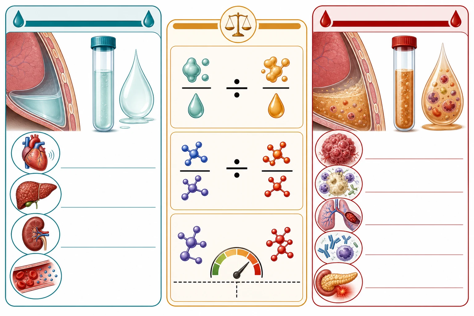
The causes are grouped by the transudate and the exudate. [1]
Transudate (systemic)
- Heart failure (the commonest — bilateral, symmetric)
- Cirrhosis (the hepatic hydrothorax — right-sided)
- Nephrotic syndrome (hypoalbuminaemia)
- Constrictive pericarditis, hypothyroidism, peritoneal dialysis
- Treat the systemic cause; drain only if symptomatic
Exudate (local)
- Bacterial pneumonia (the parapneumonic effusion)
- Malignancy (the lung, the mesothelioma, the metastasis)
- Pulmonary embolism (a common cause of an exudate)
- Tuberculosis, autoimmune (the lupus, the rheumatoid)
- Drain if symptomatic; the fluid analysis guides the diagnosis
Empyema
- A parapneumonic effusion with a positive culture or a pH below 7.2
- Needs the chest drain and the prolonged antibiotics
- May need the surgical drainage if loculated
- The pH, the glucose and the LDH guide the drainage decision
Haemothorax
- A haemorrhagic effusion from a trauma, a malignancy or a PE
- A pleural fluid haematocrit over 50 per cent of the serum haematocrit
- A chest drain; the output volume and the rate determine the thoracotomy
- Cross-link to the chest-trauma topic
Clinical presentation — the bedside assessment
The patient with a pleural effusion presents with a dyspnoea (the commonest symptom, worsening as the effusion enlarges), a pleuritic chest pain (the inflamed pleura of the parapneumonic, the malignant, or the embolic effusion), a cough (usually dry), and the symptoms of the underlying cause (the orthopnoea and the peripheral oedema of the heart failure; the fever and the productive sputum of the pneumonia; the weight loss and the haemoptysis of the malignancy; the leg swelling and the pleuritic pain of the PE). A large effusion may cause a type 1 respiratory failure from the lung compression and the V/Q mismatch. The clinical signs are the classic triad: the decreased breath sounds, the stony-dull percussion note, and the reduced vocal resonance and fremitus over the effusion; the trachea and the apex beat deviate AWAY from a large effusion (a push effect), and the reduced chest-wall expansion on the affected side completes the picture. [1]
The examination findings by the effusion size
The findings scale with the volume. A small effusion (under 500 mL) may be undetectable clinically and seen only on the imaging. A moderate effusion (500 to 1,500 mL) gives the dullness to the percussion up to the mid-scapula and the reduced breath sounds. A large effusion (over 1,500 mL) gives the stony dullness over the entire hemithorax, the absent breath sounds, the tracheal deviation away from the side, and the respiratory distress. The bilateral effusion suggests a systemic cause (the heart failure, the hypoalbuminaemia, the renal failure); the unilateral effusion suggests a local cause (the pneumonia, the malignancy, the PE, the TB). [1]
The ED approach — a stepwise workup
The emergency physician's task is to (1) recognise the effusion, (2) decide whether to drain it now, and (3) classify it as a transudate or an exudate to guide the cause and the disposition. The stepwise approach: [1]
Stabilise
Assess the ABCDE. Give oxygen for the hypoxia (the SpO₂ target 92 to 96 per cent). The large effusion with a respiratory failure needs the urgent therapeutic drainage.
History and the focused exam
Identify the systemic cause (the heart failure, the cirrhosis, the nephrotic, the malignancy, the pneumonia, the PE). Examine for the stony dullness, the reduced breath sounds, and the tracheal deviation.
The erect PA and lateral chest radiograph
The costophrenic angle blunts at ~200 mL on the PA film and at ~50 mL on the lateral film. Look for the meniscus sign, the mediastinal shift, the underlying consolidation, and the masses.
The bedside ultrasound
Confirm the anechoic fluid, measure the depth, identify the loculation/septation, mark the safe site for the thoracentesis, and assess the diaphragm and the liver/spleen to avoid the organ injury.
Decide: transudate pattern or the diagnostic tap?
A bilateral symmetric effusion with the clear heart-failure history needs no tap — treat the cause. An unexplained unilateral effusion, or any effusion where the exudate is suspected, needs the diagnostic thoracentesis.
The pleural fluid and the paired serum analysis
Send the fluid for the protein, the LDH, the glucose, the pH, the cell count, the Gram stain and culture, and the cytology. Send the simultaneous serum protein, LDH, and albumin for the Light criteria and the albumin gradient.
Classify and treat
Transudate: treat the systemic cause; drain only if the dyspnoea is severe. Exudate: identify the cause; drain if the pH <7.2, the glucose <3.3, or the culture is positive. The empyema and the haemothorax need the chest drain.
Disposition
Admit the large, symptomatic, or complicated effusion (the empyema, the malignancy, the respiratory failure). Discharge the small, asymptomatic transudative effusion with the systemic treatment and the follow-up.
Imaging by modality — what each shows
Chest radiograph (PA + lateral)
- First-line — cheap, fast, available
- Blunting at ~200 mL (PA) / ~50 mL (lateral)
- Meniscus sign, mediastinal shift, underlying consolidation
- Lateral decubitus film: as little as 5 to 10 mL (subpulmonic effusion)
- Cannot tell transudate from exudate; cannot guide the tap
Bedside ultrasound
- The ED and the ICU workhorse — real-time, no radiation
- Anechoic = simple transudate; echogenic/septated/loculated = exudate, empyema, haemothorax
- Estimates the volume (the depth, the intercostal measurement)
- Marks the safe site and the depth for the thoracentesis — lowers the pneumothorax rate
- Detects the pleural nodule/thickening (the mesothelioma, the metastasis) and the trapped lung
Contrast CT chest
- Reserved for the complex, the loculated, or the unexplained effusion
- Characterises the pleural thickening, the nodularity, the mass, the loculation
- Detects the pulmonary embolism (the CT pulmonary angiogram)
- Guides the difficult drain placement and the biopsy
Point-of-care BNP/NT-proBNP
- A high serum or pleural NT-proBNP supports a heart-failure transudate
- Avoids the diagnostic tap in a typical heart-failure effusion
- Part of the "is it heart failure?" decision
The ultrasound appearance — anechoic, septated, loculated
The ultrasound appearance predicts the fluid type. An anechoic (black) effusion is a simple fluid — usually a transudate, easy to drain. An echogenic (grey, swirling, debris) effusion is an exudate — a haemothorax, an empyema, or a malignancy. Septations (the fibrin strands crossing the collection) and loculations (the fluid divided into separate pockets) indicate a parapneumonic effusion or an empyema that has organised — these need a chest drain rather than a needle tap, and often the intrapleural fibrinolytics. A pleural peel (a thick rind of solid pleural tissue) suggests a chronic empyema or a trapped lung. [1]
Investigations — the radiograph, the ultrasound and the fluid
The chest radiograph is the first investigation — the blunting of the the costophrenic angle appears at about 200 mL of the fluid; the larger effusions produce the opacification with the meniscus sign and the mediastinal shift away from the effusion (if the mediastinum shifts TOWARDS the effusion, suspect an atelectasis or a bronchial obstruction). The bedside ultrasound confirms the effusion, estimates the volume, identifies the loculation, and guides the thoracentesis (the diagnostic tap or the therapeutic drainage). The pleural fluid analysis is the key investigation of the exudate: the protein and the LDH (the Light criteria), the pH (below 7.2 suggests the empyema or the malignancy — needs the drainage), the glucose (low in the empyema, the rheumatoid, the malignancy), the cell count (the neutrophils suggest the bacterial; the lymphocytes the TB or the malignancy), the culture and the Gram stain, the cytology (the malignant cells), and the amylase (raised in the pancreatitis and the oesophageal rupture). [1]
The pleural fluid analysis — interpreting the exudate
Once the fluid is an exudate (by the Light criteria), the individual markers narrow the cause. The pH is measured on the heparinised sample sent on ice (a blood gas syringe) — never on the residual fluid in the collection bag, where the CO₂ escapes and the pH falsely rises. The glucose falls as the inflammation intensifies; the LDH rises with the pleural inflammation; the amylase marks the pancreatitis or the oesophageal rupture. [1]
pH <7.2 or glucose <3.3
- Complicated parapneumonic effusion or empyema
- Malignancy (the pH low in ~30 per cent of malignant effusions)
- Rheumatoid pleurisy (a very low glucose, often <1.6 mmol/L)
- Tuberculous pleurisy
- Action: drain it
Glucose <2.2 / pH <7.0
- The low-risk threshold for the empyema
- Empyema or a heavily inflamed parapneumonic effusion
- Loculated on the ultrasound
- Mandatory chest drain; the antibiotics alone fail
LDH >1,000 IU/L
- Marked pleural inflammation
- Empyema, rheumatoid, malignancy, paragonimiasis
- Higher LDH = more tissue destruction = more likely to need drainage or surgery
Neutrophil-predominant
- Acute bacterial infection (the parapneumonic)
- Pulmonary embolism (the infarction)
- Pancreatitis (the left-sided sympathetic effusion)
Lymphocyte-predominant (>50%)
- Tuberculous pleurisy
- Malignancy (the lymphoma, the metastatic carcinoma)
- Chylothorax
- After a coronary artery bypass (a chylothorax or a lymphocytic effusion)
Amylase raised
- Pancreatitis (a left-sided exudate)
- Oesophageal rupture (the Boerhaave — also a low pH, food particles)
- Malignancy (some tumours secrete the amylase)
Triglycerides >1.1 mmol/L
- Chylothorax — the thoracic duct injury (post-CABG, post-oesophagectomy, lymphoma)
- Milky fluid; lymphocyte-predominant
- Conservative (NPO, MCT diet) vs surgical ligation for the high output
Cytology positive
- Malignant effusion — the lung (commonest), the breast, the mesothelioma, the lymphoma
- Adenocarcinoma cells; the sensitivity rises with the repeat sampling (60–90 per cent by three taps)
- Pleurodesis for the recurrence; the oncology referral
The light's-criteria performance and the pitfalls
The Light criteria miss an exudate in only about 2 per cent (the high sensitivity) — but they over-call an exudate in about 25 per cent of the heart-failure effusions on the diuretics (the pseudoexudate).[3] The candidate must therefore always interpret the Light criteria with the clinical picture: a heart-failure patient with a borderline exudative ratio still has a heart-failure effusion, and the albumin gradient confirms it. The chylothorax and the urinothorax can be misclassified because they have unusual compositions — the chylothorax has a high triglyceride (a clue to the thoracic-duct injury), and the urinothorax has a low pleural-fluid creatinine-to-serum-creatinine ratio.
Management — the treat-the-cause and the drainage
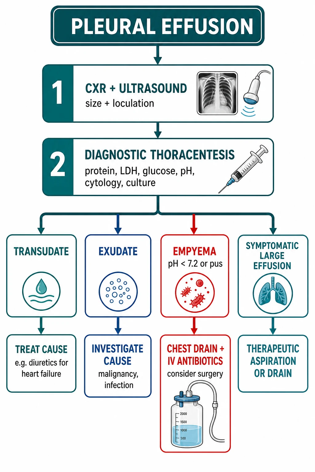
The management depends on the cause and the type. [1]
[1]The Light criteria and the effusion thresholds
The parapneumonic effusion and the empyema
The parapneumonic effusion is the pleural fluid associated with a bacterial pneumonia, and it ranges from the simple (uncomplicated, resolves with the antibiotics) through the complicated (needs the drainage) to the empyema (the frank pus — needs the chest drain and the prolonged antibiotics). The pleural fluid pH and the glucose are the key decision-makers: a pH below 7.2, a glucose below 3.3 mmol per litre, or a positive culture mandate the drainage. The empyema may be loculated (needing the intrapleural fibrinolytics — the tPA and the DNase — or the surgical VATS decortication). The antibiotic regimen covers the anaerobes and the Gram-negatives in addition to the pneumococcus: the co-amoxiclav 1.2 g intravenously or the ceftriaxone 2 g plus metronidazole 500 mg.[2]
The parapneumonic effusion — the three stages
The parapneumonic effusion evolves through three stages, and the stage dictates the management.[6]
Stage 1 — Simple (uncomplicated)
- Thin, free-flowing, sterile fluid
- pH >7.2, glucose >3.3, LDH <1,000, Gram stain negative
- Resolves with the antibiotics for the pneumonia alone
- No chest drain
Stage 2 — Complicated parapneumonic
- Fibrin deposition, early loculation
- pH <7.2, glucose <3.3, LDH >1,000, Gram stain may be positive
- Will progress to the empyema if not drained
- Chest drain + antibiotics; add the tPA + DNase if loculated
Stage 3 — Empyema
- Frank pus in the pleural space, organised loculations, a pleural peel
- Bacteria in the sediment, a positive culture, the pH low
- The chest drain (large-bore for the thick pus) + the prolonged antibiotics (4–6 weeks)
- The intrapleural tPA + DNase; the VATS decortication for the failure or the pleural peel
Thoracentesis — the diagnostic and the therapeutic tap
The thoracentesis (the pleural tap) is both a diagnostic and a therapeutic procedure. The diagnostic thoracentesis is performed when the cause is unclear (an unexplained exudative effusion) — 20 to 50 mL is aspirated and sent for the full panel. The therapeutic thoracentesis is performed when the effusion causes the dyspnoea — the fluid is drained to relieve the breathlessness, and the volume is limited to 1.5 litres per session (or stopped earlier for the chest tightness, the cough, or the chest pain) to prevent the re-expansion pulmonary oedema. [1]
The technique — needle above the rib, the 8th ICS
The patient sits upright, leaning forward over a bedside table (to open the intercostal spaces and the posterior access). The site is chosen under the ultrasound guidance — typically the 8th intercostal space in the posterior mid-scapular line for a moderate effusion, or one to two rib spaces below the upper level of the effusion (the dullness). The needle is inserted immediately above (superior to) the rib to avoid the neurovascular bundle (the intercostal vein, artery, and nerve) that runs in the costal groove below each rib. The local anaesthesia is the 1 per cent lidocaine up to 3 mg per kilogram (with the adrenaline for the non-end-artery sites), infiltrated from the skin down to the parietal pleura. The needle is advanced with the syringe attached, aspirating continuously, until the fluid is drawn — then the needle is withdrawn slightly and a larger-bore cannula or a catheter is threaded over a guidewire (the Seldinger technique). A small-bore (8 to 14 Fr) catheter is preferred for the pleural effusion (less painful than the large-bore drain, and adequate for the fluid); the large-bore drain (24 to 32 Fr) is reserved for the haemothorax and the empyema. [1]
The Seldinger technique — the stepwise
Position and prep
Sit the patient upright, leaning forward. Sterile field, skin prep, drapes. Confirm the site and depth on the ultrasound.
Local anaesthesia
Infiltrate 1 per cent lidocaine (up to 3 mg/kg) from the skin to the parietal pleura, raising a wheal, aspirating before each injection to avoid the intravascular injection.
Diagnostic aspirate
Advance a 21-gauge needle attached to a syringe, aspirating continuously, until the fluid is drawn. Aspirate 20 to 50 mL for the diagnostic panel (protein, LDH, glucose, pH, cell count, culture, cytology).
Guidewire
Pass the guidewire through the needle into the pleural space. Remove the needle, leaving the guidewire in place.
Dilate and cannulate
Make a small skin nick, dilate the tract over the guidewire, then pass the 8 to 14 Fr pigtail catheter over the guidewire into the pleural space.
Connect and drain
Remove the guidewire and the inner stiffener. Connect the catheter to a three-way tap and a drainage bag. Drain slowly — stop at 1.5 L or for any chest tightness/cough.
Secure and document
Suture the catheter in place, apply a dressing, and document the volume drained and the fluid appearance. Send the samples with the paired serum.
Post-procedure
A routine post-tap chest radiograph is NOT required after an uncomplicated ultrasound-guided tap; request one only for the chest pain, the dyspnoea, or the suspected pneumothorax.
The contraindications and the coagulopathy
There is no absolute contraindication to a diagnostic thoracentesis — even a coagulopathic patient can have a tap if the benefit outweighs the risk. The relative contraindications are the severe coagulopathy (the INR over 1.5 to 2.0, the platelet count below 50, the recent thrombolysis or anticoagulation), the overlying skin infection (the cellulitis — choose another site), and the uncooperative patient (the risk of the organ injury). The BTS advises that the routine correction of the coagulopathy before a tap is not necessary for the diagnostic aspiration, but a therapeutic tap or a chest drain in a coagulopathic patient should be discussed with the haematology. The pneumothorax rate after an ultrasound-guided tap is 2 to 6 per cent (much lower than the "blind" tap), and the routine post-tap chest radiograph is no longer required.[7]
The chest drain — the technique and the care
The chest drain for the pleural effusion (the tube thoracostomy) is placed at the safe triangle (the fourth or fifth intercostal space, the anterior axillary line), under the local anaesthesia (lidocaine 1 per cent up to 3 mg per kilogram), and connected to the underwater seal. The drain is monitored for the output, the swing (the tidalling) and the bubbling (if an air leak is present). The drain is removed when the output falls below 200 mL per day and the lung is re-expanded on the chest radiograph. The complications are the pain (the analgesia — the morphine 5 to 10 mg intravenously), the infection, the bleeding, the subcutaneous emphysema, and the re-expansion pulmonary oedema if drained too fast.[1]
The evidence — the intrapleural fibrinolytic trials
The management of the loculated empyema rests on two landmark multicentre randomised trials. The MIST1 trial (the first Multicentre Intrapleural Sepsis Trial) tested the intrapleural streptokinase against the placebo and found no benefit — the streptokinase did not reduce the mortality, the surgery, or the length of stay.[4] The lesson was that the single-agent streptokinase is ineffective. The MIST2 trial then tested the dual intrapleural therapy — the tissue plasminogen activator (tPA) plus the DNase — and found a significant improvement in the fluid drainage, the surgery rate, and the length of stay, because the tPA breaks the fibrin strands (the loculations) and the DNase degrades the neutrophil extracellular DNA (the viscosity).[5] The current BTS-endorsed regimen for the loculated empyema is the tPA 10 mg plus the DNase 5 mg intrapleurally twice daily for three days, with the chest drain in situ, escalating to the VATS decortication if the drainage fails.[6]
MIST2 — Rahman et al, NEJM 2011
Population: 454 adults with the pleural infection (the complicated parapneumonic effusion or the empyema)
Key finding
The tPA + DNase combination improved the pleural fluid drainage (a 4.3-fold increase), reduced the referral for surgery at 3 months (4 per cent vs 16 per cent), and shortened the hospital stay.
Practice change
For the loculated empyema, use the intrapleural tPA + DNase together — the combination is synergistic; neither agent alone helps. Escalate to the VATS decortication if the drainage fails.
MIST1 — Maskell et al, NEJM 2005
Population: 454 adults with the pleural infection
Key finding
No difference in the mortality, the surgery rate, the radiographic outcome, or the length of stay. A trend towards more adverse events (the bleeding, the fever) with the streptokinase.
Practice change
The intrapleural streptokinase alone is NOT effective for the pleural infection — abandon it. The dual tPA + DNase (MIST2) replaced it.
Complications and pitfalls
The complications of the pleural effusion are the respiratory failure (the hypoxia from the lung compression), the infection (the empyema), the trapped lung (the pleural peel preventing the re-expansion), and the complications of the drainage (the pain, the infection, the bleeding, the re-expansion oedema). The pitfalls are: not classifying the fluid as a transudate or an exudate before the management; draining the heart-failure effusion unnecessarily (it recurs until the heart failure is treated); missing the empyema (not checking the pH); missing the malignancy (not sending the cytology); and not recognising the PE as a cause of the exudative effusion. [1]
Prognosis and disposition
The prognosis depends on the cause — the heart failure effusion resolves with the treatment; the parapneumonic resolves with the antibiotics and the drainage; the malignant effusion is palliative (the pleurodesis for the symptom control); the empyema has a mortality of 10 to 20 per cent and a prolonged recovery. The patient is admitted if the effusion is large, symptomatic, or complicated (the empyema, the malignancy); the small, asymptomatic, transudative effusion may be managed as an outpatient with the systemic treatment and the follow-up. [1]
Special populations
The heart failure patient is the commonest transudate; treat the heart failure and drain only if the dyspnoea is severe. The cancer patient with the malignant effusion needs the palliative drainage, the pleurodesis and the oncology. The pneumonia patient with the parapneumonic effusion needs the pH check and the drainage if complicated. The renal-failure patient on the dialysis may have a hydrothorax. The post-CABG patient may have a pleural effusion from the irritation or the chylothorax. [1]
Evidence and regional guidelines
The contemporary framework is the BTS pleural disease guideline[1] and the pleural-infection evidence (the intrapleural fibrinolytics, the VATS).[2] The Light criteria, the fluid-analysis strategy and the drainage thresholds are the global standard. The antibiotic choice and the pleurodesis protocol follow the local respiratory and the oncology pathway.
ANZ practice note. The Light criteria, the pleural fluid analysis (the pH, the glucose, the LDH, the protein, the cell count, the culture, the cytology), and the drainage thresholds (the pH below 7.2) follow the BTS framework via the local respiratory pathway; the parapneumonic effusion receives the co-amoxiclav or the ceftriaxone, the empyema receives the chest drain, and the malignant effusion receives the palliative drainage and the pleurodesis. [1]
Clinical pearls — the high-yield
[1] [1] [1] [1] [1] [1] [1] [1] [1] [1] [1]SAQs
SAQ — the unexplained unilateral pleural effusion
10 minutes · 10 marks
A 68-year-old male smoker presents with three weeks of progressive dyspnoea, a 6 kg weight loss and a right-sided pleuritic chest pain. The chest radiograph shows a large right pleural effusion with the mediastinal shift to the left. A diagnostic thoracentesis returns a lymphocyte-predominant fluid: the pleural protein 48 g/L (serum 70), the pleural LDH 410 IU/L (serum 210, ULN 250), the glucose 2.0 mmol/L, the pH 7.18.
SAQ — the complicated parapneumonic effusion needing a chest drain
10 minutes · 10 marks
A 55-year-old man with type 2 diabetes presents with a five-day fever, a productive cough and a worsening dyspnoea. He is septic (HR 118, BP 95/60, RR 28, SpO₂ 91 per cent on air) with a stony-dull right base. The bedside ultrasound shows a 6 cm loculated right parapneumonic collection. A diagnostic tap returns a thick yellow fluid: the pH 7.10, the glucose 1.8 mmol/L, the LDH 1,400 IU/L, the Gram stain showing Gram-positive diplococci.
Exam pearls
- The Light criteria: pleural fluid/serum protein >0.5 OR pleural fluid/serum LDH >0.6 OR pleural fluid LDH >2/3 ULN → exudate (any one).
- Transudate = heart failure, cirrhosis, nephrotic. Exudate = pneumonia, malignancy, PE, TB.
- A pleural fluid pH below 7.2 → empyema or malignancy → drain.
- A parapneumonic effusion with a positive culture or a low pH → chest drain.
- A unilateral effusion in a cancer patient → malignant until proven otherwise (cytology).
- The pseudoexudate: heart failure on diuretics — the albumin gradient (serum–fluid albumin >1.2 g/dL) resolves it.
- A haemothorax: pleural fluid haematocrit >50 per cent of the serum haematocrit.
- Blunted costophrenic angle on the PA chest radiograph needs ~200 mL; on the lateral film only ~50 mL.
- A mediastinum shifted TOWARDS an effusion = atelectasis or a proximal bronchial obstruction, NOT a simple effusion — escalate (CT, bronchoscopy).
- Insert the thoracentesis needle ABOVE the rib (the neurovascular bundle runs below it) — never below.
- A therapeutic thoracentesis: limit to 1.5 L per session (or stop for chest tightness/cough) to avoid re-expansion pulmonary oedema.
- Chylothorax = milky fluid, triglycerides >1.1 mmol/L, thoracic duct injury (post-surgery, malignancy); pseudochylothorax = cholesterol crystals, chronic (TB, rheumatoid).
- A post-CABG effusion: early (<1 month) is bloody, exudative; late (>1 month) is a sympathetic transudate.
- Hepatic hydrothorax is right-sided in ~70 per cent — a cirrhotic with a right pleural effusion and no cardiopulmonary disease.
- PE causes a small exudative effusion in ~40 per cent — send D-dimer/Wells when a dyspnoeic patient has a unilateral effusion of unclear cause.
- Empyema necessitatis: the empyema tracking through the chest wall — a fluctuant chest-wall mass over a loculated collection.
- Heerfordt's and Light's: a transudative effusion plus ascites in Meigs syndrome (ovarian fibroma) — resolves after tumour removal. [1]
Red flags
[1]References
- [1]MacDuff A, Arnold A, Harvey J, et al. Management of spontaneous pneumothorax: British Thoracic Society Pleural Disease Guideline 2010 Thorax, 2010.PMID 20696690
- [2]Bhatnagar M, Dua R, Bhatnagar A. Pleural Infection in Adults: Integrating Medical and Surgical Management for Optimal Outcomes Thorac Surg Clin, 2026.PMID 42336508
- [3]Porcel JM, Light RW. Pleural Fluid Analysis: Are Light's Criteria Still Relevant After Half a Century? Clin Chest Med, 2021.PMID 34774168
- [4]Maskell NA, Davies CWH, Nunn AJ, et al. U.K. Controlled trial of intrapleural streptokinase for pleural infection N Engl J Med, 2005.PMID 15745977
- [5]Rahman NM, Maskell NA, West A, et al. Intrapleural use of tissue plasminogen activator and DNase in pleural infection N Engl J Med, 2011.PMID 21830966
- [6]Psallidas I, Corcoran JP, Rahman NM. Management of parapneumonic effusions and empyema Semin Respir Crit Care Med, 2014.PMID 25463162
- [7]Gordon CE, Feller-Kopman D, Balk EM, et al. Pneumothorax following thoracentesis: a systematic review and meta-analysis Arch Intern Med, 2010.PMID 20177035