EM · Bradyarrhythmias
Bradyarrhythmias and atrioventricular block in the emergency department
Also known as Bradycardia · Heart block · Complete heart block · Atrioventricular block
Symptomatic bradycardia and atrioventricular block — the symptomatic-versus-incidental decision, the atrioventricular-block classification (first degree, Mobitz I and II, complete), the bradycardia algorithm (atropine 500 micrograms to a 3 mg maximum, then transcutaneous pacing, then an adrenaline or dopamine infusion, then transvenous pacing), the reversal of the precipitant, the atropine-resistant drug-overdose bradycardia (glucagon, high-dose insulin), and the inferior-versus-anterior-infarct distinction. ACEM-primary, globally tagged.
On this page & tools
Your progress
Saved locally on this device.
Target exams
Red flags
A bradyarrhythmia is managed in the emergency department by a single bedside decision — is the slow rate causing symptoms? An incidental bradycardia is observed and its cause sought; a symptomatic bradycardia is treated down a clear ladder from atropine through transcutaneous pacing to an infusion and a transvenous wire. The Fellowship candidate must recognise the atrioventricular-block pattern, run the ladder with the right doses, and never forget that bradycardia is a symptom with a precipitant — a drug, an infarct, an electrolyte, a temperature, a vagal trigger — that must be reversed throughout.[2]
Definition and the symptomatic decision
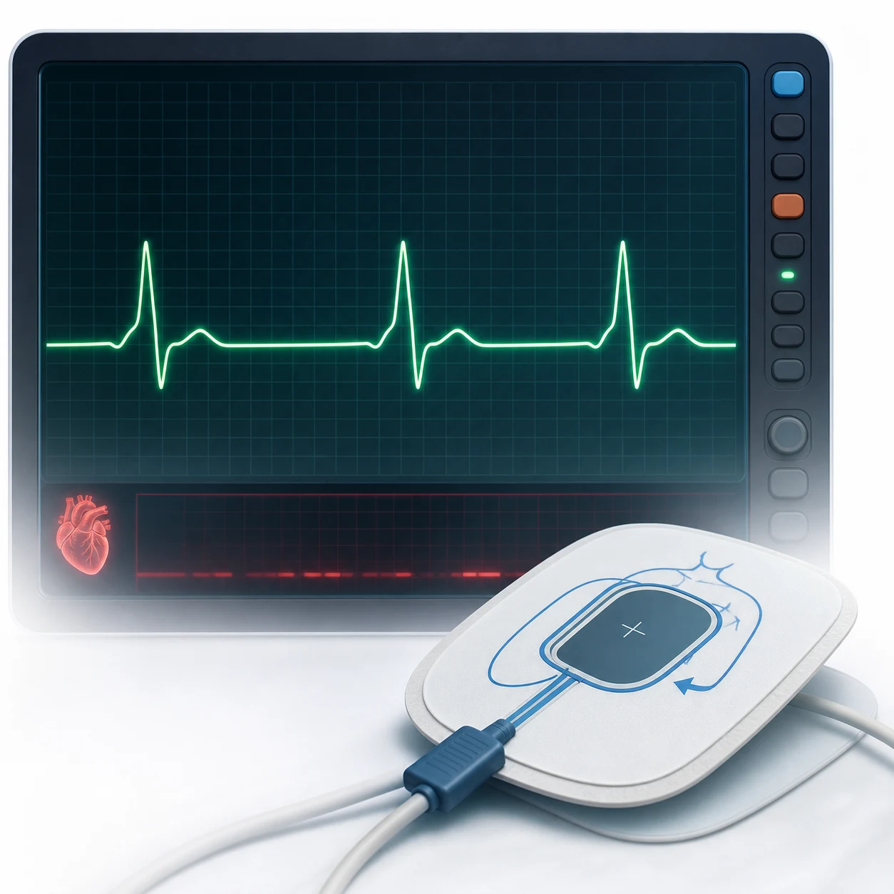
Bradycardia is a heart rate under 60 per minute, but the emergency decision is not the rate — it is whether the rate is causing symptoms. The symptomatic bradycardia produces syncope, presyncope, dizziness, hypotension, ischaemic chest pain, acute heart failure, or a pause-induced ventricular escape rhythm or arrhythmia. An asymptomatic bradycardia is investigated and its cause sought but is not a pacing emergency. This symptomatic-versus-incidental distinction is the first and the most important step. [1]
The atrioventricular-block classification
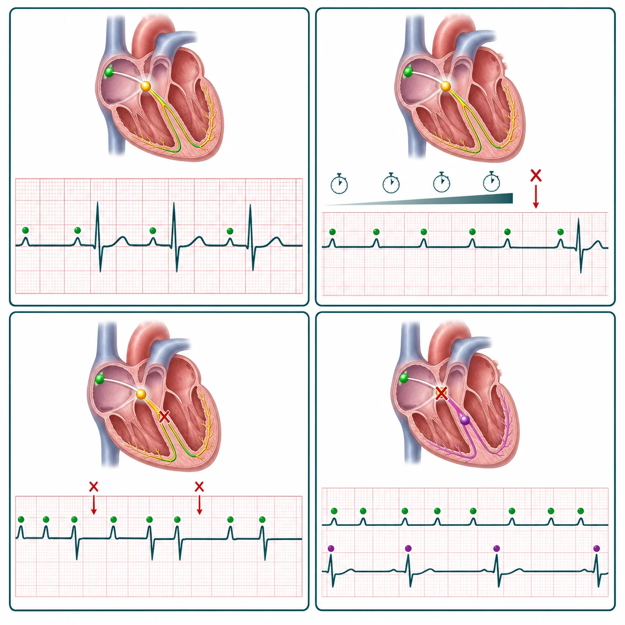
The atrioventricular block is classified on the ECG. First-degree block has a prolonged PR interval (over 200 ms) with every beat conducted. Second-degree Mobitz I (Wenckebach) has a progressive PR-lengthening until a beat is dropped, is usually at the AV node, and is generally benign. Second-degree Mobitz II drops beats without preceding PR-lengthening, is infranodal, and carries a high risk of progression to complete block and a permanent pacemaker. Third-degree (complete) heart block has the atria and the ventricles beating independently (atrioventricular dissociation), with a slow escape rhythm — narrow and atropine-responsive when the block is at the AV node (often an inferior infarct), wide and needing pacing when the block is infranodal (often an anterior infarct). [1]
First degree (PR >200 ms)
- Every P wave conducted; PR uniformly prolonged
- Often at the AV node; usually asymptomatic
- No specific treatment; observe and seek the cause
- May be normal in athletes and the elderly
Mobitz I (Wenckebach)
- Progressive PR prolongation until a beat drops
- Cycle then resets; the dropped beat has the shortest PR after it
- Block at the AV node; narrow QRS; usually benign
- Often transient (inferior MI, vagal, drug); rarely needs pacing
Mobitz II
- Sudden dropped beat WITHOUT preceding PR prolongation
- Infranodal (His-Purkinje); QRS often wide
- High risk of progression to complete block
- Needs a permanent pacemaker; temporary pacing if symptomatic
Third degree (complete)
- AV dissociation — atria and ventricles beat independently
- Escape rhythm: narrow-junctional (nodal) or wide-ventricular (infranodal)
- Narrow escape (inferior MI) often atropine-responsive
- Wide escape (anterior MI) needs urgent pacing
AV block — ECG and site
Pathophysiology — why the rate, and the infarct location, decide
The cardiac output is the product of the stroke volume and the heart rate, so a slow rate lowers the output unless the stroke volume can rise to compensate — and a fixed-rate elderly or failing ventricle cannot, so the patient hypoperfuses. This is why a young athlete tolerates a physiological bradycardia and an elderly patient collapses from the same rate. The location of the infarct determines the block: the right coronary artery supplies the atrioventricular node, so an inferior infarct produces a nodal block with a narrow-complex junctional escape that is often transient and atropine-responsive, whereas an anterior infarct that interrupts the conduction below the node produces a wide-complex ventricular escape that is high-risk and needs pacing. This inferior-versus-anterior distinction is examined because it changes the management. [1]
Causes and precipitants
A bradycardia almost always has a precipitant, and reversing it is itself the treatment. The causes are drugs (a beta-blocker, a rate-limiting calcium-channel blocker, digoxin, amiodarone, ivabradine), ischaemia (especially the inferior myocardial infarction), an increased vagal tone (vomiting, suctioning, a vagal manoeuvre, raised intracranial pressure, a vasovagal episode), hypothyroidism, hypothermia, hyperkalaemia, hypoxia, and the intrinsic conduction disease of sick sinus syndrome. A raised intracranial pressure produces the Cushing triad of bradycardia with hypertension and abnormal respirations, and is itself an emergency. [1]
Sinus bradycardia — causes organised by mechanism
Sinus bradycardia (a slow sinus rate with normal P-wave morphology and 1:1 conduction) has four broad mechanism groups, each reversible. Recognising the group at the bedside directs the antidote or the pacing decision. [1]
Increased vagal tone
- Vasovagal episode, nausea/vomiting, gagging/suctioning, Valsalva
- Pain, fear, tracheal suctioning, nasogastric insertion
- Raised intracranial pressure — the Cushing reflex
- Usually transient; resolves with the trigger or atropine
Drug-induced
- Beta-blockers, non-dihydropyridine CCBs (verapamil, diltiazem)
- Digoxin, amiodarone, ivabradine, adenosine (transient)
- Clonidine, opioids, donepezil, neostigmine
- Stop the drug; give the specific antidote (glucagon, HDI, DigFab)
Metabolic / endocrine
- Hypothyroidism (myxoedema) — profound bradycardia
- Hypothermia — Osborn J waves, slow rate
- Hyperkalaemia — wide QRS, sine-wave, bradyasystole
- Hypoxia and sleep apnoea; correct the metabolic state
Intrinsic / structural
- Sick sinus syndrome (sinus node dysfunction)
- Age-related fibrosis of the conduction system (Lev, Lenègre)
- Post-cardiac transplant (denervated node)
- Infiltrative disease (amyloid, sarcoid); needs pacing
Differential diagnosis
The slow rate has a differential, and the history, the ECG and the precipitant resolve it. [1]
Symptomatic AV block / bradycardia
- HR <60 with syncope/hypotension/ischaemia; AV block on ECG
- Atropine → transcutaneous pacing → infusion → transvenous
- Mobitz II / complete heart block → pacing
- Find and reverse the precipitant
Vasovagal (reflex) bradycardia
- A clear trigger; rapid spontaneous recovery
- Reflex vasodilation + bradycardia; reproducible
- No residual AV-block on ECG; reassurance
- Not a pacing disease
Drug toxicity (BB / CCB / digoxin)
- Overdose history; hypotension with the bradycardia
- Atropine often ineffective → glucagon, high-dose insulin
- CCB/BB: glucagon 5–10 mg then infusion; HDI; lipid
- Digoxin: digoxin-specific antibody
Raised-ICP / physiological
- Raised ICP: Cushing triad (bradycardia + hypertension)
- Athlete: asymptomatic, resolves on exertion
- Hypothyroid/hypothermic/hyperkalaemic bradycardia
- Treat the underlying state
Bedside assessment
Decide first whether the bradycardia is symptomatic, because that drives the treatment. Assess the blood pressure, the perfusion, the ischaemia and the heart failure; identify the atrioventricular-block type on the ECG; and seek the precipitant in the drug chart, the ECG, the electrolytes, the temperature and the thyroid. A focused examination also seeks the Cushing triad of a raised intracranial pressure (the bradycardia with a hypertension and an abnormal breathing pattern), the stigmata of hypothyroidism, and the signs of hypothermia — each of which changes the immediate management beyond the bradycardia ladder itself. [1]
Investigations and the targets
The 12-lead ECG (and a rhythm strip) defines the rhythm, the PR interval, the atrioventricular-block type, the QRS width of the escape rhythm, and any ischaemia or hyperkalaemia. The electrolytes (potassium, magnesium, calcium) are checked and corrected, the troponin identifies an ischaemic cause, the thyroid function and a digoxin level are sent where relevant. The PR interval threshold for first-degree block is 200 ms, and the symptomatic rate threshold is under 60 per minute. [1]
Immediate management — the bradycardia algorithm
Stabilise the airway and the breathing with oxygen, and run the ladder, escalating only as the symptoms demand. [1]
Stable versus symptomatic — the disposition fork
An asymptomatic bradycardia with a stable blood pressure and no ischaemia is admitted for observation and precipitant reversal (or discharged with outpatient follow-up if the cause is trivial and reversible, e.g. a vagal episode or a single dose of a rate-limiting drug that has been held). A symptomatic bradycardia — syncope, hypotension, ischaemia, heart failure, or a ventricular escape — is treated immediately down the ladder and admitted to a monitored bed. The fork is binary and it is the single most important decision.[3]
Assess the patient — symptomatic? (syncope, hypotension, ischaemia, heart failure, ventricular escape)
12-lead ECG + rhythm strip — classify the block; measure the PR interval and the QRS width
Send electrolytes (K, Mg, Ca), troponin, TSH, digoxin level; correct the reversible causes in parallel
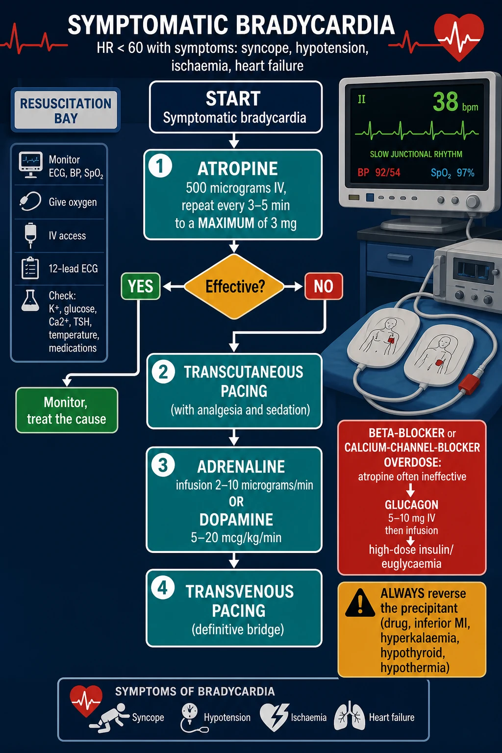
Atropine 500 mcg IV bolus, repeat every 3–5 min to a 3 mg maximum
If atropine fails or is inappropriate → transcutaneous pacing (with analgesia and sedation)
While pacing is set up → adrenaline 2–10 mcg/min (or dopamine 5–20 mcg/kg/min, or isoprenaline)
Transvenous pacing — the definitive temporary bridge
Call cardiology — permanent pacemaker decision; admit to a monitored bed
Pharmacology of the bradycardia agents
The agents of the bradycardia ladder each have a mechanism, a dose and a trap that the Fellowship candidate must know cold. [1]
Atropine
- Muscarinic antagonist — blocks vagal tone at the AV node
- 500 mcg IV, repeat q3–5 min to a 3 mg max
- Trap: a small dose (<500 mcg) paradoxically slows the rate
- Fails when the block is infranodal, drug-induced, or post-transplant
Adrenaline (epinephrine)
- β1 agonist — direct chronotropy and inotropy
- 2–10 mcg/min IV infusion; titrate to rate and BP
- Use while pacing is set up; also raises the blood pressure
- Caution in ischaemia — increases myocardial oxygen demand
Dopamine
- Dose-dependent: β1 at 5–10 mcg/kg/min; α at >10
- 5–20 mcg/kg/min IV; combine with dobutamine if low CO
- Useful when bradycardia coexists with hypotension
- Tachyarrhythmia, vasoconstriction at higher doses
Isoprenaline
- Pure β1/β2 agonist — strong chronotropy
- 1–4 mcg/min IV infusion (start low, titrate)
- Bridges to transvenous pacing; used in heart-block overdose
- β2 effect causes vasodilation — may worsen hypotension
Glucagon (BB/CCB overdose)
- Bypasses the β-receptor — activates adenylyl cyclase directly
- 5–10 mg IV bolus then 1–5 mg/hr infusion
- For BB or CCB overdose where atropine fails
- Nausea and vomiting are prominent — give an antiemetic
Transcutaneous pacing — technique and traps
Place the pacing pads in the anteroposterior position (or anterior–lateral if antero-posterior is impractical), set the demand or fixed rate to 60–80 per minute, and increase the current (output in mA) from threshold until electrical capture — a wide paced QRS following each pacing spike — and then mechanical capture — a palpable pulse matching the paced rate. The traps: failure to confirm mechanical capture (electrical without a pulse is not perfusion); failure to sedate and analgese (the pectoral contraction with each impulse is painful); and the under-sensing / over-sensing of the intrinsic rhythm in a demand mode, which can cause a non-captured patient to be labelled as "paced and stable." [1]
Pacing — the escalation
The bradycardia ladder and doses
The transcutaneous pacing is a bridge — its capture is confirmed electrically (a paced wide QRS following each pacing spike) and mechanically (a palpable pulse), and the patient is given analgesia and sedation because the muscular contraction of pacing is painful. The transvenous wire, placed under aseptic technique, is the definitive bridge to a permanent system. Reverse the precipitant throughout: stop the offending drug, reperfusion the infarct, correct the potassium, warm the hypothermic, treat the thyroid, relieve a raised intracranial pressure. [1]
Atrioventricular block in acute myocardial infarction — inferior versus anterior
The infarct location is the single most powerful predictor of the AV-block behaviour in a myocardial infarction, because the right coronary artery supplies the AV node (in 90% of people — the RCA gives the posterior descending artery in a right-dominant circulation and the AV-nodal artery) while the left anterior descending artery supplies the bundle branches and the fascicles via septal perforators. This anatomy explains why an inferior infarct produces a nodal, narrow-complex, atropine-responsive block, and an anterior infarct produces an infranodal, wide-complex, high-risk block. [1]
Inferior MI — nodal block
- RCA occlusion → AV-node ischaemia; narrow-complex junctional escape
- Usually Mobitz I or complete nodal block; often transient
- Frequently atropine-responsive; resolves with reperfusion (PCI)
- Temporary pacing if symptomatic; permanent rarely needed
- Poor prognosis only with RV infarct or cardiogenic shock
Anterior MI — infranodal block
- LAD occlusion → septal/His-Purkinje ischaemia; wide-complex escape
- Mobitz II, 2:1, or complete block; large infarct
- Atropine-resistant — the block is BELOW the node
- Needs urgent temporary then often permanent pacing
- High in-hospital mortality; reflects massive septal necrosis
Subtypes and special scenarios
The inferior-infarct AV block is nodal, narrow-complex and often transient and atropine-responsive, whereas the anterior-infarct block is wide-complex and needs pacing. Sick sinus syndrome is an intrinsic disease of the sinoatrial node with chronotropic incompetence and a permanent pacemaker. A Stokes-Adams attack is a sudden syncope from a transient high-grade block or an asystolic pause. The post-cardiac-transplant patient is denervated, so atropine is ineffective and a direct sympathomimetic or pacing is needed. [1]
The 2:1 and the high-grade atrioventricular blocks are managed as Mobitz II when the block is infranodal, because the risk of progression to a complete block is high and the escape rhythm is unreliable. The post-operative cardiac patient and the patient after a catheter ablation may develop an iatrogenic block that is often transient but is paced while it recovers. The bradycardia-tachycardia syndrome, a variant of sick sinus syndrome, alternates a pathologically slow rate with paroxysms of atrial fibrillation or flutter; here the slow node is paced before the fast rhythm is treated with an atrioventricular-node-blocking drug, because such a drug can precipitate an asystolic pause in the unprotected node. [1]
Permanent pacemaker indications
The permanent pacemaker is implanted for an irreversible conduction disease that produces symptoms or carries a high risk of asystole. The 2018 ACC/AHA/HRS bradycardia guideline and the ESC guidance converge on the following indications.[3][4]
Class I (definite)
- Symptomatic third-degree or high-grade AV block (any cause)
- Symptomatic Mobitz II (or 2:1 infranodal block)
- Symptomatic sinus-node dysfunction (sick sinus syndrome)
- Atrial fibrillation with symptomatic pauses >3 s
Class IIa (reasonable)
- Asymptomatic third-degree AV block (mean ventricular rate <40)
- Asymptomatic Mobitz II with wide QRS
- Sinus-node dysfunction with HR <40 unexplained
- Neurally-mediated syncope with documented cardioinhibitory response
Post-MI specific
- Persistent second- or third-degree AV block after anterior MI
- Transient second-/third-degree block + bundle-branch block after MI
- Permanent pacing NOT indicated for transient inferior-MI block that resolves
Not indicated
- First-degree AV block (asymptomatic)
- Asymptomatic Mobitz I at the AV node
- Reversible causes (drug, electrolyte, vagal, hypothyroid, hypothermia)
- Atrial fibrillation with a slow ventricular response from drugs
The drug-overdose bradycardia — antidote-directed therapy
In a beta-blocker, calcium-channel-blocker or digoxin overdose, atropine is often ineffective because the bradycardia is not vagally mediated — it is the direct pharmacological suppression of the node and the conduction tissue. The treatment is antidote-directed, and the bradycardia algorithm is a back-up, not the primary therapy. [1]
Beta-blocker overdose
- Bradycardia + hypotension + hypoglycaemia (esp. propranolol)
- Atropine often fails — the block is downstream of vagal tone
- Glucagon 5–10 mg IV then 1–5 mg/hr; high-dose insulin/euglycaemia
- Lipid emulsion and pacing for refractory cases
Calcium-channel-blocker overdose
- Bradycardia + hypotension + hyperglycaemia (the CCB fingerprint)
- Atropine fails; calcium chloride 1 g IV; glucagon; HDI
- High-dose insulin: 1 U/kg bolus then 0.5–1 U/kg/hr with glucose
- Lipid emulsion; ECMO / pacing in refractory shock
Digoxin toxicity
- Bradycardia, AV block, premature ventricular beats
- Visual disturbance (yellow halos), nausea, confusion
- Potassium — check FIRST (hyperkalaemia is a marker of toxicity)
- Digoxin-specific antibody (DigFab) — dose by the load or the level
Complications and pitfalls
An under-recognised pitfall is the slow atrial fibrillation that masks an underlying sick-sinus pause, which only declares itself on a longer rhythm strip or a continuous monitor — so a symptomatic pause warrants monitoring even when the index ECG looks benign. [1]
The complications are syncope with injury, asystole and sudden death, the ischaemia of a runaway paced rate, and the pain, the failure-to-capture and the infection of a transvenous lead. The pitfalls are the inverse of the algorithm: treating an asymptomatic incidental bradycardia; giving a small atropine dose; relying on atropine in a drug-toxicity or a denervated-transplant bradycardia; missing the inferior-infarct or the hyperkalaemia or the hypothyroid cause; delaying the transcutaneous pacing; and failing to sedate and analgesise the transcutaneously-paced patient. [1]
Prognosis and disposition
The prognosis turns on the cause: a reversible drug, electrolyte or ischaemic cause resolves once it is treated, whereas an intrinsic conduction disease (Mobitz II, a complete heart block, sick sinus syndrome) needs a permanent pacemaker. The symptomatic patient, the paced patient, the post-infarction patient and the drug-toxic patient are admitted to cardiology or a monitored bed; the precipitant is treated; and the pacing decision is made by cardiology. [1]
Special populations
The elderly carry the degenerative conduction disease and the polypharmacy that most often cause bradycardia, and they tolerate the atropine and the pacing less well, with a higher likelihood of a permanent system. The post-cardiac-transplant patient is denervated, so atropine is ineffective and a direct sympathomimetic or pacing is the response. The drug-overdose patient needs the antidote-directed therapy.[1] Pregnancy tolerates transcutaneous pacing, and the paediatric bradycardia is more often hypoxic or vagal than a primary conduction disease, so the response is oxygenation first.
Evidence and regional guidelines
The contemporary framework is the 2018 ACC/AHA/HRS Bradycardia and Cardiac Conduction Delay guideline and the advanced-life-support bradycardia algorithm.[3] The 2018 ESC syncope guideline addresses the pacing decision for the bradycardia-driven faint.[4] The beta-blocker and calcium-channel-blocker overdose evidence supports glucagon, high-dose insulin and lipid emulsion, consolidated in the 2017 St-Onge CCB-poisoning consensus and the 2020 Rotella beta-blocker-poisoning systematic review.[5][6][9] The BRASH syndrome clinical picture has been systematically scoped.[8] The approach to bradycardia in the injured patient — and the relative-bradycardia concept — has been reviewed.[2] The algorithm and the doses are global; the pacing pathway and the permanent-pacemaker criteria follow the ACC/AHA/HRS class framework.
2018 ACC/AHA/HRS Bradycardia Guideline
JACC / Circulation / Heart Rhythm (2019)
PMID 30412709
Society clinical-practice guideline
Population: Adults with bradycardia or conduction delay
Key finding
Codified the symptomatic bradycardia algorithm: atropine first-line, transcutaneous pacing, then an infusion (adrenaline or dopamine), then transvenous pacing. Stratified permanent-pacemaker indications by class. Recommended treating the reversible cause before committing to a permanent system.
2018 ESC Syncope Guideline
European Heart Journal (2018)
PMID 29562304
Society clinical-practice guideline
Population: Patients with syncope of suspected arrhythmic cause
Key finding
Established the risk-stratified evaluation of the bradycardic faint — prolonged monitoring, electrophysiology, and a pacemaker for the documented cardioinhibitory response. Distinguished neurally-mediated (benign) from intrinsic-node (paced) syncope.
St-Onge CCB-Poisoning Consensus
Critical Care Medicine (2017)
PMID 27749343
Expert consensus (modified Delphi)
Population: Adult calcium-channel-blocker poisoning
Key finding
Tiered the antidote ladder: calcium first, then high-dose insulin/euglycaemia, then catecholamines, then lipid emulsion; ECMO as rescue. Atropine endorsed only as a temporising measure, not definitive therapy.
Beta-blocker poisoning — systematic review
Clinical Toxicology (2020)
PMID 32310006
Systematic review
Population: Adult beta-blocker overdose
Key finding
Found low-quality evidence overall; glucagon and high-dose insulin the most-studied antidotes, with HDI increasingly first-line. Confirmed atropine is frequently ineffective and should not delay definitive antidote therapy.
BRASH syndrome — systematic scoping review
European Journal of Internal Medicine (2022)
PMID 35676108
Systematic scoping review
Population: Patients with bradycardia + renal failure + AV-blockade + shock + hyperkalaemia
Key finding
Defined BRASH as a self-perpetuating cycle; mortality high if the metabolic drivers (hyperkalaemia, acidosis) are not corrected. Pacing alone is insufficient — withdraw the offending drug and treat the electrolytes.
ANZ practice note. The symptomatic-bradycardia algorithm and the doses follow the ANZCOR/Resuscitation Council framework via local cardiology pathways; atropine 500 micrograms to a 3 mg maximum is the first-line escalation, transcutaneous pacing the bridge, and a beta-blocker or calcium-channel-blocker overdose receives glucagon and high-dose insulin/euglycaemia. [1]
Exam practice
SAQ — Complete heart block with syncope in an elderly man (inferior STEMI)
10 minutes · 10 marks
A 78-year-old man is brought to the emergency department by ambulance after a witnessed syncopal episode. He was sitting watching television when he turned pale, slumped, and lost consciousness for approximately 40 seconds with no seizure activity; he recovered quickly but remains lightheaded. On arrival he is drowsy but rousable (GCS 14), blood pressure 92/54 mmHg, heart rate 32 bpm. The cardiac monitor shows a slow, regular rhythm. The 12-lead ECG demonstrates complete atrioventricular dissociation — independent P waves at 90/min and a narrow-complex junctional escape at 32/min — with 2 mm ST elevation in leads II, III and aVF and reciprocal ST depression in I and aVL. SpO2 95 per cent on room air.
SAQ — Unstable symptomatic bradycardia from beta-blocker toxicity: running the ladder
10 minutes · 10 marks
A 58-year-old woman is brought to the resuscitation bay by ambulance after a deliberate self-poisoning approximately three hours ago with her own metoprolol — an estimated 50 tablets of 100 mg (5 g total). She is drowsy (GCS 13), profoundly bradycardic at 30 bpm, blood pressure 64/38 mmHg, RR 18, SpO2 92 per cent on room air, with cool peripheries and a capillary refill of 5 seconds. The ECG shows a slow junctional escape with a normal QRS. The bedside glucose is 3.2 mmol/L. Two 500-microgram doses of atropine have been given by the paramedics with no change in the heart rate or the blood pressure.
Exam pearls
- Symptomatic bradycardia → atropine 500 mcg to 3 mg → transcutaneous pacing → adrenaline 2–10 mcg/min or dopamine → transvenous pacing.
- Never a small atropine dose — give 500 mcg or more (a small dose slows the rate).
- Inferior-MI block is nodal, narrow-complex, atropine-responsive; anterior-MI block is wide-complex and needs pacing.
- Beta-blocker/calcium-channel-blocker overdose → glucagon and high-dose insulin; atropine is often ineffective.
- Mobitz II and complete heart block → pacing.
- Reverse the precipitant — the drug, the infarct, the potassium, the thyroid, the temperature.
- First-degree AV block = PR >200 ms with every beat conducted; usually asymptomatic, no treatment, observe.
- Mobitz I (Wenckebach) — progressive PR prolongation then a dropped beat; nodal, narrow QRS, benign, often transient, atropine-responsive; rarely needs pacing.
- Mobitz II — sudden dropped beat with NO preceding PR change; infranodal, often wide QRS, high risk of complete block; needs a permanent pacemaker.
- Complete (third-degree) heart block — AV dissociation; the escape is narrow (nodal, atropine-responsive) or wide (infranodal, needs pacing).
- 2:1 AV block cannot be classified on a single tracing — use atropine or a His-bundle study; treat an infranodal 2:1 as Mobitz II.
- Atropine dose: 500 mcg IV, repeat q3–5 min to a 3 mg max — a small dose (<500 mcg) paradoxically slows the rate via a central vagal effect.
- Transcutaneous pacing — confirm BOTH electrical (paced QRS) and mechanical (palpable pulse) capture; always sedate and analgese — it is painful.
- Adrenaline 2–10 mcg/min (or dopamine 5–20 mcg/kg/min, or isoprenaline) bridges to transvenous pacing; the transvenous wire is the definitive temporary bridge.
- Inferior-MI AV block — RCA supplies the AV node → Mobitz I, narrow escape, transient, atropine-responsive; permanent pacing rarely needed.
- Anterior-MI AV block — LAD supplies the septum/His-Purkinje → Mobitz II/complete, wide escape, atropine-resistant, needs pacing, high mortality.
- Cushing triad (bradycardia + hypertension + irregular respiration) = raised ICP — a pre-terminal sign; treat the ICP, not the rate.
- BRASH syndrome — Bradycardia + Renal failure + AV-blockade + Shock + Hyperkalaemia; treat the electrolytes and stop the drug, not pacing alone.
- Post-cardiac-transplant patient is denervated → atropine is ineffective; use a direct sympathomimetic (adrenaline/isoprenaline) or pacing.
- Bradycardia-tachycardia syndrome — pace the slow node FIRST, then rate-control the fast rhythm (avoid an asystolic pause from an AV-node-blocker in an unprotected node).
- Paediatric bradycardia is usually hypoxic, not a primary conduction disease — oxygenate first; atropine is second-line.
- Digoxin toxicity — check potassium first (hyperkalaemia is a marker); treat with digoxin-specific antibody (DigFab), not calcium (risk of "stone heart" is debated but HDI/calcium are not the antidote). [1]
Red flags
[1]References
- [1]Lashari BH, Naseer F, Bhan C. The Use of High-dose Insulin Infusion and Lipid Emulsion Therapy in Concurrent Beta-blocker and Calcium Channel Blocker Overdose Cureus, 2018.PMID 30648068
- [2]El-Menyar A, Furlan R, Asim M, et al. Scoping Review on True and Relative Bradycardia in Trauma: How to Approach Bradycardia in Traumatic Brain Injury J Cardiovasc Transl Res, 2026.PMID 42043702
- [3]Kusumoto FM, Schoenfeld MH, Barrett C, et al. 2018 ACC/AHA/HRS Guideline on the Evaluation and Management of Patients With Bradycardia and Cardiac Conduction Delay: A Report of the American College of Cardiology/American Heart Association Task Force on Clinical Practice Guidelines and the Heart Rhythm Society J Am Coll Cardiol, 2019.PMID 30412709
- [4]Brignole M, Moya A, de Lange FJ, et al. 2018 ESC Guidelines for the diagnosis and management of syncope Eur Heart J, 2018.PMID 29562304
- [5]St-Onge M, Anseeuw K, Cantrell FL, et al. Experts Consensus Recommendations for the Management of Calcium Channel Blocker Poisoning in Adults Crit Care Med, 2017.PMID 27749343
- [6]Engebretsen KM, Kaczmarek KM, Morgan J, et al. High-dose insulin therapy in beta-blocker and calcium channel-blocker poisoning Clin Toxicol (Phila), 2011.PMID 21563902
- [7]Graudins A, Lee HM, Druda D. Calcium channel antagonist and beta-blocker overdose: antidotes and adjunct therapies Br J Clin Pharmacol, 2016.PMID 26344579
- [8]Shah P, Gozun M, Keitoku K, et al. Clinical characteristics of BRASH syndrome: Systematic scoping review Eur J Intern Med, 2022.PMID 35676108
- [9]Rotella JA, Greene SL, Koutsogiannis Z, et al. Treatment for beta-blocker poisoning: a systematic review Clin Toxicol (Phila), 2020.PMID 32310006