EM · Tachyarrhythmias
Tachyarrhythmias in the emergency department
Also known as Tachycardia · SVT · Ventricular tachycardia · Atrial fibrillation
The ED tachyarrhythmias — the stability-first algorithm, the narrow-versus-broad-complex (QRS 120 ms) decision, the management of a regular narrow-complex SVT (vagal manoeuvres then adenosine 6-12-12 mg), a regular broad-complex rhythm assumed ventricular until proven otherwise (amiodarone if stable, cardioversion if unstable), atrial fibrillation, and the dangerous contraindication of atrioventricular-node blockers in pre-excited AF (Wolff-Parkinson-White). ACEM-primary, globally tagged.
On this page & tools
Your progress
Saved locally on this device.
Target exams
Red flags
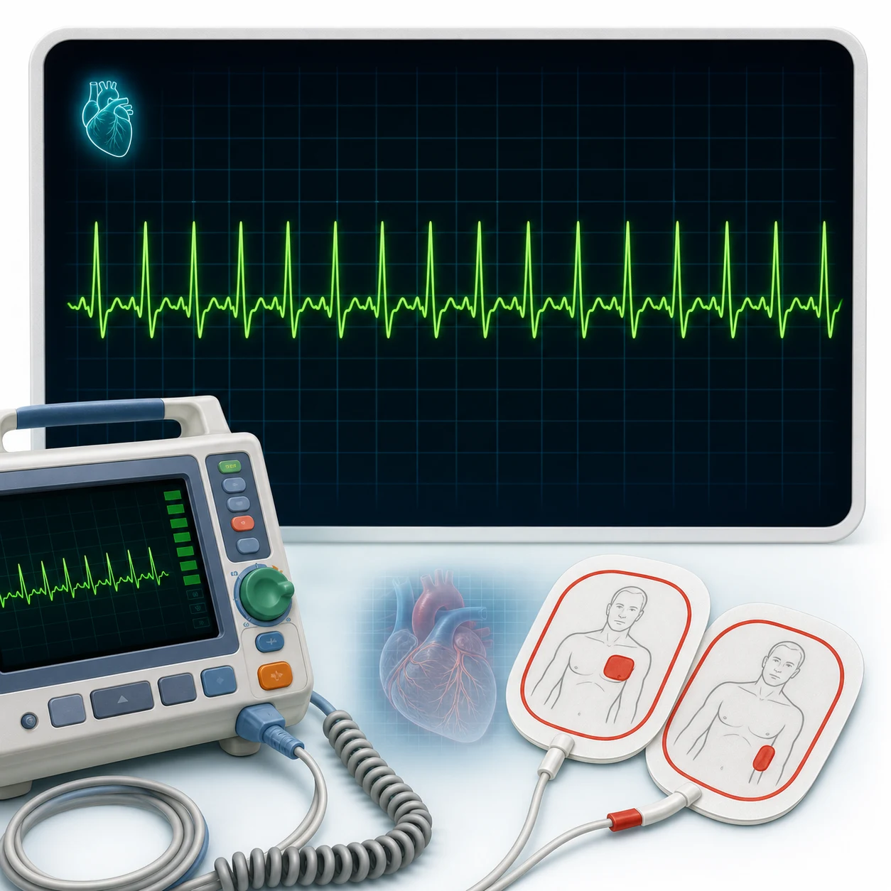
The tachyarrhythmias are managed in the emergency department by a single decision, made at the bedside before any drug: is the patient unstable? An unstable tachyarrhythmia is cardioverted whatever the rhythm; a stable one is then sorted by the QRS width and the regularity. The Fellowship candidate must know the algorithm cold, the drugs and their doses, and the two lethal traps — treating a regular broad-complex rhythm as SVT and giving verapamil, and giving an atrioventricular-node blocker to pre-excited atrial fibrillation.[1][2]
Definition and classification
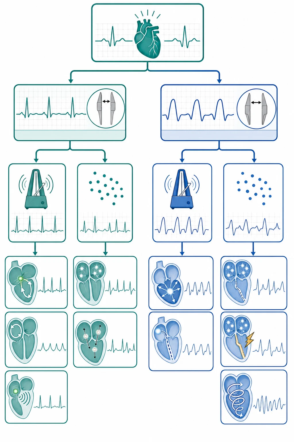
A tachyarrhythmia is any rhythm with a ventricular rate over 100 per minute, and the emergency classification turns on two features. The first is the QRS width: a narrow-complex rhythm (under 120 ms) arises above the ventricles — from the atria or the atrioventricular node — and a broad-complex rhythm (120 ms or more) is either ventricular in origin or a supraventricular rhythm conducted with aberrancy. The second is the regularity. Together, the QRS width and the regularity sort the stable tachyarrhythmia into a narrow-regular (the supraventricular tachycardias), a narrow-irregular (atrial fibrillation or flutter), a broad-regular (ventricular tachycardia, presumed), or a broad-irregular rhythm (pre-excited atrial fibrillation, or torsades de pointes). [1]
Pathophysiology — why the regular broad-complex rhythm is presumed VT
Most tachyarrhythmias are re-entrant — a self-sustaining electrical circuit — and the remainder arise from enhanced automaticity or triggered activity. The reason a regular broad-complex tachycardia in an adult is treated as ventricular tachycardia until proven otherwise is statistical and clinical: the great majority of regular broad-complex tachycardias in adults are ventricular, and treating one as a supraventricular rhythm with aberrancy — particularly by giving verapamil — can collapse an already failing ventricle, because verapamil is a negative inotrope and blocks the atrioventricular node. The diagnosis is supported by independent P waves (atrioventricular dissociation), capture and fusion beats, and concordance of the precordial QRS, but the default treatment in the emergency department is to presume VT. [1]
Clinical presentation
The patient presents with palpitations, dyspnoea, chest pain, presyncope or syncope. The instability that mandates immediate cardioversion is the presence of any one of hypotension or shock, syncope, ischaemic chest pain, acute heart failure, or an altered conscious level. A tachyarrhythmia may also present as cardiac arrest — pulseless ventricular tachycardia or fibrillation — which is defibrillated as part of the advanced-life-support algorithm. [1]
Differential diagnosis
The differential is the rhythm itself, and the ECG resolves it. [1]
Regular narrow (SVT)
- QRS <120 ms, regular, no visible P (AVNRT) or retrograde
- Haemodynamically stable usually; vagal then adenosine
- AVNRT commonest; AVRT with an accessory pathway
- Rate often 150–250
Regular broad (VT)
- QRS ≥120 ms, regular — presume VT
- Amiodarone if stable; cardiovert if unstable
- AV dissociation, capture/fusion beats support VT
- Verapamil is dangerous
AF / flutter (irregular narrow)
- Irregularly irregular, narrow QRS; flutter sawtooth
- Rate control (beta-blocker, diltiazem/verapamil)
- Anticoagulate by CHA₂DS₂-VASc
- Rhythm control if <48 h and low stroke risk
Pre-excited AF / torsades (irregular broad)
- WPW: irregular broad + delta wave — NO AV-node blockers
- Torsades: polymorphic, twisting, long QT — magnesium 2 g
- Cardiovert the unstable pre-excited AF
- Stop the QT-prolonging drug
Bedside assessment
The first assessment is the stability, because it decides between cardioversion and a drug. Look for the unstable features — hypotension, shock, syncope, ischaemic chest pain, acute heart failure, an altered conscious level — and if any is present, prepare for synchronized cardioversion at once. Then assess the QRS width and the regularity on the monitor and the 12-lead ECG, and seek the precipitant (ischaemia, electrolyte disturbance, hypoxia, sepsis, a drug, thyroid disease, structural heart disease). [1]
Investigations and the targets
The 12-lead ECG (and a rhythm strip) is the central investigation: the QRS width (the 120-ms threshold), the regularity, the P waves and their relation to the QRS (atrioventricular dissociation in VT), the axis, capture and fusion beats in VT, a delta wave and a short PR in Wolff-Parkinson-White, and the QT interval (a long QT raises torsades). The electrolytes — potassium, magnesium and calcium — are checked and corrected, the troponin identifies an ischaemic cause, the magnesium is checked in suspected torsades, and a thyroid function test and a digoxin level are sent where relevant. [1]
Immediate management — stability first, then the algorithm
Stabilise the airway and the breathing with oxygen, and decide the stability. [1]
[1]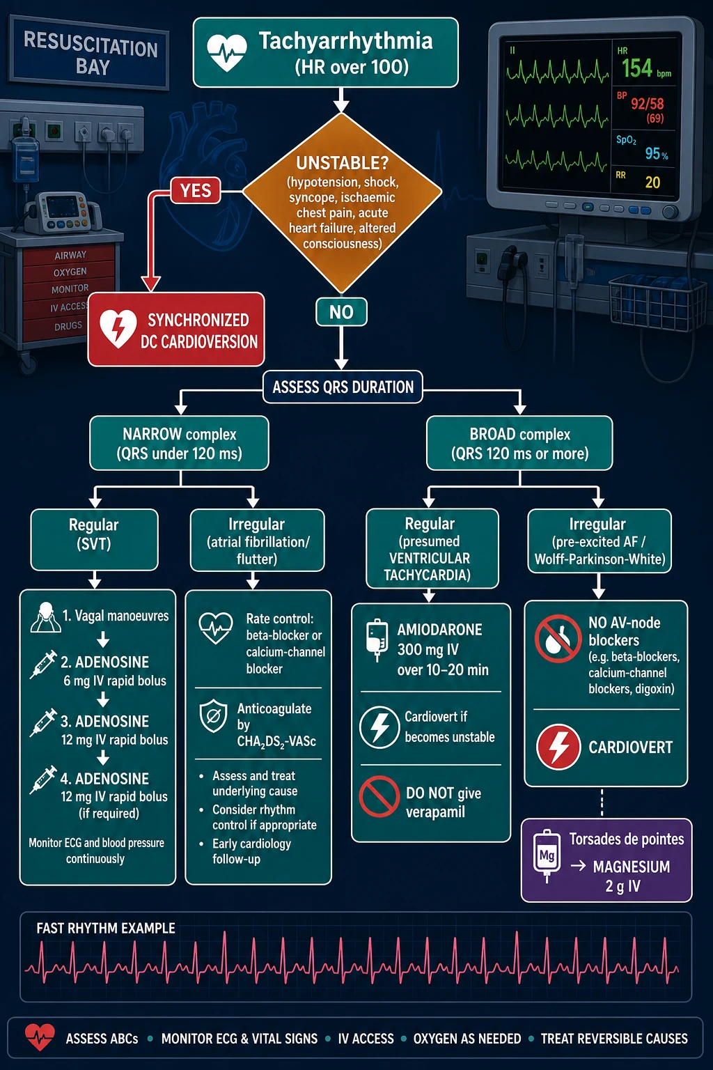
Key thresholds and doses
Adenosine terminates the common re-entrant supraventricular tachycardia by transiently blocking the atrioventricular node, and the 6-then-12-then-12 mg sequence is the standard escalation; the patient must be warned of the brief flush, dyspnoea and chest tightness, and the rhythm monitored throughout, because adenosine can produce a transient asystole.[2] It is contraindicated in asthma and in high-grade atrioventricular block. Torsades de pointes — a polymorphic broad-complex tachycardia with a twisting axis on a long QT — is treated with intravenous magnesium sulphate 2 g, withdrawal of the offending drug, and potassium correction, with overdrive pacing for recurrent episodes.
Subtypes and special scenarios
The AV-nodal-re-entrant tachycardia (AVNRT) is the commonest regular narrow-complex SVT and responds to vagal manoeuvres and adenosine; the atrioventricular-re-entrant tachycardia (AVRT) uses an accessory pathway (Wolff-Parkinson-White) and is flagged for catheter ablation. Atrial fibrillation is managed by a rate-control or a rhythm-control strategy: rate control with a beta-blocker or a rate-limiting calcium-channel blocker (digoxin in heart failure) is first-line for most; rhythm control by pharmacological or electrical cardioversion is considered when the onset is under 48 hours and the stroke risk is low, and anticoagulation is stratified on the CHA₂DS₂-VASc score (congestive heart failure, hypertension, age 65 or over, diabetes, stroke or transient ischaemic attack, vascular disease, and sex category). A cardioversion performed on a fibrillation of uncertain or prolonged duration is preceded by anticoagulation or a trans-oesophageal echocardiogram to exclude a left-atrial thrombus. Monomorphic ventricular tachycardia in structural heart disease is presumed and treated as above. Digoxin toxicity produces an atrial tachycardia with atrioventricular block and is treated with the digoxin-specific antibody fragment. The channelopathies — the long-QT syndrome (and the drug-induced QT prolongation that leads to torsades), the Brugada syndrome, and the catecholaminergic polymorphic ventricular tachycardia — produce characteristic arrhythmias and need cardiology input, an implantable defibrillator in selected cases, and the strict avoidance of the offending drug. [1]
Complications and pitfalls
The complications are the arrhythmia itself (haemodynamic collapse, cardiac arrest), thromboembolism after the cardioversion of unrecognised atrial fibrillation, amiodarone-induced hypotension, and the bronchospasm, heart block or asystole of adenosine. The pitfalls are the dangerous inverse of the algorithm: treating a regular broad-complex rhythm as SVT and giving verapamil; giving an atrioventricular-node blocker to pre-excited atrial fibrillation; giving adenosine in asthma; missing an electrolyte or drug cause; not recognising torsades; and delaying cardioversion in the unstable patient. [1]
Prognosis and disposition
The prognosis depends on the rhythm and the cause: ventricular tachycardia and fibrillation carry a high risk; the benign re-entrant supraventricular tachycardias do well. The unstable patient, the patient with ventricular tachycardia, and the patient with new atrial fibrillation needing rate or rhythm control are admitted to cardiology or a monitored bed; the precipitant is treated; and the anticoagulation decision is made by the CHA₂DS₂-VASc score and the rhythm's duration. Patients with a structurally normal heart and a clearly provoked, terminated arrhythmia may suit a short-stay pathway after cardiology review, whereas those with syncope, structural disease, a channelopathy, or a family history of sudden death are admitted for a full workup and the consideration of an implantable cardioverter-defibrillator. [1]
Each antiarrhythmic drug is itself pro-arrhythmic — amiodarone, flecainide and sotalol can each worsen a rhythm — so the response to any drug therapy is monitored on a continuous trace before the patient leaves the monitored area. [1]
Narrow-complex tachycardias in depth
A narrow-complex tachycardia (the QRS under 120 ms) is, by definition, supraventricular — the impulse uses the His–Purkinje system and the ventricles depolarise normally, so the complex is narrow. The emergency task is to separate the regular rhythms (the AV-nodal-re-entrant tachycardia, the AV-re-entrant tachycardia, the atrial tachycardia, the atrial flutter with a fixed block) from the irregular one (atrial fibrillation, and the atrial flutter with a variable block), because the regular one is terminated with adenosine while the irregular one is rate-controlled and anticoagulated. [1]
The regular narrow-complex tachycardias — the four to name
N-A-V-A
AV-nodal-re-entrant tachycardia — the commonest, a micro-re-entry circuit inside the node; no visible P or a pseudo-r′ in V1
AV-re-entrant tachycardia — a macro-circuit using an accessory pathway (WPW); orthodromic (narrow) or antidromic (broad)
A focal atrial origin; adenosine may reveal the P waves without terminating it — the diagnostic clue
Sawtooth flutter waves at 300/min, conducted 2:1 for a ventricular rate of 150 — the classic regular rate
Vagal manoeuvres — the first step in the stable SVT
The modified Valsalva manoeuvre is the first-line termination for a haemodynamically stable regular narrow-complex tachycardia, recommended by both the ESC and the ACC/AHA/HRS guidelines before any drug.[1][8] The patient is positioned supine, blows into a 10 mL syringe to move the plunger for 15 seconds (a sustained intrathoracic pressure of about 40 mmHg), and is then laid flat with the legs passively elevated to 45 degrees for 15 seconds — the augmented venous return and the reflex vagal surge increase the success rate to around 43 per cent versus 17 per cent for the standard seated strain. Carotid sinus massage is the alternative, applied for 5 to 10 seconds to one carotid at a time after auscultation for a bruit (a bruit contraindicates it in the atherosclerotic patient) — never both carotids together, and never in the elderly without first excluding significant carotid disease.
[1] [1]Adenosine — the escalation and the warnings
If the vagal manoeuvre fails, adenosine terminates the AV-node-dependent tachycardia (the AVNRT and the orthodromic AVRT) by transiently blocking the node. The dose is escalated through the 6 mg, then 12 mg, then 12 mg sequence, each as a rapid intravenous bolus through a large proximal cannula, immediately followed by a 20 mL saline flush, with the patient on a continuous cardiac monitor and a defibrillator at the bedside. The half-life is under 10 seconds, so a slow push fails — the drug is metabolised before it reaches the heart. The 6 mg dose terminates roughly 60 per cent; the first 12 mg dose raises this to over 90 per cent.[2]
The adenosine termination of a stable SVT
Confirm a regular narrow-complex tachycardia on the 12-lead and the monitor — a 12-lead first, because adenosine in a regular broad-complex rhythm presumed VT is wrong, and adenosine in pre-excited AF is dangerous.
Warn the patient — the brief flush, the chest tightness, the sense of doom, and the few seconds of asystole — and obtain consent; the warning prevents the alarmed grab and the feeling of impending death.
Draw up adenosine 6 mg and a 20 mL saline flush; attach the defibrillation pads in case of a degeneration to VF (rare, but the rhythm that adenosine unmasks can fibrillate).
Push the 6 mg rapidly through a large proximal cannula followed immediately by the flush; record a continuous rhythm strip throughout.
If the rhythm persists after 2 minutes, repeat with 12 mg; if still in the rhythm after a further 2 minutes, give a final 12 mg.
If sinus rhythm returns, observe on the monitor, seek the precipitant, and arrange the cardiology referral for the consideration of the catheter ablation.
If the tachycardia continues despite the full 6-12-12 escalation, reconsider the diagnosis — the focal atrial tachycardia and the junctional tachycardia are adenosine-resistant; give verapamil or a beta-blocker (only once VT and pre-excited AF are excluded) or seek the urgent cardiology input.
The adenosine dose ladder and its modifiers
Atrial fibrillation and atrial flutter in the emergency department
Atrial fibrillation is the commonest sustained arrhythmia and the most frequent reason for an arrhythmia-related emergency presentation. The emergency-department decision is governed by two questions: how unstable is the patient, and when did the fibrillation start — because the answer to the first decides between the cardioversion and the rate control, and the answer to the second decides the stroke risk of a cardioversion. [1]
The rate-control versus the rhythm-control decision
The AFFIRM trial established that, for the older patient with the risk factors for stroke, a rate-control strategy is non-inferior to a rhythm-control strategy for survival — and is simpler and safer.[3] The rate control is therefore the first-line strategy for most stable patients, and the rhythm control (the pharmacological or the electrical cardioversion) is reserved for the patient with the new-onset fibrillation (the onset under 48 hours, when the stroke risk of the cardioversion is low) and the significant ongoing symptoms despite the adequate rate control, or the patient in whom a tachycardia-induced cardiomyopathy is developing. The RACE II trial further showed that a lenient rate target (a resting ventricular rate under 110 per minute) is non-inferior to a strict target (under 80 at rest, under 110 with the moderate exercise) for the cardiovascular outcome in the stable ambulatory patient.[4]
AFFIRM — rate vs rhythm control in atrial fibrillation
New England Journal of Medicine, 2002
A randomised controlled trial of the rate control versus the rhythm control in 4,060 patients with the atrial fibrillation and the high stroke risk, followed for a mean of 3.5 years.
Key finding
No significant difference in the overall mortality (the primary endpoint); the rhythm-control arm had more hospitalisations and more of the adverse drug effects, with a trend to a higher mortality in the subgroup analyses.
Practice change
The rate control became the default first-line strategy for the older, high-stroke-risk patient; the rhythm control is reserved for the symptomatic, the newly-diagnosed, and the heart-failure patient. The 48-hour threshold for the anticoagulation before the cardioversion was reinforced.
RACE II — lenient vs strict rate control
New England Journal of Medicine, 2010
A randomised non-inferiority trial of the lenient (resting rate under 110) versus the strict (under 80 at rest, under 110 on exercise) rate control in 614 patients with the permanent atrial fibrillation, followed for up to three years.
Key finding
The lenient control was non-inferior for the composite of the cardiovascular death, the heart-failure hospitalisation, the stroke, the bleeding and the arrhythmia, with fewer clinic visits and easier achievement of the target.
Practice change
The lenient resting-rate target of under 110 is acceptable for the stable permanent-fibrillation patient; the strict target is reserved for the symptomatic, the heart-failure and the tachycardiomyopathy-risk patient.
Rate control
- The default for most — the beta-blocker or the diltiazem/verapamil
- Targets: lenient <110 (stable); stricter if symptomatic or in HF
- Metoprolol 5 mg IV over 2 min, repeated to 15 mg; or diltiazem 0.25 mg/kg IV
- Digoxin in the heart failure or the beta-blocker intolerance; amiodarone if both fail
- Anticoagulate by the CHA₂DS₂-VASc, independent of the strategy
Rhythm control
- Considered for the onset <48 h, the symptomatic, the young, the heart failure
- Pharmacological: flecainide, amiodarone, or the "pill-in-the-pocket"
- Electrical: the synchronised DC cardioversion (120–150 J biphasic)
- Anticoagulate for 3 weeks before if >48 h, or a TOE to exclude the LA thrombus
- Followed by 4 weeks of the anticoagulation regardless of the duration
Special scenarios
- Heart failure: amiodarone or digoxin — NOT the calcium-channel blocker
- WPW with AF: NO AV-node blocker — cardiovert
- Sepsis/thyrotoxicosis: treat the precipitant; beta-blocker for the rate
- Pregnancy: beta-blocker or digoxin; avoid the ACE-i and the amiodarone
CHA₂DS₂-VASc — the stroke-risk score for the anticoagulation decision
CHA₂DS₂-VASc
Congestive heart failure (or the LV dysfunction)
Hypertension
Age 75 or older — 2 points
Diabetes mellitus
Prior stroke, TIA or thromboembolism — 2 points
Vascular disease (prior MI, the PAD, the aortic plaque)
Age 65 to 74 — 1 point
Female sex — 1 point (but only counts if another risk factor is present)
A score of 0 in a man, or 1 in a woman with no other risk factor, is a low stroke risk and the anticoagulation is withheld; a score of 2 or more in a man, or 3 or more in a woman warrants the oral anticoagulation (a direct oral anticoagulant preferred, or the warfarin with the mechanical-valve or the moderate-to-severe mitral stenosis). The 2023 ACC/AHA/ACCP/HRS guideline reaffirmed this threshold and the preference for the direct oral anticoagulants in the non-valvular fibrillation.[10]
The AF rate-control drug doses
Atrial flutter — the sawtooth and the 2:1 trap
Atrial flutter produces the characteristic sawtooth flutter waves (the negative deflections in the inferior leads, the typical "sawtooth" of the counterclockwise cavotricuspid-isthmus-dependent flutter) at an atrial rate around 300 per minute. The commonest conduction is 2:1, giving a regular ventricular rate of 150 per minute — a regular narrow-complex tachycardia at exactly 150 should prompt the search for the flutter waves, because the atrial tachycardia masquerades as an SVT. The adenosine is diagnostic here: it transiently increases the AV block to 4:1 or 8:1, slowing the ventricular rate and unmasking the flutter waves without terminating the flutter itself. The rate control, the rhythm control and the anticoagulation follow the same principles as the atrial fibrillation; the definitive treatment is the cavotricuspid-isthmus ablation (the success rate above 90 per cent). [1]
[1]Broad-complex tachycardia — the ventricular tachycardia
The stable regular broad-complex tachycardia in an adult is ventricular tachycardia until proven otherwise. The statistical justification is overwhelming — over 80 per cent of the regular broad-complex tachycardias in adults with the structural heart disease are ventricular — and the consequence of the misdiagnosis (the verapamil-induced collapse of the failing ventricle) is catastrophic. The Brugada algorithm (the four-step criteria for the QRS morphology) and the Vereckei aVR lead analysis help distinguish the VT from the SVT-with-aberrancy, but the emergency default is to treat as VT.[12]
The ECG features that support the VT diagnosis
The diagnosis of the ventricular tachycardia is supported by the independent P waves (the atrioventricular dissociation — the atria and the ventricles fire independently), the capture beats (a normal narrow QRS among the broad ones, where a sinus impulse captures the ventricles), the fusion beats (a hybrid of the sinus and the ventricular complexes), and the concordance of the precordial QRS (all positive or all negative in V1 to V6). Other supportive features are the extreme axis deviation (the northwest axis), the AV dissociation, the delay to the nadir of the S wave of over 100 ms, and the notching of the downstroke. None of these is universally present — their absence does not exclude the VT. [1]
Ventricular tachycardia
- AV dissociation — independent P waves
- Capture and fusion beats (pathognomonic)
- Concordant precordial pattern (all + or all −)
- Extreme axis (northwest); >100 ms to the S nadir
- Markedly broad >160 ms, the initial upstroke slurred
SVT with aberrancy
- A typical RBBB or LBBB pattern
- A concordant response to the vagal manoeuvre or the adenosine
- A known bundle branch block in sinus rhythm that matches
- Onset with a premature atrial beat
- A narrower QRS, a normal axis
The stable VT — amiodarone, or the lidocaine
The haemodynamically stable ventricular tachycardia (no hypotension, no shock, no chest pain, no heart failure, no altered consciousness) is treated with the intravenous amiodarone 300 mg over 20 to 60 minutes, followed by the infusion of 900 mg over 24 hours (the 900 mg in 500 mL of the 5 per cent dextrose, run over 24 hours).[7] The amiodarone terminates about 40 to 60 per cent of the stable monomorphic VT within the first hour. The alternative is the lidocaine (lignocaine) 1 to 1.5 mg/kg intravenously (a 100 mg dose in the average adult), repeated every 5 to 10 minutes to a maximum of 3 mg/kg, followed by the infusion of 1 to 4 mg/min — useful when the amiodarone is contraindicated or the VT is clearly ischaemia-mediated. The procainamide (a class Ia agent, 20 to 50 mg/min to a maximum of 17 mg/kg) is an alternative that some guidelines prefer for the stable monomorphic VT with the preserved ejection fraction, but it requires a monitored bed and carries the hypotension risk.
[1] [1]Levine 1996 — intravenous amiodarone for hypotensive VT
Journal of the American College of Cardiology, 1996
A randomised double-blind trial of the intravenous amiodarone (a 150 mg loading dose then 0.5–1.0 mg/min) versus the bretylium in 273 patients with the recurrent sustained hypotensive ventricular tachyarrhythmias.
Key finding
The amiodarone was at least as effective as the bretylium for the termination of the arrhythmia and was better tolerated, with fewer of the hypotension events and the discontinuations for the adverse effects.
Practice change
The intravenous amiodarone became the first-line antiarrhythmic for the recurrent sustained hypotensive VT, displacing the bretylium (now withdrawn in many markets) and the lidocaine.
Pulseless ventricular tachycardia and ventricular fibrillation — the ALS pathway
The pulseless ventricular tachycardia and the ventricular fibrillation are the shockable rhythms of the cardiac arrest, managed within the advanced-life-support algorithm as a continuous loop of the high-quality CPR and the defibrillation, with the adrenaline and the amiodarone added at the third shock.[9]
The shockable-rhythm (VF/pulseless VT) ALS loop
Confirm the cardiac arrest — the unresponsive patient, the absent or the abnormal breathing, no pulse; call for the arrest team and the defibrillator; begin the high-quality chest compressions (the rate 100–120/min, the depth 5–6 cm, the full recoil, the minimised interruptions).
Attach the defibrillator pads and analyse the rhythm — the VF (the chaotic, irregular, no recognisable QRS) or the pulseless VT (the broad regular, but with no pulse) are the shockable rhythms.
Shock ONE — the unsynchronized biphasic shock at 150–200 J (or the 360 J monophasic); immediately resume the CPR for 2 minutes without a rhythm check.
Shock TWO — if the VF/VT persists after the 2 minutes of CPR, deliver the second shock at the escalating energy (200 J biphasic); resume the CPR for 2 minutes.
Shock THREE — if the VF/VT persists, give the adrenaline 1 mg intravenously/intraosseously, then the third shock; resume the CPR for 2 minutes.
After the third shock — give the amiodarone 300 mg intravenously/intraosseously as a bolus (the lidocaine 100 mg is the alternative); resume the CPR.
Continue the 2-minute cycles of the CPR-and-rhythm-check, with the adrenaline 1 mg every 3–5 minutes (every other cycle), and a further amiodarone 150 mg after the fifth shock.
Search for and treat the reversible causes — the four Hs (the hypoxia, the hypovolaemia, the hypo-/hyperkalaemia, the hypothermia) and the four Ts (the tension pneumothorax, the tamponade, the thrombosis, the toxins); the VF arrest is often ischaemia-mediated, so the early coronary angiography in the return of the spontaneous circulation.
ALIVE — amiodarone vs lidocaine for shock-resistant VF
New England Journal of Medicine, 2002
A randomised double-blind trial of the intravenous amiodarone (5 mg/kg) versus the lidocaine (1.5 mg/kg) in 347 patients with the out-of-hospital ventricular fibrillation that was resistant to the three shocks, the adrenaline and a further shock.
Key finding
A significantly higher proportion of the amiodarone-treated patients survived to the hospital admission (22.8 per cent versus 12.0 per cent); the benefit held in the witnessed arrests and the bystander-CPR subgroups.
Practice change
The amiodarone became the preferred antiarrhythmic for the shock-resistant ventricular fibrillation, displacing the lidocaine in the advanced-life-support algorithm (the lidocaine remains a recognised alternative).
ROC — amiodarone, lidocaine, or placebo in OHCA
New England Journal of Medicine, 2016
A randomised double-blind trial of the amiodarone versus the lidocaine versus the placebo in around 3,000 patients with the out-of-hospital cardiac arrest and the initial shockable rhythm, in the non-shockable rhythm subgroup and the overall cohort.
Key finding
No significant difference in the survival to discharge in the overall population, but a significant improvement in the survival to discharge in the witnessed-arrest subgroup with the amiodarone and the lidocaine over the placebo.
Practice change
The amiodarone and the lidocaine remain the recommended antiarrhythmics for the refractory shockable-rhythm arrest, with the benefit concentrated in the witnessed arrests; the placebo does not suffice in the witnessed shockable arrest.
The shockable-rhythm (VF/pulseless VT) doses in sequence
Torsades de pointes and the long-QT syndromes
The torsades de pointes is a polymorphic ventricular tachycardia on a prolonged QT interval, characterised by the QRS complexes of the varying amplitude that appear to "twist" around the isoelectric baseline. The sustained torsades causes the syncope or the cardiac arrest; the self-terminating bursts cause the dizziness and the palpitations. The QT prolongation (a corrected QT over 500 ms, or an increase of over 60 ms on a new drug) is either congenital (the long-QT syndromes — the Romano-Ward, the Jervell and Lange-Nielsen) or, far more commonly in the emergency setting, acquired from a QT-prolonging drug (the macrolides, the fluoroquinolones, the antipsychotics — especially the haloperidol and the droperidol, the methadone, the ondansetron, the sotalol, the amiodarone), the electrolyte disturbance (the hypokalaemia, the hypomagnesaemia, the hypocalcaemia), the bradycardia, and the acute neurological injury. [1]
The torsades de pointes management
Recognise the polymorphic broad-complex tachycardia with the twisting axis on the ECG; check the corrected QT in the baseline sinus beats — a QTc over 500 ms confirms.
If the patient is pulseless or the torsades is sustained and unstable — defibrillate (the unsynchronized 200 J biphasic); the synchronized cardioversion is often impractical as the QRS morphology varies.
Give the intravenous magnesium sulphate 2 g (the 20 mL of the 10 per cent solution, or the 4 mL of the 50 per cent diluted) over 1 to 2 minutes, regardless of the serum magnesium — the magnesium suppresses the early after-depolarisations that drive the torsades.<Cite id="11" />
Withdraw the offending QT-prolonging drug; check and aggressively correct the potassium to the high-normal (the 4.5–5.0 mmol/L) and the magnesium.
For the recurrent or the refractory torsades — the overdrive pacing (the temporary transvenous or the isoprenaline infusion to drive the heart rate to 100–120, shortening the QT) breaks the bradycardia-dependent torsades.
Admit to the coronary care or the ICU; the cardiology referral; the consideration of the implantable defibrillator in the congenital long-QT with the recurrent syncope despite the beta-blockade.
The Vaughan-Williams classification and the antiarrhythmic drug profiles
The antiarrhythmics are classified by the Vaughan-Williams scheme — the class I agents block the sodium channel, the class II block the beta-receptors, the class III block the potassium channel, and the class IV block the calcium channel. The digoxin is sometimes the class V. The classification is imperfect — many drugs span the classes (the amiodarone has the class I, II, III and IV activity) — but it remains the framework for the Fellowship viva. [1]
The Vaughan-Williams classes — and a representative drug for each
NO BAD KICK
A (quinidine, procainamide), B (lidocaine), C (flecainide) — the sodium-channel blockers
Metoprolol, the propranolol, the esmolol — the AV-node slowers
Amiodarone, the sotalol, the ibutilide — the repolarisation prolongers
Verapamil, the diltiazem — the AV-node slowers; NOT the dihydropyridines
The cardiac glycoside — the AV-node slower; useful in the heart-failure AF
Amiodarone (class III)
- A broad spectrum — I, II, III, IV activity
- Stable VT 300 mg over 20–60 min, then 900 mg/24 h
- VF arrest: 300 mg after the 3rd shock, 150 mg after the 5th
- Acute: hypotension (the solvent), the bradycardia
- Long-term: thyroid, pulmonary, hepatic, the corneal deposits
Lidocaine (class Ib)
- For the ischaemia-mediated VT
- 1–1.5 mg/kg IV, repeated to 3 mg/kg, then 1–4 mg/min
- VF arrest: 100 mg after the 3rd shock (alternative)
- Less effective than the amiodarone for the shock-resistant VF
- Caution in the hepatic failure and the severe heart block
Flecainide (class Ic)
- For the structurally-normal-heart AF and SVT (the pill-in-the-pocket)
- NOT in the structural or the ischaemic heart disease (pro-arrhythmic)
- Avoid in the broad-complex tachycardia of uncertain origin
- The CAST trial showed the increased mortality in the post-MI patient
Sotalol (class II + III)
- A beta-blocker with the potassium-channel activity
- For the AF and the VT in the normal heart
- Prolongs the QT — the torsades risk
- Avoid in the renal failure (the dose is renally cleared)
The synchronised cardioversion and the defibrillation — the technique
The synchronised DC cardioversion delivers the shock on the R wave of the QRS (the "sync" mode) to avoid the delivery of the shock on the T wave, which would precipitate the ventricular fibrillation. It is used for the unstable tachyarrhythmia with a QRS — the unstable SVT, the unstable atrial fibrillation and flutter, the unstable monomorphic VT — where the R wave is identifiable. The defibrillation is the unsynchronized shock, used when the R wave is not identifiable — the ventricular fibrillation, the polymorphic VT, and the pulseless VT — and is delivered at the higher energy from the first shock. [1]
The preparation for the synchronised cardioversion
Establish the IV access, the continuous cardiac monitoring, the pulse oximetry and the blood-pressure monitoring; place the defibrillation pads in the anterolateral (the sternum-apex) or the anteroposterior position.
Pre-oxygenate with the 100 per cent oxygen; draw up the sedation and the analgesia (the midazolam 2–5 mg and the fentanyl 50–100 µg, or the propofol, or the ketamine 1 mg/kg); have the airway equipment and the reversal agents at the bedside.
Set the defibrillator to the synchronised mode; confirm the synchronisation markers on the R waves on the monitor.
Select the energy — the 120–150 J biphasic for the atrial fibrillation, the 100–120 J for the flutter and the SVT, the 120–150 J for the stable monomorphic VT (escalating to the 200 J for the refractory).
Press and hold the shock button until the shock is delivered (the sync mode introduces a slight delay as the defibrillator waits for the R wave); reassess the rhythm; escalate the energy if the arrhythmia persists.
If the rhythm degenerates to the VF, switch to the unsynchronized mode and defibrillate.
The synchronised-cardioversion energy by the rhythm
The precipitants — find and reverse the cause
A tachyarrhythmia is rarely an isolated event — the precipitant drives the recurrence, and the search for it is part of the emergency management. The common precipitants are the myocardial ischaemia and infarction (the troponin, the ECG), the electrolyte disturbance (the potassium, the magnesium, the calcium — the hypokalaemia is the classic precipitant of the atrial and the ventricular arrhythmias), the hypoxia and the respiratory failure, the sepsis, the thyrotoxicosis (the thyroid function test in the new atrial fibrillation), the alcohol excess (the "holiday heart" — the binge-drinking-induced atrial fibrillation), the sympathomimetic drugs (the cocaine, the amphetamines, the excessive caffeine), the pulmonary embolism (the cause of both the sinus tachycardia and the atrial fibrillation), the recreational and the prescribed drug withdrawal, and the structural heart disease (the cardiomyopathies, the valvular disease, the channelopathies). [1]
[1]The disposition and the safety net
The unstable patient, the patient with the ventricular tachycardia, the patient with the pre-excited atrial fibrillation, and the patient with the new atrial fibrillation needing the rate or the rhythm control are admitted to the monitored bed (the coronary care or the high-dependency unit). The patient with the clearly provoked and terminated benign SVT (the young, the structurally-normal heart, the clear precipitant removed) may suit the short-stay or the discharged-after-the-cardiology-review pathway, with the referral for the catheter ablation (the curative treatment for the AVNRT, the AVRT and the atrial flutter). The patient with the syncope, the structural heart disease, the channelopathy, or the family history of the sudden death is admitted for the full workup and the consideration of the implantable cardioverter-defibrillator. Every patient discharged after a tachyarrhythmia receives a written safety net — the return-if symptoms and the cardiology follow-up — because the recurrence and the first presentation of the channelopathy can be the cardiac arrest. [1]
Special populations
In pregnancy, adenosine is safe for the mother and the fetus, and amiodarone is avoided where possible because of its fetal thyroid effects; a synchronized cardioversion is also safe in pregnancy when the rhythm is unstable. The elderly and those with structural heart disease more often have ventricular tachycardia, decompensate at lower rates, and tolerate the rate-control and the anticoagulation less well, with a higher bleeding risk. The patient with a known accessory pathway (Wolff-Parkinson-White) is flagged for catheter ablation, and the electrolyte-depleted or the septic patient has the precipitant corrected alongside the rhythm itself. [1]
Evidence and regional guidelines
The contemporary framework is the advanced-life-support tachyarrhythmia algorithm (Resuscitation Council) and the 2019 ESC supraventricular-tachycardia guideline[1] with the ESC ventricular-arrhythmia guidance. The adenosine dosing is supported by emergency-department studies.[2] The algorithm and the drug doses are global; the cardioversion energies and the anticoagulation pathway are local.
ANZ practice note. The stability-first algorithm and the drug doses follow the ANZCOR/Resuscitation Council framework via local cardiology pathways; adenosine 6 then 12 then 12 mg is the SVT escalation, amiodarone 300 mg the stable-VT choice, and pre-excited atrial fibrillation is cardioverted rather than treated with an AV-node blocker. [1]
SAQs — exam practice
SAQ — Unstable AVNRT in the resuscitation bay
10 minutes · 10 marks
A 34-year-old man is brought to the resuscitation bay with two hours of sudden-onset palpitations, dyspnoea and central chest tightness. He has no past cardiac history and takes no regular medication. On arrival he is diaphoretic, anxious and cool peripherally: GCS 15, BP 82/50, HR 224 (regular), RR 26, SpO2 93 per cent on room air. The cardiac monitor shows a regular narrow-complex tachycardia at 224 per minute with no visible P waves; the 12-lead confirms a QRS of 90 ms with no pre-excitation or delta wave. He describes ongoing central chest tightness. Two large-bore cannulae are sited and the defibrillator is at the bedside.
SAQ — New-onset atrial fibrillation with rapid ventricular response
10 minutes · 10 marks
A 76-year-old woman presents with six hours of sudden-onset palpitations, dyspnoea and exertional central chest tightness that began at rest. Her past history includes the hypertension, the type 2 diabetes and a transient ischaemic attack two years ago; her medications are perindopril 5 mg, amlodipine 10 mg, metformin 1 g twice daily and aspirin 100 mg daily. On arrival she is alert and warm peripherally: BP 104/68, HR 148 (irregularly irregular), RR 24, SpO2 94 per cent on room air, afebrile. The 12-lead ECG shows the atrial fibrillation with a narrow QRS and a ventricular rate of 140–155, no acute ischaemic change. The onset was clearly six hours ago.
Exam pearls
- Unstable → synchronized cardioversion (any tachyarrhythmia).
- Regular broad complex = VT until proven otherwise → amiodarone 300 mg if stable; verapamil is dangerous.
- Irregular broad complex (pre-excited AF, WPW) → NO AV-node blockers → cardiovert.
- Stable narrow regular (SVT) → vagal then adenosine 6→12→12 mg.
- Adenosine: warn of the flush; contraindicated in asthma; can cause brief asystole.
- Torsades de pointes → magnesium 2 g, stop the offending drug, correct the potassium. [1]
Red flags
[1]References
- [1]Brugada J, Katritsis DG, Arbelo E, et al. 2019 ESC Guidelines for the management of patients with supraventricular tachycardiaThe Task Force for the management of patients with supraventricular tachycardia of the European Society of Cardiology (ESC) Eur Heart J, 2020.PMID 31504425
- [2]Sert ET, Yilmaz MS, Ay MO, et al. Initial 12 mg Versus 6 mg Adenosine for Supraventricular Tachycardia in the Emergency Department Acad Emerg Med, 2026.PMID 42057249
- [3]Wyse DG, Waldo AL, DiMarco JP, et al. (AFFIRM Investigators) A comparison of rate control and rhythm control in patients with atrial fibrillation N Engl J Med, 2002.PMID 12466506
- [4]Van Gelder IC, Groenveld HF, Crijns HJGM, et al. (RACE II Investigators) Lenient versus strict rate control in patients with atrial fibrillation N Engl J Med, 2010.PMID 20231232
- [5]Dorian P, Cass D, Schwartz B, Cooper R, Gelaznikas R, Barr A (ALIVE Investigators) Amiodarone as compared with lidocaine for shock-resistant ventricular fibrillation N Engl J Med, 2002.PMID 11907287
- [6]Kudenchuk PJ, Brown SP, Daya M, et al. (ROC Investigators) Amiodarone, Lidocaine, or Placebo in Out-of-Hospital Cardiac Arrest N Engl J Med, 2016.PMID 27043165
- [7]Levine JH, Massumi A, Scheinman MM, et al. (intravenous amiodarone multicenter investigators) Intravenous amiodarone for recurrent sustained hypotensive ventricular tachyarrhythmias. Intravenous Amiodarone Multicenter Trial Group J Am Coll Cardiol, 1996.PMID 8522712
- [8]Page RL, Joglar JA, Caldwell MA, et al. 2015 ACC/AHA/HRS Guideline for the Management of Adult Patients With Supraventricular Tachycardia: Executive Summary: A Report of the American College of Cardiology/American Heart Association Task Force on Clinical Practice Guidelines and the Heart Rhythm Society J Am Coll Cardiol, 2016.PMID 26409258
- [9]Soar J, Donnino MW, Maconochie I, et al. European Resuscitation Council Guidelines for Resuscitation: 2018 Update - Antiarrhythmic drugs for cardiac arrest Resuscitation, 2019.PMID 30496838
- [10]Joglar JA, Chung MK, Armbruster AL, et al. 2023 ACC/AHA/ACCP/HRS Guideline for the Diagnosis and Management of Atrial Fibrillation: A Report of the American College of Cardiology/American Heart Association Joint Committee on Clinical Practice Guidelines Circulation, 2024.PMID 38033089
- [11]Tzivoni D, Banai S, Schuger C, et al. Terminology of torsades de pointes Cardiovasc Drugs Ther, 1991.PMID 1854659
- [12]Antunes E, Brugada J, Steurer G, Andries E, Brugada P The differential diagnosis of a regular tachycardia with a wide QRS complex on the 12-lead ECG: ventricular tachycardia, supraventricular tachycardia with aberrant intraventricular conduction, and supraventricular tachycardia with anterograde conduction over an accessory pathway Pacing Clin Electrophysiol, 1994.PMID 7991423