EM · Syncope (approach)
Syncope — the emergency department approach and risk stratification
Also known as Fainting · Blackout · Transient loss of consciousness · Collapse
Syncope — the transient loss of consciousness from the cerebral hypoperfusion, the classification (the reflex, the orthostatic, the cardiac, the cerebrovascular), the critical cardiac-versus-non-cardiac distinction, the San Francisco Syncope Rule for the risk stratification, the ESC high-risk features, the investigation strategy (the ECG, the glucose, the bedside echo), the management (the treat-the-cause, the admission for the high-risk, the discharge with the advice for the low-risk), and the syncope-versus-seizure differential. ACEM-primary, globally tagged.
On this page & tools
Your progress
Saved locally on this device.
Target exams
Red flags
Syncope is the transient loss of consciousness from a global cerebral hypoperfusion, characterised by a rapid onset, a short duration and a spontaneous complete recovery. It is one of the commonest presentations to the emergency department (up to 3 per cent of the attendances), and the Fellowship candidate's task is not to diagnose the syncope (the patient has already woken up) but to risk-stratify it — to separate the low-risk reflex syncope that goes home with the advice from the high-risk cardiac syncope that goes to the cardiology with the monitoring. The error is not missing a rare diagnosis; it is sending home a patient with a cardiac syncope who arrests on the way out.[1][2]

Definition and classification
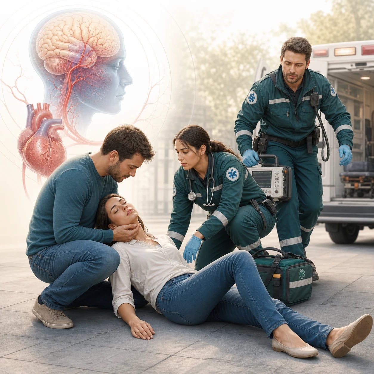
Syncope is a transient loss of consciousness (a TLOC) from a global cerebral hypoperfusion, with a rapid onset, a short duration (usually under 30 seconds) and a spontaneous, complete recovery. It is classified by the mechanism. The reflex (the neurally-mediated) syncope — the vasovagal (the commonest, triggered by the emotion, the pain, the prolonged standing, the warm environment), the situational (the cough, the micturition, the defecation, the post-prandial) and the carotid sinus hypersensitivity — is the low-risk type. The orthostatic syncope (from a postural hypotension — the medication, the dehydration, the autonomic failure, the elderly) is also usually low-risk. The cardiac syncope — from an arrhythmia (the bradycardia, the tachycardia, the conduction block), a structural heart disease (the aortic stenosis, the hypertrophic cardiomyopathy, the pulmonary embolism, the cardiac tamponade) or a mechanical cause — is the high-risk type. The cerebrovascular mimic (the vertebrobasilar TIA, the subclavian steal) is rare and over-diagnosed. [1]
Causes by category — the structured framework
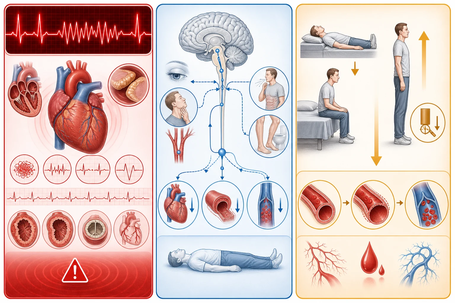
The Fellowship candidate must hold the four-category framework during the assessment — the reflex (the neurally-mediated), the orthostatic, the cardiac, and the cerebrovascular. The cardiac causes dominate the risk; the reflex causes dominate the numbers. Roughly a third to a half of the syncope in the emergency department is reflex, a further portion is cardiac, and the remainder is orthostatic or unexplained after the workup.[3][4]
Reflex (neurally-mediated)
- Vasovagal — the commonest cause overall (emotion, pain, prolonged standing, warm environment)
- Situational — micturition, defecation, cough, post-prandial, hair-grooming
- Carotid sinus hypersensitivity — the older patient, tight collar, head turn
- Low risk; characteristic prodrome; upright position; reproducible
Orthostatic
- Hypovolaemia — dehydration, haemorrhage, vomiting, diarrhoea
- Drug-induced — antihypertensives, diuretics, alpha-blockers, dopaminergic agents
- Autonomic failure — primary (Parkinson, PAF), secondary (diabetes, alcohol)
- Usually low risk; reproduces on standing; reversible when the cause is corrected
Cardiac — arrhythmia
- Bradycardia — sinus node dysfunction, AV block (Mobitz II, complete), slow AF
- Tachycardia — VT, SVT, AF with a rapid response, Torsades
- The commonest cardiac cause; often no prodrome; may occur in the supine position
- High risk; the ECG, the monitoring and the cardiology are essential
Cardiac — structural
- Aortic stenosis, hypertrophic cardiomyopathy — the exertional syncope
- Pulmonary embolism, pulmonary hypertension, the cardiac tamponade
- Aortic dissection, myocardial infarction, arrhythmogenic cardiomyopathy
- High risk; the exertional or supine faint; the bedside echo screens
The critical cardiac-versus-non-cardiac distinction
The single most important clinical decision is whether the syncope is cardiac or non-cardiac, because the cardiac syncope has a 6-month mortality of over 10 per cent (it may be the presenting feature of a life-threatening arrhythmia or a structural disease), while the non-cardiac reflex syncope has an excellent prognosis. The history is the diagnostic tool: a syncope on exertion (the aortic stenosis, the hypertrophic cardiomyopathy, the pulmonary hypertension), a syncope in the supine position (the arrhythmia), a syncope with a chest pain or a palpitation (the arrhythmia, the ischaemia, the PE), a syncope with no prodrome (the abrupt cardiac syncope), and a family history of the sudden cardiac death — these are the cardiac features. The vasovagal syncope, by contrast, has a characteristic prodrome (the nausea, the warmth, the sweating, the visual dimming, the lightheadedness) and a trigger (the emotion, the pain, the prolonged standing), and it occurs in the upright position. [1]
History — the diagnostic weapon
The history is the single most powerful diagnostic tool in the syncope — more than any investigation. A focused history alone identifies the likely mechanism in over half of the cases.[3] The candidate must ask about the position (the upright position favours the reflex or the orthostatic; the supine position favours the cardiac), the trigger (the emotion, the pain, the prolonged standing and the warm environment favour the vasovagal; the exertion favours the structural or the arrhythmic), the prodrome (the nausea, the warmth, the diaphoresis and the visual dimming favour the vasovagal; the abrupt loss with no prodrome favours the cardiac), the associated symptoms (the chest pain, the palpitation and the dyspnoea favour the cardiac or the pulmonary embolism), and the family history (the sudden cardiac death at a young age, the hereditary channelopathy).
Vasovagal / reflex
- Upright position; a clear trigger (emotion, pain, standing)
- A characteristic prodrome — warmth, nausea, diaphoresis, visual dimming
- A rapid, complete recovery; no post-event confusion
- Reproducible over time; a benign prognosis
Orthostatic
- On standing up; the elderly on multiple medications
- Lightheadedness; blurred vision; a minimal prodrome
- Reproduced by the orthostatic vital signs
- Correct the volume, adjust the drug, treat the autonomic cause
Cardiac — arrhythmia
- Supine position or any position; an abrupt onset; no prodrome
- Palpitation before the collapse; may occur in the sleep
- The ECG may show the cause or may be normal (paroxysmal)
- Needs the monitoring; a high risk of the recurrence and the sudden death
Cardiac — structural
- Exertional syncope — the classic red flag
- Chest pain, dyspnoea, a murmur on the examination
- Aortic stenosis, HCM, pulmonary hypertension, PE
- The bedside echo screens; the cardiology referral is mandatory
Examination — the structured search for the cause
The examination targets the mechanism and the injury. The vital signs (with the orthostatic measurement), the cardiovascular (the murmur of the aortic stenosis or the HCM, the signs of the heart failure), the neurological (a focal deficit suggesting a stroke or a subclavian steal), the carotids (a bruit before any massage), the rectal (the melaena of the occult gastrointestinal bleed), and the injury survey (the head injury, the fracture, the facial laceration from the fall) are the structured components. [1]
Reassess the ABCs and the bedside glucose — a hypotension or a bradycardia needs the immediate treatment
Orthostatic vital signs — measure the lying and the standing blood pressure and heart rate; a drop over 20 mmHg systolic or over 10 mmHg diastolic with the symptoms confirms the orthostatic syncope
Auscultate the heart — an ejection systolic murmur radiating to the carotids (the aortic stenosis), a murmur that augments with the Valsalva (the hypertrophic cardiomyopathy)
Examine for the heart failure — the raised JVP, the basal crackles, the peripheral oedema
Neurological examination — a focal deficit, a tongue bite, a post-event confusion, a nystagmus
Carotid sinus massage (if indicated) — in the monitored setting, no bruit, no stroke history, no recent MI; a pause over 3 seconds or a BP drop over 50 mmHg reproduces the carotid sinus hypersensitivity
Injury survey — the head injury, the fracture, the laceration from the fall; the elderly syncope commonly presents as a fall
Differential diagnosis — syncope and the mimics
The transient loss of consciousness has a differential, and the Fellowship candidate must distinguish the true syncope from the mimics. [1]
Syncope (true)
- Transient loss of consciousness from the cerebral hypoperfusion
- Rapid onset, short duration, spontaneous recovery
- No post-event confusion (unlike the seizure)
- Reflex, orthostatic or cardiac
Seizure
- A tonic-clonic movement, a tongue bite, a urinary incontinence
- A post-ictal confusion (the syncope has none)
- An aura or a déjà vu
- A prolonged recovery (minutes vs seconds)
Cardiac arrest (aborted)
- A circulatory arrest requiring the resuscitation
- May be mislabelled as a syncope if the recovery is rapid
- The ECG, the troponin and the echo are mandatory
- The highest-risk presentation
Other TLOC
- A hypoglycaemia (the bedside glucose excludes it)
- A psychogenic pseudosyncope (no loss of the consciousness — the eyes are closed, no injury)
- A drop attack (a sudden fall without a loss of consciousness)
- A cataplexy (the narcolepsy association)
Investigations — the ECG and the risk factors
The ECG is the single most important investigation — it is performed on every syncope patient and it may reveal the conduction abnormality (the complete heart block, the bifascicular block, the Mobitz II), the arrhythmia (the atrial fibrillation, the VT), the prolonged QT (the long-QT syndrome), the Brugada pattern, the ischaemia, or the pre-excitation (the WPW). The bedside glucose excludes the hypoglycaemia. The full blood count (the anaemia contributing to the orthostatic syncope), the electrolytes (the hyponatraemia, the hyperkalaemia — both pro-arrhythmic), and the troponin (if the chest pain or the ischaemia is suspected) are sent. The bedside echo screens for the structural heart disease (the aortic stenosis, the hypertrophic cardiomyopathy, the pulmonary hypertension, the reduced EF). The orthostatic vital signs (the lying and the standing blood pressure — a drop of over 20 mmHg systolic or over 10 mmHg diastolic on standing, with symptoms, confirms the orthostatic syncope). The carotid sinus massage (in the monitored setting, with no bruit and no stroke history — a pause over 3 seconds or a drop over 50 mmHg confirms the carotid sinus hypersensitivity). The CT head is NOT routine — it is performed only if a head injury, a focal neurology, or a subarachnoid haemorrhage is suspected. [1]
The ECG — mandatory on every syncope patient
The ECG is performed on every patient presenting with a syncope — without exception — and it is the highest-yield single investigation. Most tracings are normal, but the abnormal ECG is a high-risk feature that mandates the admission.[3] The candidate must actively search for the deadly patterns, not merely confirm that the tracing is in the sinus.
Conduction disease
- Bifascicular block (RBBB + LAFB or LPFB) — a high risk of the complete block
- Mobitz II and the third-degree AV block — a pacing indication
- Sinus pauses over 3 s, the sick sinus syndrome, the paroxysmal AV block
- A new bundle-branch block after a syncope is high-risk
Brugada pattern
- A coved ST elevation in V1 to V3 (type 1) — the coved-downslope
- A risk of the polymorphic VT and the sudden cardiac death
- May be intermittent — record the right-precordial leads in the high position
- A family history of the sudden death raises the suspicion
Long QT
- QTc over 470 ms (men) or 480 ms (women), or over 500 ms unequivocal
- A risk of the Torsades de pointes — a syncope or a sudden death
- Drugs (macrolides, antipsychotics, methadone), the electrolytes (low K, Mg, Ca)
- Congenital (Romano-Ward, Jervell-Lange-Nielsen) in the young
WPW / pre-excitation
- A short PR, a delta wave, a widened QRS
- A risk of the pre-excited AF degenerating to the VF
- A syncope with the WPW is high-risk — the pathway ablation
- Look for the delta wave in the lead V1 and the short PR
Ischaemia / structural
- The ST changes of the acute ischaemia — the syncope may be the arrhythmia of the MI
- Q waves, T inversion, LVH with the strain — the structural substrate
- The bradyarrhythmia of the inferior MI, the VT of the scarred ventricle
- The low voltage of the pericardial effusion or the tamponade
Risk stratification — the San Francisco Syncope Rule
The San Francisco Syncope Rule identifies the high-risk patient who needs the admission. The five criteria — any one of which flags the patient as the high-risk: [1]
San Francisco Syncope Rule — any one flags the high-risk
CHESS
A history or the signs of the heart failure
A severe anaemia contributing to the syncope
Any abnormality — the conduction, the arrhythmia, the ischaemia, the QT, the Brugada
At the presentation or with the event
A low systolic blood pressure at the triage
A patient with any one of these five has a serious-outcome risk of about 10 per cent over 7 to 30 days, and warrants the admission.[2] The ESC high-risk features are similar and broader: the structural heart disease, the abnormal ECG, the palpitations at the time of the syncope, the syncope in the supine position or on exertion, the sudden and the abrupt loss of consciousness without a prodrome, the frequent recurrence, and the family history of the sudden cardiac death.[1]
Risk stratification tools compared — the SFSR, the CSRS and the ROSE
No single rule is perfect; the SFSR is sensitive but poorly specific, and it was derived for the short-term serious outcome. The Canadian Syncope Risk Score (CSRS) is a validated, multicentre tool that stratifies the 30-day serious-outcome risk into the very-low through high bands, and it performs better than the SFSR in the contemporary validation.[6][5] The ROSE rule (the Red Flags in Syncope) is an alternative used in the UK. The candidate should know one rule well and understand its limitations. The Canadian Syncope Arrhythmia Risk Score (CSARS) is a complementary tool that targets the short-term arrhythmic risk specifically and flags the patient who warrants the prolonged monitoring.[8]
San Francisco Syncope Rule
- Five criteria — CHF, Hct under 30, abnormal ECG, SOB, SBP under 90
- Any one flags the high-risk; sensitive (~96–98%) but non-specific
- Designed for the 7-day serious outcome
- Simple and widely taught; over-admits
Canadian Syncope Risk Score
- Nine variables — the age, the sex, the BP, the troponin, the ECG abnormality
- Stratifies into the very-low to high 30-day risk bands
- Better calibrated than the SFSR in the multicentre validation
- The high band — admit; the very-low band — the safe discharge
ROSE rule (UK)
- BNP, the gut motility, the bradycardia, the rectal bleed, the ECG, the SaO2
- Identifies the high-risk missed by the SFSR
- Less commonly used outside the UK
- The BNP is not routinely available in the ED
ESC high-risk features
- The structural heart disease, the abnormal ECG, the palpitation at the event
- Supine or exertional syncope; the abrupt no-prodrome faint
- The family history of the sudden death; the frequent recurrence
- A clinical-judgement framework, not a numeric score
Immediate management — treat the cause and the risk-stratify
The management follows the risk stratification. [1]
[1]The syncope targets and the thresholds
Disposition algorithm — admit, observe or discharge
The disposition follows the risk stratification. Three pathways: the high-risk patient is admitted for the cardiac monitoring and the cardiology assessment; the intermediate-risk patient is observed in the ED short-stay or the chest-pain unit for 6 to 12 hours with the serial ECGs; the low-risk patient is discharged with the advice and the primary-care follow-up. [1]
Resuscitate, take the history, examine, and obtain the 12-lead ECG and the bedside glucose on every patient
Apply the San Francisco Syncope Rule and the ESC high-risk features; consider the Canadian Syncope Risk Score
High-risk (any SFSR criterion, any ESC feature, an abnormal ECG, the structural disease) — admit for the cardiac monitoring, the troponin, the echo and the cardiology assessment
Intermediate-risk (older, recurrent, an unclear mechanism, an isolated borderline feature) — observe 6 to 12 hours in the short-stay unit with the serial ECGs and the reassessment
Low-risk (a clear vasovagal, a normal ECG, no structural disease, no high-risk feature) — discharge with the syncope advice and the primary-care follow-up in 7 days
Discharge safety-net — return immediately for the chest pain, the palpitation, the recurrence or the new shortness of breath
Document the risk assessment, the rule applied, the advice given and the follow-up arranged
Complications and pitfalls
The complications are the recurrent syncope (the injury from the fall — the fracture, the head injury), the sudden cardiac death from an untreated cardiac syncope, and the complications of the over-investigation (the unnecessary CT, the unnecessary admission, the anxiety). The pitfalls are: not recognising the cardiac syncope (the exertional, the supine, the no-prodrome, the abnormal ECG); over-relying on the single ECG (the arrhythmia may be paroxysmal — the monitoring is needed); not checking the bedside glucose; performing a routine CT head on every syncope (it is low-yield and not recommended); not performing the orthostatic vital signs; and discharging a high-risk patient without the cardiology assessment. [1]
Prognosis and disposition
The prognosis depends on the cause. The reflex syncope has an excellent prognosis; the cardiac syncope has a 6-month mortality of over 10 per cent;[7] the unexplained syncope (after the ED workup) is intermediate. The disposition: the high-risk patient is admitted to the cardiology or the monitored bed; the low-risk patient is discharged with the advice and the GP follow-up; the intermediate-risk patient may be observed in the ED's short-stay unit for 6 to 12 hours with the serial ECGs and the monitoring.
Special populations
The elderly patient with the syncope has a broader differential (the medication-induced orthostatic, the carotid sinus hypersensitivity, the arrhythmia, the structural heart disease, the aortic stenosis) and a higher threshold for the workup. The cardiac history patient (the prior MI, the reduced EF, the structural disease) is a high-risk presentation — the cardiology is involved early. The athlete with the syncope is a high-risk presentation (the hypertrophic cardiomyopathy, the arrhythmogenic cardiomyopathy, the long-QT, the WPW) — the sports cardiology and the restriction from the sport are the considerations. The pregnant patient has a higher baseline vagal tone and a higher orthostatic risk from the vasodilation. [1]
The landmark evidence and the guidelines
2018 ESC Guidelines for the diagnosis and management of syncope
European Heart Journal (2018)
PMID 29562304
Society clinical-practice guideline
Population: All patients with the transient loss of consciousness
Key finding
Codified the four-category classification (reflex, orthostatic, cardiac, cerebrovascular), the structured history-and-ECG evaluation, the high-risk features mandating the admission, and the management of the recurrent reflex syncope (the counter-pressure manoeuvres, the fludrocortisone, the selective pacing in the cardioinhibitory type). Established that the routine CT head, the carotid ultrasound and the EEG are NOT indicated without a focal neurological pointer.
2017 ACC/AHA/HRS Guideline for the Evaluation and Management of Patients with Syncope
Circulation (2017)
PMID 28280231
Society clinical-practice guideline
Population: Adults with the suspected syncope
Key finding
Aligned with the ESC on the structured evaluation, the cardiac-versus-non-cardiac distinction and the risk-stratified disposition. Recommended the ECG for every patient, the selective echocardiography and the structured history as the cornerstone. Emphasised the shared decision-making and the reduction of the unnecessary testing.
Derivation of the San Francisco Syncope Rule
Annals of Emergency Medicine (2004)
PMID 14747812
Prospective cohort derivation
Population: 684 ED syncope patients
Key finding
Derived the five-variable rule (CHF, Hct under 30, abnormal ECG, shortness of breath, SBP under 90) that identified the patients at risk of a serious outcome within 7 days with a sensitivity of 96 per cent and a specificity of 62 per cent. Any one criterion flagged the patient as high-risk.
External validation of the San Francisco Syncope Rule
Annals of Emergency Medicine (2007)
PMID 17210201
Prospective multicentre validation
Population: 716 ED syncope patients
Key finding
The SFSR underperformed its derivation sensitivity in the external validation (89 per cent), missing a small number of the serious outcomes. The rule is a useful adjunct but does not replace the clinical judgement, and the abnormal ECG remains the strongest single predictor.
Multicenter ED Validation of the Canadian Syncope Risk Score
JAMA Internal Medicine (2020)
PMID 32202605
Prospective multicentre cohort
Population: 3819 ED syncope patients across 9 Canadian sites
Key finding
Validated the CSRS as a well-calibrated 30-day serious-outcome prediction tool, stratifying the patients into the very-low to high risk. Outperformed the SFSR in the discrimination and allowed the confident discharge of the very-low-risk band. The troponin and the ECG abnormality were the dominant predictors.
Incidence and prognosis of syncope — the Framingham Heart Study
New England Journal of Medicine (2002)
PMID 12239256
Prospective population cohort
Population: 7814 Framingham subjects over 26 years
Key finding
The lifetime cumulative incidence of the syncope was 6 per cent in the middle-aged and 11 per cent in the elderly. The cardiac syncope doubled the all-cause mortality and the risk of the non-fatal and fatal cardiovascular events, confirming the cardiac syncope as a marker of the underlying structural or arrhythmic disease.
ACC/AHA/HRS versus ESC guidelines for the diagnosis and management of syncope
Journal of the American College of Cardiology (2019)
PMID 31699282
Comparative guideline review
Population: Patients with the syncope
Key finding
Compared the two major guidelines and found them broadly aligned on the evaluation, the risk stratification and the disposition, with the minor differences in the emphasis on the tilt-testing and the prolonged monitoring. The ECG and the history remain the cornerstone of both.
Evidence and regional guidelines
The contemporary framework is the ESC syncope guideline (2018) and the ACC/AHA/HRS syncope guideline, which are broadly aligned.[1] The San Francisco Syncope Rule is the validated risk-stratification tool for the emergency department.[2] The risk-stratification approach and the admission criteria are global; the local protocol governs the monitoring, the cardiology referral, and the follow-up pathway.
ANZ practice note. The San Francisco Syncope Rule and the ESC risk factors guide the admission; the ECG is mandatory on every syncope patient; the high-risk patient (the abnormal ECG, the structural heart disease, the exertional syncope, the family history of the sudden cardiac death) is admitted for the cardiac monitoring and the cardiology assessment, and the low-risk reflex syncope is discharged with the advice and the GP follow-up. [1]
SAQ — The exertional syncope and the high-risk ECG
10 minutes · 10 marks
A 58-year-old man is brought to the emergency department after collapsing while climbing a flight of stairs. He recovered fully within 30 seconds and has no chest pain, but he describes a brief palpitation just before the collapse. His father died suddenly at the age of 50. The examination is normal, the blood pressure is 128 over 76, and the 12-lead ECG shows a bifascicular block (a right bundle branch block with a left anterior fascicular block).
SAQ — The recurrent syncope in the elderly patient on multiple medications
10 minutes · 10 marks
A 76-year-old woman who takes amlodipine, furosemide and doxazosin for the hypertension presents after her third syncopal episode in two months, all on standing up from the chair in the morning. She recovers fully each time within the seconds, has no chest pain or palpitation, and the examination, the 12-lead ECG and the bedside glucose are normal. The lying blood pressure is 138 over 82, and on standing it falls to 104 over 64 with the reproducing lightheadedness.
Exam pearls
- The cardiac syncope is the high-risk: the exertional, the supine, the no-prodrome, the abnormal ECG, the structural heart disease, the family history of the sudden cardiac death.
- The San Francisco Syncope Rule: any one of CHF, Hct <30, abnormal ECG, SOB, SBP <90 → admit.
- The ECG is mandatory on every syncope patient — the arrhythmia, the conduction block, the long-QT, the Brugada.
- The vasovagal syncope has a prodrome; the cardiac syncope does not.
- The seizure has a post-ictal confusion; the syncope does not.
- The CT head is NOT routine — only for the head injury, the focal neurology, or the SAH suspicion.
- The orthostatic vitals: >20 mmHg systolic drop with symptoms on standing.
- The reflex syncope has a prodrome and a trigger; the cardiac syncope does not — the abrupt no-prodrome faint is cardiac until proven otherwise.
- The position is diagnostic: the upright faint is reflex or orthostatic; the supine faint is arrhythmic.
- The carotid sinus massage is performed only in the monitored setting, with no bruit and no prior stroke — a pause over 3 seconds or a BP drop over 50 mmHg reproduces the carotid sinus hypersensitivity.
- The bedside glucose is checked on every syncope — the hypoglycaemia is a mimic, and it is reversible in seconds.
- The troponin is sent if the chest pain, the ischaemia or the cardiac features are present — a routine troponin on every syncope is low-yield.
- The CT head is NOT routine for the syncope — only for the head injury, the focal neurology, the anticoagulation with a head strike, or the suspected subarachnoid haemorrhage.
- The Canadian Syncope Risk Score stratifies the 30-day risk into bands and outperforms the SFSR in the calibration — know one rule well.
- The bifascicular block with a syncope is a pacing problem — admit for the monitoring even if the index ECG is stable.
- The long-QT syncope — check the QTc, stop the offending drug, correct the potassium and the magnesium, and involve the cardiology.
- The Brugada pattern — the coved ST elevation in V1 to V3 — with a syncope is high-risk for the sudden death; record the right-precordial leads in the high position.
- The syncope in the athlete — the hypertrophic cardiomyopathy, the arrhythmogenic right-ventricular cardiomyopathy, the long-QT, the WPW — restrict from the sport and refer to the sports cardiology.
- The elderly syncope frequently presents as a fall — the unwitnessed collapse with a head injury in the older patient may be a syncope or an arrhythmia, and the workup is the same.
- Document the risk stratification, the rule applied, the advice given and the follow-up — the medicolegal protection for the discharge.
- The recurrent syncope with an injury (the fracture, the head injury, the driving incident) needs a definitive workup, not a repeat discharge. [1]
Red flags
[1]References
- [1]Goldberger ZD, Petek BJ, Brignole M, et al. ACC/AHA/HRS Versus ESC Guidelines for the Diagnosis and Management of Syncope: JACC Guideline Comparison J Am Coll Cardiol, 2019.PMID 31699282
- [2]Quinn JV, Stiell IG, McDermott DA, et al. Derivation of the San Francisco Syncope Rule to predict patients with short-term serious outcomes Ann Emerg Med, 2004.PMID 14747812
- [3]Brignole M, Moya A, de Lange FJ, et al. 2018 ESC Guidelines for the diagnosis and management of syncope Eur Heart J, 2018.PMID 29562304
- [4]Shen WK, Sheldon RS, Benditt DG, et al. 2017 ACC/AHA/HRS Guideline for the Evaluation and Management of Patients With Syncope: A Report of the American College of Cardiology/American Heart Association Task Force on Clinical Practice Guidelines and the Heart Rhythm Society Circulation, 2017.PMID 28280231
- [5]Sun BC, Mangione CM, Merchant G, et al. External validation of the San Francisco Syncope Rule Ann Emerg Med, 2007.PMID 17210201
- [6]Thiruganasambandamoorthy V, Sivilotti MLA, Le Sage N, et al. Multicenter Emergency Department Validation of the Canadian Syncope Risk Score JAMA Intern Med, 2020.PMID 32202605
- [7]Soteriades ES, Evans JC, Larson MG, et al. Incidence and prognosis of syncope N Engl J Med, 2002.PMID 12239256
- [8]Thiruganasambandamoorthy V, Stiell IG, Sivilotti MLA, et al. Predicting Short-term Risk of Arrhythmia among Patients With Syncope: The Canadian Syncope Arrhythmia Risk Score Acad Emerg Med, 2017.PMID 28791782