EM · Cervical spine injury & clearance
Cervical spine injury and clearance in trauma
Also known as C-spine injury · Cervical spine clearance · Spinal cord injury · NEXUS criteria
Cervical spine injury and clearance — the NEXUS and the Canadian C-spine clinical decision rules for the safe clearance of the C-spine without the imaging, the CT as the standard imaging (replacing the plain radiographs), the spinal-cord-injury syndromes (the complete, the central cord, the anterior cord, the Brown-Sequard), the SCIWORA, the immobilisation (the collar, the head blocks, the manual in-line stabilisation), the methylprednisolone controversy (no longer recommended by the AANS), and the neurosurgical referral. ACEM-primary, globally tagged.
On this page & tools
Your progress
Saved locally on this device.
Target exams
Red flags

Cervical spine injury complicates up to 5 per cent of the major trauma presentations and up to 10 per cent of the head-injured patients, and the Fellowship candidate must know the clinical decision rules for the safe clearance, the imaging strategy (the CT as the standard, the MRI for the ligamentous and the cord), the spinal-cord-injury syndromes, and the management — because the missed C-spine injury with a secondary cord damage is the preventable disaster that ends careers.[1][2]
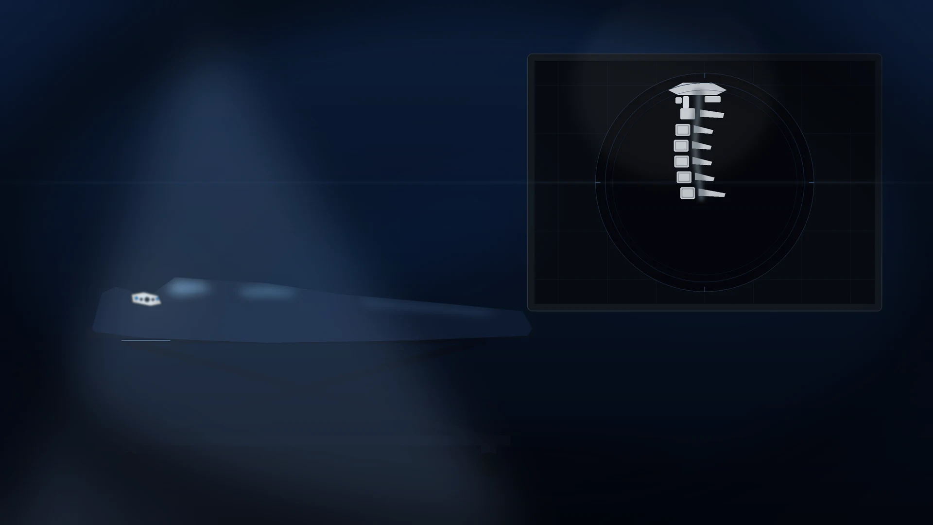
The immobilisation — from the first contact
The C-spine is immobilised from the first pre-hospital contact in any patient with a mechanism compatible with a spinal injury (the high-speed collision, the fall from a height, the diving injury, the assault). The rigid cervical collar, the head blocks and the tape (or the sandbags) are the standard. The manual in-line stabilisation (the MILS) is used during the intubation — a second operator holds the head in the neutral position while the intubator performs the RSI. The collar is NOT removed until the C-spine is cleared — clinically (by the decision rules) or radiologically (by the CT or the MRI). [1]
The equipment — the collar, the board, the vacuum mattress
The rigid cervical collar (the Ambu Perfit, the Stifneck, the Aspen) limits the flexion and the extension but does NOT eliminate the movement — it allows roughly 30 per cent of the normal flexion-extension even when correctly fitted, which is why it is always paired with the head blocks and the tape. The collar must be sized correctly: too large and it flexes the neck; too small and it fails to control the movement. The head blocks (or the rolled blankets, or the sandbags) on each side plus the tape across the forehead and the chin secure the head to the spinal board. The combination — the collar, the head blocks, the tape — is the triple immobilisation that is the pre-hospital standard. [1]
The scoop stretcher (the orthopaedic stretcher) is the preferred device for the lifting and the transfer — it splits longitudinally so it slides under the patient with the minimal log-roll, reducing the spinal movement compared with the rigid board. The long spine board (the LSB) is for the extrication only — it is NOT a transport device, because the prolonged time on the hard board causes the pressure injury (the sacral and the heel ulcers develop within 30 minutes on an uncushioned board), the pain, and the respiratory restriction. The current ATLS and the ANZ trauma guidance is: transfer off the board within 30 minutes, onto the scoop or the vacuum mattress for the transport. [1]
The log-roll is the manoeuvre for the examination and the transfer of the supine trauma patient while the spinal alignment is maintained. The patient is rolled as a single unit (the head, the shoulders, the pelvis, the legs moving together) by a minimum of four staff — one at the head (the team leader, controlling the head and the neck), two at the trunk, and one at the legs. The 15-degree log-roll is used for the scooping and the examination of the back; the full 90-degree roll is used for the transfer onto the board. The log-roll is avoided in the suspected thoracolumbar fracture — the patient is lifted on the scoop instead, because the roll pivots on the unstable segment. [1]
[1]The pre-hospital and the ED immobilisation sequence
1
2
3
4
5
Differential diagnosis — the clearance rules and the imaging strategy
The first decision is whether to image or to clear clinically. The two validated clinical decision rules guide this. [1]
NEXUS (5 criteria)
- ALL 5 must be present to clear without imaging
- No midline cervical tenderness
- No focal neurological deficit
- Normal level of alertness (no intoxication, no GCS <15)
- No painful distracting injury
- If any one is absent, the patient is imaged
Canadian C-spine rule
- More specific (fewer unnecessary scans); the age over 65, the dangerous mechanism, the paresthesias are the high-risk factors
- If none of the high-risk, then the low-risk factor (a simple rear-end, sitting, ambulatory, delayed onset of pain, no midline tenderness) allows the assessment of the rotation
- The patient who can rotate 45 degrees left and right is cleared without imaging
- More sensitive and more specific than NEXUS
CT (the standard)
- The CT of the cervical spine (with the head, the chest, the abdomen and the pelvis as the trauma series)
- Replaced the plain radiographs (the sensitivity of the 3-view is only about 50 per cent)
- Sensitivity for the bony injury over 99 per cent
- Does NOT exclude the purely ligamentous injury
MRI (the definitive)
- The MRI for the suspected ligamentous or the cord injury
- The SCIWORA (the paediatric cord injury with a normal CT)
- The central cord syndrome with a normal CT
- The persistent midline tenderness despite a normal CT (the obtunded or the symptomatic patient)
The NEXUS criteria — the five low-risk criteria
The NEXUS (the National Emergency X-Radiography Utilization Study) low-risk criteria, published by Hoffman and colleagues in 2000, is the older and the simpler of the two rules. The rule says: a stable, alert trauma patient can have the C-spine cleared clinically (without any imaging) if ALL FIVE of the following are present.[3] The mnemonic is the NSAID in reverse — but most remember them as the five. The candidate must be able to recite them precisely.
The NEXUS five — NSAID without the D, or just learn them
NEXUS
A full neurological examination of the upper and the lower limbs (the motor, the sensory, the reflexes) is normal. Any focal deficit — a weakness, a sensory level, a hyperreflexia — is imaging.
No clinical evidence of alcohol or drug intoxication. The intoxicated patient cannot reliably report the pain, and the intoxication itself may have caused a fall that injured the spine.
A normal level of alertness — the GCS of 15, no reduced consciousness from a head injury, no post-ictal state, no dementia that prevents a reliable examination. The candidate must be fully oriented and cooperative.
No painful injury elsewhere that would distract the patient from the cervical tenderness — a long-bone fracture, a large laceration, a burn, a crush injury, an acute abdomen. The threshold is the clinical judgement of the examiner.
No midline cervical-spine tenderness on the palpation of the spinous processes from the occiput to T1. The tenderness off the midline (the paraspinal muscles) does NOT count — only the midline posterior elements.
The Canadian C-spine rule — the three high-risk factors
The Canadian C-spine rule, published by Stiell and colleagues in 2001, is the more recent, the more specific, and the more clinically useful of the two rules. It applies only to the alert (GCS 15) and the stable trauma patient in whom the C-spine injury is a concern. The rule is a three-step decision tree that asks, in order: the high-risk factors, the low-risk factors, and the rotation.[4]
The Canadian C-spine rule — the three-step decision
1
2
3
NEXUS
- Sensitivity ~99%, specificity ~13%
- Simpler to apply — five binary criteria
- Validated across the age range including the elderly
- Includes the paediatric population (down to the neonate in the original study)
- The most widely taught and the most internationally adopted rule
- Tends to over-image — the low specificity
Canadian C-spine rule
- Sensitivity ~100%, specificity ~45%
- More complex — a three-step decision tree with the rotation
- More specific — images fewer patients for the same sensitivity
- Validated only in the alert and the stable patient (GCS 15)
- Validated in adults; the paediatric extension is down to 16 years
- Preferred by the ANZ and the Canadian trauma guidance where applicable
NEXUS — the five low-risk criteria to rule out the cervical-spine injury
New England Journal of Medicine
A prospective, multi-centre, observational study of 34,069 patients with blunt trauma who had a cervical-spine radiograph, validating the five low-risk criteria to rule out the injury without imaging.
Key finding
The NEXUS criteria had a sensitivity of 99.0 per cent (8 of 818 clinically significant injuries were missed — a 1 per cent miss rate) and a specificity of 12.9 per cent, reducing the radiography rate by roughly 12 per cent.
Practice change
Established the five-criterion rule as the most widely adopted clinical decision rule for the cervical-spine clearance, though the low specificity led to the over-use of the plain radiographs and, later, the CT.
The Canadian C-spine rule — versus the NEXUS
Annals of Emergency Medicine
A prospective cohort study of 8,283 alert and stable trauma patients comparing the Canadian C-spine rule and the NEXUS criteria head-to-head for the radiography in the emergency department.
Key finding
The Canadian C-spine rule was more sensitive (99.4 per cent vs 90.7 per cent for NEXUS in this cohort) and more specific (45.1 per cent vs 36.9 per cent), with a lower imaging rate.
Practice change
Established the Canadian C-spine rule as the superior rule where it can be applied, and is now the preferred rule in the ANZ and the Canadian trauma protocols.
The spinal cord injury syndromes
The spinal cord injury is classified as the complete (the total loss of the motor and the sensory function below the level — the worst prognosis) and the incomplete (some preserved function). The incomplete syndromes are examined because they test the anatomical knowledge of the cord tracts. [1]
The incomplete cord syndromes
CORD
The upper-limb weakness greater than the lower-limb; after a hyperextension in the elderly (the cervical spondylosis); the most common incomplete syndrome
The bilateral motor loss and the pain/temperature loss, with the preserved proprioception and the vibration (the posterior columns spared); from the anterior spinal artery occlusion or the flexion injury; the worst incomplete prognosis
The ipsilateral motor and proprioception loss with the contralateral pain/temperature loss (the hemisection); from the penetrating injury; the best incomplete prognosis
The loss of the proprioception and the vibration with the preserved motor; rare; from the hyperextension
The SCIWORA (the spinal cord injury without a radiographic abnormality) is a paediatric entity — the flexible paediatric spine allows the cord to be injured (the stretch, the contusion) without the bony fracture or the dislocation on the CT. The MRI shows the cord oedema or the haemorrhage. The child with a neurological deficit after a trauma has a spinal cord injury until proven otherwise, even if the CT is normal. [1]
The SCIWORA — the paediatric cord injury with the normal CT
The SCIWORA (the spinal cord injury without a radiographic abnormality) was first described before the CT era, when it meant "no abnormality on the plain films." With the modern CT, the definition has shifted: the SCIWORA is a spinal cord injury with a normal CT (the bony injury is absent) — and the MRI is the diagnostic test, showing the cord oedema, the haemorrhage, or the ligamentous injury. The SCIWORA is overwhelmingly a paediatric entity for two reasons: the paediatric spine is more elastic than the spinal cord (the ligaments stretch up to 5 cm before they tear, but the cord tears at 0.6 cm), so the spine can deform and recoil without a fracture while the cord is stretched and contused; and the relatively large head and the weak neck muscles of the younger child amplify the flexion and the extension forces on the high cervical spine. [1]
The SCIWORA presents in the child (typically under 8 years) after a high-energy mechanism (the motor vehicle crash, the pedestrian struck, the fall from a height, the bicycle handlebar, the sport) with a neurological deficit — a weakness, a sensory level, a paraesthesia, a transient paralysis that may have recovered by the time of the presentation (the "sting" phenomenon). The examination may show a complete or an incomplete cord syndrome. The delayed SCIWORA (the onset of the deficit hours to days after the injury) is a recognised and a feared pattern — the child who walks in and later develops the paralysis. [1]
[1] [1]The PECARN paediatric head-injury rule — and the paediatric C-spine
The PECARN (the Pediatric Emergency Care Applied Research Network) rule, published by Kuppermann and colleagues in 2009, is the validated clinical decision rule for the paediatric head injury — it identifies the children at a very low risk of a clinically important traumatic brain injury in whom the CT can be avoided.[6] The rule is age-stratified (under 2 years, and 2 years and over) and is the most widely used paediatric head-injury rule in the world. While the PECARN rule addresses the brain, the paediatric C-spine clearance is a separate and a more contentious question.
PECARN — under 2 years
- A GCS of less than 15 — image
- A palpable skull fracture — image
- A occipital, parietal or temporal scalp haematoma — image
- A loss of consciousness for 5 minutes or longer — image
- Not acting normally per the parent, or a severe mechanism — consider
- NONE of the above → a very low risk (under 0.02%) — observation only
PECARN — 2 years and over
- A GCS of less than 15 — image
- A sign of a basilar skull fracture — image
- A loss of consciousness — image
- A history of vomiting — image
- A severe headache, or a severe mechanism — consider
- NONE of the above → a very low risk (under 0.05%) — observation only
Paediatric C-spine
- The NEXUS is validated down to the paediatric age group but the data are sparse for the under-8 age group
- The Canadian C-spine rule is validated only down to 16 years
- The children under 8 with a significant mechanism are generally imaged if any criterion fails
- The paediatric C-spine CT carries a lifetime cancer risk (the radiosensitive thyroid) — the judicious use is the principle
- The MRI is the first-line for the suspected SCIWORA in the child with a normal CT
Immediate management — the immobilisation and the cord protection
[1]The C-spine targets and the doses
The clearance of the obtunded patient
The clearance of the obtunded or the intubated patient is the most contentious area, because the clinical decision rules cannot be applied (the patient cannot report the tenderness or the pain). The CT alone (a normal high-quality CT) is now considered sufficient for the clearance in most major trauma centres (the sensitivity for the clinically significant injury is over 99 per cent). The MRI was previously mandatory for the obtunded patient with a normal CT, but the MRI has a high false-positive rate (it shows the ligamentous oedema that is not a clinically significant injury), and the sedation and the transfer risks of the MRI outweigh the benefit in most cases. The current consensus is: the normal high-quality CT clears the obtunded patient, and the MRI is reserved for the patient with a neurological deficit that is unexplained by the CT.[1][2]
Complications and pitfalls
The complications are the secondary cord injury (the preventable — from the hypoxia, the hypotension, the inadequate immobilisation), the spinal shock (the hypotension and the bradycardia from the loss of the sympathetic tone, lasting up to 1 to 2 weeks), the autonomic dysreflexia (the later complication of the cord injury at T6 or above — the hypertensive crisis from the noxious stimulus below the level), the respiratory failure (the diaphragm — the C3 to C5; the high cervical injury needs the ventilation), the deep vein thrombosis, the pressure injury, and the neurogenic bowel and bladder. The pitfalls are: removing the collar before the clearance; relying on the plain radiographs (the sensitivity is too low); not considering the SCIWORA in the paediatric patient with a neurological deficit and a normal CT; giving the methylprednisolone as a standard (it is no longer recommended); not maintaining the MAP at 85 to 90 for the cord perfusion; and not referring the neurosurgery early for the unstable fracture or the cord compression. [1]
Prognosis and disposition
The prognosis depends on the level and the completeness of the cord injury. The complete cord injury (the ASIA A — no motor or sensory function below the level) has a poor prognosis for the recovery. The incomplete syndromes (the ASIA B to D) have a variable but a generally better prognosis — the central cord syndrome often recovers partially; the Brown-Sequard may recover substantially; the anterior cord is the worst. The patient is admitted to the neurosurgery, the intensive care or the spinal injuries unit. The early decompression (within 24 hours) of the cord compression from a fracture or a haematoma improves the neurological outcome in selected cases. [1]
Special populations
The elderly patient (the cervical spondylosis) is at a higher risk of the central cord syndrome from a low-energy hyperextension (a fall from a standing height). The paediatric patient is at a higher risk of the SCIWORA and the high-cervical injury (the relatively large head, the weak neck muscles, the ligamentous laxity). The pregnant patient is managed with the left-lateral tilt and the modified immobilisation (the collar may not fit in the later pregnancy). The anticoagulated patient has a higher risk of the spinal epidural haematoma (a rare but a catastrophic complication that needs the urgent MRI and the surgical decompression within 24 hours to preserve the cord function). The early reversal of the anticoagulation in the suspected cord injury is the standard. [1]
Evidence and regional guidelines
The contemporary framework is the ATLS and the local trauma protocol for the C-spine clearance, the NEXUS and the Canadian C-spine rule for the clinical decision-making, and the CT as the standard imaging.[1] The methylprednisolone is no longer recommended by the AANS and the ATLS. The MRI is reserved for the unexplained neurological deficit.[2]
ANZ practice note. The C-spine clearance follows the ATLS/EMST framework via the local trauma protocol; the NEXUS or the Canadian C-spine rule are used for the clinical clearance; the CT of the cervical spine is the standard imaging (the plain radiographs are no longer used); the MRI is reserved for the unexplained neurological deficit or the suspected ligamentous injury; the methylprednisolone is not routinely given; and the neurosurgery is referred for any cord injury or an unstable fracture. [1]
SAQ — Clinical decision rules for C-spine clearance
10 minutes · 10 marks
A 42-year-old man presents to your emergency department 30 minutes after a high-speed motor vehicle crash at 110 km/h on a highway. He was the restrained driver, the vehicle rolled over, and he was extracted by paramedics in a rigid cervical collar with head blocks. On arrival his GCS is 15, he is not intoxicated, his vital signs are normal, and he complains of midline cervical tenderness at the C5 level and paraesthesia in both hands. He has no other injuries.
SAQ — Complications of prolonged cervical spine immobilisation
10 minutes · 10 marks
A 78-year-old woman is admitted to your emergency department after a fall from a standing height. She has a Glasgow Coma Scale of 14, and because of her dangerous mechanism and inability to cooperate fully she remains in a rigid cervical collar and on a scoop stretcher pending CT. Six hours later she is still in the collar. The nurse asks whether the collar can stay on overnight while she waits for an inpatient bed.
Exam pearls
- NEXUS: all 5 present to clear without imaging (no tenderness, no deficit, alert, no intoxication, no distracting injury).
- Canadian C-spine: the age over 65, the dangerous mechanism, the paresthesias are the high-risk; the rotation 45 degrees clears the low-risk.
- The CT replaces the plain radiographs — the sensitivity over 99 per cent for the bony injury.
- A normal CT does NOT exclude the purely ligamentous injury — the MRI is the definitive test.
- The central cord syndrome: the upper-limb weakness greater than the lower-limb, after a hyperextension in the elderly — the commonest incomplete cord syndrome.
- The SCIWORA: the paediatric cord injury with a normal CT — the MRI is the diagnostic test.
- The methylprednisolone is NO LONGER recommended by the AANS/ATLS — a local decision, not a standard.
- The MAP target 85 to 90 for the cord perfusion — the noradrenaline for the spinal shock. [1]
Red flags
[1]References
- [1]Anderson GA, Schroeder GD, Rasouli MR, et al. Cervical spine clearance in adult trauma patients: What youneed to know J Trauma Acute Care Surg, 2026.PMID 42224714
- [2]de Oliveira Manduca Palmiero H, Dall'Ava DO, Cunha MP, et al. Cervical Spine Clearance in Adult and Pediatric Trauma: A Systematic Review J Emerg Med, 2026.PMID 41955954
- [3]Hoffman JR, Mower WR, Wolfson AB, Todd KH, Zucker MI, et al. Is generic prescribing acceptable in epilepsy? Drug Saf, 2000.PMID 11005701
- [4]Stiell IG, Wells GA, Vandemheen KL, Clement CM, Lesiuk H, et al. Nitric oxide and inflammatory mediators in the perpetuation of osteoarthritis Curr Rheumatol Rep, 2001.PMID 11709117
- [5]Stiell IG, Lesiuk H, Wells GA, McKnight RD, Brison R, et al. Critical factors for designing programs to increase the supply and retention of rural primary care physicians JAMA, 2001.PMID 11559288
- [6]Kuppermann N, Holmes JF, Dayan PS, Hoyle JD Jr, Atabaki SM, et al. Noninvasive functional and structural connectivity mapping of the human thalamocortical system Cereb Cortex, 2010.PMID 19729393
- [7]Panczykowski DM, Tomycz ND, Okonkwo DO, et al. Autism and the broad autism phenotype: familial patterns and intergenerational transmission J Neurodev Disord, 2013.PMID 23639131
- [8]Stiell IG, Wells GA, Vandemheen K, Clement C, Lesiuk H, et al. The arterial communication between the gastrocnemius muscle heads: a fresh cadaveric study and clinical implications Plast Reconstr Surg, 2000.PMID 10626976
- [9]Bracken MB, Shepard MJ, Holford TR, et al. Cyclic vomiting syndrome: timing, targets, and treatment--a basic science perspective Dig Dis Sci, 1999.PMID 10490037