EM · Damage control resuscitation
Damage control resuscitation in trauma
Also known as DCR · Haemostatic resuscitation · Trauma haemorrhage resuscitation · Permissive hypotension
Damage control resuscitation — the lethal triad of trauma (hypothermia, acidosis, coagulopathy), the permissive hypotension principle (SBP 80 to 90 until the bleeding is controlled), the haemostatic resuscitation with a balanced blood-product ratio (1:1:1), the tranexamic acid within 3 hours (the CRASH-2 trial), the calcium replacement, the damage-control surgery (control the bleeding, control the contamination, temporary closure), and the massive haemorrhage protocol. ACEM-primary, globally tagged.
On this page & tools
Your progress
Saved locally on this device.
8 MCQs with explanations
Target exams
Red flags
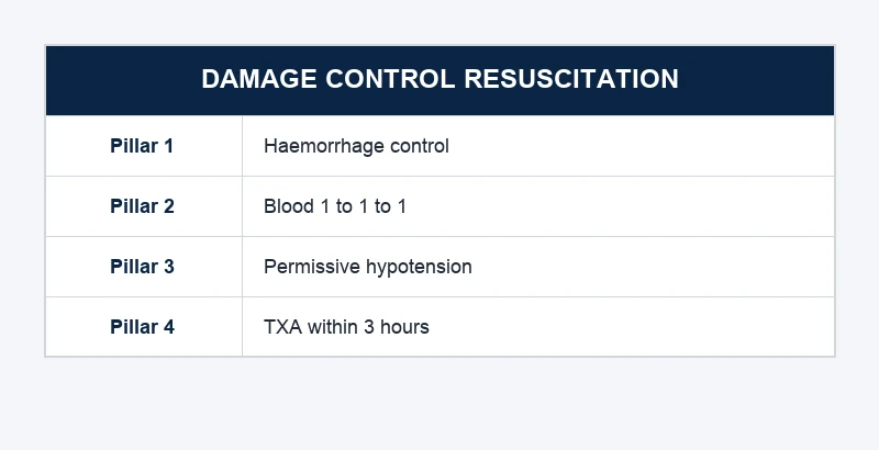
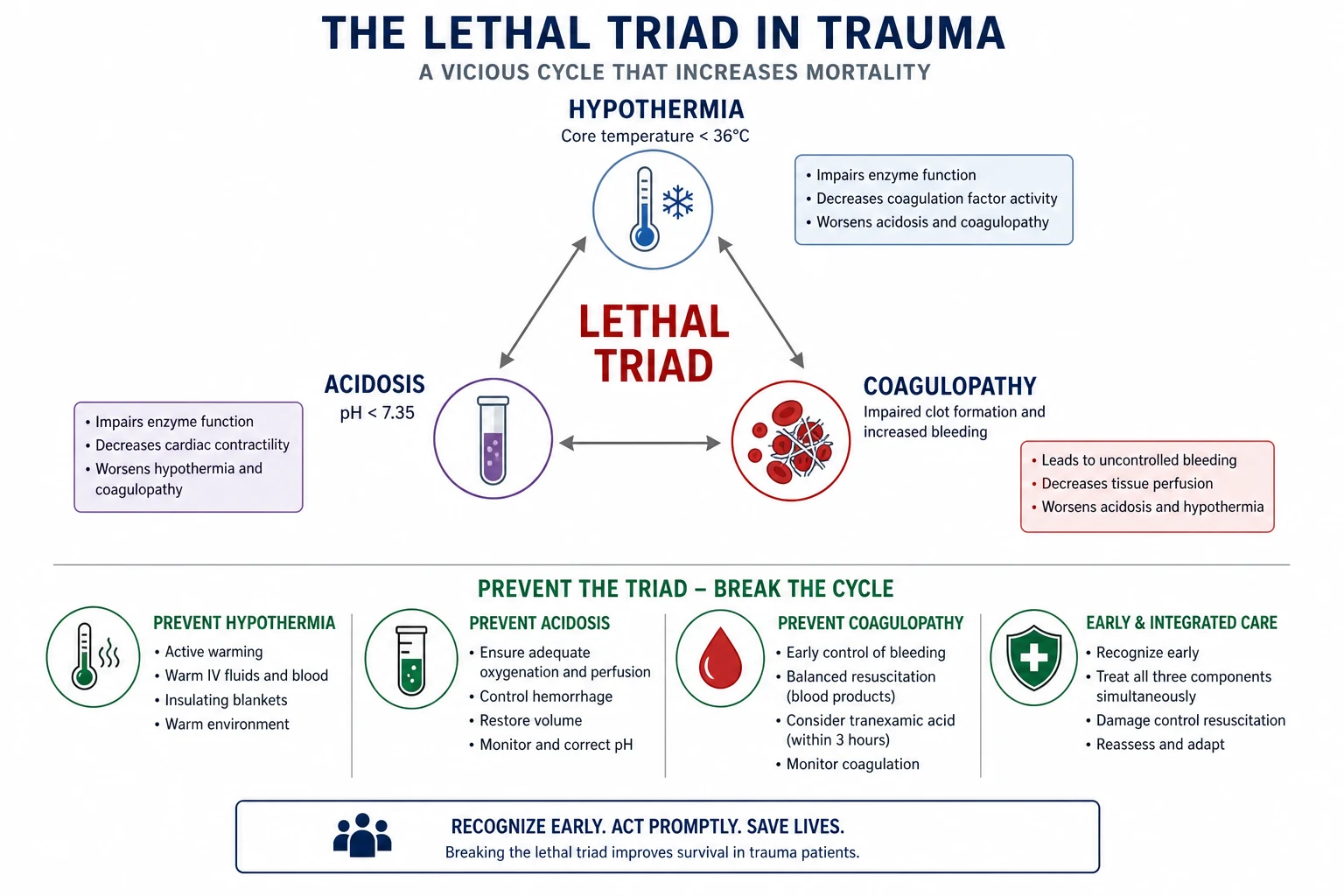
Damage control resuscitation is the paradigm that shifted the trauma mortality in the last two decades: the recognition that the bleeding trauma patient dies not of a single problem but of a lethal triad — hypothermia, acidosis and coagulopathy — that each feeds the other in a spiral to death, and that the resuscitation must target the haemostasis rather than the blood pressure. The Fellowship candidate must understand the permissive-hypotension principle, the haemostatic resuscitation with the blood products in a balanced ratio, the tranexamic acid, and the damage-control surgery, because the application of these — early, in the emergency department and the theatre — is what breaks the spiral and saves the life.[1][2]
The four pillars of damage control resuscitation
The damage control resuscitation is built on the four pillars that together break the lethal triad and the spiral of death.[14][15] The Fellowship candidate must understand each pillar, the evidence behind it, and the practical steps to deliver it, because the survival of the major trauma patient depends on the simultaneous application of all four — the failure of any one (resuscitating to a normal blood pressure, the crystalloid instead of the blood products, the late tranexamic acid, the definitive surgery on a cold and coagulopathic patient) restarts the spiral. The four pillars are:
1. Permissive hypotension
- Resuscitate to SBP 80 to 90 (or a MAP of 65) until the bleeding is controlled
- A higher pressure disrupts the soft clot and worsens the bleeding
- Contraindicated in the TBI — keep the SBP at 110 or above
- Modified in the ischaemic heart disease and the elderly
2. Haemostatic resuscitation
- Blood products in a 1:1:1 ratio (PRBC : FFP : platelets) from the start
- Minimise the crystalloid — under 1 L, in 250 mL aliquots
- Avoid the dilutional coagulopathy, the acidosis and the hypothermia of the saline
- Add the cryoprecipitate for the low fibrinogen, the calcium for the citrate toxicity
3. Damage control surgery
- Haemorrhage control, contamination control, temporary closure
- Pack, ligate, balloon-tamponade, angio-embolise — not the definitive repair
- Open abdomen with the vacuum dressing; the planned 24-to-48-hour relook
- The definitive surgery after the rewarming and the correction in the ICU
4. The pharmacology
- Tranexamic acid 1 g IV within 3 hours (best within the first hour)
- Calcium chloride after every four units of the blood products
- Reversal of the anticoagulants (the PCC, the andexanet, the vitamin K)
- The POCUS-guided, the viscoelastic-guided factor concentrate in the modern protocol
The lethal triad — the spiral to death
The trauma patient with a major haemorrhage develops three interlocking physiological derangements that, if uncorrected, spiral to an irreversible death. The hypothermia (from the exposure, the blood loss and the cold fluids) impairs the coagulation cascade and the platelet function. The acidosis (from the shock, the tissue hypoperfusion and the lactataemia) further disables the coagulation enzymes and the catecholamine receptors. The coagulopathy (from the consumption of the clotting factors, the dilution by the crystalloid, and the hypothermia and the acidosis) produces the ongoing bleeding that deepens the hypothermia and the acidosis. Each derangement feeds the other, and the spiral tightens with every unit of crystalloid and every minute of uncontrolled bleeding. The damage control resuscitation breaks the spiral by warming the patient, giving the blood products instead of the crystalloid, and surgically controlling the bleeding before the triad becomes irreversible. [1]
[1]Acute traumatic coagulopathy — the coagulopathy is not iatrogenic
A long-held teaching was that the trauma coagulopathy was the consequence of the resuscitation — the consumption of the factors, the dilution by the crystalloid, and the hypothermia and the acidosis. The contemporary evidence shows that one in four of the severely injured trauma patients arrives in the emergency department with a coagulopathy already established — the acute coagulopathy of trauma (ACT) — before any fluid has been given.[13] The mechanism is the hypoperfusion-driven activation of the protein C pathway: the tissue hypoperfusion from the shock exposes the thrombomodulin on the endothelium, which binds the thrombin and diverts it from the procoagulant to the anticoagulant pathway; the activated protein C consumes the factors Va and VIIIa, and the clot becomes friable. The hyperfibrinolysis (the breakdown of the clot) is the second mechanism — the tissue injury releases the tissue plasminogen activator, and the clot lyses faster than it forms. The tranexamic acid targets this hyperfibrinolysis (it inhibits the plasminogen activation), and this is the mechanistic rationale for the early tranexamic acid in the major trauma.
[1]Consumption coagulopathy
- The clotting factors and the platelets used up in the massive bleeding
- The mechanism of the classical disseminated intravascular coagulation
- Treated with the 1:1:1 blood-product replacement
- The part of the lethal triad
Dilutional coagulopathy
- The crystalloid and the packed red cells (without the plasma and the platelets) dilute the factors
- The iatrogenic coagulopathy of the over-resuscitation
- Prevented by the haemostatic resuscitation (1:1:1) and the minimum crystalloid
- Avoidable — the central argument against the saline-first approach
Acute coagulopathy of trauma (ACT)
- The endogenous coagulopathy from the shock and the tissue hypoperfusion
- The protein C pathway activation; the hyperfibrinolysis
- Present on the arrival in one in four of the severely injured
- Targeted by the tranexamic acid and the early plasma
Hypothermia–acidosis coagulopathy
- The hypothermia (below 35 °C) impairs the clotting enzymes and the platelets
- The acidosis (pH below 7.2) further disables the enzymes
- Reversed by the warming, the correction of the pH and the blood products
- The reason the rewarming is a pillar of the resuscitation
The lethal triad — the quantitative thresholds and the targets
The lethal triad is not a vague concept; each component has a quantitative threshold beyond which the mortality rises sharply, and a corresponding target that the resuscitation aims for. The Fellowship candidate must know the numbers — they are the examination favourites and the practical resuscitation goals. [1]
The lethal triad — the thresholds and the targets
Differential diagnosis — the causes of the traumatic haemorrhage
The traumatic haemorrhage is classified by the source, because the source determines the surgical approach. [1]
Compressible haemorrhage
- Limb, scalp, external — direct pressure, tourniquet, packing
- The easiest to control in the ED
- A tourniquet for the limb, a pelvic binder for the pelvis
- Direct pressure for the scalp and the external wound
Non-compressible (torso)
- Intra-abdominal, intrathoracic, retroperitoneal
- Not controllable by the external pressure
- FAST + CT → damage-control surgery or angio-embolisation
- The pelvic binder for the unstable pelvic fracture
Cavity haemorrhage
- Haemothorax → chest drain; massive → thoracotomy
- Haemoperitoneum → laparotomy
- The massive-haemorrhage protocol activated
- Each cavity has its approach
The anticoagulated bleeder
- Warfarin/DOAC + trauma → rapid reversal
- PCC/andexanet early; vitamin K for the warfarin
- Higher mortality at every injury severity
- Reversal is part of the haemostasis
The massive haemorrhage protocol — the activation and the delivery
The massive haemorrhage protocol (MHP), also called the massive transfusion protocol (MTP), is the institutional pre-defined pathway that delivers the blood products in a 1:1:1 ratio with the minimum delay. The activation is the trigger — the senior clinician calls the blood bank on the dedicated hotline, and the protocol kicks in. The pack (the "shock pack", the "code red pack") is delivered: one unit of the packed red cells, one of the fresh-frozen plasma, and one of the platelets (or, in some institutions, a six-unit pack of the red cells, the six of the plasma and the one apheresis platelet pool). The tranexamic acid, the calcium, the cryoprecipitate and the warming are the simultaneous pharmaceutical adjuncts. The protocol continues until the bleeding is controlled, the haemodynamics are stable, and the laboratory targets are met — then it is stood down. [1]
The massive haemorrhage protocol — the activation-to-delivery sequence
1
2
3
4
5
6
7
Permissive hypotension — the principle
The traditional resuscitation aimed for a normal blood pressure; the damage control resuscitation aims for permissive hypotension — a systolic blood pressure of 80 to 90 mmHg, enough for the consciousness and the urine output, but deliberately below the normal — until the bleeding is surgically controlled.[2] The rationale: a normal blood pressure disrupts the soft clot that is tamponading the bleeding vessel (the same principle as the ruptured AAA); a lower pressure preserves the clot and the tamponade. The crystalloid that raises the pressure also dilutes the clotting factors, lowers the temperature (the cold fluids), and worsens the acidosis (the saline has a pH of 6.0). The permissive hypotension is contraindicated in the traumatic brain injury (where the cerebral perfusion pressure is paramount — the SBP must be 110 or above) and it is modified in the ischaemic heart disease (where the hypotension may provoke the ischaemia). In the isolated torso or the limb haemorrhage without a brain injury, the permissive hypotension is the standard until the operating theatre.
Permissive hypotension — the standard
- SBP 80 to 90 mmHg, or a MAP of 65 — until the bleeding is controlled
- Preserves the soft clot and the tamponade; avoids the clot disruption
- Minimises the crystalloid and the dilution
- The indication: the isolated torso or the limb haemorrhage without the TBI
Normotensive resuscitation — the exception
- SBP at 110 or above, MAP at 75 or above
- Indicated in the TBI (the cerebral perfusion is paramount)
- Indicated in the suspected spinal cord injury (the cord perfusion)
- Modified in the ischaemic heart disease (the coronary perfusion)
The endpoint — the conscious level
- The BP is titrated to the consciousness, not the absolute number
- The alert, the talking patient is the perfused patient
- The loss of the consciousness is the trigger for the blood (not the fluid)
- The radial pulse (a SBP of at least 90) is the bedside marker
The evidence and the controversy
- The systematic reviews show a trend to the reduced mortality
- The trials are mixed; the contemporary practice is the permissive approach
- The DROWN and the POLAR trials informed the contemporary thresholds
- The hypotension is RELATIVE — never let the patient exsanguinate
The haemostatic resuscitation — the blood products in a balanced ratio
The haemostatic resuscitation replaces the traditional crystalloid-first approach with a blood-product-first strategy. The massive haemorrhage protocol delivers the packed red cells, the fresh-frozen plasma and the platelets in a 1:1:1 ratio (one unit of each, per pack), aiming for the whole-blood-like profile that supports the clotting and the oxygen carriage. The evidence for the 1:1:1 ratio is the PROPPR trial, which showed a trend towards the reduced mortality at 24 hours with the 1:1:1 (versus the 1:1:2) ratio. The protocol also includes the calcium replacement — the massive transfusion causes a hypocalcaemia from the citrate (the anticoagulant in the stored blood), and a low ionised calcium impairs the cardiac contractility and the clotting; the calcium chloride 10 mL of 10 per cent intravenously (or the calcium gluconate 10 mL of 10 per cent) is given after every four units of the blood products. [1]
The damage control targets
The blood products — the contents, the ratios and the targets
Each blood product has a specific role in the haemostatic resuscitation. The Fellowship candidate must know the contents, the unit volume, the storage lesion, and the laboratory target of each, because the goal-directed resuscitation aims for the whole-blood profile (the haematocrit, the clotting factors, the platelets, the fibrinogen) that the uncontrolled bleeding depletes. [1]
Packed red cells (PRBC)
- The oxygen carriage; the target haemoglobin above 70 to 80 g/L (above 90 in the ischaemic heart disease)
- A unit of PRBC is ~300 mL; raises the haemoglobin by ~10 g/L
- Stored in the SAGM additive; the storage lesion (the potassium, the acid, the free haemoglobin) worsens with the age
- The O-negative for the immediate need; the type-specific as soon as available
Fresh-frozen plasma (FFP)
- The clotting factors; the target INR below 1.5
- A unit of FFP is ~250 mL; the thawing time of 20 to 30 minutes (the thawed plasma or the liquid plasma for the speed)
- The ABO-compatible; the TRALI risk (the transfusion-related acute lung injury)
- The 1:1 ratio with the PRBC — the early plasma is the key to the survival
Platelets
- The primary haemostasis; the target platelet count above 50 × 10⁹/L (above 100 in the active bleeding)
- A pool of platelets is ~200 mL (a single apheresis unit); raises the count by ~20 to 40 × 10⁹/L
- Stored at the room temperature (the bacterial-contamination risk); the 5-day shelf life
- One apheresis unit equates to the 1:1 ratio with six units of the PRBC
Cryoprecipitate
- The fibrinogen, the factor VIII, the von Willebrand factor, the factor XIII
- The target fibrinogen above 1.5 to 2.0 g/L (the threshold in the trauma)
- Two pools (10 units) raise the fibrinogen by ~1.0 g/L
- Given when the fibrinogen is low or guided by the viscoelastic test (the FIBTEM, the functional fibrinogen)
PROPPR — the 1:1:1 versus the 1:1:2 plasma-to-platelet-to-red-cell ratio (JAMA 2015)
JAMA
PMID 25647203
Key finding
A multicentre randomised trial of 680 severely injured trauma patients with the major bleeding, comparing the 1:1:1 ratio (the plasma, the platelets, the red cells) against the 1:1:2 ratio. The 24-hour and the 30-day mortality were not significantly different, but the death by the exsanguination within 24 hours fell from 15 per cent to 9 per cent in the 1:1:1 group. No difference in the complications.
Practice change
The 1:1:1 ratio is the reasonable starting ratio for the massive transfusion, on the strength of the reduction in the exsanguination death. The 1:1:2 is not inferior for the overall survival, but the modern protocols default to the 1:1:1 for the early packs.
MATTERs — the Military Application of Tranexamic Acid in Trauma Emergency Resuscitation (Archives of Surgery 2012)
Archives of Surgery
PMID 22301964
Key finding
A retrospective military registry study of 896 casualties with the combat injury requiring the massive transfusion, comparing the tranexamic acid (293 received it) against the no-tranexamic-acid. The tranexamic acid reduced the mortality from 17.4 per cent to 13.7 per cent overall, and in the subgroup requiring the massive transfusion (over 10 units) the mortality fell from 27.6 per cent to 12.4 per cent — a near halving. The thromboembolic events were higher in the tranexamic-acid group (11 per cent vs 6 per cent).
Practice change
In the military and the major-bleeding setting, the tranexamic acid has a substantial mortality benefit, especially in the massive-transfusion subgroup. The CRASH-2 and the MATTERs together established the tranexamic acid as the standard of care in the traumatic haemorrhage.
Minimise the crystalloid — under one litre, in 250 mL aliquots [1]
The traditional ATLS taught the two-litre crystalloid bolus on the arrival of the trauma patient — the modern damage control resuscitation rejects this. The crystalloid is the resuscitation's enemy in the actively bleeding patient. The saline (and the Hartmann, the PlasmaLyte) dilute the clotting factors and the platelets (the dilutional coagulopathy); they are cold (the storage at the room temperature, sometimes the refrigerator) and they lower the body temperature; the saline has a pH of 6.0 and worsens the acidosis (and the hyperchloraemia causes the renal vasoconstriction and the AKI). The contemporary practice: under one litre of the crystalloid in total, given in the 250 mL aliquots titrated to the conscious level and the radial pulse, with the rapid switch to the blood products (the 1:1:1) as the resuscitation fluid. [1]
[1]0.9% saline
- pH of 6.0 — worsens the acidosis
- High chloride (154 mmol/L) — the hyperchloraemic metabolic acidosis
- The renal vasoconstriction — the AKI risk
- Avoid in the trauma resuscitation
Hartmann / Ringer lactate
- Balanced, near-physiological; the lactate is metabolised to the bicarbonate
- Lower chloride — less of the hyperchloraemic acidosis
- The preferred crystalloid if the crystalloid must be given
- Still a crystalloid — minimise the total volume
PlasmaLyte
- The most balanced of the crystalloids; the acetate and the gluconate buffers
- The least acidifying; the least renal toxicity
- More expensive; not universally available
- A reasonable choice for the small-volume crystalloid adjunct
The hypertonic saline
- Small volume, the high sodium — the historical interest for the trauma
- The trials (the TBI, the shock) showed no benefit over the isotonic
- Not the standard of the contemporary practice
- Reserved for the symptomatic hyponatraemia
The calcium — the citrate toxicity of the massive transfusion
The calcium is the silent killer of the massive transfusion. The stored blood products contain the citrate as the anticoagulant (it chelates the calcium and prevents the clotting in the bag). On the transfusion, the citrate is rapidly metabolised by the liver — but in the setting of the massive transfusion, the citrate load overwhelms the hepatic metabolism, and the ionised calcium falls. The hypocalcaemia (an ionised calcium below 1.0 mmol/L) impairs the cardiac contractility (the cardiogenic shock on top of the haemorrhagic shock), the vasopressor response (the catecholamines need the calcium), and the coagulation (the factor IV — the calcium is the essential cofactor in the clotting cascade). The calcium chloride 10 mL of 10 per cent (6.8 mmol of the calcium) or the calcium gluconate 10 mL of 10 per cent (2.2 mmol) is given intravenously after every four units of the blood products, with the ionised-calcium target of 1.0 to 1.3 mmol/L. The calcium chloride is preferred in the arrest or the severe hypocalcaemia (it releases three times more of the ionised calcium per millimole), but it is vesicant if extravasated — give it through a central line or a large peripheral cannula. [1]
[1]The electrolyte and the metabolic derangements of the massive transfusion
The rewarming — break the hypothermia arm of the lethal triad
The hypothermia is the silent contributor to the lethal triad. The trauma patient arrives cold (the exposure, the blood loss, the cold environment), and the resuscitation makes it worse (the cold fluids, the cold blood products, the open body cavity). The hypothermia (a core temperature below 35 °C) impairs the coagulation cascade (the clotting enzymes are temperature-dependent, and below 33 °C they are virtually disabled), the platelet function (the cold platelets do not aggregate), and the hepatic metabolism (the citrate metabolism slows, worsening the hypocalcaemia). The rewarming is therefore a pillar of the damage control resuscitation — and it must be ACTIVE, not passive. [1]
The active rewarming of the trauma patient — the layered approach
1
2
3
4
5
The tranexamic acid — the CRASH-2 trial
The tranexamic acid is the one pharmacological agent with a proven mortality benefit in the traumatic haemorrhage. The CRASH-2 trial — a landmark international randomised controlled trial of over 20,000 trauma patients — showed that tranexamic acid 1 g intravenously within 3 hours of the injury reduced the all-cause mortality from 13.5 to 14.5 per cent (an absolute reduction of 1.5 per cent, a number-needed-to-treat of about 120).[1] The critical nuance: the benefit is time-dependent — the tranexamic acid given within the first hour has the greatest effect; given between 1 and 3 hours it still helps; given after 3 hours it may increase the mortality (from the increased thrombotic risk without the haemostatic benefit). This is why the tranexamic acid is given early — in the emergency department, within the first hour, as part of the massive-haemorrhage protocol.
CRASH-2 — the tranexamic acid in the traumatic bleeding (Lancet 2010)
Lancet
PMID 20554319
Key finding
An international randomised placebo-controlled trial of 20,211 trauma patients with, or at risk of, the significant bleeding, comparing the tranexamic acid (a 1 g loading dose over 10 minutes, then 1 g over 8 hours) against the placebo within 8 hours of the injury. The all-cause mortality fell from 16.0 per cent to 14.5 per cent, with the largest benefit when the drug was given within the first hour (a 32 per cent relative reduction in the bleeding death). The benefit was lost and the harm possible beyond 3 hours.
Practice change
The tranexamic acid 1 g IV over 10 min then 1 g over 8 h, given as early as possible after the injury (and within 3 hours), is the standard of care in the traumatic haemorrhage. The drug is cheap, safe, easy to give, and has a clear mortality benefit — there is no excuse for the late or the omitted tranexamic acid in the major trauma.
CRASH-3 — the tranexamic acid in the traumatic brain injury (Lancet 2019)
Lancet
PMID 31470090
Key finding
A randomised placebo-controlled trial of 12,737 trauma patients with the traumatic brain injury and a GCS of up to 15, comparing the tranexamic acid against the placebo within 3 hours of the injury. No overall mortality difference, but a significant reduction in the mild-to-moderate TBI (the GCS 9 to 15) head-injury-related death when given early. No increase in the adverse events (the stroke, the MI, the thromboembolism, the seizures).
Practice change
The tranexamic acid within 3 h (and ideally within the first hour) is safe and beneficial in the mild-to-moderate traumatic brain injury; the benefit in the severe TBI (GCS 3 to 8) was less clear, but the drug is not harmful. The pragmatic implication: give the tranexamic acid to the major trauma patient with the head injury within 3 h, without waiting for the final head-injury classification.
PATCH-TBI — the pre-hospital tranexamic acid in the traumatic brain injury (NEJM 2020)
New England Journal of Medicine
PMID 33095850
Key finding
A randomised placebo-controlled trial of 1,063 trauma patients with the moderate-to-severe TBI (a GCS of 3 to 12 and any intracranial haemorrhage), comparing the pre-hospital tranexamic acid (1 g bolus within 2 hours of injury) against the placebo. The 28-day mortality was similar, with a non-significant trend to the lower mortality in the tranexamic-acid group. No increase in the thromboembolic events.
Practice change
The pre-hospital tranexamic acid is safe in the moderate-to-severe TBI; the mortality benefit is not established in this subgroup, but the harm is not demonstrated. The pragmatic practice: give the tranexamic acid early, including in the suspected TBI, on the strength of the CRASH-2 and the CRASH-3 evidence.
Immediate management — the protocol
[1]The damage-control surgery
The damage-control surgery is the surgical counterpart of the resuscitation — it prioritises the haemorrhage control and the contamination control over the definitive repair, because the definitive surgery on a cold, acidotic, coagulopathic patient kills. The three objectives: control the bleeding (the packing, the ligation, the balloon tamponade, the angio-embolisation); control the contamination (the simple closure or the stapling of the bowel perforations, the resection without the anastomosis); and the temporary abdominal closure (a vacuum dressing, a Bogota bag — the abdomen is left open to prevent the abdominal compartment syndrome). The patient is then transferred to the intensive care for the rewarming, the correction of the coagulopathy and the acidosis, and the optimisation, before the planned return to the theatre for the definitive surgery (the 24-to-48-hour relook). [1]
The viscoelastic haemostatic assay — the TEG and the ROTEM
The viscoelastic haemostatic assay (VHA) — the thromboelastography (TEG) and the rotational thromboelastometry (ROTEM) — is the modern, dynamic, whole-blood test of the coagulation that has replaced the static INR and APTT in the goal-directed trauma resuscitation.[8][10][11] The traditional coagulation tests (the INR, the APTT, the fibrinogen) measure the plasma clotting in a tube at a single time point; they take 30 to 60 minutes to return; they do not distinguish the clotting-factor deficiency from the platelet dysfunction from the hyperfibrinolysis. The viscoelastic test measures the whole-blood clot formation and the breakdown in real time, returns the actionable result in 10 to 30 minutes, and identifies the specific defect — the clotting-factor deficiency, the platelet dysfunction, the low fibrinogen, or the hyperfibrinolysis — that the goal-directed therapy then targets. The VHA is now part of the European and the EAST guidelines on the trauma haemorrhage, and the major trauma centres have the TEG or the ROTEM in the emergency department.[14][15]
The static tests (INR, APTT)
- Plasma clotting in a tube; a single time point
- Slow — 30 to 60 minutes to return
- Do NOT distinguish the factor deficiency from the platelet dysfunction from the fibrinolysis
- The INR is affected by the warfarin and the liver disease (not the trauma coagulopathy)
The TEG (thromboelastography)
- Whole-blood clot formation and the breakdown in real time
- The R time (the clot initiation), the K time (the clot kinetics), the alpha angle (the clot strengthening), the MA (the maximum amplitude — the clot strength), the LY30 (the lysis at 30 min — the fibrinolysis)
- The actionable result in 10 to 30 minutes
- Guides the factor concentrate, the platelets, the fibrinogen and the antifibrinolytics
The ROTEM (rotational thromboelastometry)
- The same principle as the TEG, with the slightly different nomenclature
- The CT (the clotting time), the CFT (the clot formation time), the alpha angle, the MCF (the maximum clot firmness), the ML (the maximum lysis)
- The EXTEM (the extrinsic), the INTEM (the intrinsic), the FIBTEM (the fibrinogen-only), the APTEM (the hyperfibrinolysis test)
- The European preference; the TEG is the North American preference
The goal-directed algorithm
- The abnormal R/CT → the FFP or the prothrombin complex concentrate
- The abnormal K/CFT or the low alpha → the cryoprecipitate or the fibrinogen concentrate
- The low MA/MCF → the platelets
- The high LY30/ML → the tranexamic acid
The TEG-guided goal-directed resuscitation — the algorithm
1
2
3
4
5
The ROTEM-guided goal-directed resuscitation — the algorithm
1
2
3
4
5
Goal-directed resuscitation endpoints — beyond the blood pressure
The blood pressure is a poor endpoint of the trauma resuscitation — the permissive hypotension deliberately targets a low BP, and a normal BP may coexist with the ongoing hypoperfusion (the compensated shock). The contemporary resuscitation uses the macro-circulatory and the micro-circulatory endpoints that better reflect the tissue perfusion. The lactate is the workhorse — it reflects the anaerobic metabolism of the shock, and the lactate clearance (a fall of at least 20 per cent in 2 hours, or the normalisation within 24 hours) is the marker of the adequate resuscitation. The base excess (a base deficit of −5 or worse on the arrival, with the persistent deficit) is the prognostic marker of the severe shock. The venous-to-arterial CO2 gap (above 6 mmHg) and the central venous oxygen saturation (below 70 per cent) are the advanced endpoints used in the ICU. The viscoelastic assay guides the haemostatic endpoint — the trace normalises when the coagulopathy is corrected. [1]
The goal-directed resuscitation endpoints
Pre-hospital damage control resuscitation
The damage control resuscitation begins before the arrival — in the pre-hospital phase, by the paramedics, the retrieval teams and the aeromedical services. The principles are the same — the minimisation of the crystalloid, the permissive hypotension, the early tranexamic acid, the warming — but the practicalities differ. The tourniquet for the exsanguinating limb haemorrhage (the CAT, the MAT) has been the single biggest lesson from the military and the civilian mass-casualty experience — the pre-hospital tourniquet saves lives. The pelvic binder for the suspected pelvic fracture. The tranexamic acid given by the paramedics within the first hour (the pre-hospital PATCH-TBI and the CRASH-2 evidence). The minimised crystalloid (the permissive hypotension to the radial pulse). The warming (the blankets, the heated fluids). The whole blood or the plasma (the pre-hospital blood product resuscitation is the contemporary practice in the mature retrieval systems). The "scoop and run" (the minimal scene time, the rapid transport to the major trauma centre) is the principle — the time on the scene is the time lost to the definitive care. [1]
[1]Complications and pitfalls
The complications of the uncorrected lethal triad are the multi-organ failure and the death. The complications of the over-resuscitation are the pulmonary oedema, the abdominal compartment syndrome, and the dilutional coagulopathy. The pitfalls are the inverse of the protocol: resuscitating to a normal blood pressure in the actively bleeding patient (disrupting the clot); giving the large-volume crystalloid (diluting the factors, lowering the temperature, worsening the acidosis); delaying the tranexamic acid beyond 3 hours (the harmful window); not giving the calcium (the citrate-induced hypocalcaemia); not warming the patient (the hypothermia feeds the triad); and not activating the massive-haemorrhage protocol early enough. [1]
Abdominal compartment syndrome
- An intra-abdominal pressure above 20 mmHg with the new organ failure
- The over-resuscitation, the open abdomen packed, the ileus — the risk factors
- The oliguria, the high airway pressures, the falling CO2 clearance — the bedside clues
- Decompress: open the abdomen, the percutaneous drain, the sedation and the paralysis
TRALI (transfusion-related acute lung injury)
- The donor anti-leucocyte antibodies against the recipient leucocytes
- Bilateral pulmonary oedema within 6 hours of the transfusion
- Treated with the supportive care (the oxygen, the PEEP) — NOT the diuretics
- Reported to the blood bank; the donor flagged for the future donation
TACO (transfusion-associated circulatory overload)
- The circulatory overload from the rapid transfusion in the susceptible
- The elderly, the heart failure, the renal failure — the risk factors
- Pulmonary oedema with the hypertension — distinguish from the TRALI
- Treated with the oxygen, the diuretics, the slowed transfusion
The hyperkalaemia from the old PRBC
- The storage lesion — the extracellular potassium rises with the age
- A unit over 21 days has the extracellular potassium of 30 to 50 mmol/L
- The rapid transfusion of the old blood can cause the arrest
- Use the freshest blood for the massive transfusion; consider the washed cells
Prognosis and disposition
The mortality of the major traumatic haemorrhage depends on the injury, the time to the theatre, and the correction of the lethal triad. The patient is taken to the theatre for the damage-control surgery and then to the intensive care for the rewarming and the correction, before the planned relook. The tranexamic acid, the permissive hypotension and the 1:1:1 ratio each contribute to the survival benefit. [1]
Special populations
The traumatic brain injury patient is the exception to the permissive hypotension — the SBP must be 110 or above to protect the cerebral perfusion. The anticoagulated patient is reversed early (the PCC, the andexanet, the vitamin K). The pregnant trauma patient is managed with the left-lateral tilt and the fetal monitoring; the permissive hypotension is modified (the fetus is sensitive to the hypotension). The paediatric trauma patient uses the weight-based blood-product volumes and the paediatric trauma team. [1]
Evidence and regional guidelines
The contemporary framework is the damage-control-resuscitation evidence: the CRASH-2 trial (the tranexamic acid)[1] and the permissive-hypotension evidence.[2] The PROPPR trial (the 1:1:1 ratio) and the institutional massive-haemorrhage protocols. The ATLS and the local trauma protocol govern the activation and the targets. The pharmacological and the surgical principles are global; the exact protocol, the blood-product ratios and the theatre pathway are local.
ANZ practice note. The damage-control-resuscitation principles follow the ATLS/EMST framework via the local trauma and the transfusion-medicine pathway; the tranexamic acid 1 g is given in the emergency department within the first hour, the massive-haemorrhage protocol delivers the 1:1:1 blood products, the permissive hypotension (SBP 80 to 90) is applied to the non-TBI haemorrhage, and the damage-control surgery is performed by the trauma team in the theatre. [1]
Exam pearls
- The lethal triad: hypothermia + acidosis + coagulopathy — the spiral of death; the resuscitation breaks it.
- Permissive hypotension SBP 80 to 90 (NOT the TBI — SBP ≥110) — do not resuscitate to normal until the bleeding is controlled.
- 1:1:1 blood products (PRBC : FFP : platelets) — not the crystalloid.
- Tranexamic acid 1 g IV within 3 hours (best within 1 hour) — the CRASH-2 mortality benefit; harmful after 3 hours.
- Calcium chloride 10 mL of 10 per cent after every 4 units — the citrate-induced hypocalcaemia.
- Warm the patient — the hypothermia worsens the coagulopathy.
- Damage-control surgery: control the bleeding, control the contamination, temporary closure → ICU → relook.
- The four pillars: permissive hypotension + haemostatic resuscitation + damage-control surgery + the pharmacology (the TXA, the calcium, the anticoagulant reversal).
- The acute coagulopathy of trauma (the ACT) is endogenous — present on the arrival in one in four of the severely injured, from the protein C pathway and the hyperfibrinolysis; targeted by the early tranexamic acid.
- Minimise the crystalloid — under 1 L, in 250 mL aliquots — the saline dilutes, is cold, and has a pH of 6.0; switch to the blood products (1:1:1) as the resuscitation fluid.
- The viscoelastic assay (the TEG / the ROTEM) is the modern, dynamic, whole-blood test — the goal-directed resuscitation targets the specific defect (the R/CT → the FFP/PCC; the alpha/FIBTEM → the fibrinogen; the MA/MCF → the platelets; the LY30/APTEM → the TXA).
- The citrate-induced hypocalcaemia — give the calcium chloride 10 mL of 10 per cent after every four units; the ionised calcium target of 1.0 to 1.3 mmol/L.
- The endpoints — the lactate clearance (≥20 per cent in 2 h), the base excess (>−5), the temperature (≥36 °C), the ionised calcium (≥1.0).
- The TBI is the exception — the SBP must be 110 or above; the permissive hypotension is contraindicated; the tranexamic acid within 3 h is safe (the CRASH-3, the PATCH-TBI).
- The pre-hospital DCR — the scoop-and-run, the early tourniquet, the paramedic-administered tranexamic acid in the first hour, the minimal crystalloid.
- The PROPPR trial (the 1:1:1) reduced the exsanguination death at 24 h from 15 to 9 per cent; the MATTERs (the military) showed a near-halving of the mortality in the massive-transfusion subgroup with the tranexamic acid.
- The anticoagulated trauma patient — the early reversal (the PCC for the warfarin, the andexanet or the PCC for the DOAC, the vitamin K for the warfarin) is part of the haemostasis. [1]
Red flags
[1]Short-answer questions (ACEM Fellowship practice)
SAQ — DCR principles in a shocked polytrauma patient
10 minutes · 10 marks
A 32-year-old man is brought to the trauma bay 20 minutes after a high-speed motorcycle crash. He is pale, diaphoretic, and confused (GCS 13). His BP is 74/48, HR 138, RR 28, SpO2 95% on 15 L O2 via non-rebreather. Examination reveals a deformed pelvis, a grossly deformed open right tibia–fibula fracture with ongoing external bleeding, and a tense, distended abdomen. The FAST is positive in the right upper quadrant. Lactate 7.8 mmol/L, INR 1.8, ionised calcium 0.88 mmol/L, core temperature 34.6 degrees C. There is no clinical evidence of a traumatic brain injury.
SAQ — Activation and delivery of the massive transfusion protocol
10 minutes · 10 marks
A 45-year-old pedestrian is brought to the trauma bay 35 minutes after being struck by a car. He is agitated (GCS 14), BP 78/52, HR 134, RR 30, SpO2 96% on 15 L O2. There is an obvious flail segment on the right with a clinically large right haemothorax, an unstable pelvis, and bilateral open tibial fractures. A right-sided chest drain immediately drains 1500 mL of blood, with ongoing fresh bleeding of ~250 mL/min. The massive transfusion protocol has just been activated. Lactate 6.9 mmol/L, ionised calcium 0.86 mmol/L, temperature 34.8 degrees C, INR 1.7, fibrinogen 1.2 g/L. He takes no regular medications. The trauma team and the blood bank are present.
References
- [1]CRASH-2 trial collaborators. Effects of tranexamic acid on death, vascular occlusive events, and blood transfusion in trauma patients with significant haemorrhage (CRASH-2): a randomised, placebo-controlled trial Lancet, 2010.PMID 20554319
- [2]Indorewala Y, Sharma A, Bhatt N, et al. Permissive hypotension in adult trauma: A systematic review of outcomes across clinical settings, injury type, and resuscitation strategies Am J Emerg Med, 2026.PMID 42030689
- [3]Holcomb JB, Tilley BC, Baraniuk S, et al.; PROPPR Study Group. Association of +3179G/A insulin-like growth factor-1 receptor polymorphism and insulin-like growth factor-1 serum level with systemic lupus erythematosus Lupus, 2013.PMID 23989734
- [4]Morrison JJ, Dubose JJ, Rasmussen TE, Midwinter MJ. Visualization of plastid movement in the pollen tube of Arabidopsis thaliana Plant Signal Behav, 2012.PMID 22301964
- [5]CRASH-3 trial collaborators. Computational approaches for inferring 3D conformations of chromatin from chromosome conformation capture data Methods, 2020.PMID 31470090
- [6]ROWELL SE, Meier EN, Bulger EM, et al. Cross-sectional and prospective associations between sleep regularity and metabolic health in the Hispanic Community Health Study/Study of Latinos Sleep, 2021.PMID 33095850
- [7]Gonzalez EA, Moore FA, Holcomb JB, et al. Contributions of mood, pain catastrophizing, and cold hyperalgesia in acute and chronic low back pain: a comparison with pain-free controls Clin J Pain, 2014.PMID 24145929
- [8]Schöchl H, Maegele M, Solomon C, et al. Instant mentoring: sharing wisdom and getting advice online with e-mentoring J Acad Nutr Diet, 2013.PMID 23582461
- [9]Davenport R, Curry N, Manson J, et al. Generality of shear thickening in dense suspensions Nat Mater, 2010.PMID 20118945
- [10]Gonzalez E, Moore EE, Moore HB, et al. REI/SH3BP5 protein family: New GEFs for Rab11 Cell Cycle, 2016.PMID 26745340
- [11]Schöchl H, Nienaber U, Maegele M, et al. Cerebral aspergillus infection in pediatric acute lymphoblastic leukemia induction therapy Indian J Med Paediatr Oncol, 2012.PMID 23580827
- [12]Kashuk JL, Moore EE, Sawyer M, et al. An excess of catalytically required motions inhibits the scavenger decapping enzyme Nat Chem Biol, 2015.PMID 26258763
- [13]Brohi K, Cohen MJ, Ganter MT, et al. Nottingham trial of faecal occult blood testing for colorectal cancer: a 20-year follow-up Gut, 2012.PMID 22052062
- [14]Cannon JW, Khan MA, Raja AS, et al. Unravelling the structure of electrocatalytically active Fe-N complexes in carbon for the oxygen reduction reaction Angew Chem Int Ed Engl, 2014.PMID 25115803
- [15]Spahn DR, Bouillon B, Cerny V, et al. A signal integration model of thymic selection and natural regulatory T cell commitment J Immunol, 2014.PMID 25392533