EM · Deep vein thrombosis
Deep vein thrombosis (emergency department diagnosis and management)
Also known as DVT · Venous thrombosis · Deep venous thrombosis
Deep vein thrombosis — the Wells-DVT risk score (the clinical signs, the risk factors, the alternative diagnosis — reproduced), the D-dimer and the compression ultrasound strategy, the anticoagulation management (the apixaban or the rivaroxaban first-line, the enoxaparin, the warfarin), the compression stockings, the PE cross-link, and the Wells-DVT-versus-clinical-probability approach. ACEM-primary, globally tagged.
On this page & tools
Your progress
Saved locally on this device.
Target exams
Red flags
Deep vein thrombosis and the pulmonary embolism it gives rise to are one disease — the venous thromboembolism — and the emergency physician manages the DVT to prevent the PE. The Fellowship candidate must apply the Wells-DVT score, choose between the D-dimer and the compression ultrasound by the probability, and start the anticoagulation with the right drug and the right dose.[1][2]
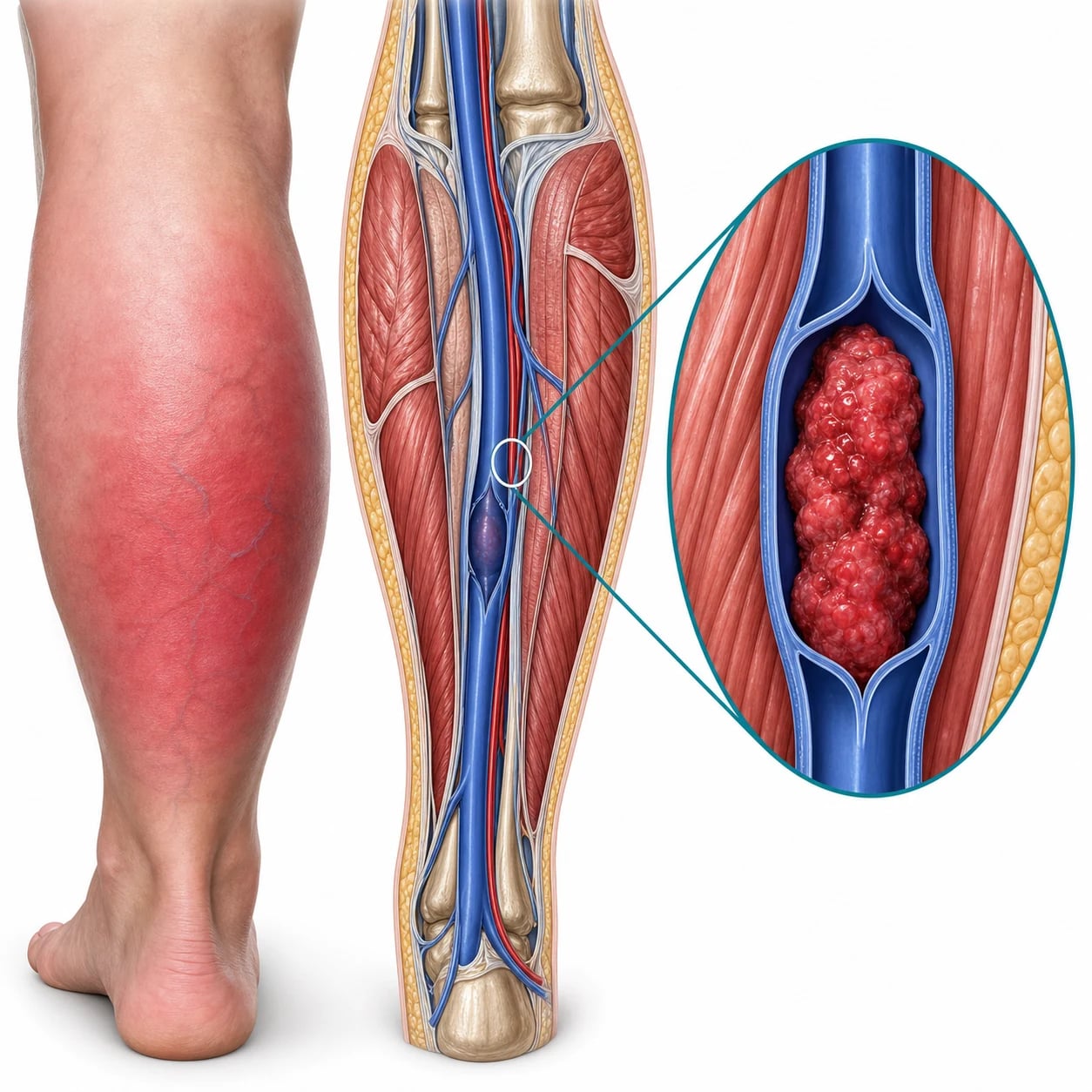
Pathophysiology — Virchow's triad
The DVT arises from the Virchow's triad: the venous stasis (the immobilisation, the post-operative bed rest, the long-haul flight, the obesity, the heart failure), the endothelial injury (the surgery, the trauma, the catheter, the previous thrombosis), and the hypercoagulability (the active cancer, the thrombophilia, the oestrogen, the pregnancy, the nephrotic syndrome). The thrombus forms most commonly in the deep veins of the calf, and may propagate proximally to the popliteal, the femoral and the iliac veins; the proximal propagation is the risk factor for the PE. [1]
Clinical features of the deep vein thrombosis
The DVT presents most often as the unilateral leg swelling, the pain and the tenderness of the calf or the thigh, with the warmth and the erythema over the affected segment. The clinical signs are neither sensitive nor specific on their own — roughly half of the symptomatic proximal DVTs have minimal bedside signs, and a quarter of the swollen legs that look like a DVT turn out to be something else. The Fellowship candidate must therefore measure, compare and quantify, and then apply the Wells-DVT score rather than make the diagnosis on the gestalt alone. [1]
The bedside signs to elicit
Differential diagnosis — the swollen leg
The swollen leg has a differential, and the Wells-DVT score helps separate the likely DVT from the mimics. [1]
Deep vein thrombosis
- Unilateral calf/thigh swelling, tenderness, warmth, erythema
- Wells-DVT score → D-dimer → ultrasound
- Risk factors: cancer, surgery, immobilisation, pregnancy
- Anticoagulation: apixaban or rivaroxaban first-line
Cellulitis
- Fever, erythema, warmth, clearly demarcated margin
- Leukocytosis, raised CRP; no proximal swelling
- Antibiotics (flucloxacillin), not anticoagulation
- A D-dimer may be falsely raised
Baker cyst rupture
- Sudden calf pain; a history of knee arthritis
- May mimic a DVT; the ultrasound shows the cyst
- No proximal swelling; no collateral veins
- Symptomatic management
Post-thrombotic syndrome
- Chronic leg swelling, pain, venous eczema, ulceration
- A history of a previous DVT
- No acute change; no new thrombus on the ultrasound
- Compression stockings, wound care
The Wells-DVT score — the clinical probability
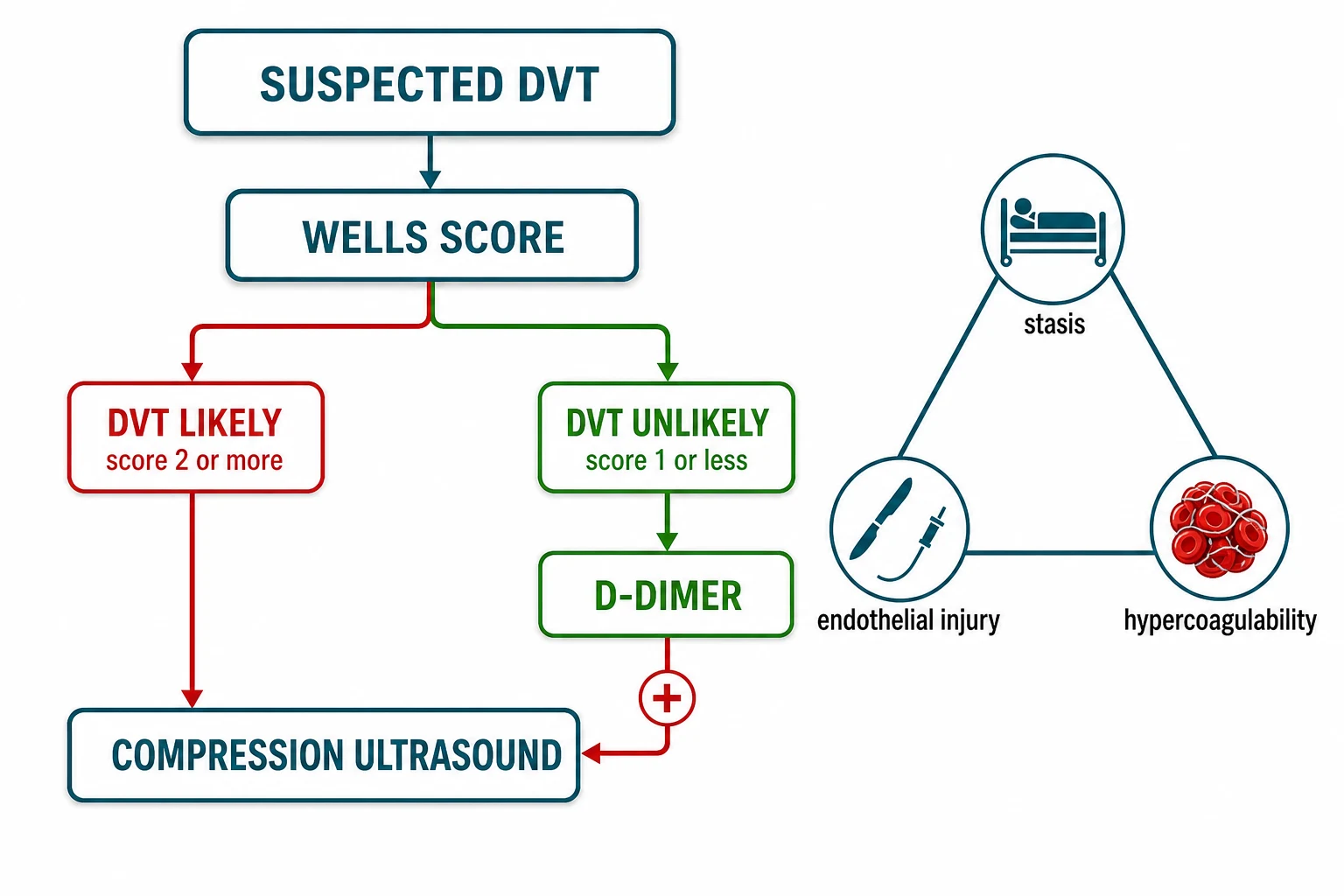
The Wells-DVT score estimates the clinical probability of a DVT from the signs and the risk factors, and it guides the investigation. The Fellowship candidate must reproduce its components. [1]
Wells-DVT — the clinical probability score
Wells
Active cancer (treatment within 6 months or palliative) — 1 point
Recently bedridden (over 3 days) or major surgery within 12 weeks, or paralysis/paresis — 1 point
Along the distribution of the deep venous system — 1 point
Entire leg swollen (1) / calf swelling over 3 cm difference (1) / pitting oedema (1)
Collateral superficial veins (not varicose) — 1 point
Previously documented DVT — 1 point
An alternative diagnosis as likely or more likely than the DVT — minus 2 points
The score: 0 or less is the low probability (the D-dimer may rule out); 1 to 2 is the moderate; 3 or more is the high (goes straight to the ultrasound, or the anticoagulation is started while awaiting the ultrasound). The clinical probability then guides the D-dimer or the ultrasound decision, exactly as in the PE workup (cross-link). [1]
The dichotomised (two-level) Wells-DVT — the modern operational rule
The score is most often applied in the two-level form that mirrors the PE pathway and simplifies the disposition. A score of 0 or 1 — DVT unlikely — sends the patient to the D-dimer, and a negative D-dimer excludes the DVT without the ultrasound. A score of 2 or more — DVT likely — bypasses the D-dimer (it will be positive in the majority and adds nothing) and goes straight to the compression ultrasound, with the anticoagulation started if the delay to the scan is more than a few hours. The three-level form (the low, the moderate, the high) is still taught and is equivalent, but the two-level is the cleaner operational rule.[3]
The ED diagnostic pathway for the suspected DVT
Step 1 — Score the Wells-DVT
Apply the Wells-DVT score at the bedside from the signs, the risk factors and the alternative-diagnosis judgement. The score splits the patient into the DVT-unlikely (0–1) and the DVT-likely (2 or more).
Step 2a — DVT-unlikely: the D-dimer
Send a quantitative D-dimer (the high-sensitivity assay). A D-dimer below the cut-off (500 micrograms per litre, or the age-adjusted cut-off of age multiplied by 10 in the over-fifty) excludes the DVT — discharge with the safety-net advice and no ultrasound. A D-dimer above the cut-off proceeds to the ultrasound.
Step 2b — DVT-likely: the compression ultrasound
Bypass the D-dimer and go straight to the proximal compression ultrasound. Start the empirical anticoagulation (a DOAC) if the ultrasound will be delayed beyond four hours or it is after hours and the scan is booked for the morning — do not leave a likely DVT untreated overnight.
Step 3 — The ultrasound and the action
A non-compressible proximal deep vein confirms the DVT — start or continue the anticoagulation. A negative proximal ultrasound with a positive D-dimer in the DVT-likely patient is repeated at one week to catch a propagating calf thrombus; if the repeat is negative the DVT is excluded. A negative proximal ultrasound with a negative D-dimer (the DVT-unlikely pathway) excludes the DVT without the repeat.
Step 4 — Look for the PE and the cause
In every confirmed DVT, screen for the PE symptoms (the pleuritic pain, the dyspnoea, the syncope) and image if present. Address the provoking factor (the immobility, the recent surgery, the oestrogen, the cancer) and, in the unprovoked DVT, consider the limited malignancy screen and the thrombophilia testing in the selected patient.
Investigations — the D-dimer and the ultrasound
The D-dimer is used in the low- and the moderate-probability patient — a negative result excludes the DVT; a positive result prompts the ultrasound. The D-dimer is raised in many conditions (the infection, the inflammation, the malignancy, the pregnancy, the post-operative state) and is therefore not diagnostic when positive. The compression ultrasonography (the proximal-leg venous ultrasound, or the whole-leg ultrasound) is the diagnostic test — a non-compressible vein confirms the thrombus. A negative ultrasound in the setting of a high Wells-DVT and a raised D-dimer is repeated at 7 days to catch a propagating calf thrombus. [1]
The compression ultrasound — the diagnostic standard
The compression ultrasonography is the diagnostic test for the DVT. The technique is the two-point or the whole-leg compression: the operator places the probe transverse over the common femoral and the popliteal veins (the two-point) or the entire deep system (the whole-leg), and presses with the probe — a non-compressible vein is the DVT. The healthy vein collapses completely under the probe pressure; the thrombosed vein stays open and round. The absence of the colour flow and the loss of the respiratory variation support the diagnosis. [1]
The two strategies differ in what they miss and what they imply:[16]
Proximal (two-point) ultrasound
- Common femoral + popliteal vein compression; the practical ED standard
- Misses the isolated calf (distal) DVT — hence the one-week repeat if the D-dimer is positive
- High specificity (over 95 per cent); a positive test means the DVT
- Lower inter-observer variability; reproducible by the trained sonographer or the POCUS operator
Whole-leg ultrasound
- Examines the entire deep system including the calf veins
- Detects the distal DVT in one pass — no repeat needed
- Higher sensitivity but more incidental distal findings; may over-diagnose the harmless calf clot
- Operator-dependent; longer; less available out of hours
Isolated distal (calf) DVT
- Below the popliteal; many do not propagate and do not embolise
- Selective treatment: anticoagulate if symptomatic, severe, or propagating; surveil with the serial ultrasound if not
- The risk is the proximal propagation — the reason for the one-week repeat in the proximal strategy
- Do NOT anticoagulate every asymptomatic calf clot — the bleeds outweigh the benefit
The D-dimer — the rule-OUT test
The D-dimer is a high-sensitivity, low-specificity test — its power is to exclude the DVT, not to confirm it. A negative D-dimer in the low- or moderate-probability patient excludes the DVT with a false-negative rate below 1 per cent; a positive D-dimer is unhelpful because it is raised by anything that turns over the fibrin (the infection, the inflammation, the malignancy, the pregnancy, the post-operative state, the trauma, the age). The age-adjusted D-dimer (the cut-off of age multiplied by 10 micrograms per litre in the over-fifty) improves the specificity without the loss of the sensitivity and reduces the unnecessary ultrasounds in the elderly. The D-dimer is NEVER used alone in the DVT-likely patient — it will be positive in the majority and does not move the management. [1]
Immediate management — the anticoagulation
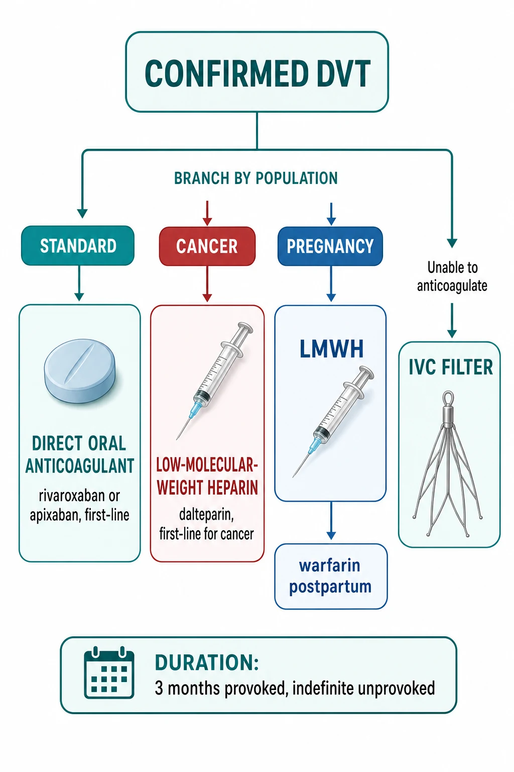
The anticoagulation is started as soon as the diagnosis is confirmed or the probability is high. The direct oral anticoagulants are now the first-line for the uncomplicated proximal DVT.[1]
[1]The DVT targets and the doses
The compression stockings (the graduated elastic compression, the knee-high, 30 to 40 mmHg) reduce the post-thrombotic syndrome (the chronic leg swelling, the pain, the ulceration that follows 20 to 50 per cent of the proximal DVTs). The patient is advised to mobilise — the bed rest is no longer recommended after the anticoagulation is started. [1]
The anticoagulant agents — the head-to-head
The contemporary DVT is treated with the direct oral anticoagulant as the first-line; the warfarin and the LMWH retain the niche roles. The Fellowship viva rewards the candidate who can name the drug, the dose, the loading and the maintenance, and the indication for each.[6][7][8][9]
Apixaban (factor Xa)
- 10 mg orally twice daily for 7 days, then 5 mg twice daily
- No initial parenteral agent — started directly
- No routine monitoring; the lowest major-bleed rate of the DOACs
- Dose-reduce in the severe renal impairment, the age over 80, the weight under 60 kg
Rivaroxaban (factor Xa)
- 15 mg orally twice daily for 21 days, then 20 mg daily
- No initial parenteral agent — started directly
- Take with the food (improves the absorption of the higher doses)
- Avoid in the severe renal impairment and the pregnancy
Dabigatran (direct thrombin)
- 5 to 10 days of the parenteral anticoagulation FIRST, then 150 mg twice daily
- Cannot be started directly — the lead-in LMWH or unfractionated heparin is mandatory
- A prodrug; the dyspepsia is the common gastric side effect
- Reversible with the idarucizumab
Edoxaban (factor Xa)
- 5 to 10 days of the parenteral anticoagulation FIRST, then 60 mg daily
- Cannot be started directly — the lead-in heparin
- Less effective if the creatinine clearance is very high (the low-efficacy zone)
- Reversible with the andexanet alfa (shared with apixaban and rivaroxaban)
Warfarin (vitamin K antagonist)
- Overlapped with the heparin for at least 5 days and until the INR is 2 to 3 for 24 hours
- The choice for the antiphospholipid syndrome (the triple-positive) and the mechanical valve
- Requires the INR monitoring; the diet, the drugs and the genes interact
- Slow onset; pro-thrombotic in the first days via the protein C depletion
LMWH — enoxaparin
- 1 mg per kilogram subcutaneously twice daily, or 1.5 mg per kilogram once daily
- The choice for the cancer-associated thrombosis, the pregnancy, the DOAC contraindication
- No monitoring in the standard patient; the anti-Xa level in the renal failure and the pregnancy
- Avoid in the heparin-induced thrombocytopenia and the severe renal failure
Starting the warfarin (when the DOAC is contraindicated) — the overlap and the bridge
Start the heparin (the LMWH enoxaparin 1 mg per kilogram twice daily, or the unfractionated heparin infusion) on the day of the diagnosis — immediately, not after the scan if the Wells is high.
Start the warfarin on the same day or the day after — the 5 mg loading dose for the first two days, then adjusted to the INR. Do NOT delay the warfarin, but do NOT rely on it alone in the first week.
Continue BOTH the heparin and the warfarin for a minimum of 5 days AND until the INR is 2.0 or above for 24 hours — both conditions must be met before the heparin is stopped. The overlap covers the pro-thrombotic window of the warfarin (the protein C depletion).
Stop the heparin once the overlap criteria are met; continue the warfarin to the INR target of 2.0 to 3.0. Educate the patient on the diet, the drugs, the bleeding signs and the INR monitoring schedule.
In the antiphospholipid syndrome or the unusual thrombosis, involve the haematology team early — the DOAC may be inferior and the warfarin with the INR 2 to 3 is the standard.
The inferior vena cava filter — the exception, not the rule
The IVC filter traps the embolus before it reaches the lungs; it does not treat the thrombus and it does not replace the anticoagulation. The only absolute indication is the acute DVT or the PE with a contraindication to the anticoagulation — the active life-threatening bleed, the recent intracranial haemorrhage, the planned emergency surgery, or the heparin-induced thrombocytopenia before the non-heparin anticoagulant is established. The PREVENT trial showed that the prophylactic filter in the high-risk anticoagulated patient reduced the early symptomatic PE but increased the late DVT recurrence — so the filter is not added to the anticoagulated patient.[5][16]
[1]The duration — provoked versus unprovoked
Provoked DVT (reversible trigger)
- Surgery, the immobilisation, the fracture, the long-haul flight, the oestrogen
- 3 months of the anticoagulation is the standard — no shorter, no longer
- Stop the oestrogen; address the reversible factor; no extended therapy
- The recurrence after a provoked DVT is low
Unprovoked DVT
- No reversible trigger identified — the higher recurrence risk
- At least 3 months, then reassess; the extended (often indefinite) therapy for most
- The bleeding risk and the patient preference weigh in the continuation
- The Kearon trial showed the extended therapy halved the recurrence
Cancer-associated DVT
- The active cancer drives the ongoing risk
- Extended (the LMWH or the DOAC) for as long as the cancer is active
- The CLOT trial established the LMWH over the warfarin
- Reassess the risk–benefit at each follow-up
Recurrent (second) unprovoked DVT
- Indefinite anticoagulation is the standard after a second unprovoked event
- The recurrence risk is high; the benefit of the extended therapy is clear
- Consider the thrombophilia screen and the antiphospholipid workup
- The bleeding risk dictates the agent and the dose
Subtypes and special scenarios
The iliofemoral DVT (the entire leg swollen, the phlegmasia alba dolens, or the phlegmasia cerulea dolens — the limb-threatening ischaemia from the massive venous obstruction) is a high-risk presentation that may need the catheter-directed thrombolysis, not just the anticoagulation. The upper-extremity DVT (from the central line, the pacemaker, the thrombophilia, the Paget-Schroetter syndrome — the effort thrombosis in the young athlete) is managed with the anticoagulation and the removal of the catheter or the thoracic-outlet decompression. The cancer-associated thrombosis is managed with the LMWH (historically) or the DOACs (increasingly), with a longer duration. The pregnancy-associated DVT uses the LMWH (the DOACs and the warfarin are contraindicated in pregnancy). [1]
Complications and pitfalls
The complications are the PE (the proximal propagation and the embolism — cross-link to the PE topic), the post-thrombotic syndrome (the chronic venous insufficiency), the recurrence (the risk is highest in the first year after the event), and the bleeding from the anticoagulation. The pitfalls are: not starting the anticoagulation early (waiting for the ultrasound in the high-probability patient); using the warfarin without the initial heparin (the warfarin alone is pro-thrombotic in the first few days — the protein C depletion); under-treating the cancer-associated thrombosis (the warfarin is inferior to the LMWH in the active cancer); not investigating the unprovoked DVT for the underlying malignancy or the thrombophilia; and not addressing the proximal DVT risk for the PE. [1]
Prognosis and disposition
The prognosis is excellent with the early anticoagulation — the recurrence rate is about 5 per cent in the first year and falls thereafter; the post-thrombotic syndrome affects 20 to 50 per cent of the proximal DVTs. The patient is managed as an outpatient (the DOACs) or a short-stay (the LMWH loading then the warfarin); the admission is reserved for the massive iliofemoral DVT, the severe symptoms, the comorbidity, or the social indication. The follow-up includes the INR monitoring (if on warfarin), the compression stockings, and the unprovoked-DVT malignancy screen. [1]
Special populations
The cancer patient with the DVT is managed with the LMWH or the DOACs and a longer duration. The pregnant patient receives the LMWH (the DOACs and the warfarin are contraindicated). The antiphospholipid syndrome patient is managed with the warfarin (the DOACs may be inferior in the triple-positive antiphospholipid syndrome). The elderly and the renal-failure patient need the dose-adjusted LMWH or the reduced-dose DOAC. [1]
The landmark DVT trials — the evidence base
The Fellowship viva expects the candidate to name the trials behind the DOAC-first strategy, the cancer-LMWH tradition, the IVC filter caveat and the stocking controversy. The trials below are the high-yield set. [1]
Wells — value of the pretest probability in the DVT (Lancet 1997)
Lancet
PMID 9428249
Key finding
A prospective cohort of 529 patients with the suspected DVT, deriving and validating a clinical prediction rule that combined the signs, the risk factors and the alternative-diagnosis judgement into a low-, moderate- and high-probability score, which then guided the D-dimer and the ultrasound strategy.
Practice change
The Wells-DVT score is the global standard for the pretest-probability step — the entire modern pathway rests on it.
EINSTEIN-DVT — oral rivaroxaban for the symptomatic DVT (NEJM 2010)
New England Journal of Medicine
PMID 21128814
Key finding
A randomised open-label non-inferiority trial of 3,449 patients with the acute symptomatic DVT comparing the oral rivaroxaban (15 mg twice daily for 3 weeks, then 20 mg daily) against the enoxaparin–warfarin standard. Rivaroxaban was non-inferior for the recurrent VTE (2.1 versus 3.0 per cent) with a similar major-bleed rate and no routine monitoring.
Practice change
The single-drug oral rivaroxaban regimen — no parenteral lead-in, no INR monitoring — is the evidence for the DOAC-first DVT strategy.
AMPLIFY — oral apixaban for the acute VTE (NEJM 2013)
New England Journal of Medicine
PMID 23808982
Key finding
A randomised double-blind non-inferiority trial of 5,395 patients with the acute VTE (DVT or PE) comparing the oral apixaban (10 mg twice daily for 7 days, then 5 mg twice daily) against the conventional enoxaparin–warfarin. Apixaban was non-inferior for the recurrent VTE or the PE-related death (2.3 versus 2.7 per cent) and significantly reduced the major bleeding (0.6 versus 1.8 per cent).
Practice change
The single-drug oral apixaban regimen with the lowest major-bleed rate is the first-line for the uncomplicated DVT.
RE-COVER — dabigatran for the acute VTE (NEJM 2009)
New England Journal of Medicine
PMID 19966341
Key finding
A randomised double-blind non-inferiority trial of 2,539 patients with the acute VTE comparing the oral dabigatran 150 mg twice daily against the warfarin, both preceded by the parenteral anticoagulation. Dabigatran was non-inferior for the recurrent VTE (2.4 versus 2.1 per cent) with a similar major-bleed rate.
Practice change
Dabigatran is effective but CANNOT be started directly — it requires the 5 to 10 day parenteral lead-in, which is its practical limitation versus apixaban and rivaroxaban.
Hokusai-VTE — edoxaban for the symptomatic VTE (NEJM 2013)
New England Journal of Medicine
PMID 23991658
Key finding
A randomised double-blind non-inferiority trial of 8,240 patients with the symptomatic VTE comparing the oral edoxaban 60 mg daily against the warfarin, both preceded by the parenteral anticoagulation. Edoxaban was non-inferior for the recurrent VTE (3.2 versus 3.5 per cent) with less major bleeding.
Practice change
Edoxaban joins dabigatran as a once-daily oral agent that needs the parenteral lead-in; it is less effective when the renal function is very good (the high-clearance zone).
CLOT — dalteparin versus warfarin in the cancer-associated thrombosis (NEJM 2003)
New England Journal of Medicine
PMID 12853587
Key finding
A randomised open-label trial of 676 patients with the cancer and the acute DVT or PE comparing the dalteparin (200 IU per kilogram daily for one month then 75 to 80 per cent) against the warfarin for six months. The dalteparin reduced the recurrent VTE from 17 per cent to 9 per cent, with no difference in the bleeding.
Practice change
The historical basis for the LMWH as the first-line in the cancer-associated thrombosis — the basis of the older guideline that the candidate must still reproduce.
CATCH — tinzaparin versus warfarin in the cancer-associated thrombosis (JAMA 2015)
JAMA
PMID 26284719
Key finding
A randomised open-label non-inferiority trial of 900 patients with the cancer and the acute VTE comparing the tinzaparin 175 IU per kilogram daily against the warfarin for six months. The tinzaparin did NOT meet the non-inferiority margin for the recurrent VTE but reduced the clinically relevant non-major bleeding; no difference in the overall survival.
Practice change
The LMWH and the warfarin are close in the cancer patient; the DOACs (apixaban, rivaroxaban) are now increasingly used and are the contemporary first-line in the selected cancer patient.
PREVENT — the vena cava filter with the anticoagulation (NEJM 1998)
New England Journal of Medicine
PMID 9459643
Key finding
A randomised trial of 400 patients with the proximal DVT and the planned anticoagulation, randomised to the IVC filter or no filter (all anticoagulated). The filter reduced the symptomatic PE at 12 days (1.1 versus 4.8 per cent) but increased the recurrent DVT at two years (21 versus 11 per cent), with no difference in the mortality.
Practice change
The prophylactic filter in the anticoagulated patient prevents the early PE at the cost of the late DVT — the filter is reserved for the contraindication to the anticoagulation, not added to it.
Kearon — 3 months versus extended anticoagulation for the first unprovoked VTE (NEJM 1999)
New England Journal of Medicine
PMID 10089183
Key finding
A randomised double-blind trial of 162 patients with the first unprovoked proximal DVT, all on the warfarin, randomised after 3 months to either stop or continue indefinitely. The extended anticoagulation reduced the recurrent VTE from 18 per cent to under 3 per cent over the follow-up, with a small increase in the bleeding.
Practice change
The unprovoked DVT recurs — the extended (often indefinite) anticoagulation is the standard after the first unprovoked event, balanced against the bleeding risk.
SOX — elastic compression stockings and the post-thrombotic syndrome (Lancet 2014)
Lancet
PMID 24315521
Key finding
A randomised placebo-controlled trial of 803 patients with the first proximal DVT comparing the active elastic compression stockings (30 to 40 mmHg) against the placebo stockings for two years. There was NO difference in the post-thrombotic syndrome (around 31 per cent in both arms).
Practice change
The routine compression stocking to prevent the post-thrombotic syndrome is NOT supported; the stockings remain for the symptomatic relief of the chronic swelling, not for the prevention.
CALISTO — fondaparinux for the superficial-vein thrombosis (NEJM 2010)
New England Journal of Medicine
PMID 20860504
Key finding
A randomised double-blind trial of 3,002 patients with the lower-limb superficial-vein thrombosis comparing the fondaparinux 2.5 mg daily for 45 days against the placebo. The fondaparinux reduced the composite of the extension, the DVT and the PE from 6 per cent to 1 per cent, with no excess bleeding.
Practice change
The symptomatic superficial-vein thrombosis near the saphenofemoral junction is treated with the fondaparinux (or the DOAC) — it is a DVT-equivalent, not a varicose-vein problem.
CASSINI — rivaroxaban prophylaxis in the high-risk cancer patient (NEJM 2019)
New England Journal of Medicine
PMID 30786186
Key finding
A randomised double-blind trial of 841 ambulatory cancer patients with a high Khorana score comparing the rivaroxaban 10 mg daily against the placebo for 180 days. The rivaroxaban reduced the VTE at 6 months from 9 to 4 per cent, with no significant excess in the major bleeding.
Practice change
The primary thromboprophylaxis with the rivaroxaban is an option in the high-risk ambulatory cancer patient — the balance of the benefit and the bleeding is patient-specific.
BRIDGE — perioperative bridging in the AF on warfarin (NEJM 2015)
New England Journal of Medicine
PMID 26095867
Key finding
A randomised double-blind trial of 1,884 patients on the warfarin for the atrial fibrillation undergoing the elective procedure, comparing the bridging LMWH against the placebo. The bridging did NOT reduce the arterial thromboembolism and significantly increased the major bleeding.
Practice change
The routine bridging of the warfarin for the procedure is NOT needed in the low-bleed-risk AF; the bridging is reserved for the very high thrombotic risk (the mechanical valve, the recent VTE, the antiphospholipid syndrome).
2016 CHEST Guideline — the antithrombotic therapy for the VTE (Chest 2016)
Chest
PMID 26867832
Key finding
The multidisciplinary CHEST guideline that codifies the DOAC as the first-line for the DVT and the PE (over the vitamin K antagonist), the non-superiority of the IVC filter with the anticoagulation, the selective treatment of the distal DVT, the against-the-routine use of the compression stockings for the prevention of the post-thrombotic syndrome, and the duration framework (3 months provoked, extended for the unprovoked and the cancer-associated).
Practice change
The single most cited contemporary framework for the DVT management — the synthesis the Fellowship examiner wants.
Evidence and regional guidelines
The contemporary framework is the DOAC-first strategy for the uncomplicated proximal DVT,[1] and the LMWH for the cancer-associated and the pregnancy-associated thrombosis.[2] The Wells-DVT score and the D-dimer/ultrasound strategy are the global standard. The drug choice, the duration and the follow-up follow the local haematology and the thrombosis pathway.
ANZ practice note. The Wells-DVT score, the D-dimer and the compression ultrasound follow the local thrombosis pathway; the DOAC (apixaban or rivaroxaban) is the first-line for the uncomplicated DVT, the LMWH for the cancer-associated and the pregnancy-associated, and the compression stockings reduce the post-thrombotic syndrome. [1]
The post-thrombotic syndrome — the chronic legacy
The post-thrombotic syndrome affects 20 to 50 per cent of the proximal DVTs, developing over months to years, and presents as the chronic leg heaviness, the swelling, the pain on standing, the venous eczema and the lipodermatosclerosis, culminating in the venous ulcer above the medial malleolus. The mechanism is the venous hypertension from the valvular destruction and the residual obstruction. The early anticoagulation and the adequate duration reduce it; the routine compression stocking does NOT prevent it (the SOX trial) but relieves the symptoms once present. [1]
[1] [1]Exam practice
SAQ — The swollen leg: applying the Wells-DVT score and the diagnostic pathway
10 minutes · 10 marks
A 62-year-old man presents to the emergency department with a two-day history of swelling and pain in the left calf. He had an uncomplicated total knee replacement three weeks ago. On examination the left calf measures 4 cm larger than the right at 10 cm below the tibial tuberosity, with tenderness along the popliteal vein and pitting oedema confined to the symptomatic leg. He is afebrile, HR 92, BP 138/84, RR 18, SpO2 97 per cent on room air. There is no erythema, no clear demarcated margin, and no history of trauma. The registrar asks whether a D-dimer should be sent to rule the clot out.
SAQ — Anticoagulation choice in the cancer-associated deep vein thrombosis
10 minutes · 10 marks
A 71-year-old woman with metastatic pancreatic cancer on chemotherapy is diagnosed with an acute proximal deep vein thrombosis of the right femoral vein on compression ultrasound. She has no chest pain, no dyspnoea and no signs of pulmonary embolism. Her creatinine is 78 micromol per litre, platelet count 210 x 10^9/L, haemoglobin 112 g/L. She takes paracetamol only and has had no bleeding. The registrar asks which anticoagulant to start, and for how long.
Exam pearls
- Wells-DVT: active cancer, bedridden/immobilisation/paralysis, bed rest/surgery, localised tenderness, leg swelling (entire/calf/oedema), collateral superficial veins, previous DVT, alternative diagnosis (−2).
- Score ≤0 = low → D-dimer; ≥3 = high → ultrasound or start the anticoagulation.
- Apixaban 10 mg bid x 7 d then 5 mg bid; or rivaroxaban 15 mg bid x 21 d then 20 mg od.
- LMWH (enoxaparin 1.5 mg/kg daily) for cancer and pregnancy.
- 3 months provoked; 6 months or indefinite unprovoked/cancer.
- Mobilise (no bed rest) once anticoagulated.
- Compression stockings reduce the post-thrombotic syndrome. [1]
Red flags
[1]References
- [1]Kumbhani DJ, Cannon CP, Piazza G, et al. Direct Oral Anticoagulants in Primary and Secondary Prevention of Thrombotic Events: 2026 ACC Scientific Statement: A Report of the American College of Cardiology J Am Coll Cardiol, 2026.PMID 42377292
- [2]Stals MAM, Takada T, Kraaijpoel N, et al. Noninvasive diagnostic work-up for suspected acute pulmonary embolism during pregnancy: a systematic review and meta-analysis of individual patient data J Thromb Haemost, 2023.PMID 36696189
- [3]Wells PS, Anderson DR, Bormanis J, et al. Value of assessment of pretest probability of deep-vein thrombosis in clinical management Lancet, 1997.PMID 9428249
- [4]Kearon C, Gent M, Hirsh J, et al. A comparison of three months of anticoagulation with extended anticoagulation for a first episode of idiopathic venous thromboembolism N Engl J Med, 1999.PMID 10089183
- [5]Decousus H, Leizorovicz A, Parent F, et al. A clinical trial of vena caval filters in the prevention of pulmonary embolism in patients with proximal deep-vein thrombosis. Prévention du Risque d'Embolie Pulmonaire par Interruption Cave Study Group N Engl J Med, 1998.PMID 9459643
- [6]Bauersachs R, Berkowitz SD, Brenner B, et al. Oral rivaroxaban for symptomatic venous thromboembolism N Engl J Med, 2010.PMID 21128814
- [7]Agnelli G, Buller HR, Cohen A, et al. Oral apixaban for the treatment of acute venous thromboembolism N Engl J Med, 2013.PMID 23808982
- [8]Schulman S, Kearon C, Kakkar AK, et al. Dabigatran versus warfarin in the treatment of acute venous thromboembolism N Engl J Med, 2009.PMID 19966341
- [9]Büller HR, Décousus H, Grosso MA, et al. Edoxaban versus warfarin for the treatment of symptomatic venous thromboembolism N Engl J Med, 2013.PMID 23991658
- [10]Lee AY, Levine MN, Baker RI, et al. Low-molecular-weight heparin versus a coumarin for the prevention of recurrent venous thromboembolism in patients with cancer N Engl J Med, 2003.PMID 12853587
- [11]Lee AYY, Kamphuisen PW, Meyer G, et al. Tinzaparin vs Warfarin for Treatment of Acute Venous Thromboembolism in Patients With Active Cancer: A Randomized Clinical Trial JAMA, 2015.PMID 26284719
- [12]Decousus H, Prandoni P, Mismetti P, et al. Fondaparinux for the treatment of superficial-vein thrombosis in the legs N Engl J Med, 2010.PMID 20860504
- [13]Khorana AA, Soff GA, Kakkar AK, et al. Rivaroxaban for Thromboprophylaxis in High-Risk Ambulatory Patients with Cancer N Engl J Med, 2019.PMID 30786186
- [14]Douketis JD, Spyropoulos AC, Kaatz S, et al. Perioperative Bridging Anticoagulation in Patients with Atrial Fibrillation N Engl J Med, 2015.PMID 26095867
- [15]Kahn SR, Shapiro S, Wells PS, et al. Compression stockings to prevent post-thrombotic syndrome: a randomised placebo-controlled trial Lancet, 2014.PMID 24315521
- [16]Kearon C, Akl EA, Ornelas J, et al. Antithrombotic Therapy for VTE Disease: CHEST Guideline and Expert Panel Report Chest, 2016.PMID 26867832