EM · Acute limb ischaemia
Acute limb ischaemia (the emergency of the 6 Ps)
Also known as ALI · Acute arterial occlusion · The 6 Ps · Acute leg ischaemia
Acute limb ischaemia — the 6 Ps (pain, pallor, pulseless, paraesthesia, paralysis, perishing cold), the embolic versus the thrombotic cause, the Rutherford classification (viable through irreversible), the immediate anticoagulation (unfractionated heparin 80 units per kilogram bolus then 18 units per kilogram per hour infusion), the definitive management (the Fogarty embolectomy for the embolic, the catheter-directed thrombolysis for the thrombotic), the 6-hour window for the limb salvage, and the fasciotomy for the reperfusion. ACEM-primary, globally tagged.
On this page & tools
Your progress
Saved locally on this device.
Target exams
Red flags
Acute limb ischaemia is the sudden decrease in the limb perfusion that threatens the tissue viability, and it is one of the few true vascular emergencies where minutes matter — the limb that is not revascularised within 6 hours may be irreversibly lost. The Fellowship candidate must recognise the 6 Ps at the bedside, start the immediate anticoagulation, and make the embolic-versus-thrombotic distinction that decides the surgical approach.[1][2]
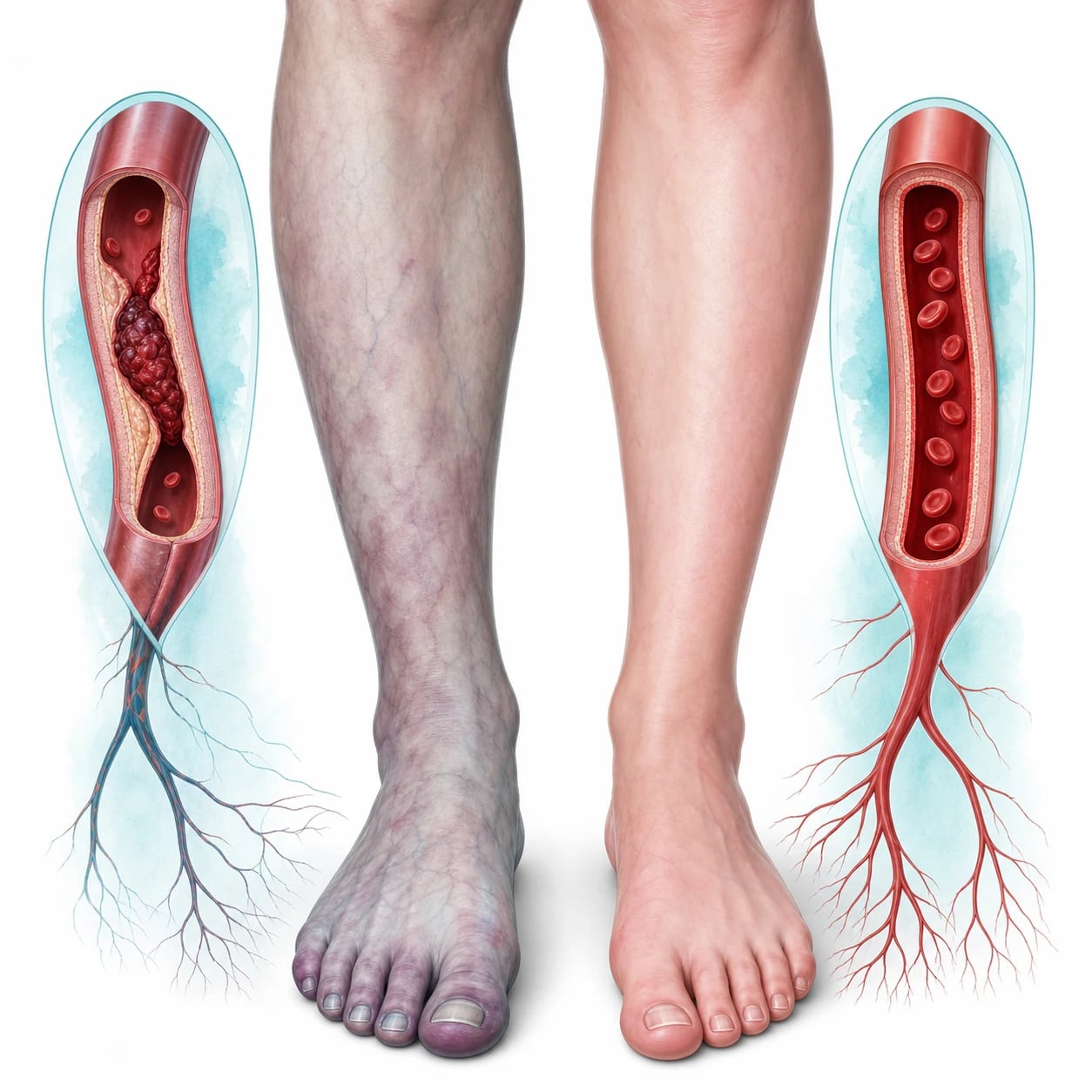
The 6 Ps — the clinical diagnosis
The clinical diagnosis of the acute limb ischaemia is made at the bedside by the 6 Ps: the pain (sudden, severe, often disproportionate to the appearance), the pallor (the limb is white or waxy, not cyanotic — the cyanosis suggests the venous, not the arterial, obstruction), the pulselessness (the absent distal pulse, confirmed by the Doppler if palpable pulses are not felt), the paraesthesia (the numbness from the nerve ischaemia), the paralysis (the muscle weakness or the fixed flexion from the motor nerve and the muscle ischaemia), and the perishing cold (the limb is cold to the touch below the level of the obstruction). The paraesthesia and the paralysis are the critical signs — they indicate the threatened limb that needs the immediate revascularisation, not just the anticoagulation. The level of the obstruction is inferred from the line of the demarcation (the cold, the pale, the pulseless border) and the pulse examination (the femoral, the popliteal, the posterior tibial and the dorsalis pedis). [1]
Differential diagnosis — the embolic versus the thrombotic
The cause is either an embolus or a thrombosis, and the distinction guides the surgical approach. [1]
Embolus (commoner)
- Atrial fibrillation (the commonest source), post-MI mural thrombus, valvular, aortic
- No history of the claudication or the prior PAD
- A sudden, dramatic presentation (the 6 Ps in full)
- Fogarty catheter embolectomy; look for and treat the source
Thrombosis (on PAD)
- A pre-existing PAD with an acute-on-chronic occlusion
- A history of the claudication, the bypass graft or the known stenosis
- A less dramatic onset (collateral vessels partially compensate)
- Catheter-directed thrombolysis or the bypass; the heparin as a bridge
Graft thrombosis
- A prosthetic or a vein bypass graft that has thrombosed
- A history of a prior vascular surgery
- May present as a subacute or an acute occlusion
- Thrombolysis or the graft revision
Trauma / dissection
- A traumatic occlusion (the knee dislocation — the popliteal artery)
- An aortic dissection extending to the iliac/femoral
- The mechanism and the associated injuries give the diagnosis
- The vascular surgery and the trauma team
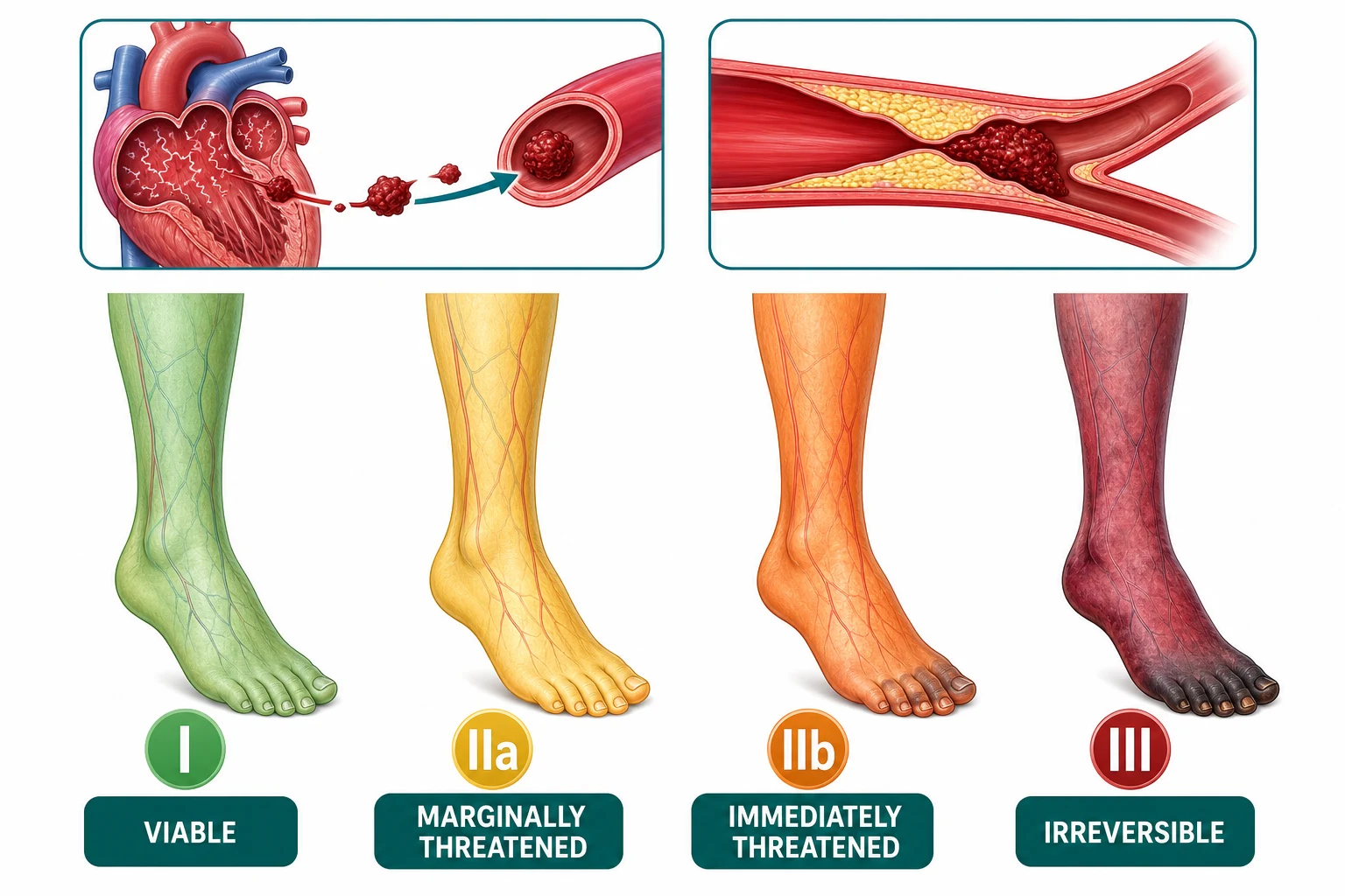
The Rutherford classification — the severity and the urgency
The Rutherford classification grades the severity and determines the urgency: [1]
| Category | Description | Motor/Sensory | Doppler (arterial) | Urgency |
|---|---|---|---|---|
| I (Viable) | Not immediately threatened | Normal | Audible | Elective workup |
| IIa (Marginally threatened) | Salvageable if promptly treated | Mild sensory loss, no motor loss | Inaudible | Urgent (hours) |
| IIb (Immediately threatened) | Salvageable with the immediate revascularisation | Sensory and motor loss | Inaudible | Emergency (immediate) |
| III (Irreversible) | Major permanent tissue loss | Paralysis, anaesthesia | Inaudible | Amputation or palliation |
The category IIb — with the motor and the sensory loss — is the immediately threatened limb that needs the revascularisation within minutes, not hours. [1]
Investigations — the bedside and the angiography
The bedside Doppler confirms the absent arterial signal and assesses the venous flow. The ABI (the ankle-brachial index) is less than 0.4 in the severe ischaemia. The bloods include the full blood count, the coagulation, the urea and electrolytes (the rhabdomyolysis from the reperfusion — the creatine kinase rises), the group and crossmatch, and the glucose. The ECG screens for the atrial fibrillation (the embolic source) and the ischaemia (the mural thrombus). The CT angiogram (if the patient is stable and the diagnosis needs confirmation) defines the level and the extent of the occlusion and guides the intervention. In the unstable or the category IIb patient, the angiogram is skipped and the patient goes directly to the theatre. [1]
Immediate management — the heparin and the revascularisation
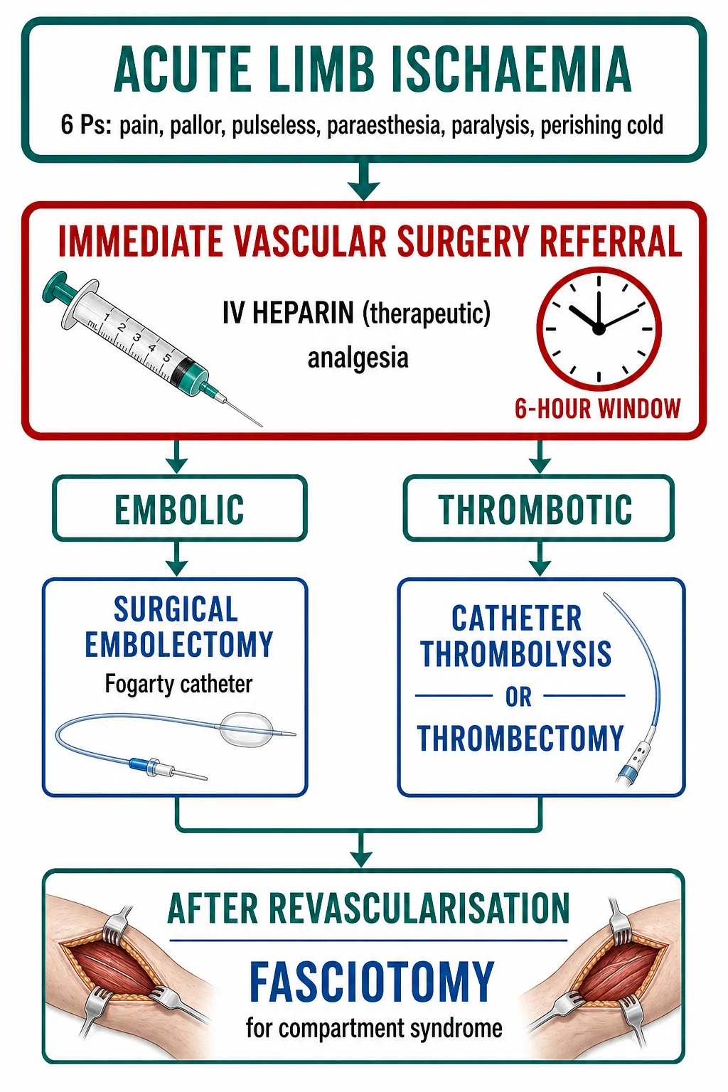
The management has two components: the immediate anticoagulation to prevent the thrombus propagation and the definitive revascularisation to restore the flow. [1]
[1]The ALI targets and the doses
The fasciotomy — the reperfusion injury
After the revascularisation, the ischaemic limb may swell as the blood returns and the reperfusion injury develops — the compartment syndrome, the rhabdomyolysis, the hyperkalaemia and the acidosis. A prophylactic fasciotomy (the fascial release of the four compartments of the leg — the anterior, the lateral, the deep posterior and the superficial posterior) is performed in the severely ischaemic or the delayed-presentation case to prevent the compartment syndrome. The creatine kinase and the myoglobin are monitored (the rhabdomyolysis is managed with the intravenous fluids to a urine output of 1 to 1.5 mL per kg per hour). The hyperkalaemia and the acidosis from the reperfusion are corrected. [1]
Complications and pitfalls
The complications are the limb loss (the amputation), the reperfusion injury (the compartment syndrome, the rhabdomyolysis, the systemic inflammatory response, the multi-organ failure from the release of the potassium, the myoglobin and the acid), the recurrence (the recurrent embolus or thrombosis), the bleeding from the heparin or the thrombolysis, and the infection. The pitfalls are: not starting the heparin immediately (waiting for the vascular surgeon); missing the motor and the sensory loss (the category IIb); delaying the revascularisation beyond the 6-hour window; not performing the prophylactic fasciotomy; and not investigating and treating the embolic source (the atrial fibrillation — the long-term anticoagulation prevents the recurrence). [1]
Prognosis and disposition
The limb-salvage rate is over 80 per cent for the category I and IIa with the prompt revascularisation; the mortality is 10 to 15 per cent (from the comorbidity and the reperfusion injury). The patient is admitted to the vascular surgical service or the intensive care (for the severe case with the reperfusion injury); the long-term management addresses the PAD (the antiplatelet, the statin, the risk-factor modification)[8] and the embolic source (the anticoagulation for the atrial fibrillation).
Special populations
The diabetic patient with the PAD and the neuropathy may present with a less dramatic picture (the pain is blunted by the neuropathy) — the diagnosis may be delayed. The elderly with the severe PAD and the comorbidity may not tolerate the major revascularisation — the primary amputation may be the better option. The anticoagulated patient (the warfarin or the DOAC) with the acute limb ischaemia may still thrombose or embolise (the sub-therapeutic INR) — the heparin is still given and the anticoagulation is reviewed. [1]
Evidence and regional guidelines
The contemporary framework is the vascular-surgical guideline (the Rutherford classification, the heparin-first, the Fogarty embolectomy for the embolus, the thrombolysis for the thrombosis, and the fasciotomy for the reperfusion).[1][2] The drug doses and the revascularisation pathway follow the local vascular-surgical protocol.
ANZ practice note. The heparin-first (80 U/kg bolus then 18 U/kg/h), the Fogarty embolectomy for the embolic and the thrombolysis for the thrombotic, the 6-hour window, and the prophylactic fasciotomy follow the local vascular-surgical pathway; the atrial fibrillation is the commonest embolic source and the long-term anticoagulation prevents the recurrence. [1]
Pathophysiology — the 6-hour window and the no-reflow
The skeletal muscle is the most ischaemia-sensitive tissue of the limb. The sequence of the metabolic and the electrophysiological failure is stereotyped and time-locked: the motor nerve fails first (the conduction block at 2 to 4 hours), then the sensory nerve (the paraesthesia progressing to the anaesthesia), then the skeletal muscle (the irreversible necrosis beginning at 4 to 6 hours), and finally the skin and the subcutaneous tissue (the skin is the most tolerant — it survives up to 12 hours). The 6-hour window for the limb salvage is set by the muscle: an ischaemic limb that is not revascularised within 6 hours of the onset is at a high risk of the irreversible myonecrosis and the amputation.[3]
The corollary, which the examiners test, is that the pain and the pallor are reversible; the paralysis and the paraesthesia signal that the nerve injury is already underway — these are the signs that convert a category IIa limb into a category IIb limb and that demand the revascularisation in minutes rather than hours. After the revascularisation, the reperfusion injury (the washout of the potassium, the myoglobin, the lactate and the acid from the dead muscle; the neutrophil-mediated endothelial injury; the capillary leak and the compartment swelling) produces the no-reflow phenomenon — the blood returns to the major artery but cannot perfuse the capillary bed because the capillaries are occluded by the swollen endothelium and the microthrombi. This is why a category III limb may not recover even after a technically-successful revascularisation. [1]
[1]The causes — the embolism, the thrombosis, and the trauma
Acute limb ischaemia is the sudden arterial occlusion, and the cause falls into one of three buckets: an embolus (the commonest, roughly 80 per cent in the historical series), a thrombosis of a pre-existing atherosclerotic stenosis or a graft, and a traumatic occlusion (the blunt, the penetrating or the iatrogenic). The distinction decides the operation: the embolus is removed by the Fogarty catheter embolectomy; the thrombosis on the PAD substrate is treated by the catheter-directed thrombolysis or the bypass; the trauma needs the vascular repair.[2]
Embolus (~80%)
- Sudden, no collateral, the full 6 Ps; the commonest cause
- Atrial fibrillation the #1 source; then the post-MI mural thrombus, the prosthetic valve, the aortic atheroma, the paradoxical (DVT through a PFO)
- No prior claudication; a normal contralateral limb; the embolus lodges at the arterial bifurcation (the femoral, the popliteal, the aortic saddle)
- Fogarty catheter embolectomy; the heparin; the search and the treatment of the source
Thrombosis (~15%)
- Acute-on-chronic on a pre-existing PAD stenosis; the collateral vessels partially compensate
- A history of the claudication, the prior revascularisation, or the known stenosis
- A less dramatic onset; the ABI often chronically low; the occlusion at the site of the worst stenosis
- Catheter-directed thrombolysis or the surgical bypass; the heparin as a bridge; the Watanabe / the thrombangiitis obliterans (Buerger disease) in the young male smoker
Graft thrombosis
- A thrombosed prosthetic or vein bypass graft — a subset of the thrombotic cause
- A history of the prior vascular surgery (the femoropopliteal, the femorotibial, the axillofemoral)
- May present as a subacute or an acute occlusion; the graft is the " Achilles heel" of the revascularised patient
- Thrombolysis (if the acute occlusion and no contraindication) or the graft revision / the jump graft
Trauma / dissection
- A traumatic occlusion — the knee dislocation (the popliteal artery), the supracondylar femur fracture, the penetrating injury, the arterial catheterisation
- An aortic dissection extending to the iliac or the femoral — the pulse differential and the tearing back pain
- The mechanism and the associated injuries give the diagnosis; consider the compartment pressure
- The vascular repair or the stent; the trauma team; the orthopaedics for the fracture
The embolus — the sources and the search
The atrial fibrillation is the single commonest source of the peripheral embolus, accounting for over half of the embolic acute limb ischaemia. The left atrial appendage thrombus dislodges, traverses the aorta, and lodges at an arterial bifurcation where the lumen narrows — the femoral bifurcation (the commonest site, around 40 per cent), the popliteal bifurcation (around 20 per cent), the aortic saddle embolus (the bifurcation, around 15 per cent, producing the bilateral leg ischaemia), and the upper-limb sites (the brachial, around 10 per cent). The other sources are the post-infarct mural thrombus (the akinetic left-ventricular wall within the first weeks of the anterior MI), the prosthetic or the diseased native valve (the vegetations and the calcific emboli), the aortic atheroma (the mobile protruding plaque in the descending thoracic aorta), and the paradoxical embolus (a venous thrombus traversing a patent foramen ovale, especially in the young patient with the simultaneous DVT).[3]
[1]The thrombosis — the substrate and the triggers
The thrombosis-on-PAD cause requires a pre-existing stenosis that acutely occludes — the patient typically has the claudication, the low ABI, and the collateral vessels that blunt the severity (a category II rather than a category III presentation). The triggers of the acute occlusion in the borderline stenosis are the dehydration, the low-output state (the heart failure, the sepsis), the hypercoagulable state (the malignancy, the myeloproliferative disorder, the nephrotic syndrome), and the graft stenosis (the intimal hyperplasia at the distal anastomosis, the commonest cause of the late graft failure). The thrombangiitis obliterans (the Buerger disease) — the small- and medium-artery vasculitis of the young male heavy smoker, presenting with the distal upper-limb ischaemia, the claudication of the arch of the foot, and the superficial thrombophlebitis — is the rare thrombotic cause to know for the Fellowship viva.[7]
The trauma — the popliteal and the iatrogenic
The popliteal artery injury is the high-yield traumatic cause: the artery is tethered at the adductor hiatus and the soleal arch, and the posterior knee dislocation (or the tibial plateau fracture, the supracondylar femur) stretches and tears it. Every posterior knee dislocation gets the hard signs check (the pulselessness, the pallor, the paresthesia, the paralysis, the poikilothermia, the expanding haematoma, the bruit) and, in the modern era, the ABI measurement — an ABI below 0.9 mandates the CT angiogram even if the pulse is palpable (the intimal flap may be present with a preserved pulse initially). The iatrogenic causes are the post-catheterisation femoral occlusion (the access-site thrombosis or the dissection), the intra-arterial drug injection (the particulate and the vasospastic occlusion, often with the digital ischaemia), and the aortic dissection extending to the iliac branches. [1]
[1]The 6 Ps — each one explained and graded
The 6 Ps are the bedside diagnosis; the Fellowship candidate must be able to grade each one and to translate the grade into the Rutherford category. [1]
Pain
- Sudden, severe, often disproportionate to the appearance; the calf is the commonest site in the lower limb
- May be absent in the diabetic (the neuropathy) and in the complete anaesthesia of the late presentation
- The first P to appear and the last to disappear (until the nerve dies)
Pallor
- The limb is white or waxy, not cyanotic — the cyanosis suggests the venous obstruction (the phlegmasia)
- The line of the demarcation localises the level: the groin for the iliac, the thigh for the common femoral, the knee for the popliteal
- The " sub-dermal mottling" (the fixed livedoid pattern) is a sign of the irreversible ischaemia
Pulselessness
- The absent distal pulse, confirmed by the handheld Doppler if the pulse is not palpable
- The contralateral pulse comparison is critical — the absent contralateral pulse suggests the dissection or the Leriche
- A palpable pulse does NOT exclude the ischaemia (the intimal flap, the proximal embolus with the collateral flow)
Paraesthesia
- The numbness from the sensory nerve ischaemia — the critical sign that the nerve is failing
- Graded as the mild (the toe numbness, the category IIa) or the profound (the stocking-distribution anaesthesia, the category IIb)
- The sign that converts the threatened-but-viable limb into the immediately-threatened limb
Paralysis
- The muscle weakness or the fixed flexion contracture — the most ominous sign of the myonecrosis
- The toe-flexion or the foot-drop, progressing to the rigid fixed-flexion (the late, the irreversible)
- Any motor loss is the category IIb or III — the immediate revascularisation, not the heparin-and-wait
Perishing cold
- The limb is cold below the level of the obstruction; the temperature transition is the localisation clue
- Confirmed by the back of the examiner's hand, comparing the two limbs
- The capillary refill over 3 seconds; the slow venous refilling after the blanching
The Rutherford classification — the categories and the action
The Rutherford category (the revised standards of 1997) is the universal language that drives the urgency and the operation. The Fellowship viva will present a limb and ask for the category, the urgency, and the intervention.[6]
| Category | Prognosis | Sensory | Motor | Arterial Doppler | Venous Doppler | Urgency | Intervention |
|---|---|---|---|---|---|---|---|
| I (Viable) | Not immediately threatened | Normal | Normal | Audible | Audible | Elective (workup) | Heparin; outpatient workup; the revascularisation may be deferred |
| IIa (Marginally threatened) | Salvageable if promptly treated | Minimal (toe) or none | None | Inaudible | Audible | Urgent (within hours) | Heparin; the angiography and the revascularisation within 6 hours |
| IIb (Immediately threatened) | Salvageable with the immediate revascularisation | More than the toe; the rest pain | Mild to moderate | Inaudible | Audible | Emergency (immediate) | Heparin; the immediate surgical embolectomy or the bypass; no time for the angiogram |
| III (Irreversible) | Major permanent tissue loss; the amputation | Profound anaesthesia | Profound paralysis (rigor) | Inaudible | Inaudible | Amputation or the palliation | The primary amputation or the comfort care; the revascularisation is futile |
The diagnosis — clinical, Doppler, and the imaging decision
The diagnosis is clinical in the first instance — the 6 Ps and the pulse examination localise the level and grade the severity (the Rutherford category) before any imaging. The handheld continuous-wave Doppler confirms the absent arterial signal and, critically, the presence of the venous (biphasic) signal — the venous Doppler distinguishes the salvageable limb (the IIb, the venous outflow preserved) from the irreversible limb (the III, the venous outflow lost). [1]
The imaging decision hinges on the stability and the Rutherford category:[3]
Category I / IIa (stable)
- The CT angiogram of the lower-limb arteries from the aortic bifurcation to the ankle
- Defines the level and the extent of the occlusion; distinguishes the embolus (the sharp cut-off at a bifurcation) from the thrombosis (the diffuse disease, the collaterals)
- Guides the intervention — the embolectomy site, the thrombolysis target, the bypass plan
- A contraindication to the contrast (the severe renal failure) — the MR angiogram or the on-table angiogram
Category IIb (unstable / the immediate)
- NO CT angiogram — the time is the limb; the IIb limb goes straight to the theatre
- The on-table angiogram after the surgical embolectomy confirms the result and identifies the residual occlusion
- The patient is consented and transferred to the theatre while the heparin infuses
- The vascular surgeon is mobilised in parallel, not in series
Category III (irreversible)
- The imaging is for the amputation planning, not the revascularisation
- The plain X-ray may show the calcified arteries (the medial calcinosis of the diabetes); the CT defines the level of the amputation
- The clinical assessment (the rigor, the anaesthesia, the absent venous Doppler) is the diagnosis
- The decision is the primary amputation or the palliation
The management by the Rutherford category — heparin first, then the revascularisation
Every category (except the irreversible III) gets the immediate unfractionated heparin before any imaging or the surgical referral. The heparin does not lyse the clot — it stops the propagation and preserves the collateral flow — and it is the single intervention that buys time for the definitive revascularisation. [1]
The acute limb ischaemia first hour
The recognition — the 6 Ps at the bedside; the pulse examination and the handheld Doppler; the assignment of the Rutherford category (I, IIa, IIb or III) before any imaging.
The immediate heparin — the unfractionated heparin 80 units per kilogram intravenous bolus, then the infusion of 18 units per kilogram per hour, targeting the APTT 1.5 to 2.5 times the control. Do NOT wait for the vascular surgeon.
The analgesia — the morphine 5 to 10 mg intravenously (or the fentanyl 50 to 100 micrograms), titrated; the limb is kept level (not elevated — the elevation worsens the ischaemia) and exposed (not warmed — the warmth increases the metabolic demand).
The bloods, the ECG and the group-and-crossmatch; the IV access (the two large-bore cannulae); the fluid resuscitation if the patient is hypovolaemic (the hypovolaemia worsens the ischaemia).
The vascular-surgical referral — state the category (the I, the IIa, the IIb or the III), the level of the obstruction, the time of the onset, and the comorbidity; the surgeon decides the embolectomy, the thrombolysis or the bypass.
The imaging decision — the CT angiogram if the I or the IIa and the patient is stable; straight to the theatre if the IIb; the on-table angiogram after the embolectomy.
The reperfusion vigil — after the revascularisation, the potassium, the creatine kinase, the urine output (the myoglobinuria) and the compartment pressures are monitored; the prophylactic fasciotomy is performed for the severely ischaemic or the delayed-presentation case.
Category I and IIa — the heparin and the urgent revascularisation within 6 hours
The category I (the viable) and the category IIa (the marginally threatened) limbs are managed by the heparin first, then the CT angiogram, then the revascularisation within 6 hours. The revascularisation choice depends on the cause: an embolus at a bifurcation is treated by the Fogarty catheter embolectomy (the open surgical, the balloon catheter); a thrombosis on the PAD is treated by the catheter-directed thrombolysis (the tissue plasminogen activator infused locally over 24 to 48 hours) or the surgical bypass; a graft thrombosis is treated by the thrombolysis or the graft revision.[3][5]
Category IIb — the immediate surgical embolectomy, no time for the angiogram
The category IIb (the immediately threatened) limb is the surgical emergency — the immediate surgical exploration and the Fogarty embolectomy (for the embolic cause) or the surgical bypass (for the thrombotic cause), without the prior CT angiogram. The heparin is given in the emergency department; the patient is transferred directly to the theatre; the on-table angiogram confirms the result after the embolectomy. The prophylactic fasciotomy is performed at the time of the revascularisation for the IIb limb (the high risk of the reperfusion compartment syndrome). [1]
Category III — the amputation or the palliation
The category III (the irreversible) limb — the fixed flexion contracture, the profound anaesthesia, the rigor, the absent venous Doppler — is treated by the primary amputation (the guillotine or the definitive, at the level defined by the viable tissue) or, in the moribund patient, the palliative comfort care. The revascularisation of a III limb is futile and dangerous (the reperfusion of the dead muscle flushes the potassium and the myoglobin into the circulation). The one exception is the very recent onset of the III signs (within 1 to 2 hours) where a last-ditch revascularisation with the prophylactic fasciotomy may be considered. [1]
The Rutherford categories and the intervention
The Fogarty catheter embolectomy — the technique for the embolus
The Fogarty catheter embolectomy is the definitive operation for the embolic cause. The technique: the local or the general anaesthesia; an arteriotomy at the common femoral (for the iliac or the femoral embolus) or the exposure of the popliteal (for the popliteal embolus); the Fogarty balloon catheter (the size 3 to 4 French for the femoral, 2 to 3 for the popliteal, 2 for the tibial) is passed distally beyond the embolus, the balloon is inflated with the saline, and the catheter is withdrawn, extracting the embolus and the secondary thrombus; the procedure is repeated proximally and distally until the back-bleeding and the forward-flow are restored; the on-table angiogram confirms the complete clearance and identifies the residual clot (which may require the catheter-directed thrombolysis); the arteriotomy is closed; the prophylactic fasciotomy is performed if the ischaemia was prolonged. The Fogarty catheter (described by Thomas Fogarty in 1963) transformed the embolic limb from the 50 per cent amputation rate of the pre-catheter era to the 80 to 90 per cent limb-salvage rate of the modern era. [1]
[1]The catheter-directed thrombolysis — the technique and the evidence for the thrombosis
The catheter-directed thrombolysis (the CDT) is the definitive treatment for the thrombotic cause (the thrombosis on the PAD, the graft thrombosis) and for the residual clot after the Fogarty embolectomy. The technique: the arterial puncture (the contralateral femoral, the antegrade); the selective catheter is embedded in the thrombus; the tissue plasminogen activator (the alteplase) is infused at 0.5 to 1 mg per hour (or the pulse-spray lacing bolus followed by the infusion) for 24 to 48 hours, with the concomitant heparin at the sub-therapeutic dose (to prevent the pericatheter thrombosis); the angiogram is repeated every 12 to 24 hours to monitor the lysis and to reposition the catheter; the infusion is stopped when the flow is restored or the contraindication develops. The patient is monitored in the high-dependency or the intensive care for the bleeding (the intracranial, the gastrointestinal, the access-site). [1]
The evidence base for the CDT versus the surgery is the STILE trial (1994) and the TOPAS trial (1998). The STILE trial showed that the thrombolysis and the surgery had the similar amputation-free survival, but the thrombolysis was better for the less-than-14-day ischaemia (the acute) and the surgery was better for the more-than-14-day ischaemia (the chronic).[4] The TOPAS trial confirmed that the recombinant urokinase and the surgery had the similar amputation-free survival at six months, with the thrombolysis avoiding the open surgery in around a third.[5] The synthesis: the thrombolysis is preferred for the acute thrombosis (under 14 days) and the surgery for the chronic or the embolic. The thrombolysis is contraindicated in the IIb limb (no time for the 24-hour infusion), in the active bleeding, in the recent stroke, in the recent surgery, and in the uncontrolled hypertension.
Catheter-directed thrombolysis
- For the acute thrombotic (under 14 days) occlusion on the PAD or the graft
- The alteplase 0.5 to 1 mg/h locally for 24 to 48 hours; the heparin at the sub-therapeutic dose
- Avoids the open surgery in around a third (TOPAS); better for the acute than the chronic (STILE)
- Contraindicated in the IIb (no time), the active bleeding, the recent stroke or surgery, the severe hypertension
Surgical embolectomy / bypass
- For the embolic (the Fogarty) and the IIb limb (no time for the lysis)
- The Fogarty catheter embolectomy via the arteriotomy; the on-table angiogram; the bypass for the diffuse disease
- Faster (the immediate restoration of the flow); the lower bleeding risk than the thrombolysis
- Preferred for the chronic (more than 14 days) occlusion and the embolus at a bifurcation
Percutaneous mechanical thrombectomy
- The adjunct or the alternative — the aspiration or the rheolytic thrombectomy to debulk the clot
- Faster than the CDT (the immediate debulking); reduces the lytic dose and the bleeding
- May be combined with the low-dose CDT (the "pharmacomechanical thrombolysis")
- The distal embolisation and the haemolysis (the device-specific complications)
The reperfusion injury and the fasciotomy — the four compartments
After the revascularisation, the ischaemic limb reperfuses and the reperfusion injury develops in two waves. The systemic wave (the washout of the potassium, the lactate, the myoglobin and the acid from the dead muscle; the release of the inflammatory mediators) produces the hyperkalaemia, the metabolic acidosis, the myoglobinuric acute kidney injury and the systemic inflammatory response that may progress to the multi-organ failure. The local wave (the capillary leak, the neutrophil-mediated endothelial injury, the swelling of the revascularised muscle) produces the reperfusion compartment syndrome — the raised pressure in the closed fascial compartments that occludes the capillary flow and re-ischaemias the just-revascularised muscle.[1]
The prophylactic fasciotomy (the open fascial release of the four compartments of the leg — the anterior, the lateral, the deep posterior and the superficial posterior) is performed at the time of the revascularisation for the high-risk limb: the category IIb, the ischaemia over 4 to 6 hours, the combined arterial and venous injury, the prolonged hypotension, and the post-thrombolysis or the post-embolectomy limb. The fasciotomy is bilateral (the medial and the lateral incisions) and the wounds are left open (the delayed primary closure or the skin graft). [1]
The reperfusion management — the first 24 hours
The monitoring — the cardiac rhythm (the hyperkalaemia and the arrhythmia), the urine output (the myoglobinuria), the creatine kinase and the potassium trend, the limb compartments (the tenseness, the pain on the passive stretch).
The hyperkalaemia — the calcium gluconate (the membrane stabilisation), the insulin-dextrose and the salbutamol (the intracellular shift), and the bicarbonate (the acidosis); the dialysis for the refractory.
The myoglobinuria — the intravenous fluids to the urine output of 1 to 1.5 mL per kg per hour (the forced diuresis), the sodium bicarbonate for the alkalinisation of the urine (the myoglobin is less nephrotoxic at the alkaline pH); the mannitol (controversial — the osmotic diuresis and the free-radical scavenging).
The compartment monitoring — the clinical signs (the pain out of proportion, the pain on the passive stretch, the paraesthesia, the paralysis) or the compartment pressure (the delta — the diastolic minus the compartment pressure — below 30 mmHg mandates the fasciotomy).
The fasciotomy — the two-incision four-compartment release (the anterolateral and the posteromedial); the wounds left open; the delayed primary closure or the skin graft at 48 to 72 hours.
The source workup and the long-term secondary prevention — the echocardiogram (the mural thrombus, the AF), the anticoagulation (the recurrence prevention), the antiplatelet and the statin (the PAD), the risk-factor modification.
The upper-limb acute ischaemia — the different aetiology
The upper limb is the less common site (around 10 to 20 per cent of the acute limb ischaemia) and the better collateralised (the rich scapular and the thoracoacromial anastomoses). The aetiology differs: the cardiac embolus is still the commonest (the AF, the mural thrombus), but the subclavian / axillary causes are prominent — the thoracic outlet obstruction (the cervical rib compressing the subclavian, the post-stenotic aneurysm with the distal embolisation), the axillary artery trauma (the anterior shoulder dislocation, the humeral neck fracture), the intra-arterial drug injection, and the vasospastic / the collagen-vascular (the Raynaud, the scleroderma). The upper-limb ischaemia is better tolerated (the collaterals) and the limb-salvage rate is higher, but the digital ischaemia from the embolisation or the vasculitis may still be limb-threatening. The management is the same — the heparin first, the embolectomy or the thrombolysis, the search and the treatment of the source.[3]
The special scenarios revisited
The diabetic
- The neuropathy blunts the pain; the presentation is delayed (the "no pain" trap)
- The medial calcinosis makes the ankle pressure unreliable (the incompressible artery); use the toe pressure and the toe-brachial index
- The combined arterial and the venous disease; the higher risk of the amputation
- The heparin and the revascularisation as standard; the aggressive wound care after
The elderly / the frail
- The severe comorbidity may not tolerate the major revascularisation; the primary amputation or the palliation is weighed
- The higher perioperative mortality (the 10 to 15 per cent overall; higher in the elderly with the comorbidity)
- The less dramatic picture (the blunted inflammatory response); the higher index of the suspicion
- The shared decision-making; the functional outcome and the quality of life weigh heavily
The anticoagulated
- The warfarin or the DOAC does NOT prevent the limb ischaemia — the sub-therapeutic level (the missed dose, the interaction) allows the thrombus or the embolus
- The heparin is still given (the warfarin reversed if the surgery is needed; the DOAC partially reversed or awaited)
- The higher bleeding risk for the thrombolysis — the thrombectomy or the surgery preferred
- The review of the anticoagulation — the adherence, the interaction, the level
The pregnant
- The rare cause — the hypercoagulable state, the rare event
- The heparin (not the warfarin — the teratogenic; not the DOAC); the embolectomy if the IIb
- The thrombolysis is relatively contraindicated (the bleeding); the surgical or the mechanical thrombectomy preferred
- The multidisciplinary — the obstetrics, the haematology, the vascular
The pitfalls — the examiner's traps
[1] [1] [1]The landmark trials — the evidence base
STILE — surgery versus thrombolysis for the lower-extremity ischaemia
Annals of Surgery, 1994
A multicentre randomised trial of the immediate surgery versus the catheter-directed thrombolysis (recombinant tissue plasminogen activator) in 393 patients with the lower-extremity ischaemia (under and over 14 days).
Key finding
The trial was stopped early — the thrombolysis carried a higher amputation-and-death rate at one year in the over-14-day (chronic) group, but a trend to the better outcome in the under-14-day (acute) group. No overall difference; the subgroup signal favoured the thrombolysis for the acute and the surgery for the chronic.
Practice change
The catheter-directed thrombolysis is reasonable for the acute (under 14 days) thrombotic occlusion; the surgery is preferred for the chronic and the embolic. The IIb limb (no time for the 24-hour infusion) goes straight to the theatre.
TOPAS — recombinant urokinase versus surgery for the acute leg occlusion
New England Journal of Medicine, 1998
A multicentre randomised trial of the recombinant urokinase versus the vascular surgery as the initial treatment of the acute arterial occlusion of the legs (under 14 days) in 544 patients.
Key finding
The amputation-free survival at six months was similar (around 72 per cent in both arms); the thrombolysis avoided the open surgery in around a third. The intracranial bleed rate was around 1.5 per cent with the urokinase.
Practice change
The catheter-directed thrombolysis is a valid alternative to the surgery for the acute thrombotic occlusion, with the similar limb-salvage and the avoidance of the open surgery in the subset — at the cost of the bleeding risk.
Rutherford — the revised standards for the lower-extremity ischaemia
Journal of Vascular Surgery, 1997
The consensus standards and the reporting criteria for the lower-extremity ischaemia, the revision of the original Rutherford categories.
Key finding
Defined the seven-category (I, IIa, IIb, III) classification based on the motor, the sensory, the arterial Doppler and the venous Doppler, the universal language for the urgency and the intervention.
Practice change
The Rutherford category drives the urgency (the I elective, the IIa hours, the IIb immediate, the III the amputation) and the intervention. The candidate must assign the category at the bedside.
TASC II — the Inter-Society Consensus on the PAD
Journal of Vascular Surgery, 2007
The TransAtlantic Inter-Society Consensus on the management of the peripheral arterial disease, the comprehensive document covering the epidemiology, the investigation and the treatment.
Key finding
Provided the lesion-classification (the TASC A to D, the extent and the location) that drives the endovascular-versus-surgical decision for the chronic PAD; the acute limb ischaemia chapter defined the heparin-first and the revascularisation pathway.
Practice change
The TASC lesion classification is the language for the chronic-PAD revascularisation; the acute-limb-ischaemia chapter is the reference for the ED pathway.
ESVS 2020 — the Acute Limb Ischaemia Guidelines
European Journal of Vascular and Endovascular Surgery, 2020
The European Society for Vascular Surgery clinical practice guidelines on the management of the acute limb ischaemia — the comprehensive contemporary document.
Key finding
Reaffirmed the systemic heparin for every viable or threatened limb; the surgical embolectomy for the embolic and the IIb; the catheter-directed thrombolysis for the thrombotic and the under-14-day; the prophylactic fasciotomy for the high-risk reperfusion.
Practice change
The ESVS 2020 is the contemporary reference — the heparin first, the Fogarty for the embolus, the lysis for the thrombosis, the fasciotomy for the reperfusion, and the source treatment for the recurrence prevention.
Additional red flags
[1]SAQs — exam practice
SAQ — Acute arterial occlusion with Rutherford IIb: the embolus in the atrial fibrillation
10 minutes · 10 marks
A 71-year-old woman with chronic atrial fibrillation on apixaban 5 mg twice daily (self-administered erratically) presents 3 hours after the sudden onset of severe left calf and foot pain at rest. The leg is pale and perishingly cold below the knee, with a sharp line of demarcation at the mid-calf. The left femoral pulse is normal, but the popliteal, posterior tibial and dorsalis pedis pulses are absent. She has numbness across the dorsum of the foot and is unable to dorsiflex the toes against gravity. The handheld Doppler shows no arterial signal in the foot but a clear venous (monophasic) signal. BP 132/78, HR 96 irregularly irregular, SpO2 97 per cent on room air, capillary refill over 6 seconds. Bloods: Hb 132, platelets 240, INR 1.3, creatinine 88, CK 340, glucose 7.1. The ECG confirms the atrial fibrillation.
SAQ — Catheter-directed thrombolysis: the thrombotic occlusion on the PAD substrate
10 minutes · 10 marks
A 64-year-old man with a four-year history of the right-calf claudication (the baseline ABI 0.55) and known femoropopliteal occlusive disease on the aspirin and the atorvastatin presents 8 hours after the sudden worsening of his right calf pain to the severe rest pain that prevented sleep. The right leg is pale and cool below the mid-calf; the popliteal and the pedal pulses are absent (a weak popliteal was previously documented). The sensation is intact throughout and the motor power is full; the handheld Doppler shows the absent arterial signal but a clear venous (biphasic) signal at both ankles. The CT angiogram confirms the acute-on-chronic occlusion of the superficial femoral artery at the adductor canal, with the extensive collateral formation. BP 144/86, HR 88 regular. He had a drug-eluting coronary stent placed 6 weeks ago and remains on the dual antiplatelet therapy (the aspirin and the clopidogrel).
Exam pearls
- The 6 Ps: Pain, Pallor, Pulseless, Paraesthesia, Paralysis, Perishing cold.
- The 6-hour window for the limb salvage — the muscle necrosis is irreversible after 6 hours of the ischaemia.
- Heparin immediately: 80 U/kg bolus then 18 U/kg/h, APTT 1.5 to 2.5 times the control — do NOT wait for the surgeon.
- Paralysis and paraesthesia = the threatened limb (Rutherford IIb) → the immediate surgical revascularisation, NOT the CT angiogram.
- Embolic (AF) → the Fogarty embolectomy; thrombotic (PAD) → the catheter-directed thrombolysis; the IIb → the surgery, never the lysis.
- The femoral bifurcation is the commonest embolic site; the aortic saddle embolus causes the bilateral leg ischaemia.
- Atrial fibrillation is the commonest embolic source (over half) — the ECG and the echocardiogram; the anticoagulate long-term to prevent the recurrence.
- The paradoxical embolus (the DVT through a PFO) — the young patient with the simultaneous DVT; the bubble study and the TEE.
- The Rutherford I: the Doppler audible, elective workup; the IIb: the sensory + motor loss, straight to theatre; the III: the rigor and the absent venous Doppler, the amputation.
- The IIb limb has NO time for the CT angiogram — the on-table angiogram after the embolectomy confirms the result.
- The catheter-directed thrombolysis is for the acute (under 14 days) thrombotic occlusion — the STILE and TOPAS evidence; never the IIb (the 24-hour infusion is too slow).
- The thrombolysis contraindications: the active bleeding, the recent stroke, the recent surgery, the severe hypertension — run the checklist; the intracranial bleed is the feared complication.
- The fasciotomy after the revascularisation for the IIb, the over-6-hour ischaemia, the combined arterial-venous injury — the four compartments via the two-incision technique.
- The reperfusion injury: the hyperkalaemia, the myoglobinuria, the metabolic acidosis — the calcium, the insulin-dextrose, the bicarbonate, the forced diuresis to 1 to 1.5 mL/kg/h.
- The compartment syndrome: the pain out of proportion, the pain on the passive stretch (the earliest); the delta pressure (the diastolic minus the compartment) below 30 mmHg → the fasciotomy.
- The venous gangrene (the phlegmasia) is cyanotic and swollen; the arterial ischaemia is pale and shrivelled — the wrong operation is catastrophic.
- The popliteal artery injury after the posterior knee dislocation — the ABI below 0.9 mandates the CT angiogram even if the pulse is palpable.
- The Buerger disease (the thrombangiitis obliterans) — the young male heavy smoker, the distal upper-limb ischaemia, the claudication of the arch of the foot, the superficial thrombophlebitis. [1]
Red flags
[1]References
- [1]Boucher N, Basmaji J, Chowdhury A, et al. Anaesthesia for vascular emergencies - a state of the art review Anaesthesia, 2023.PMID 36308289
- [2]Brewster DC. Acute peripheral arterial occlusion Cardiol Clin, 1991.PMID 1833055
- [3]Björck M, Earnshaw JJ, Acosta S, et al. Editor's Choice - European Society for Vascular Surgery (ESVS) 2020 Clinical Practice Guidelines on the Management of Acute Limb Ischaemia Eur J Vasc Endovasc Surg, 2020.PMID 31899099
- [4]The STILE Investigators. Results of a prospective randomized trial evaluating surgery versus thrombolysis for ischemia of the lower extremity. The STILE trial Ann Surg, 1994.PMID 8092895
- [5]Ouriel K, Veith FJ, Sasahara AA, for the Thrombolysis or Peripheral Arterial Surgery (TOPAS) Investigators. A comparison of recombinant urokinase with vascular surgery as initial treatment for acute arterial occlusion of the legs. Thrombolysis or Peripheral Arterial Surgery (TOPAS) Investigators N Engl J Med, 1998.PMID 9545358
- [6]Rutherford RB, Baker JD, Ernst C, et al. Recommended standards for reports dealing with lower extremity ischemia: revised version J Vasc Surg, 1997.PMID 9308598
- [7]Norgren L, Hiatt WR, Dormandy JA, et al. Inter-Society Consensus for the Management of Peripheral Arterial Disease (TASC II) J Vasc Surg, 2007.PMID 17223489
- [8]Conte MS, Bradbury AW, Kolh P, et al. Global Vascular Guidelines on the Management of Chronic Limb-Threatening Ischemia Eur J Vasc Endovasc Surg, 2019.PMID 31182334