EM · Pulmonary embolism
Pulmonary embolism (acute, in the emergency department)
Also known as PE · Massive pulmonary embolism · Venous thromboembolism · Submassive PE
Acute pulmonary embolism — the PE–DVT continuum and the Virchow risk factors, the right-ventricular-afterload pathophysiology that kills in massive PE, the Wells and PERC risk-stratification tools, the workup (D-dimer, CTPA, V/Q, echo, ECG), the risk-tiered management (massive PE with shock to thrombolysis or embolectomy; submassive and low-risk to anticoagulation with a DOAC), the cautious-fluids-in-shock rule, and the pregnancy pathway. ACEM-primary, globally tagged.
On this page & tools
Your progress
Saved locally on this device.
Target exams
Red flags
Acute pulmonary embolism is obstruction of the pulmonary arterial tree by embolic thrombus — almost always dislodged from a deep vein thrombosis — and it spans a spectrum from an incidental small embolus in a well patient to a massive embolus that kills by acute right-ventricular failure. The Fellowship candidate must apply the risk-stratification tools (Wells and PERC) to reach the diagnosis efficiently in the stable patient, recognise the high-risk PE by its haemodynamics, and deliver reperfusion — thrombolysis or embolectomy — without delay, while avoiding the fluid-bolus error that worsens the failing right ventricle.[1][2]
Definition and classification
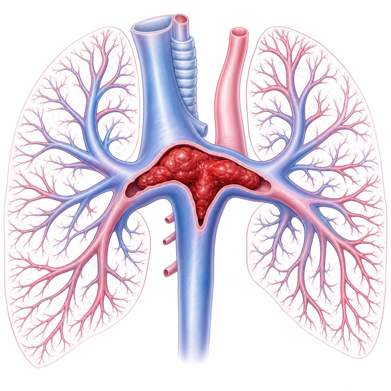
A pulmonary embolism and the deep-vein thrombosis it arises from are one disease — venous thromboembolism. The emergency classification is by haemodynamic impact, because it drives treatment. A massive (high-risk) PE presents with hypotension or shock. A submassive (intermediate-risk) PE is normotensive but shows right-ventricular dysfunction or strain. A low-risk PE is stable with no right-heart strain. The distinction between massive and the rest is the blood pressure: a shocked patient has a high-risk PE until proven otherwise. [1]
Epidemiology and risk factors
Pulmonary embolism is common and lethal, and the risk follows Virchow's triad of venous stasis, endothelial injury and hypercoagulability. The recognised risks are immobility, recent surgery or trauma, active malignancy, pregnancy and the combined oral contraceptive pill, a proven deep-vein thrombosis, a personal or family history of venous thromboembolism, an inherited or acquired thrombophilia, an indwelling central venous catheter, obesity and smoking. Identifying a provoking risk factor shapes both the workup and the duration of later anticoagulation. [1]
Pathophysiology — why the right ventricle decides survival
The embolus obstructs the pulmonary vascular bed, raising the pulmonary vascular resistance and so the right-ventricular afterload. The right ventricle is a thin-walled, afterload-sensitive pump, and as the afterload climbs it dilates and fails — and right-ventricular failure, not the hypoxia alone, is what kills the patient with a massive embolism. The obstruction also creates ventilation–perfusion mismatch and dead space, producing hypoxia, and reflex bronchoconstriction and vasoactive mediators worsen both the hypoxia and the afterload. This is the rationale for two non-obvious treatments: reperfusion (thrombolysis or embolectomy) to unload the right ventricle by removing the obstruction, and cautious fluids (a large bolus further distends and fails the right ventricle).[2]
Clinical presentation
The classic presentation is sudden pleuritic chest pain, breathlessness, syncope or haemoptysis, with tachycardia, tachypnoea and hypoxia — but a normal oxygen saturation does not exclude it. A massive embolism presents with hypotension, shock, or cardiac arrest, and pulmonary embolism is one of the leading reversible causes of pulseless-electrical-activity arrest. Atypical presentations are common and dangerous: isolated syncope, new dyspnoea alone in an otherwise well patient, or sudden collapse with no chest pain. [1]
Differential diagnosis
The sudden dyspnoea or pleuritic pain has a differential, and the risk-stratification tools plus the imaging resolve it. [1]
Pulmonary embolism
- Sudden dyspnoea/pleuritic pain, syncope; DVT signs; risk factors
- Wells/PERC → D-dimer → CTPA/VQ
- ECG: sinus tach, ± S1Q3T3, RBBB; echo RV strain
- Massive: hypotension, shock, PEA arrest
Pneumonia
- Fever, purulent sputum, progressive onset
- Focal consolidation; septic features
- CXR: consolidation; leukocytosis
- Antibiotics
Pneumothorax
- Sudden pleuritic pain; reduced air entry, hyperresonance
- CXR: pleural line; USS: absent sliding
- Tension: shock (clinical, decompress)
- No DVT signs
ACS / dissection
- ACS: crescendo pressure pain, ECG, troponin
- Dissection: tearing, migrating, widened mediastinum
- Distinguish by ECG, troponin, mediastinum
- Distinct pathway
Investigations and the risk-stratification tools
The workup is risk-stratified to avoid over-imaging. The Wells score estimates the pre-test probability from seven variables — clinical signs of a deep-vein thrombosis, the PE being the most likely diagnosis, a heart rate over 100, immobility or recent surgery, a previous venous thromboembolism, haemoptysis, and malignancy — into a low, moderate or high probability. A low-risk patient may first be tested with the Pulmonary Embolism Rule-out Criteria (PERC), a set of eight findings (age under 50, pulse under 100, oxygen saturation at least 95 per cent, no haemoptysis, no oestrogen use, no surgery or trauma, no prior venous thromboembolism, no unilateral leg swelling); if all are negative the PE is excluded without any blood test.[1] A low- or moderate-risk patient without a PERC rule-out has a D-dimer, which, if normal, excludes the diagnosis, but which is raised in many conditions and so is not diagnostic when positive. A high-probability, or a D-dimer-positive, patient goes to imaging: CT pulmonary angiography is the gold standard (the filling defect in the pulmonary artery), with attention to contrast nephropathy; a ventilation–perfusion scan is preferred in pregnancy or with a contrast allergy. The ECG most often shows sinus tachycardia; the classical S1Q3T3, a right-bundle-branch block and right-axis deviation, and T-wave inversion in V1 to V4 are non-specific. The bedside echocardiogram in the unstable patient (too sick for a CT) shows right-ventricular strain and McConnell's sign (right-ventricular free-wall hypokinesia with apical sparing). The troponin and the BNP are markers of right-ventricular strain, and the blood gas shows hypoxia with a respiratory alkalosis.
The Wells score and the revised Geneva score — the pre-test probability tools
The clinical pre-test probability is the gateway to every other test, because no single investigation rules PE in or out across all comers. The Wells score is the dominant tool in ANZ, the UK and North America; it is a seven-variable weighted score that asks the clinician, crucially, to judge whether an alternative diagnosis is less likely than PE (the single most heavily weighted item and the most subjective).[3] The points are: clinical signs of a DVT (3 points), PE the most likely diagnosis (3 points), heart rate over 100 (1.5 points), immobilisation or surgery in the last four weeks (1.5 points), previous VTE (1.5 points), haemoptysis (1 point), and active malignancy (1 point). The traditional three-tier read is low (under 2), moderate (2 to 6), high (over 6); the modern two-tier read is PE-unlikely (under 4) or PE-likely (4 or more), which simplifies the downstream testing. The revised Geneva score is the principal alternative — fully standardised and objective, with no "most likely diagnosis" judgement — and it performs similarly; the candidate should recognise both and know the local preference.
PERC — ruling PE out without a single blood test
The Pulmonary Embolism Rule-out Criteria (PERC) are applied only to a patient judged low-risk on clinical grounds. All eight criteria must be negative: age under 50, pulse under 100, oxygen saturation at least 95 per cent on room air, no haemoptysis, no oestrogen use, no recent surgery or trauma requiring hospitalisation, no prior VTE, and no unilateral leg swelling. When all eight are absent, the post-test probability of PE is below the test threshold and the patient is discharged without a D-dimer or imaging.[1] PERC is a rule to spare the low-risk patient from cascading investigation; it must never be applied to a moderate- or high-risk patient, and a single positive criterion invalidates it.
The ED diagnostic algorithm for suspected pulmonary embolism
Step 1 — Is the patient haemodynamically stable?
A hypotensive or shocked patient (systolic blood pressure under 90, or a drop of 40 from baseline, or clinical shock) bypasses the diagnostic algorithm: this is a suspected massive (high-risk) PE. Perform a bedside echo for RV strain and proceed to bedside diagnostics and empiric reperfusion — do not send an unstable patient to the CT scanner.
Step 2 — Estimate the pre-test probability (Wells or Geneva)
Score the patient. A PE-unlikely or low-probability patient goes to PERC then D-dimer; a PE-likely or high-probability patient skips straight to imaging.
Step 3 — Apply PERC if low-risk
If all eight PERC criteria are negative in a low-risk patient, PE is ruled out — no D-dimer, no imaging, discharge with safety-net advice.
Step 4 — D-dimer if PERC fails or is not applicable
A D-dimer below the cutoff (500 micrograms per litre, or age multiplied by 10 in the over-50) rules out PE in a low- or moderate-probability patient. A raised D-dimer is never diagnostic — it sends the patient to imaging.
Step 5 — Image: CTPA or V/Q scan
CTPA is first-line — fast, sensitive, and it identifies alternative diagnoses. V/Q is preferred in pregnancy (after a normal chest X-ray), in contrast allergy, and in renal impairment where iodinated contrast is undesirable.
Step 6 — The bedside echo for the unstable patient
A dilated RV with septal bowing and McConnell sign in the correct clinical context justifies empiric thrombolysis when the CT is unsafe.
The D-dimer — sensitive, not specific
The D-dimer is a degradation product of cross-linked fibrin, and it is raised by any fibrin formation and breakdown — infection, malignancy, pregnancy, surgery, inflammation, trauma, and advancing age all elevate it. Its strength is its negative predictive value in the low-probability patient; its weakness is its non-specificity. A raised D-dimer mandates imaging, not a diagnosis. The high-sensitivity assays are preferred; a point-of-care D-dimer is acceptable only if it is a validated high-sensitivity device. [1]
CTPA versus V/Q scan — choosing the imaging
CT pulmonary angiography
- First-line in most EDs; fast, available, sensitive
- Shows the filling defect in the pulmonary artery and identifies alternative diagnoses
- Radiation dose to breast and lung higher than V/Q; iodinated-contrast risk
- Preferred when the CXR is abnormal or another intrathoracic diagnosis is suspected
V/Q scan
- Preferred in pregnancy (normal CXR), contrast allergy, renal impairment
- Lower radiation; no contrast nephropathy
- Reported as a probability (high, intermediate, low, normal), not a yes or no
- Less useful when the CXR is abnormal (pneumonia, effusion, oedema) — more non-diagnostic
Bedside echo (unstable)
- For the patient too unstable for CT
- RV strain: RV dilatation, RV-to-LV ratio over 0.9, septal bowing, McConnell sign
- Justifies empiric thrombolysis in the correct clinical context
- Does not confirm PE — also seen in RV infarct, ARDS, severe COPD
The PIOPED study established the interpretive framework for the V/Q scan — a high-probability scan in a high-probability patient confirms PE; a normal scan excludes it; the large "intermediate" group remained the limitation, and it is this non-diagnostic zone that drove the shift to CTPA.[5]
The ECG in PE — non-specific but informative
The ECG is never diagnostic of PE, but it carries prognostic weight and excludes mimics. The classical S1Q3T3 (a deep S in lead I, a Q in III, and inverted T waves in III) is famous, specific when present, but insensitive — it appears in only about 1 in 10. The commoner and more useful findings are sinus tachycardia (the near-universal default), an incomplete or complete right-bundle-branch block, right-axis deviation, S waves in I and aVL, a QR in V1, and T-wave inversion in the right precordial leads V1 to V4 (the pattern that most strongly correlates with RV strain and a worse prognosis). A right-heart-strain pattern on ECG, alongside a raised troponin or BNP, flags the intermediate-risk patient for closer monitoring. [1]
[1]The bedside echo — McConnell's sign and the RV
The focused echo is decisive in the unstable patient who cannot reach the CT. The right-ventricular strain pattern is a dilated, hypokinetic right ventricle with an RV-to-LV end-diastolic diameter ratio over 0.9, paradoxical septal bowing into the left ventricle in systole, a reduced tricuspid annular plane systolic excursion (TAPSE under 1.6 cm), and an elevated estimated pulmonary-artery systolic pressure. McConnell's sign — hypokinesia of the RV free wall with sparing of the apex — is characteristic though not pathognomonic (it also appears in RV infarct and ARDS). A completely normal RV in a haemodynamically unstable patient argues against a massive PE as the cause and redirects the search. [1]
Troponin and BNP — the biomarkers of RV strain
A raised cardiac troponin (I or T) and an elevated BNP or NT-proBNP are not diagnostic of PE — they are markers of right-ventricular myocardial strain and microinfarction. Their value is in risk stratification: a normotensive patient with RV dysfunction on imaging AND a raised troponin or BNP is intermediate-high-risk and warrants admission to a monitored bed, anticoagulation, and a low threshold for escalation if they deteriorate. A normal troponin and BNP identify the intermediate-low-risk patient with a far more benign course.[10]
[1]Immediate management — risk-stratified, and the massive PE first
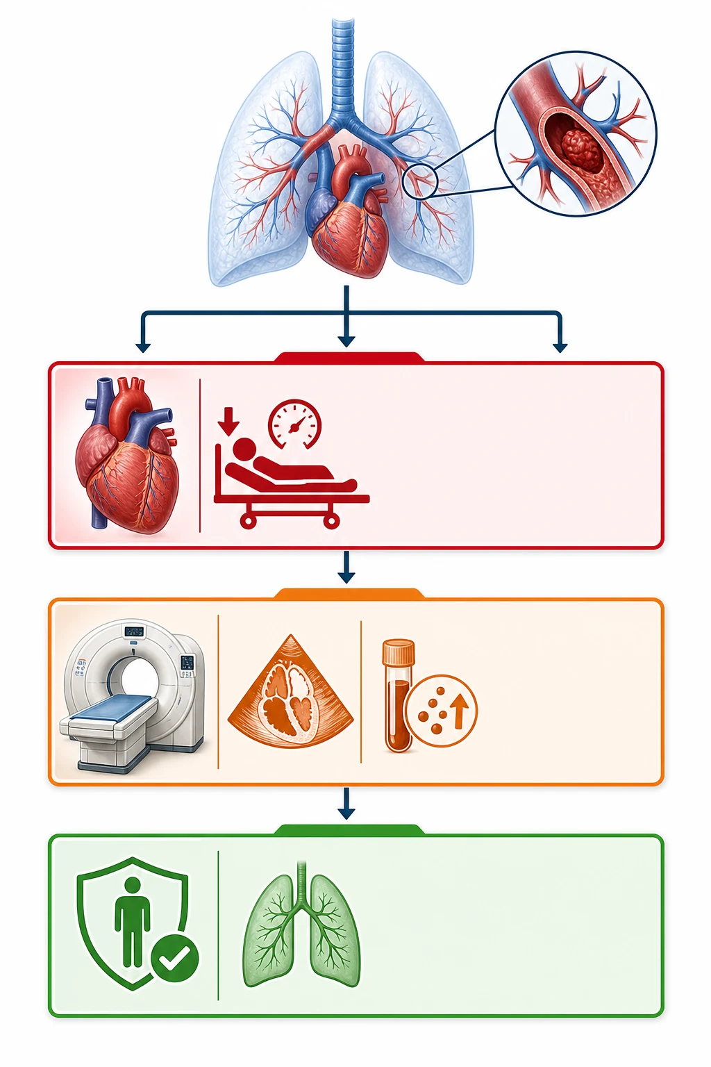
Stabilise the airway and the breathing with oxygen, and stratify by the blood pressure. [1]
[1]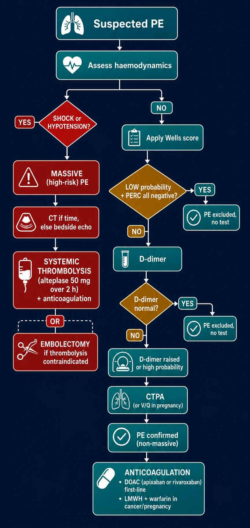
For anticoagulation, a direct oral anticoagulant is now first-line for most low- and intermediate-risk PE and needs no initial parenteral agent — apixaban 10 mg twice daily for seven days then 5 mg twice daily, or rivaroxaban 15 mg twice daily for twenty-one days then 20 mg daily. Low-molecular-weight heparin (enoxaparin 1 mg per kilogram twice daily) followed by warfarin or a DOAC is the alternative for the cancer and the pregnant patient, and intravenous unfractionated heparin is chosen when rapid reversal may be needed or in renal failure. The duration is about three months for a provoked event and indefinite for an unprovoked or cancer-associated one. The absolute contraindications to thrombolysis are the same as for the acute coronary syndrome — a prior intracranial haemorrhage, a recent ischaemic stroke, an intracranial lesion, and active bleeding — and in their presence an embolectomy is the route to reperfusion. A pulmonary embolism suspected behind a cardiac arrest warrants thrombolysis during the resuscitation. [1]
Massive PE — the resuscitation that unloads the right ventricle
The shocked patient with a PE is dying of right-ventricular failure, and every intervention must be judged by its effect on the right ventricle. Oxygen relieves the hypoxic pulmonary vasoconstriction that is adding to the afterload; a vasopressor (noradrenaline first-line, with vasopressin or adrenaline as alternatives) restores the coronary perfusion pressure to the right ventricle, which is failing partly because its own wall is underperfused; a cautious fluid bolus (250 mL aliquots, reassessing) may help a genuinely underfilled ventricle but a large bolus pushes the distended RV past the flat part of the Frank-Starling curve and into worsening failure; and reperfusion (systemic thrombolysis, or embolectomy when thrombolysis is contraindicated) is the only treatment that removes the obstruction and unloads the pump. [1]
The first 30 minutes — suspected massive (high-risk) PE
0 min — Recognise the high-risk PE
Unexplained hypotension, shock, syncope, or PEA arrest in a patient with risk factors and a compatible story. Do NOT wait for a CTPA — this is a clinical diagnosis in the unstable patient.
0 to 5 min — Airway, oxygen, monitoring
High-flow oxygen to hold the SpO₂ at 94 per cent or more; full monitoring (ECG, SpO₂, non-invasive blood pressure every 3 minutes); two large-bore cannulae; blood for troponin, BNP, lactate, group and save, coagulation and renal function.
5 to 10 min — The cautious fluid challenge
A 250 mL aliquot of balanced crystalloid, reassessed for response. A large bolus (500 mL or more) worsens the failing RV and is a common and dangerous error. If the pressure does not rise, start the vasopressor.
5 to 10 min — Bedside echo
Confirm the RV strain pattern (RV dilatation, septal bowing, McConnell sign). This is the imaging that justifies empiric reperfusion when the CT is unsafe.
10 to 20 min — Vasopressor and anticoagulation
Noradrenaline titrated to a mean arterial pressure of 65 mmHg or more; unfractionated heparin 80 units per kilogram bolus then 18 units per kilogram per hour (UFH is chosen because it is rapidly reversible, which matters if thrombolysis or embolectomy follows).
15 to 30 min — Reperfusion
Systemic alteplase — the PE dose is 50 to 100 mg intravenously over 2 hours (100 mg is the older dose; the 50 mg half-dose is increasingly preferred for the higher-bleeding-risk patient). If thrombolysis is contraindicated or fails, activate the surgical or catheter embolectomy pathway.
Thrombolysis for PE — the dose and the decision
Systemic alteplase is the standard fibrinolytic for the massive PE. The original dose was 100 mg over 2 hours; the contemporary ESC-favoured dose is 50 mg over 2 hours (or a weight-based 0.6 mg per kilogram bolus, capped at 50 mg), which retains efficacy with less bleeding. Tenecteplase, a single-bolus fibrinolytic, is an alternative with logistic advantages and comparable outcome data. The candidate must know the absolute contraindications — prior intracranial haemorrhage, ischaemic stroke within 6 months, intracranial or spinal lesion, active bleeding, recent brain or spinal surgery, recent major trauma — and that a relative contraindication is weighed against the lethality of the untreated massive PE. In a cardiac arrest with suspected PE, thrombolysis is given during CPR and, if the arrest reverses, the resuscitation continues with full post-ROSC care. [1]
Catheter-directed and surgical reperfusion
When systemic thrombolysis is contraindicated, has failed, or the bleeding risk is prohibitive, catheter-directed thrombolysis (ultrasound-assisted or standard, delivering a fraction of the systemic alteplase dose directly into the clot) and surgical pulmonary embolectomy are the alternatives. Catheter-directed therapy achieves clot lysis with lower systemic fibrinolytic exposure and is the emerging standard in centres with the capability; surgical embolectomy, on cardiopulmonary bypass, is reserved for the patient in whom catheter therapy is unavailable or has failed, or for a large central clot. An inferior-vena-cava filter is placed when anticoagulation is absolutely contraindicated or has failed (recurrent PE despite therapeutic anticoagulation); it is a bridge to the resumption of anticoagulation, not a substitute for it, and a retrievable filter is preferred. [1]
Anticoagulation — the DOACs and the exceptions
For the haemodynamically stable PE (low- and intermediate-risk), a direct oral anticoagulant is first-line globally, with no requirement for initial parenteral heparin. Apixaban 10 mg twice daily for 7 days then 5 mg twice daily, and rivaroxaban 15 mg twice daily for 21 days then 20 mg daily, are the two single-drug DOAC regimens; both were validated against conventional enoxaparin-warfarin in large non-inferiority trials (AMPLIFY for apixaban, EINSTEIN-PE for rivaroxaban) with less major bleeding.[11][9] The principal exceptions — where a low-molecular-weight heparin (enoxaparin 1 mg per kilogram twice daily) or unfractionated heparin is preferred — are cancer (LMWH historically, though the DOACs now rival it), pregnancy (LMWH throughout), severe renal impairment (UFH, intravenous and titrated), antiphospholipid syndrome (warfarin, because of the DOAC failure signal), and the patient in whom thrombolysis or embolectomy may be needed (UFH, because it is rapidly reversible). The duration is 3 months for a provoked event, 6 months for an unprovoked event, and indefinite for recurrent unprovoked or cancer-associated VTE.
Low-risk PE
- Normotensive; no RV strain; normal troponin and BNP
- DOAC (apixaban or rivaroxaban) as a single agent
- Consider outpatient or early discharge in selected patients
- 3 months if provoked; longer if unprovoked
Intermediate-risk PE
- Normotensive but RV strain on imaging or raised biomarker
- Anticoagulate (DOAC) and monitor in a high-acuity bed
- Reserve thrombolysis (50 mg alteplase) for deterioration
- Watch for haemodynamic decompensation in the first 48 to 72 hours
Massive (high-risk) PE
- Hypotension, shock, or PEA arrest
- Reperfusion: alteplase 50 to 100 mg over 2 hours
- Cautious fluids plus noradrenaline; bedside echo
- Embolectomy if thrombolysis contraindicated or it fails
Pulmonary embolism in pregnancy — the radiation-aware pathway
Pregnancy is a prothrombotic state (increased factors II, VII, VIII, X and fibrinogen, reduced protein S, venous stasis from the gravid uterus, and a roughly five-fold rise in VTE risk), and PE remains a leading cause of maternal death. The workup is distorted: the D-dimer is physiologically raised and unhelpful; the Wells and Geneva scores perform well but the threshold to image is lower; a V/Q scan is preferred over CTPA when the chest X-ray is normal (lower radiation to maternal breast and to the fetus, and comparable accuracy); and bilateral leg compression ultrasonography may identify a DVT and, if positive in the right context, obviate the need for pulmonary imaging. The anticoagulant is weight-based low-molecular-weight heparin — enoxaparin 1 mg per kilogram twice daily — throughout pregnancy and for six weeks postpartum (a minimum of three months total); unfractionated heparin is used around delivery or if rapid reversal is needed. Warfarin is avoided throughout pregnancy because it is teratogenic (first trimester) and causes fetal and placental bleeding (second and third trimester and at delivery); it may be used postpartum and is safe in lactation. A massive PE in pregnancy warrants systemic thrombolysis (the maternal benefit outweighs the fetal bleeding risk, which is modest) and, where time and expertise allow, catheter-directed lysis or surgical embolectomy as alternatives that limit systemic fibrinolysis. [1]
The pregnancy PE pathway — the radiation-aware sequence
Apply the Wells score (it performs as well as in the non-pregnant); a low score plus a PERC-negative is reassuring, but pregnancy lowers the imaging threshold.
Perform a chest X-ray first — a normal CXR favours a V/Q scan (the preferred modality for radiation economy); an abnormal CXR favours CTPA.
Image with a V/Q scan (or a perfusion-only scan) when the CXR is normal; reserve CTPA for the abnormal CXR or the haemodynamically unstable patient.
Anticoagulate with weight-based LMWH (enoxaparin 1 mg per kilogram twice daily) — DOACs and warfarin are avoided in pregnancy; UFH is used near delivery.
Plan the peripartum switch: stop LMWH 24 hours before a planned delivery or neuraxial anaesthesia; use UFH for the shortest reversible window; resume LMWH postpartum and continue for at least 6 weeks postpartum.
Reserve systemic thrombolysis for the life-threatening massive PE — give alteplase 50 mg, with catheter-directed lysis or surgical embolectomy as alternatives.
Subtypes and special scenarios
A massive PE in cardiac arrest — typically a pulseless-electrical-activity rhythm — warrants thrombolysis during the cardiopulmonary resuscitation. Pregnancy distorts the workup (a D-dimer is unhelpful, a V/Q scan is preferred to a CT for the radiation, and weight-based low-molecular-weight heparin is the anticoagulant, with a DOAC deferred). The cancer patient needs long-term anticoagulation. The post-operative patient carries a high bleeding risk that complicates thrombolysis. An isolated subsegmental PE is contentious; it is anticoagulated when symptomatic or in a patient with risk factors. [1]
Complications and pitfalls
The complications are right-ventricular failure and arrest, recurrence, pulmonary infarction, the later chronic thromboembolic pulmonary hypertension, bleeding from anticoagulation or thrombolysis, and heparin-induced thrombocytopenia. The pitfalls are the dangerous inverse of the workup: misreading a normal oxygen saturation or D-dimer as excluding the PE; over-relying on the ECG; missing a massive PE behind an unexplained hypotension; giving a large fluid bolus to the shocked patient; delaying thrombolysis in an arrest; and not anticoagulating or not investigating the cause after a provoked event. [1]
A specific later complication is chronic thromboembolic pulmonary hypertension, which presents weeks to months afterwards with progressive dyspnoea and right-heart failure and is treated, in selected centres, by pulmonary endarterectomy; the implication for the emergency physician is to warn the discharged patient to return with new or progressive breathlessness. A second pitfall worth stating plainly is the failure to investigate an unprovoked PE for an underlying cause — an occult malignancy or a thrombophilia — because the first presentation may be the only clue, and the workup (a focused cancer screen and, in selected patients, thrombophilia testing weeks after the anticoagulation has settled) changes the long-term management. [1]
Prognosis and disposition
The mortality of a massive untreated pulmonary embolism is high — up to 30 to 50 per cent — and falls sharply with prompt reperfusion; a low-risk PE is managed with anticoagulation and, in selected patients, discharge. The patient is admitted for anticoagulation, the duration of which is about three months for a provoked event and longer for an unprovoked or a cancer-associated one; an unprovoked PE prompts a search for an occult cancer or a thrombophilia; and an inferior-vena-cava filter is reserved for the patient in whom anticoagulation is contraindicated. [1]
Special populations
Pregnancy uses the radiation-aware pathway — a ventilation–perfusion scan over a CT, weight-based low-molecular-weight heparin (enoxaparin 1 mg per kilogram twice daily, withheld at the time of delivery), and a DOAC deferred until after delivery — and reserves systemic thrombolysis for a life-threatening massive PE. Cancer warrants long-term anticoagulation. Renal failure dictates unfractionated heparin and careful dose-adjustment. The elderly more often present atypically and carry a higher bleeding risk with anticoagulation and thrombolysis alike, so the risk-stratification and the reperfusion decision weigh the bleeding risk explicitly. [1]
Pivotal trials and the evidence base
Wells — derivation of the PE clinical model (Thromb Haemost 2000)
Thrombosis and Haemostasis
PMID 10744147
Key finding
A prospective cohort in patients with suspected PE that derived a seven-variable clinical prediction rule (clinical signs of DVT, PE the likeliest diagnosis, heart rate over 100, immobilisation or surgery, previous VTE, haemoptysis, malignancy) and combined it with the SimpliRED D-dimer to categorise pre-test probability.
Practice change
Established the Wells score that still anchors the ED PE workup worldwide; the rule is most heavily weighted on the clinician's judgement that PE is the most likely diagnosis.<Cite id='3' />
Christopher Study — Wells plus D-dimer plus CT (JAMA 2006)
JAMA
PMID 16403929
Key finding
A multicentre management study of 3,306 patients with suspected PE using a simplified dichotomised Wells rule (PE-unlikely under 4 points versus PE-likely). Patients with a PE-unlikely score and a normal D-dimer were managed without imaging or anticoagulation; the three-month thromboembolic risk was 0.5 per cent, well within the safety margin.
Practice change
Validated the two-tier Wells plus D-dimer rule-out strategy that is the backbone of the modern ED algorithm.<Cite id='4' />
PIOPED — the ventilation-perfusion scan (JAMA 1990)
JAMA
PMID 2332918
Key finding
A prospective multicentre study of 931 patients with suspected PE comparing V/Q scanning against pulmonary angiography. A high-probability scan confirmed PE and a normal scan excluded it, but only a minority of scans were high- or low-probability, leaving a large intermediate-probability group needing further imaging.
Practice change
Defined the interpretive categories of the V/Q scan and exposed its non-diagnostic middle ground — the limitation that drove the later shift to CTPA, while preserving the V/Q scan as the modality of choice in pregnancy and contrast allergy.<Cite id='5' />
ADJUST-PE — the age-adjusted D-dimer (JAMA 2014)
JAMA
PMID 24643601
Key finding
A multicentre prospective study of 3,346 patients over 50 with suspected PE and a non-high Wells score, using an age-adjusted D-dimer cutoff (age multiplied by 10 micrograms per litre) instead of the fixed 500. The age-adjusted cutoff ruled out PE in an additional 19 per cent of over-50s (and around 30 per cent of over-75s) with a three-month venous-thromboembolism rate of 0.3 per cent — non-inferior to the fixed cutoff.
Practice change
The age-adjusted D-dimer safely spares about one in five older ED patients a CTPA and is endorsed by the ESC guideline.<Cite id='6' />
MAPPET-3 — alteplase in submassive PE (NEJM 2002)
New England Journal of Medicine
PMID 12374874
Key finding
A randomised trial of 256 patients with submassive PE (RV strain, normotensive) comparing heparin plus alteplase against heparin alone. The primary endpoint of in-hospital death or clinical deterioration requiring treatment escalation fell from 24.6 per cent to 10.2 per cent, largely by a reduction in the need for salvage thrombolysis, with a non-significant trend to more bleeding.
Practice change
Provided the first proof that reperfusion in submassive PE reduces clinical deterioration — but with a bleeding signal that tempered routine use and set up the PEITHO trial.<Cite id='7' />
PEITHO — tenecteplase in intermediate-risk PE (NEJM 2014)
New England Journal of Medicine
PMID 24716681
Key finding
A double-blind randomised trial of 1,006 normotensive patients with intermediate-risk PE (RV strain plus a positive troponin), comparing tenecteplase against placebo on top of anticoagulation. Tenecteplase reduced the primary composite of death or haemodynamic decompensation at 7 days from 5.6 to 2.6 per cent, but increased major bleeding (11.5 to 2.4 per cent) and intracranial haemorrhage (2.4 to 0.2 per cent), with no difference in mortality.
Practice change
Routine full-dose thrombolysis in intermediate-risk PE is NOT warranted — the bleeding cost offsets the haemodynamic benefit. Reserve thrombolysis (preferably at the lower 50 mg alteplase dose) for the intermediate-risk patient who deteriorates.<Cite id='8' />
EINSTEIN-PE — rivaroxaban for symptomatic PE (NEJM 2012)
New England Journal of Medicine
PMID 22449293
Key finding
A randomised non-inferiority trial of 4,832 patients with symptomatic PE comparing oral rivaroxaban (15 mg twice daily for three weeks, then 20 mg daily) against standard enoxaparin-warfarin. Rivaroxaban was non-inferior for recurrent VTE (2.1 versus 1.8 per cent) with less major bleeding (1.1 versus 2.2 per cent) and no heparin lead-in.
Practice change
Established the single-drug rivaroxaban regimen — no parenteral heparin, no INR monitoring — as first-line for the stable PE, and underpinned the DOAC-first-line recommendation in the ESC guideline.<Cite id='9' />
AMPLIFY — apixaban for acute VTE (NEJM 2013)
New England Journal of Medicine
PMID 23808982
Key finding
A randomised non-inferiority trial of 5,395 patients with acute VTE comparing oral apixaban (10 mg twice daily for 7 days, then 5 mg twice daily) against conventional enoxaparin-warfarin. Apixaban was non-inferior for recurrent VTE or PE-related death (2.3 versus 2.7 per cent) and reduced major bleeding (0.6 versus 1.8 per cent), again with no parenteral lead-in.
Practice change
Together with EINSTEIN-PE, established the single-drug DOAC as the standard for the stable PE — apixaban and rivaroxaban are the two dominant first-line agents.<Cite id='11' />
2019 ESC PE guideline — the contemporary framework (Eur Heart J 2020)
European Heart Journal
PMID 31504429
Key finding
The multidisciplinary ESC and ERS task-force guideline that codifies risk stratification across the haemodynamic, RV-imaging and biomarker axes; recommends the DOACs as first-line for low- and intermediate-risk PE; reserves systemic thrombolysis for the high-risk (shocked) PE and the deteriorating intermediate-risk PE (at the reduced dose); recommends the V/Q scan in pregnancy and the age-adjusted D-dimer in the over-50s; and sets the indications for catheter-directed and surgical reperfusion and for IVC filters.
Practice change
The single document that governs contemporary ED PE practice across Europe, ANZ and much of the world — the candidate should know its risk-stratification axes and its thrombolysis recommendations cold.<Cite id='10' />
Evidence and regional guidelines
The contemporary framework is the 2019 ESC acute-PE guideline; the rule-out evidence is the PERC criteria.[1] The high-risk-stratification and the reperfusion evidence is summarised in the high-risk-PE outcome literature.[2] The DOACs are first-line globally; the thrombolysis dose and the pregnancy pathway are regional and protocol-specific.
ANZ practice note. The risk-stratified workup and the DOAC-first-line anticoagulation follow the ESC framework via local pathways; a massive PE receives alteplase 50 mg over 2 hours or, where contraindicated, an embolectomy via the local cardiology and cardiothoracic service, and the pregnancy pathway uses a V/Q scan and weight-based low-molecular-weight heparin. [1]
Exam practice
SAQ — Massive pulmonary embolism in shock: reperfusion and the right ventricle
10 minutes · 10 marks
A 67-year-old man is brought to the emergency department by ambulance twenty minutes after the sudden onset of severe breathlessness and a syncopal episode at home. He had an uncomplicated total knee replacement ten days ago. In the resuscitation bay he is pale, diaphoretic and drowsy (GCS 13), with a respiratory rate of 32, SpO2 88 per cent on 15 L oxygen via a non-rebreather mask, BP 74/48 (MAP 57), HR 128 in sinus tachycardia. The JVP is visibly distended to the angle of the jaw. The chest is clear to auscultation. The bedside echocardiogram shows a dilated, hypokinetic right ventricle with paradoxical septal bowing, an RV-to-LV ratio of 1.4 and McConnell sign. The lactate is 5.2 mmol/L. The registrar asks whether to give a 1-litre fluid bolus and take the patient to the CT scanner for a CTPA.
SAQ — Submassive pulmonary embolism: risk stratification and the thrombolysis decision
10 minutes · 10 marks
A 58-year-old woman presents to the emergency department two hours after the sudden onset of left-sided pleuritic chest pain and breathlessness. She is alert and haemodynamically stable: BP 118/74, HR 104, RR 24, SpO2 94 per cent on room air, afebrile. She takes the combined oral contraceptive pill and her mother had a deep-vein thrombosis. The Wells score is 4.5 (PE-likely). The D-dimer is 2,400 micrograms per litre. The CTPA shows filling defects in the right lower-lobe and segmental left pulmonary arteries. The bedside echocardiogram shows a dilated right ventricle with an RV-to-LV ratio of 1.2 and McConnell sign. The high-sensitivity troponin is 64 ng/L (upper reference limit 14); the BNP is 410 pg/mL. She remains normotensive throughout her emergency department stay.
Exam pearls
- Massive PE = shock → thrombolysis (alteplase 50 mg over 2 hours) + anticoagulation, or embolectomy if contraindicated.
- Wells → D-dimer (low-risk) → CTPA; PERC to rule out without a blood test.
- A normal SpO₂ and a normal ECG do not exclude a PE.
- ECG: S1Q3T3, right-bundle-branch block; echo: right-ventricular strain, McConnell's sign.
- Cautious fluids in the shocked PE — a large bolus fails the right ventricle.
- DOAC first-line; pregnancy = V/Q scan + low-molecular-weight heparin.
- A pulmonary embolism is a leading reversible cause of pulseless-electrical-activity arrest — thrombolysis during CPR if suspected. [1]
Additional high-yield clinical pearls
[1] [1] [1] [1] [1] [1]Red flags
[1] [1]References
- [1]Kline JA, Courtney DM, Kabrhel C, et al. Prospective multicenter evaluation of the pulmonary embolism rule-out criteria J Thromb Haemost, 2008.PMID 18318689
- [2]Ebner M, Böttler M, Røsjø E, et al. Outcome of patients with different clinical presentations of high-risk pulmonary embolism Eur Heart J Acute Cardiovasc Care, 2021.PMID 34125186
- [3]Wells PS, Anderson DR, Rodger M, et al. Derivation of a simple clinical model to categorize patients probability of pulmonary embolism: increasing the models utility with the SimpliRED D-dimer Thromb Haemost, 2000.PMID 10744147
- [4]van Belle A, Buller HR, Huisman MV, et al. Effectiveness of managing suspected pulmonary embolism using an algorithm combining clinical probability, D-dimer testing, and computed tomography JAMA, 2006.PMID 16403929
- [5]The PIOPED Investigators. Value of the ventilation/perfusion scan in acute pulmonary embolism. Results of the prospective investigation of pulmonary embolism diagnosis (PIOPED) JAMA, 1990.PMID 2332918
- [6]Righini M, Van Es J, Den Exter PL, et al. Age-adjusted D-dimer cutoff levels to rule out pulmonary embolism: the ADJUST-PE study JAMA, 2014.PMID 24643601
- [7]Konstantinides S, Geibel A, Heusel G, et al. Heparin plus alteplase compared with heparin alone in patients with submassive pulmonary embolism N Engl J Med, 2002.PMID 12374874
- [8]Meyer G, Vicaut E, Danays T, et al. Fibrinolysis for patients with intermediate-risk pulmonary embolism N Engl J Med, 2014.PMID 24716681
- [9]Buller HR, Prins MH, Lensin AW, et al. Oral rivaroxaban for the treatment of symptomatic pulmonary embolism N Engl J Med, 2012.PMID 22449293
- [10]Konstantinides SV, Meyer G, Becattini C, et al. 2019 ESC Guidelines for the diagnosis and management of acute pulmonary embolism developed in collaboration with the European Respiratory Society (ERS) Eur Heart J, 2020.PMID 31504429
- [11]Agnelli G, Buller HR, Cohen A, et al. Oral apixaban for the treatment of acute venous thromboembolism N Engl J Med, 2013.PMID 23808982