EM · Hypertensive emergency
Hypertensive emergency
Also known as Hypertensive crisis · Malignant hypertension · Accelerated hypertension
Severe blood-pressure elevation with acute end-organ damage — the distinction between a hypertensive emergency and urgency (end-organ damage, not the number), the affected organs (brain, heart, kidney, retina, pregnancy, aorta), the rationale for controlled lowering (the shifted cerebral autoregulation curve), the titratable intravenous agents with doses and per-scenario targets, the magnesium-for-eclampsia and phaeochromocytoma-alpha-first rules, and the agents to avoid. ACEM-primary, globally tagged.
On this page & tools
Your progress
Saved locally on this device.
Target exams
Red flags
A hypertensive emergency is severe blood-pressure elevation complicated by acute end-organ damage — to the brain, heart, kidney, retina, the gravid uterus, or the aorta. The single most important principle, and the one examiners test relentlessly, is that the emergency is defined by the end-organ damage, not by the number on the cuff: a patient at 220 over 130 with no organ damage has a hypertensive urgency and is managed as an outpatient with oral agents, whereas a patient at 180 over 120 with pulmonary oedema or encephalopathy has an emergency that needs titratable intravenous therapy in a monitored bed.[1] The second principle, equally stressed, is that the pressure must be lowered gradually, because the chronic hypertensive's cerebral autoregulation curve has shifted, and an abrupt drop causes cerebral and coronary hypoperfusion.
Definition and classification
"Hypertensive crisis" is the umbrella term for a severely elevated blood pressure, typically above 180 systolic or 120 diastolic. It divides into two. A hypertensive emergency has acute ongoing end-organ damage — hypertensive encephalopathy, acute pulmonary oedema, acute coronary syndrome, aortic dissection, acute kidney injury with proteinuria or haematuria, microangiopathic haemolytic anaemia, retinal haemorrhages or papilloedema, or eclampsia. A hypertensive urgency has the severe pressure without end-organ damage; it is managed over 24 to 48 hours with oral agents, not as an emergency. The historical terms "malignant" and "accelerated" hypertension describe severe hypertension with retinopathy and renal failure and are now folded into the emergency category. [1]
Types of hypertensive emergency — by the threatened organ
The classification the examiner wants is organ-based, because each organ dictates its own agent, its own target, and its own speed of lowering. The classic trigger threshold is a systolic above 180 and/or a diastolic above 120, but it is the acute target-organ damage — not the number — that makes the emergency. Seven patterns are examined: [1]
- Hypertensive encephalopathy — cerebral oedema from a pressure that has overrun autoregulation; headache, visual disturbance, confusion, seizures, and (on MRI) the posterior reversible encephalopathy syndrome. Lower by 20 to 25 per cent over the first hour, cautiously, with labetalol or nicardipine.
- Intracerebral haemorrhage — bleeding driven by the systolic pressure; the haematoma expands while the pressure is high, so a controlled reduction to a systolic of 140 (after the first hour) limits growth.[4][5]
- Aortic dissection — propagation is driven by the rate of rise of pressure (dP/dt), so a beta-blocker first to a heart rate of 60 to 70, then a vasodilator to a systolic of 100 to 120.[6]
- Acute coronary syndrome — the afterload raises myocardial oxygen demand and the ischaemia worsens; a systolic target around 140, with glyceryl trinitrate and a beta-blocker.
- Acute pulmonary oedema — the high afterload backs the failing left ventricle up; preload- and afterload-reduction with nitrates and non-invasive ventilation, target a systolic around 140.
- Pre-eclampsia, eclampsia and HELLP — pregnancy beyond 20 weeks; magnesium for seizures, labetalol or hydralazine for the pressure, and delivery as the definitive treatment.[2][3]
- Catecholamine crisis — phaeochromocytoma, cocaine, MDMA, monoamine-oxidase-inhibitor food and drug interactions, and clonidine withdrawal; alpha-blockade first, never a beta-blocker first.
The same blood pressure produces very different emergencies. A systolic of 200 in a dissection is an immediate aggressive-lowering problem; the same 200 in an ischaemic stroke is permissively tolerated. The organ decides the rule. [1]
Encephalopathy
- Headache, confusion, visual loss, seizures; PRES on MRI
- Lower 20–25% first hour, cautiously
- Labetalol or nicardipine; avoid nitroprusside (raises ICP)
- Exclude ischaemic/haemorrhagic stroke with CT
Intracerebral haemorrhage
- Focal deficit ± reduced GCS; CT confirms
- Target SBP <140 mmHg within 1 h (INTERACT2/ATACH2 range)
- Nicardipine or labetalol infusion
- Avoid overshoot — cerebral ischaemia
Aortic dissection
- Tearing chest/back pain, pulse deficit, widened mediastinum
- Beta-blocker FIRST to HR 60–70, then vasodilator
- SBP 100–120 rapidly
- CT angiography is definitive; surgical referral
Acute coronary syndrome
- Ischaemic chest pain; ECG changes; troponin rise
- GTN infusion + beta-blocker
- Target SBP ~140
- Avoid nitroprusside — coronary steal
Acute pulmonary oedema
- Dyspnoea, crackles, hypoxia; bilateral infiltrates
- GTN infusion + NIV; preload reduction
- Target SBP ~140
- Avoid pure beta-blocker if low-output; labetalol if tachycardic
Pre-eclampsia / HELLP
- Pregnancy >20 wks: BP + proteinuria ± HELLP
- Magnesium sulphate load 4 g then 1 g/h
- Labetalol or hydralazine; target <160/110
- Delivery is definitive
Catecholamine crisis
- Pheo, cocaine, MAOI, clonidine withdrawal
- Phentolamine (alpha) FIRST, then beta-blockade
- Lower to a safe perfusing pressure
- Avoid beta-blocker first — unopposed alpha
Epidemiology and risk factors
Hypertensive emergencies are uncommon but carry a high short-term mortality when untreated, and they most often arise in a patient with known hypertension who is non-adherent to therapy or who has an acute precipitant. Recognised precipitants and secondary causes include renal artery stenosis, chronic kidney disease, pre-eclampsia, phaeochromocytoma, withdrawal of clonidine or a beta-blocker, cocaine or amphetamine use, and glucocorticoid therapy. Identifying the precipitant is part of the emergency workup because it changes the agent and the disposition. [1]
Pathophysiology — why "lower slowly" is the law

An abrupt rise in systemic vascular resistance produces sustained endothelial injury and fibrinoid necrosis of the small arterioles, which causes the end-organ damage: cerebral oedema and haemorrhage, renal ischaemia with proteinuria, retinal haemorrhage and papilloedema, and afterload-driven cardiac failure. The defence against this is autoregulation — the brain and the heart maintain a constant perfusion across a range of pressures by adjusting vascular resistance. In the chronic hypertensive, the cerebral autoregulation curve shifts to the right, so the brain tolerates high pressures but becomes dependent on them: the floor of autoregulation is higher than normal, and lowering the pressure below that floor produces cerebral hypoperfusion, ischaemic stroke and cortical blindness. The same logic applies to the coronary circulation. This is the entire rationale for controlled lowering. [1]
[1]Clinical presentation
The presentation reflects the affected organ. Brain — headache, visual disturbance, confusion, seizures, or a focal deficit, the syndrome of hypertensive encephalopathy; haemorrhagic or ischaemic stroke may coexist. Heart — ischaemic chest pain (acute coronary syndrome), dyspnoea (acute pulmonary oedema), or the tearing pain of aortic dissection. Kidney — oliguria, haematuria, and an acute rise in creatinine. Retina — blurred vision with papilloedema, haemorrhages and exudates on fundoscopy. Pregnancy — headache, visual disturbance, epigastric pain and clonus, the syndrome of severe pre-eclampsia or eclampsia. Pain, anxiety and agitation can transiently raise the blood pressure, so the measurement is repeated after the patient is settled and assessed in both arms. [1]
Differential diagnosis
The first task is to separate the emergency from the urgency and from the mimics. [1]
Hypertensive emergency
- Severe BP with acute end-organ damage (brain/heart/kidney/retina/pregnancy/aorta)
- Admit to HDU/ICU; titratable IV agent; lower 10–20% first hour
- Fundoscopy, urinalysis, troponin, creatinine, CT brain
- Identify and treat the precipitant
Hypertensive urgency
- Severe BP without end-organ damage
- Lower over 24–48 h with oral agents (e.g. amlodipine, oral labetalol)
- Not an emergency; no IV line needed
- Adherence review and outpatient follow-up
Phaeochromocytoma crisis
- Episodic severe BP with catecholamine features (palpitations, sweating, headache, pallor)
- Alpha-blockade FIRST (phentolamine), then beta-blockade
- Plasma/urine metanephrines later
- Beta-blocker first is dangerous — unopposed alpha
Pre-eclampsia / eclampsia
- Pregnancy >20 weeks: BP + proteinuria ± end-organ features
- Magnesium sulphate for seizure prophylaxis/treatment
- Hydralazine or labetalol for BP; deliver definitively
- A pregnancy-specific pathway, not a generic one
Bedside assessment
Record the blood pressure in both arms after a period of rest, and repeat it. Perform fundoscopy for papilloedema, haemorrhages and exudates; examine the nervous system (the conscious level and any focal deficit); examine the cardiovascular system for pulmonary oedema, a new murmur, or pulse deficits that suggest dissection; and check the urine for protein and blood. In every woman of reproductive age, a pregnancy test is part of the workup, because pre-eclampsia has its own management pathway. [1]
Investigations and the targets
The investigations screen for end-organ damage and for the precipitant: an ECG (left-ventricular strain, ischaemia), a chest radiograph (pulmonary oedema, a widened mediastinum), urea, creatinine and electrolytes, a full blood count (the microangiopathic haemolytic anaemia of malignant hypertension shows schistocytes), troponin and NT-proBNP, a bedside urinalysis (proteinuria and haematuria — the renal marker), and a CT brain when encephalopathy or a focal deficit is present. Plasma or urinary metanephrines are sent later if phaeochromocytoma is suspected. [1]
Blood-pressure targets by scenario
Immediate management — a titratable agent, lowered gradually
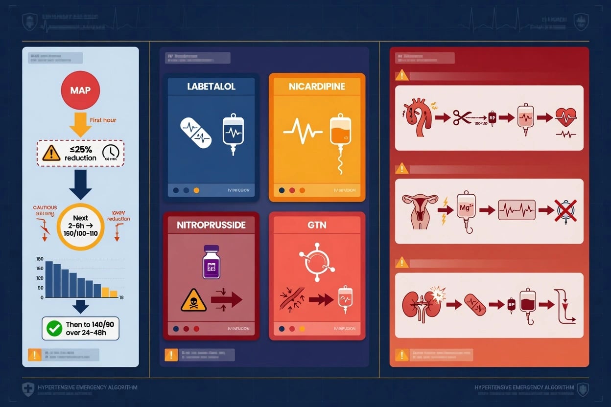
Resuscitate and treat the end-organ threat first: pulmonary oedema gets oxygen, non-invasive ventilation and a nitrate; the acute coronary syndrome gets its bundle; the dissection gets a beta-blocker first; the eclampsia gets magnesium. The blood pressure itself is lowered with a titratable intravenous agent, never with a bolus of an unpredictable oral drug. [1]
[1]Sodium nitroprusside is rarely used today because of cyanide and thiocyanate toxicity and because it raises intracranial pressure, and it is avoided when intracranial pressure is high. Sublingual nifedipine is forbidden — its onset is unpredictable and it causes precipitant cerebral hypoperfusion. Acute diuresis and clonidine have no role. [1]
[1]Special scenarios that override the general rule
Three scenarios have their own pathway and are favourite examination traps. Aortic dissection is the one hypertensive emergency lowered aggressively — a beta-blocker first to a heart rate of 60 to 70, then a vasodilator to a systolic of 100 to 120 — because propagation is driven by dP/dt, not by the pressure alone.[1] Pre-eclampsia and eclampsia are managed with magnesium sulphate for seizure prophylaxis and treatment (a 4 g intravenous loading dose over 5 to 10 minutes, then 1 g per hour), with hydralazine or labetalol for the blood pressure and definitive treatment by delivery.[2][3] Acute ischaemic stroke uses permissive hypertension — the pressure is lowered only when it exceeds about 220 over 120, or to permit thrombolysis — because the ischaemic penumbra depends on the collateral pressure.
Management by organ-specific pathway
Once the end-organ threat is identified, the resuscitation and the agent are chosen for that organ. The general principle holds — a titratable intravenous agent through an arterial line, in a monitored bed, lowered gradually — but the target and the speed are organ-specific. The common sequence is: resuscitate (airway, oxygen, IV access), confirm the end-organ damage with bedside testing and imaging, set the target for that organ, start the agent, and watch for overshoot. [1]
General emergency management sequence
Confirm severe BP in both arms after a period of rest; repeat the reading
Screen for end-organ damage: GCS, focal deficit, fundoscopy, chest, urine, ECG
Pregnancy test in every woman of reproductive age
Admit to HDU/ICU with arterial-line monitoring
Choose the agent for the organ; set the organ-specific target
Lower 20–25% in the first hour (except dissection — aggressive; stroke — permissive)
Reassess every 5–15 min; titrate to target; watch for overshoot and hypotension
Investigate and treat the precipitant; transition to oral once stable for 24 h
Intravenous agents and their place
The workhorse agents are labetalol, nicardipine, clevidipine, glyceryl trinitrate, and hydralazine. Each has a place, a dose, and a contraindication. Clevidipine, an ultra-short-acting dihydropyridine metabolised by esterases (not the kidney or liver), has become a favoured agent in the monitored setting because of its rapid onset and offset.[1]
Labetalol
- Alpha- and non-selective beta-blocker
- 20–80 mg IV q10 min, or 0.5–2 mg/min infusion
- Good: pregnancy, stroke, dissection, encephalopathy
- Avoid: severe asthma, bradycardia, heart block, severe LV failure
Nicardipine
- Dihydropyridine calcium-channel blocker
- 5–15 mg/h infusion (start 5 mg/h, titrate)
- Good: stroke, renal disease, general use
- Avoid: advanced aortic stenosis; reflex tachycardia
Clevidipine
- Ultra-short-acting CCB; esterase-metabolised
- 1–32 mg/h infusion
- Good: rapid titration, perioperative, most emergencies
- Avoid: egg/soy allergy; lipid-load with prolonged use
Glyceryl trinitrate
- Venous greater than arterial dilator; coronary dilator
- 5–200 mcg/min infusion
- Good: acute coronary syndrome, pulmonary oedema
- Avoid: RV infarct, PDE-5 inhibitors; tachyphylaxis
Hydralazine
- Direct arterial vasodilator
- 5–10 mg IV q20–30 min
- Good: pregnancy (second-line)
- Avoid: CAD, dissection; reflex tachycardia, unpredictable
Sodium nitroprusside
- Potent arterial/venous dilator
- 0.25–10 mcg/kg/min
- Reserved: refractory cases only
- Avoid: raised ICP, renal failure; cyanide/thiocyanate toxicity
Intracerebral haemorrhage — the evidence
For intracerebral haemorrhage the systolic target is 140 mmHg after the first hour, because haematoma expansion tracks with the pressure. The two landmark trials define the modern range: [1]
INTERACT2
Multicentre RCT; 2839 patients with spontaneous ICH within 6 h and SBP 150–220; intensive lowering to <140 vs guideline <180.
Key finding
No significant reduction in death/major disability (52% vs 55.6%, p=0.06), but improved functional outcome (modified Rankin) with intensive lowering. Supported SBP <140.
ATACH2
International RCT; 1000 patients with ICH and SBP >180; intensive (110–140) vs standard (140–180) lowering within 4.5 h.
Key finding
No benefit in death/disability; higher rate of renal adverse events with intensive lowering (9% vs 4%). Lower limit around 140, not 110, is safer.
The synthesis the examiner wants is: lower to a systolic of 140, not below 110, and not too fast. The first hour is for a controlled reduction; the overshoot, the hypotension, and the renal injury are the harms of being too aggressive. [1]
[1]Aortic dissection — heart rate before pressure
In dissection the entry tear propagates with each systolic ejection, so the rate of pressure rise (dP/dt) matters more than the absolute pressure. The sequence is beta-blocker first to drop the heart rate to 60 to 70, and only then a vasodilator to drop the systolic to 100 to 120. Giving a vasodilator first — without the beta-blocker — produces a reflex tachycardia that increases dP/dt and accelerates propagation, the exact opposite of the intent. The 2014 ESC aortic-disease guideline codifies this sequence.[6]
[1]Acute coronary syndrome and pulmonary oedema
In the acute coronary syndrome and acute pulmonary oedema, the high afterload is the immediate threat to the ischaemic or failing ventricle, and the agent of choice is glyceryl trinitrate — a coronary and venodilator that drops preload and afterload while dilating the culprit coronary bed. The systolic target is around 140. A short-acting beta-blocker (esmolol) is added for the tachycardic ischaemic patient once the pressure is controlled. Pure beta-blockade in established pulmonary oedema with a low-output state is dangerous — the negative inotropy can precipitate cardiogenic shock — so labetalol (which retains some vasodilating alpha effect) is preferred if a beta-blocker is unavoidable. [1]
[1]Catecholamine crisis — phaeochromocytoma, cocaine and the clonidine-withdrawal trap
The catecholamine crises share a final common pathway: a surge of alpha-mediated vasoconstriction that drives the pressure to extreme levels with the classical triad of headache, sweating and palpitation (with pallor in the phaeochromocytoma). The causes are phaeochromocytoma, cocaine and amphetamine intoxication, MAOI tyramine and drug interactions, and clonidine or beta-blocker withdrawal. The one inviolable rule is alpha-blockade first: a beta-blocker given before alpha-blockade leaves the alpha vasoconstriction unopposed and the pressure rises further, sometimes catastrophically. The agent of choice is phentolamine (a non-selective alpha-blocker, 1 to 5 mg intravenously, repeated or infused) or a rapid-onset calcium-channel blocker (nicardipine or clevidipine). Once the alpha receptors are blocked and the pressure controlled, a beta-blocker may be added for the tachycardia — never before. [1]
Catecholamine crisis sequence
Recognise the triad: paroxysmal severe hypertension, headache, sweating, palpitation
Alpha-blockade first: phentolamine 1–5 mg IV, or nicardipine/clevidipine infusion
Add beta-blockade ONLY after alpha-blockade for reflex tachycardia
Do NOT give a beta-blocker first — unopposed alpha worsens the pressure
Send plasma/urine metanephrines for phaeochromocytoma (after stabilisation)
Identify and remove the precipitant (cocaine, MAOI interaction, clonidine withdrawal)
In cocaine intoxication, the alpha surge and the monoamine reuptake blockade produce both hypertension and coronary vasospasm. Benzodiazepines are first-line (they calm the central sympathetic drive); phentolamine controls the alpha effect. Beta-blockers have historically been avoided for the theoretical risk of unopposed alpha, though contemporary data suggest they are not as dangerous as once feared — the conservative viva answer remains "benzodiazepines and phentolamine, beta-blockers with caution." [1]
Hypertensive urgency — no end-organ damage, oral over 24 to 48 hours
Hypertensive urgency is a severe blood pressure (typically above 180 systolic or 110 to 120 diastolic) without acute target-organ damage. It is the more common of the two hypertensive crises, and the one most often over-treated. The management is gradual lowering over 24 to 48 hours with oral agents, in a ward or an observation unit, with close follow-up — never an intravenous bolus, never sublingual nifedipine, and never a rapid drop. The patient is reassured and rested (pain and anxiety are common confounders), the blood pressure is repeated, and an oral agent is started or uptitrated: amlodipine 5 to 10 mg, captopril 12.5 to 25 mg (with a check of the creatinine and potassium), oral labetalol 100 to 200 mg, felodipine, or a low-dose clonidine patch. The precipitant — non-adherence, a high-salt meal, a new NSAID, alcohol, cocaine — is sought and addressed. [1]
[1]Emergency
- Acute end-organ damage present
- IV titratable agent in HDU/ICU
- Lower 20–25% first hour (organ-specific)
- Admit; arterial-line monitoring
Urgency
- No end-organ damage
- Oral agent; ward or observation unit
- Lower over 24–48 h
- Discharge with 24–72 h follow-up
Complications and pitfalls
The complications follow the affected organ: ischaemic or haemorrhagic stroke (from under- or over-treatment), acute kidney injury, the acute coronary syndrome, aortic dissection, an eclamptic seizure, retinal blindness, and the posterior reversible encephalopathy syndrome. The recurring pitfalls are treating the number instead of the organ; lowering the pressure too fast and causing cerebral or coronary hypoperfusion; using sublingual nifedipine; giving a beta-blocker before alpha-blockade in phaeochromocytoma; missing a pregnancy; and missing an aortic dissection that needs aggressive, not gradual, lowering. [1]
Prognosis and disposition
Untreated, a hypertensive emergency carried a one-year mortality above ninety per cent in the pre-treatment era; with modern controlled therapy the in-hospital mortality is much lower. Every patient is admitted to a high-dependency or intensive-care bed with arterial-line monitoring, treated with a titratable intravenous agent, and transitioned to oral therapy once stable. The underlying cause is then investigated and treated — a secondary hypertension, non-adherence, or a drug precipitant — and the long-term antihypertensive regimen is optimised before discharge. [1]
Special populations
Pregnancy follows the pre-eclampsia pathway with magnesium, fetal monitoring and delivery.[2] The elderly tolerate rapid lowering poorly and are lowered gently, with a lower first-hour target and close neurological monitoring. Chronic kidney disease raises the creatinine early and dictates careful agent and dose choice. Acute ischaemic stroke patients are managed with permissive hypertension unless the pressure is extreme or thrombolysis is planned. Across all of these groups the principle is unchanged: protect the threatened organ by lowering the pressure gradually, never below the patient's raised autoregulation floor.
Evidence and regional guidelines
The contemporary framework is the 2024 European Society of Hypertension guideline[1] and the AHA scientific statements on hypertensive emergencies. The pregnancy pathway follows the SOMANZ 2023 guideline[2] and the ESC/ESH peripartum position paper.[3] The agent choices are broadly global, but the stroke and pregnancy thresholds are protocol-specific — local stroke and obstetric pathways govern the exact numbers.
ANZ practice note. The agent and target framework follows the ESH 2024 guideline; the pregnancy pathway follows SOMANZ (magnesium sulphate, hydralazine or labetalol, and delivery), which is the ANZ obstetric-medicine standard and the one a Fellowship viva examines in the ANZ context. [1]
SAQs — exam practice
SAQ — Malignant hypertension with retinopathy, renal failure and microangiopathic haemolysis
10 minutes · 10 marks
A 52-year-old man with a five-year history of untreated hypertension presents to the emergency department with a three-day history of throbbing headache, blurred vision and progressive oliguria. He is confused (GCS 14) and dyspnoeic. Blood pressure is 226 over 144 in both arms, heart rate 108 in sinus rhythm, respiratory rate 26, oxygen saturation 93 per cent on room air. Fundoscopy reveals bilateral papilloedema, flame-shaped haemorrhages and cotton-wool spots. The full blood count shows haemoglobin 86 g per litre with schistocytes on the film and platelets of 68 by 10 to the ninth per litre; creatinine is 286 micromol per litre (baseline 95 four months ago), potassium 5.6, and the urine dipstick shows protein and blood. The lactate dehydrogenase is 620 and the troponin is mildly raised. The non-contrast CT brain shows no haemorrhage.
SAQ — Hypertensive encephalopathy with posterior reversible encephalopathy syndrome
10 minutes · 10 marks
A 68-year-old woman with a long-standing history of hypertension, non-adherent with her amlodipine for two weeks, is brought to the emergency department after a witnessed generalised tonic-clonic seizure at home. She is drowsy (GCS 13), confused, and complaining of headache and loss of vision. Blood pressure is 218 over 126 in both arms, heart rate 102 in sinus rhythm, respiratory rate 22, oxygen saturation 96 per cent on room air, capillary glucose 6.8 mmol per litre. There is no focal limb weakness and no neck stiffness. Fundoscopy reveals bilateral papilloedema. The non-contrast CT brain shows no intracranial haemorrhage and no mass. The magnetic resonance imaging of the brain, obtained because the confusion and visual loss persisted, demonstrates symmetric T2-FLAIR hyperintensity in the parieto-occipital white matter.
Exam pearls
- Emergency = end-organ damage; urgency = no end-organ damage — the number alone never decides it.
- Lower 10 to 20 per cent in the first hour, to about 160 over 100 over 2 to 6 hours — the one exception is aortic dissection (aggressive to a systolic of 100 to 120, beta-blocker first).
- Titratable intravenous agents: labetalol (pregnancy, stroke, dissection), glyceryl trinitrate (acute coronary syndrome, pulmonary oedema), nicardipine (general, stroke, renal).
- No sublingual nifedipine; no clonidine acutely.
- Eclampsia: magnesium sulphate 4 g load then 1 g per hour; hydralazine or labetalol for the blood pressure; delivery is definitive.
- Phaeochromocytoma: alpha-blocker (phentolamine) first, never a beta-blocker first.
- ICH target — systolic 140, not below 110: INTERACT2 showed benefit to <140; ATACH2 showed harm below 110 (renal adverse events). The safe floor is 140.[4][5]
- Aortic dissection — heart rate before pressure: beta-blocker first to HR 60 to 70 (esmolol or labetalol), then vasodilator to SBP 100 to 120. A vasodilator first accelerates propagation (the dP/dt trap).[6]
- Acute ischaemic stroke — permissive hypertension: lower only if SBP >220 or DBP >120, or to permit thrombolysis (target <185/<110 before, <180/<105 after). The penumbra depends on the collateral pressure.
- Acute coronary syndrome — systolic ~140: glyceryl trinitrate (a coronary dilator) plus a short-acting beta-blocker; avoid nitroprusside (coronary steal).
- Catecholamine crisis — alpha first, beta never first: phentolamine, then a beta-blocker for the tachycardia; cocaine gets benzodiazepines and phentolamine.
- Clevidipine is the modern ultra-short-acting agent (esterase-metabolised, 1 to 32 mg/h) for rapid titration in the monitored bed.[1]
- Hypertensive urgency is not an emergency: lower over 24 to 48 h with an oral agent (amlodipine, captopril, oral labetalol); no IV line, no sublingual nifedipine.
- Autoregulation is the rationale for everything: in chronic hypertension the cerebral curve shifts right — the brain tolerates high pressure but needs it, so lowering below the floor causes cerebral and coronary hypoperfusion.
- Always check both arms and a pregnancy test: a pulse or pressure difference suggests dissection; a missed pregnancy is a missed eclampsia.
- Reversible causes to find: renal artery stenosis, phaeochromocytoma, coarctation, pre-eclampsia, drug withdrawal (clonidine, beta-blocker), cocaine, glucocorticoids, NSAIDs.
Red flags
References
- [1]Kreutz R, Dobrowolski P, Prejbisz A, et al. 2024 European Society of Hypertension clinical practice guidelines for the management of arterial hypertension Eur J Intern Med, 2024.PMID 38914505
- [2]Shanmugalingam R, Hossain T, Makris A, et al. A summary of the 2023 Society of Obstetric Medicine of Australia and New Zealand (SOMANZ) hypertension in pregnancy guideline Med J Aust, 2024.PMID 38763516
- [3]Cífková R, Maier C, Lalic N, et al. Peripartum management of hypertension: a position paper of the ESC Council on Hypertension and the European Society of Hypertension Eur Heart J Cardiovasc Pharmacother, 2020.PMID 31841131
- [4]Qureshi AI, Palesch YY, Barsan WG, et al. Intensive Blood-Pressure Lowering in Patients with Acute Cerebral Hemorrhage N Engl J Med, 2016.PMID 27276234
- [5]Anderson CS, Heeley E, Huang Y, et al. Rapid blood-pressure lowering in patients with acute intracerebral hemorrhage N Engl J Med, 2013.PMID 23713578
- [6]Erbel R, Aboyans V, Boileau C, et al. 2014 ESC Guidelines on the diagnosis and treatment of aortic diseases: Document covering acute and chronic aortic diseases of the thoracic and abdominal aorta of the adult. The Task Force for the Diagnosis and Treatment of Aortic Diseases of the European Society of Cardiology (ESC) Eur Heart J, 2014.PMID 25173340