EM · Aortic dissection
Aortic dissection
Also known as Acute aortic dissection · Stanford Type A dissection · Acute aortic syndrome
Acute aortic dissection — intimal tear and the propagating false lumen, the Stanford/DeBakey classification, the sudden tearing migrating pain, the aortic dissection detection risk score (ADD-RS), the beta-blocker-first haemodynamic sequence with heart-rate and blood-pressure targets, surgery for Type A and medical-or-TEVAR for Type B, malperfusion syndromes, and the dissection-into-the-right-coronary-ostium trap. ACEM-primary, globally tagged.
On this page & tools
Your progress
Saved locally on this device.
5 MCQs with explanations
Target exams
Red flags
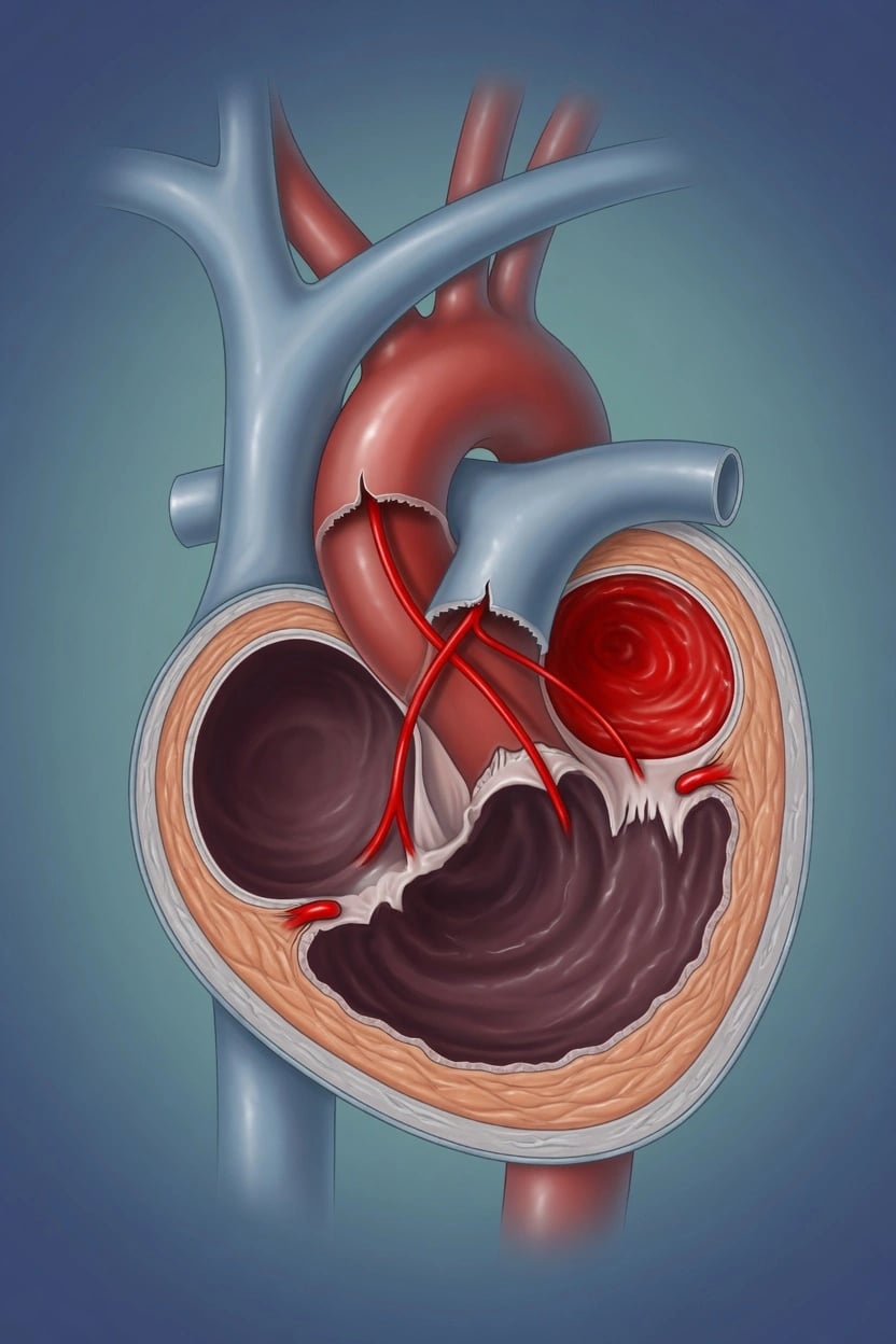
Acute aortic dissection is a tear in the aortic intima that lets pulsatile blood enter the media and cleave the wall, creating a propagating false lumen that can rupture into the pericardium, obstruct a branch artery, or compress the true lumen. It is the prototype of the acute aortic syndrome and one of the few diagnoses in medicine where a single wrong assumption — that the chest pain is myocardial infarction — leads to a fatal error: thrombolysis of a dissection that has extended into the right coronary ostium.[1][2] Untreated Type A dissection kills roughly 1 to 2 per cent of patients per hour, so the Fellowship candidate must recognise the presentation at the door, apply the risk score, control the haemodynamics in the correct sequence, and move to definitive imaging and surgery without delay.
Definition and the acute aortic syndrome
Dissection is one expression of a spectrum. The acute aortic syndrome comprises four entities that can overlap and that share the same emergency imperative: classic dissection (an intimal tear with a re-entering false lumen), intramural haematoma (a bleed into the media with no intimal flap, visible as crescentic high attenuation in the wall), penetrating atherosclerotic ulcer (an ulcer that burrows from the lumen into the media), and traumatic aortic injury. They are distinguished on computed tomography angiography but managed with the same initial haemodynamic logic, because what propagates all of them is the same: the force of each systolic pulse acting on a weakened wall. [1]
Classification
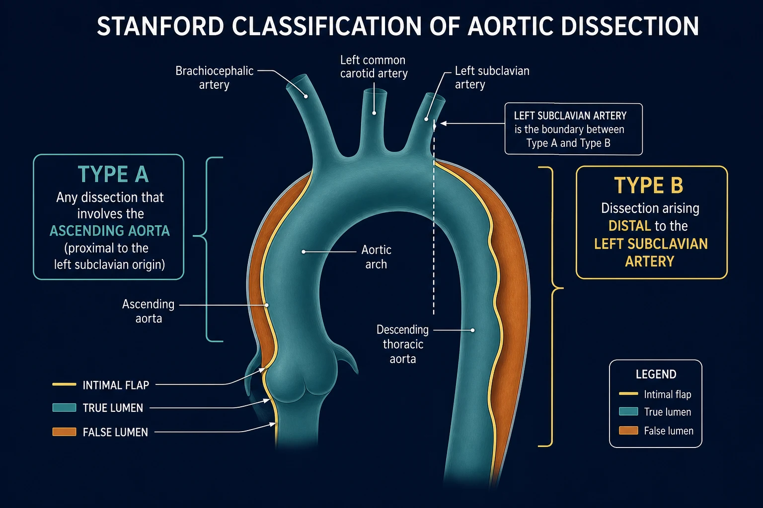
Two classifications coexist; Stanford determines management and is the one to know cold. [1]
Stanford Type A
ascending aorta involved
- Any dissection involving the ascending aorta, regardless of where the intimal tear sits
- About two-thirds of cases
- Immediate surgical emergency — untreated mortality 1–2% per hour
- May cause aortic regurgitation, coronary ostial occlusion, tamponade
Stanford Type B
distal to left subclavian
- Dissection begins distal to the left subclavian artery and does not involve the ascending aorta
- About one-third of cases
- Uncomplicated: medical management (rate and pressure control)
- Complicated (rupture, malperfusion, rapid expansion, refractory pain/HTN): TEVAR
The DeBakey classification is finer-grained — Type I (ascending and descending), Type II (ascending only), Type III (descending, distal to the subclavian) — and appears in vivas, but it does not change the emergency management, which turns on the single question: is the ascending aorta involved? [1]
Epidemiology and risk factors
Dissection is uncommon but not rare, with an incidence around 2.6 to 3.5 per 100,000 per year, a male predominance, and a peak between the fifth and seventh decades.[1] The dominant risk factor is long-standing hypertension. A smaller but high-yield group has a structurally vulnerable aorta from a connective-tissue disorder — Marfan syndrome, vascular-type Ehlers-Danlos, and Loeys-Dietz syndrome — or a bicuspid aortic valve, Turner syndrome, a known thoracic aneurysm, or prior aortic surgery. Two precipitants are worth remembering for the atypical presentation: pregnancy (particularly the third trimester and peripartum, in the setting of a bicuspid or connective-tissue aorta) and cocaine. Blunt deceleration trauma produces a distinct injury pattern at the ligamentum arteriosum covered under traumatic aortic injury.
Pathophysiology
Dissection propagates because of the rate of pressure rise within the aorta with each beat — the dP/dt — acting on a wall whose tensile strength has failed. By Laplace's law, wall tension is proportional to pressure times radius, so an aneurysmal, dilated aorta carries greater wall stress at any given pressure, which is why aneurysm is itself a risk factor. Once the intima tears, each systolic jet drives blood into the media, lengthening the dissection; each diastole allows the false lumen to stagnate and clot. This single mechanism — that propagation is driven by dP/dt, not by absolute pressure alone — is the entire rationale for the management sequence: you lower heart rate and contractility first, with a beta-blocker, so that dP/dt falls, and only then lower pressure with a vasodilator. [1]
[1]The false lumen harms the patient in three ways: it ruptures (into the pericardium causing tamponade, or into the mediastinum or pleura causing haemothorax); it compresses the true lumen and starves the branches that depend on it; or it re-enters the true lumen, creating a double-barrelled aorta. The branch-obstruction phenotype is malperfusion, and it dominates the atypical presentations. [1]
Clinical presentation
The classic presentation is unmistakeable when it is classic: pain that is sudden, maximal at the moment of onset, tearing or ripping in quality, and migrating as the dissection extends — typically from the chest to the back or abdomen.[1] In IRAD, pain is present in about 96 per cent, is sudden in about 85 per cent, is described as tearing or ripping in about half, and migrates in a minority. A blood-pressure or pulse differential between the arms (greater than 20 mmHg) is present in roughly a fifth to a third. Hypertension at presentation is common in Type B; in Type A the picture may be normotension or hypotension, and the combination of hypotension, a new diastolic murmur of aortic regurgitation, and a widened mediastinum is Type A with tamponade until proven otherwise.
The dangerous presentations are the atypical and painless ones, and examiners test them deliberately: painless syncope; an acute stroke or paraplegia** from carotid or spinal malperfusion; an acute coronary syndrome from dissection into the right coronary ostium; acute mesenteric or limb ischaemia; and sudden cardiac arrest with tamponade. Any of these in a patient with a risk factor is dissection until excluded. [1]
Differential diagnosis
The differential is the dangerous cause of chest pain, and the task is to distinguish them at the bedside by the tempo and character of the pain and the accompanying signs. [1]
Aortic dissection
- Sudden, maximal at onset, tearing, migrating
- BP/pulse differential, new murmur, focal neuro signs
- Widened mediastinum on CXR (but may be normal)
- Pain not relieved by anything
Acute coronary syndrome
- Gradual crescendo, pressure/tightness, radiates to arm/jaw
- ECG changes, troponin rise; no BP differential
- Pain may ease with GTN/opioid
- Risk factors for ischaemia
Pulmonary embolism
- Sudden pleuritic pain, dyspnoea, syncope
- Hypoxia, tachycardia; DVT signs; right-heart strain on ECG
- Normal aortic silhouette
- Wells/D-dimer/CTPA pathway
Boerhaave (oesophageal rupture)
- Vomiting then excruciating pain
- Surgical emphysema, pleural effusion
- History of instrumentation/forceful vomiting
- Pneumomediastinum on CXR/CT
The decisive discriminating feature is the tempo: dissection pain is sudden and maximal at onset, whereas ischaemic pain builds. The trap, of course, is that dissection into the right coronary ostium produces an inferior STEMI on the ECG, and it is precisely here that thrombolysis kills. [1]
Bedside assessment
Assess for the high-risk examination features of the detection score: a four-limb blood pressure looking for a differential; palpation of all pulses bilaterally looking for a deficit; auscultation for a new aortic regurgitation murmur (a blowing early diastolic murmur in Type A from loss of valve coaptation); a focused neurological examination for a stroke or cord syndrome; and an abdominal examination for mesenteric ischaemia. In the unstable patient, look for the signs of tamponade — Beck's triad, distended neck veins, muffled sounds — and prepare for deterioration. [1]
Investigations and the aortic dissection detection risk score
The first investigation is the ECG, and its purpose is as much to avoid a fatal error as to confirm a diagnosis: it is often non-specific, but an inferior STEMI coexisting with a widened mediastinum must be treated as dissection into the right coronary ostium until imaging proves otherwise.[1][2]
The Aortic Dissection Detection Risk Score (ADD-RS) is the validated, guideline-based tool that risk-stratifies the patient at the door and triggers imaging.[4] It scores three groups, one point for any feature present in each, for a maximum of three:
ADD-RS — three groups, one point each
ADD
Marfan/connective-tissue disease, family history of aortic disease, known aortic valve disease, known thoracic aneurysm, or recent aortic manipulation (including cardiac surgery)
Chest, back or abdominal pain that is abrupt in onset, tearing or ripping, or migrating
A perfusion deficit (pulse deficit, systolic BP differential greater than 20 mmHg, or a focal neurological deficit with pain), a new murmur with pain, or hypotension/shock
A score of 2 or more is high risk and mandates immediate computed tomography angiography; the score is sensitive but not specific, and a low score in a patient you still suspect should still be imaged — the score raises the threshold to scan, it does not grant permission to dismiss.[4]
Investigation thresholds at a glance
Computed tomography angiography is first-line, with a sensitivity and specificity approaching 100 per cent; it shows the intimal flap, distinguishes true from false lumen (the false lumen is typically larger, lower-flow, and may show the "cobweb" sign), defines branch involvement, and reveals rupture. The chest radiograph is abnormal in around 60 per cent — widened mediastinum, abnormal aortic contour, an apical cap, a left pleural effusion, or deviation of a nasogastric tube — but is normal in up to about 18 per cent, so a normal film never excludes dissection.[1] D-dimer is sensitive with a high negative predictive value and has a role in rule-out in low-risk patients only; in a high-risk patient it must never delay computed tomography. Transoesophageal echo is reserved for the unstable patient who cannot safely go to the scanner and for intra-operative confirmation, and a bedside transthoracic echo looks specifically for a pericardial effusion, tamponade, and aortic root dilation.
Immediate management — the beta-blocker-first sequence
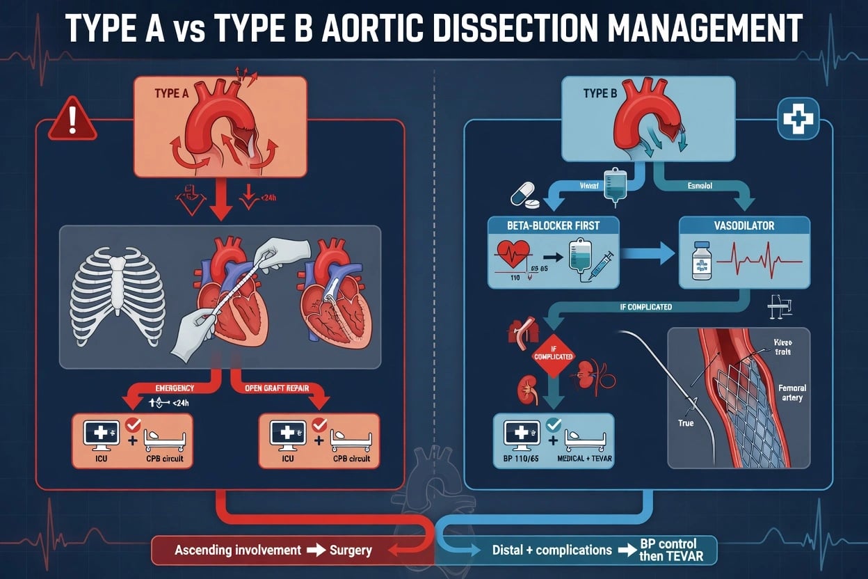
Resuscitation and the specific therapy begin together. Establish two large-bore cannulae, crossmatch with massive-haemorrhage readiness, give adequate analgesia, and start the haemodynamic sequence that reduces dP/dt. The cardinal rule, tested at every level, is: lower the heart rate first with a beta-blocker, and only then add a vasodilator. [1]
[1]Doses: labetalol 10 to 20 mg intravenously, repeated every ten minutes to effect (or as an infusion starting at 1 to 2 mg per minute); or esmolol 500 micrograms per kilogram over one minute as a loading dose, then 50 to 200 micrograms per kilogram per minute by infusion (esmolol's short half-life suits the titratable resuscitation). Once the rate is controlled, glyceryl trinitrate 5 to 200 micrograms per minute (sodium nitroprusside is an alternative but carries cyanide toxicity and raised intracranial pressure risk). The targets are a heart rate of 60 to 70 and a systolic blood pressure of 100 to 120 mmHg, titrated to organ perfusion. In the hypotensive patient — typically Type A with tamponade — do not pursue these targets; resuscitate, avoid pericardiocentesis unless in arrest (drainage provokes re-bleed), and move to emergency surgery. [1]
Definitive management — surgery, TEVAR, and the uncomplicated Type B
Definitive treatment follows the Stanford type. Type A is a surgical emergency: immediate cardiothoracic referral for open repair, with in-hospital mortality still around 17 to 26 per cent even with surgery.[1][2] The untreated mortality is 1 to 2 per cent per hour for the first day, so the call to the surgeon is part of the resuscitation, not a step after it.
Type B divides into uncomplicated and complicated. The uncomplicated Type B is managed medically with rate and pressure control, analgesia, and surveillance imaging; thrombolysis is contraindicated. A complicated Type B — defined by rupture, malperfusion, rapid expansion, or refractory pain or hypertension — goes to thoracic endovascular aortic repair (TEVAR). Two trials underpin the endovascular evidence: INSTEAD-XL showed that TEVAR plus medical therapy produced favourable aortic remodelling and better long-term outcomes than medical therapy alone in stable Type B,[3] and ADSORB confirmed that endovascular repair of uncomplicated Type B promotes false-lumen thrombosis and remodelling.[5] The indication for TEVAR in the uncomplicated patient remains selective and is a guideline judgement, not routine.
Malperfusion and complications
Malperfusion is the branch-obstruction phenotype and the source of the atypical presentations. Coronary malperfusion (typically the right coronary ostium) produces an inferior STEMI. Carotid malperfusion produces a stroke. Spinal malperfusion produces paraplegia. Renal malperfusion produces acute kidney injury; mesenteric malperfusion produces ischaemic abdominal pain out of proportion and is easily missed; limb malperfusion produces a cold, pulseless, painful leg. Static malperfusion from true-lumen compression often needs endovascular fenestration or stenting in addition to the distal repair. Other complications are rupture and tamponade, acute aortic regurgitation from root involvement, and — after repair — spinal cord ischaemia, renal failure, and the long-term risk of re-dissection or aneurysm in the residual aorta. [1]
Pitfalls
The recurring, fatal errors are well described and each maps to a concrete defence. Thrombolysing the inferior STEMI — defend by checking the mediastinum and a BP differential in every inferior STEMI. Giving GTN before a beta-blocker — defend by knowing the dP/dt rationale. Trusting a normal chest film or a reassuring D-dimer in a high-risk patient — defend by treating ADD-RS 2 or more as CTA-now, regardless. Delaying computed tomography for "more tests" — defend by making the scan part of the resuscitation. Pericardiocentesis in the non-arrest tamponade — defend by recognising that drainage provokes re-bleed and that the answer is surgery. And forgetting the four-limb blood pressure, which is the single bedside sign most likely to reveal the diagnosis. [1]
Prognosis and disposition
Type A carries an in-hospital mortality of roughly 17 to 26 per cent even with operative repair, and Type B around 10 per cent.[1] Every patient is admitted to a critical-care environment; Type A goes to theatre and then cardiac intensive care, complicated Type B to an endovascular/intensive-care pathway, and uncomplicated Type B to a monitored bed with imaging surveillance. Long-term survival depends on persistent blood-pressure control and lifelong surveillance imaging for aneurysmal degeneration of the residual aorta, which is why discharge planning includes aggressive antihypertensive optimisation and a vascular follow-up pathway.
Special populations
In pregnancy, a dissection (often in a patient with a bicuspid valve or connective-tissue disease) is managed with the same beta-blocker-first sequence; labetalol is the preferred agent, the timing of delivery is a multidisciplinary decision, and Type A remains surgical. The elderly more often present atypically and have a higher rupture risk alongside comorbidity. Patients with connective-tissue disease are younger, may dissect on a near-normal aorta, and recur — they need specialist long-term surveillance. The iatrogenic and traumatic groups have their own injury patterns and are covered under their respective topics. [1]
Evidence and regional guidelines
The evidence base is registry-led and guideline-driven. The International Registry of Acute Aortic Dissection (IRAD) remains the foundational epidemiology.[1] The 2022 ACC/AHA aortic disease guideline sets out the diagnostic and management framework used here.[2] The detection score is validated in Rogers and colleagues.[4] Endovascular evidence comes from INSTEAD-XL[3] and ADSORB.[5] Regionally, there is no dedicated Australasian dissection guideline, so ANZ practice follows the ESC and ACC/AHA positions; the United Kingdom follows the ESC and vascular-society guidance; the United States follows ACC/AHA; Europe follows ESC. The drugs, the targets, and the surgical imperative are global; the guideline citation is regional.
ANZ practice note. In Australia and New Zealand there is no dedicated ACEM- or ANZ-specific dissection guideline; the working standard is the ACC/AHA 2022 aortic disease guideline and the ESC position, with local cardiothoracic and vascular pathways governing Type A repair and Type B TEVAR access. The beta-blocker-first sequence and the ADD-RS threshold are universal. [1]
SAQs — exam practice
SAQ — Stanford Type A dissection with tamponade
10 minutes · 10 marks
A 67-year-old man presents to the resuscitation bay with sudden, tearing chest pain radiating to the interscapular area, maximal at onset. On examination he is diaphoretic, GCS 14, with a blood pressure of 78/45 mmHg (equal in both arms), heart rate 118 bpm in sinus rhythm, SpO2 94% on room air. The JVP is visibly distended to the angle of the jaw at 45 degrees, heart sounds are quiet, and there is a loud early-diastolic decrescendo murmur at the left sternal edge. The chest X-ray shows a widened mediastinum (10.2 cm) and a small left pleural effusion. A bedside transthoracic echo demonstrates a large circumferential pericardial effusion with right ventricular diastolic collapse. ECG shows non-specific ST-T changes, no STEMI. The cardiothoracic surgeon is 45 minutes away.
SAQ — Type B dissection with malperfusion syndrome
10 minutes · 10 marks
A 58-year-old man with poorly controlled hypertension (usual SBP 170) presents one hour after sudden onset of tearing interscapular pain, now accompanied by severe, cramping, poorly localised abdominal pain that is markedly disproportionate to his soft, non-tender abdomen. He is alert, BP 168/95 and 152/90 (right and left), HR 102, lactate 4.8 mmol/L and rising at repeat, creatinine 165 (baseline 95). His right leg is cool from mid-thigh, with absent popliteal and pedal pulses and a Dopplerable but weak femoral pulse. CTA chest/abdomen/pelvis demonstrates a Stanford Type B dissection originating just distal to the left subclavian artery, with the false lumen compressing the true lumen at the renal and mesenteric origins and the right common iliac, plus a 3 cm proximal descending aortic diameter.
Exam pearls
- Sudden, tearing, migrating pain; beta-blocker before vasodilator; inferior STEMI plus a widened mediastinum — do not thrombolyse.
- A normal chest radiograph in up to 1 in 5; a normal D-dimer does not help in the high-risk patient.
- ADD-RS of 2 or more means computed tomography now; the score raises the threshold to scan, never permission to dismiss.
- Type A is surgery; Type B is medical unless complicated, and complicated means rupture, malperfusion, rapid expansion, or refractory pain or hypertension.
- The false lumen is larger, lower-flow, and may show the cobweb sign; the true lumen is compressed.
- The single bedside sign most likely to reveal the diagnosis is the four-limb blood pressure and pulse differential. [1]
The ED first 30 minutes — a workflow
The emergency department management of suspected dissection runs on parallel tracks: the resuscitation track, the diagnostic track, and the disposition track, all moving at once. The following steps are the viva-correct sequence that a Fellowship examiner expects.[2][6]
Suspected aortic dissection — the ED first 30 minutes
Triage to the resuscitation bay; high-flow oxygen if hypoxic; full monitoring (three-lead ECG, continuous SpO2, non-invasive BP cycled every 3 minutes on at least one arm); two large-bore (14–16 G) cannulae.
Take the focused history: the onset (sudden and maximal-at-onset is the discriminator), the quality (tearing/ripping), the radiation and migration (chest to back to abdomen), and the risk factors (hypertension, connective-tissue disease, known aneurysm, pregnancy, cocaine, recent aortic surgery).
Perform the targeted examination: four-limb blood pressure (a differential >20 mmHg is the single most useful bedside sign), palpate all pulses bilaterally for a deficit, auscultate for a new early-diastolic murmur of aortic regurgitation, screen for a focal neurological deficit (stroke, cord), and examine the abdomen for mesenteric ischaemia.
Calculate the ADD-RS across its three groups (at-risk condition, dangerous pain, deficit exam). A score of 2 or more mandates immediate CT angiography; a score of 0 or 1 in a patient you still suspect is still scanned.
Obtain the 12-lead ECG urgently — primarily to detect an inferior STEMI that coexists with dissection into the right coronary ostium (the thrombolysis trap), and never to "rule dissection in".
Send bloods (group and crossmatch with massive-haemorrhage readiness, full blood count, coagulation, electrolytes, troponin, lactate) and a D-dimer, but do NOT wait for results to image a high-risk patient.
Start the haemodynamic sequence — beta-blocker FIRST (esmolol or labetalol IV) to a heart rate of 60–70 bpm, then add a vasodilator (GTN infusion) to a systolic of 100–120 mmHg, titrated to organ perfusion. Give adequate opioid analgesia.
Obtain computed tomography angiography of the chest, abdomen and pelvis — the diagnostic gold standard — once the patient is stable enough to travel; in the truly unstable patient, use bedside transthoracic echo to look for tamponade and aortic root dilation, and involve cardiothoracic surgery before the scan.
Refer to cardiothoracic surgery for Type A (immediate) or vascular/endovascular for complicated Type B, and arrange critical-care admission. Make the surgical call as part of the resuscitation, not after it.
The haemodynamic sequence — the viva walk-through
Examiners test this sequence relentlessly because the rationale — the dP/dt mechanism — is the single most important concept in the disease. The heart rate must fall before the pressure falls.[1][2]
Beta-blocker-first haemodynamic control — titration sequence
Give the beta-blocker first: esmolol 500 mcg/kg IV loading dose over 1 minute, then 50–200 mcg/kg/min infusion (short half-life of ~9 minutes suits titration); OR labetalol 10–20 mg IV every 10 minutes to effect, or infusion 1–2 mg/min. Titrate to a heart rate of 60–70 bpm.
Only once rate is controlled, add the vasodilator: glyceryl trinitrate 5–200 mcg/min IV infusion (preferred; no cyanide toxicity), titrated to a systolic of 100–120 mmHg. Sodium nitroprusside 0.3–3 mcg/kg/min is an alternative but carries cyanide toxicity and raised intracranial pressure risk.
Reassess organ perfusion — the target pressures are only targets: titrate to maintained urine output, conscious level, and peripheral perfusion. Do not chase a number that loses the patient.
In the hypotensive patient (typically Type A with tamponade), do NOT pursue these targets — resuscitate, withhold vasodilators and beta-blockade, and move to emergency surgery. Avoid pericardiocentesis unless in arrest (drainage provokes re-bleed).
Drug dosing — beta-blocker and vasodilator reference
Esmolol
beta-1 selective, ultra-short
- Loading 500 mcg/kg IV over 1 min, then 50–200 mcg/kg/min infusion
- Half-life ~9 min — titratable, rapidly reversible
- Preferred where haemodynamics are borderline or uncertain
- Targets HR 60–70 bpm
Labetalol
non-selective beta + alpha-1 block
- 10–20 mg IV every 10 min to effect, or infusion 1–2 mg/min
- Dual action lowers both rate and pressure in one drug
- Often first-line in ANZ/UK units
- Avoid in severe asthma / bradycardia
Glyceryl trinitrate
vasodilator — add AFTER beta-blockade
- 5–200 mcg/min IV infusion, titrated to SBP 100–120 mmHg
- No cyanide toxicity; may cause headache and tachyphylaxis
- Must never be started before the heart rate is controlled
- Also provides coronary vasodilation (useful if ACS coexists)
Sodium nitroprusside
vasodilator — alternative
- 0.3–3 mcg/kg/min IV
- Powerful arterial and venous dilator; very rapid onset
- Cyanide toxicity with prolonged/high-dose use; raises ICP
- Largely supplanted by GTN in modern practice
The complicated Type B — when to TEVAR
The uncomplicated Type B is managed medically. The decision point is the complicated Type B, where endovascular repair (TEVAR) is the standard of care. The four complications to name in a viva are rupture, malperfusion, rapid expansion, and refractory pain or hypertension.[3][5][1]
Uncomplicated Type B
medical management
- No rupture, no malperfusion, no rapid expansion, pain/HTN controllable
- Beta-blocker-first rate and pressure control (SBP 100–120, HR 60–70)
- Adequate analgesia, monitored critical-care bed
- Surveillance imaging (CTA at intervals); lifelong BP control
- Thrombolysis contraindicated
Complicated Type B
TEVAR indicated
- Rupture or impending rupture (haemothorax, retroperitoneal bleed)
- Malperfusion — mesenteric, renal, spinal, limb ischaemia
- Rapid expansion on serial imaging, or aneurysmal degeneration
- Refractory pain or hypertension despite maximal medical therapy
- Thoracic endovascular aortic repair (TEVAR) — covers the entry tear, promotes false-lumen thrombosis and aortic remodelling
The trials in brief
The dissection evidence base is registry-led and guideline-driven, with a small number of randomised trials addressing the endovascular question. The Fellowship candidate should know each by name and one-line finding.[1][3][5][6][1]
IRAD — International Registry of Acute Aortic Dissection
JAMA, 2000
Prospective multinational registry of 464 patients with acute aortic dissection across 12 international referral centres, defining the modern epidemiology, presentation, and outcomes.
Key finding
Pain present in ~96%, sudden onset in ~85%, tearing/ripping in ~50%, migrating in a minority; in-hospital mortality 17.4% Type A, 10.7% Type B — surgery reduced mortality versus medical therapy in Type A.
Practice change
Established the contemporary descriptive epidemiology of dissection that underpins every guideline since.
INSTEAD-XL — TEVAR in stable Type B
Circulation: Cardiovascular Interventions, 2013
Randomised trial of thoracic endovascular repair plus medical therapy versus medical therapy alone in 140 patients with stable, uncomplicated Type B dissection, with extended long-term follow-up.
Key finding
TEVAR plus medical therapy produced favourable aortic remodelling and a survival benefit at 5 years versus optimal medical therapy alone.
Practice change
Provided the evidence for a selective role of TEVAR in uncomplicated Type B — not routine, but considered.
ADSORB — endovascular repair of uncomplicated Type B
European Journal of Vascular and Endovascular Surgery, 2014
Randomised trial of endovascular repair plus medical therapy versus medical therapy alone in 61 patients with acute uncomplicated Type B dissection.
Key finding
Endovascular repair promoted false-lumen thrombosis and favourable aortic remodelling at 1 year; no significant difference in mortality or rupture.
Practice change
Confirmed the remodelling benefit of TEVAR; supported its selective use in uncomplicated disease rather than routine deployment.
VIRTUE Registry — the malperfusion insight
Journal of the American College of Cardiology, 2013
Prospective multicentre registry of 103 patients with complicated Type B dissection managed with a unified endovascular strategy (valiant thoracic stent graft), tracking malperfusion resolution.
Key finding
Endovascular therapy relieved static and dynamic malperfusion with acceptable midterm aortic-specific survival, supporting TEVAR as first-line for complicated Type B.
Practice change
Established TEVAR (with fenestration/stenting for static malperfusion) as the standard for complicated Type B dissection.
EACTS/STS 2024 and ACC/AHA 2022 guidelines
European Journal of Cardio-Thoracic Surgery / JTCVS, 2022–2024
Multidisciplinary consensus guidelines synthesising registry and randomised evidence into diagnostic and management recommendations across all acute aortic syndromes.
Key finding
Confirm ADD-RS-based risk stratification, CT angiography as first-line imaging, beta-blocker-first haemodynamic control, urgent surgery for Type A, and TEVAR for complicated Type B.
Practice change
Codify the universal management framework used in ANZ, UK, US, and European practice.
Intramural haematoma and penetrating atherosclerotic ulcer
Intramural haematoma (IMH) and penetrating atherosclerotic ulcer (PAU) are the two non-classic expressions of the acute aortic syndrome that share the dissection management logic. IMH is a bleed into the media without an intimal flap, seen as crescentic high-attenuation wall thickening on non-contrast CT; like dissection it is classified by Stanford and an ascending IMH behaves like a Type A — it can progress to frank dissection or rupture, and is managed surgically. PAU is an ulcer burrowing from the lumen into a diseased media, typically in an elderly, hypertensive patient with diffuse atherosclerosis; deep or expanding ulcers may progress to pseudoaneurysm or rupture and warrant endovascular repair. The initial ED management of both is identical to classic dissection: beta-blocker-first rate and pressure control and urgent imaging.[2][6]
Classic dissection
- Intimal tear with a propagating false lumen
- Intimal flap visible on CTA; true and false lumen
- False lumen larger, lower-flow; "cobweb" sign
- Stanford A = surgery; B = medical/TEVAR
Intramural haematoma
- Bleed into media with NO intimal flap
- Crescentic high-attenuation wall thickening on non-contrast CT
- Ascending IMH behaves like Type A — may progress or rupture
- Managed by Stanford type — ascending = surgery
Penetrating atherosclerotic ulcer
- Ulcer burrowing from lumen into diseased media
- Elderly, hypertensive, diffuse atherosclerosis
- May form pseudoaneurysm or rupture
- Deep/expanding ulcers warrant TEVAR
A basket of high-yield pearls
[1] [1] [1] [1] [1] [1] [1] [1] [1] [1] [1] [1] [1] [1] [1]Additional red flags
[1]Red flags
[1]References
- [1]Hagan PG, Nienaber CA, Isselbacher EM, et al. The International Registry of Acute Aortic Dissection (IRAD): new insights into an old disease JAMA, 2000.PMID 10685714
- [2]Isselbacher EM, Preventza O, Hamilton Black J, et al. 2022 ACC/AHA guideline for the diagnosis and management of aortic disease: A report of the American Heart Association/American College of Cardiology Joint Committee on Clinical Practice Guidelines J Thorac Cardiovasc Surg, 2023.PMID 37389507
- [3]Nienaber CA, Kische S, Rousseau H, et al. Endovascular repair of type B aortic dissection: long-term results of the randomized investigation of stent grafts in aortic dissection trial Circ Cardiovasc Interv, 2013.PMID 23922146
- [4]Rogers AM, Hermann LK, Booher AM, et al. Sensitivity of the aortic dissection detection risk score, a novel guideline-based tool for identification of acute aortic dissection at initial presentation: results from the international registry of acute aortic dissection Circulation, 2011.PMID 21555704
- [5]Brunkwall J, Kasprzak P, Verhoeven E, et al. Endovascular repair of acute uncomplicated aortic type B dissection promotes aortic remodelling: 1 year results of the ADSORB trial Eur J Vasc Endovasc Surg, 2014.PMID 24962744
- [6]Czerny M, Grabenwöger M, Berger T, et al. Correction: Men's knowledge, attitude, and barriers towards emergency contraception: A facility based cross-sectional study at King Saud University Medical City PLoS One, 2024.PMID 38696534