EM · Acute decompensated heart failure
Acute decompensated heart failure and cardiogenic pulmonary oedema
Also known as Acute pulmonary oedema · Cardiogenic pulmonary oedema · Acute heart failure · Flash pulmonary oedema
Acute decompensated heart failure and cardiogenic pulmonary oedema — the pump-failure pathophysiology, the wet/dry × warm/cold phenotype classification, the acute-pulmonary-oedema bundle (oxygen, non-invasive ventilation, nitrates, a loop diuretic) with doses, the role of nitrates in the hypertensive patient and inotropes in the cold-shocked patient, the precipitant search, and the BNP/NT-proBNP and chest-radiograph diagnosis. ACEM-primary, globally tagged.
On this page & tools
Your progress
Saved locally on this device.
Target exams
Red flags
Acute decompensated heart failure is the rapid onset or worsening of the heart-failure syndrome that needs urgent treatment, and its most dramatic expression is cardiogenic pulmonary oedema — the failing left ventricle cannot handle the venous return, the left-atrial and ventricular filling pressures climb, and the rising hydrostatic pressure forces fluid out through the alveolar capillaries into the lung. The Fellowship candidate must deliver the acute-oedema bundle — oxygen, non-invasive ventilation, a nitrate and a loop diuretic — while reading the patient's haemodynamic phenotype, because the same presentation is managed very differently in the warm, hypertensive patient and in the cold, shocked one.[1]

Definition and classification
Acute heart failure is the rapid onset of, or worsening of, heart-failure symptoms and signs needing urgent therapy. It embraces several overlapping syndromes: acute decompensated chronic heart failure (the commonest), acute pulmonary oedema (cardiogenic), cardiogenic shock, and isolated right-heart failure. Two classifications matter. The first is by ejection fraction — HFrEF (40 per cent or below), HFmrEF (41 to 49), and HFpEF (50 or above) — because long-term therapy differs. The second, more useful to the emergency physician, is by haemodynamic phenotype: the patient is wet or dry (congested or not) and warm or cold (well perfused or hypoperfused). The wet–warm patient is the common decompensated case; the wet–cold patient is in cardiogenic shock and is managed entirely differently. [1]
The Forrester and Stevenson haemodynamic classifications
Two complementary classifications convert the bedside phenotype into a management quadrant, and the Fellowship candidate must be able to draw both. Forrester's original four subsets, derived from invasive haemodynamic data in acute myocardial infarction, plot cardiac output against pulmonary-capillary wedge pressure: subset I (warm and dry, wedge < 18, no pulmonary congestion), subset II (warm and wet, wedge > 18, pulmonary oedema), subset III (cold and dry, wedge < 18, hypovolaemic hypoperfusion), and subset IV (cold and wet, wedge > 18, cardiogenic shock).[6] The Stevenson–Nohria clinical modification replaces the pulmonary-artery catheter with the bedside examination, using congestion (wet or dry, from the JVP, crackles, oedema, and the bedside echo) against perfusion (warm or cold, from the pulse pressure, extremity warmth, and the mental state and urine output).[7] The clinical grid is what guides emergency therapy, because the catheter is now reserved for the haemodynamically opaque patient.
Warm–wet (commonest)
- Congested + well perfused; usually hypertensive
- Pulmonary oedema, raised JVP, S3 — SBP typically > 110
- Therapy: O₂ + NIV + nitrate infusion + IV loop diuretic
- Best prognosis; the "classic" acute-oedema bundle patient
Warm–dry
- Not congested, well perfused — often over-diuresed or low-output HFpEF
- Look for a non-cardiac cause of dyspnoea
- Therapy: treat the cause; cautious fluids if hypovolaemic
- Beware over-diuresis — check the JVP and the echo before more diuretic
Cold–wet (cardiogenic shock)
- Congested + hypoperfused; SBP often < 90, cold clammy, oliguric
- Low-output state — the dangerous end of the spectrum
- Therapy: NO nitrates — inotrope (dobutamine, milrinone) ± vasopressor (noradrenaline), MCS
- Highest mortality; escalate to ICU and find the cause (MI, myocarditis, valve)
Cold–dry
- Not congested + hypoperfused — hypovolaemic or over-diuresed
- Therapy: cautious fluid challenge; stop diuretics
- May need a small fluid bolus with reassessment
- PA catheter may help if the volume status is unclear
Stevenson phenotype at a glance
Epidemiology and precipitants
Acute heart failure is a leading cause of hospital admission in older adults and carries a substantial in-hospital mortality and a high 30-day readmission rate. An acute decompensation almost always has a precipitant, and finding it is part of the emergency workup: an acute coronary syndrome (the commonest), an arrhythmia (especially atrial fibrillation), non-adherence to drugs, fluids or a low-salt diet, an infection, anaemia, renal failure, a thyroid disorder, or an offending drug (an NSAID, a calcium-channel blocker, or a negative inotrope). [1]
Identifying and reversing the precipitant is itself treatment, because the decompensation often resolves once the trigger — an ischaemic event, a fast atrial arrhythmia, or a salt-and-fluid excess — is removed, and because a decompensation without a clear precipitant carries a worse prognosis and prompts a search for a new cause such as a silent infarction or an unrecognised arrhythmia. [1]
A precipitant is found in roughly two-thirds of admissions, and a structured search is mandatory because reversing it is itself treatment. The commonest single cause is ischaemia (an acute coronary syndrome), followed by an arrhythmia (most often new or fast atrial fibrillation), and these two account for the majority of identifiable triggers. [1]
Cardiac
- Acute coronary syndrome (commonest — troponin, ECG)
- Atrial fibrillation or other tachyarrhythmia (loss of atrial kick + fast rate)
- Bradyarrhythmia / heart block
- Acute valve failure (e.g. papillary-muscle rupture, endocarditis)
- Progressive pump failure or myocarditis
Patient / systemic
- Non-adherence to diuretic, ACEi/ARNI or a low-salt diet
- Dietary or iatrogenic fluid/sodium excess
- Infection (pneumonia, UTI, sepsis) — raises demand and cytokines
- Anaemia (demand ischaemia)
- Renal failure, thyroid disease (thyrotoxicosis), pregnancy
Iatrogenic / drugs
- NSAIDs — sodium and water retention, vasoconstriction
- Calcium-channel blockers, thiazolidinediones (pioglitazone)
- Negative inotropes (some beta-blockers, diltiazem, verapamil)
- Excess fluid resuscitation or blood transfusion
- Recent withdrawal of chronic GDMT (e.g. stopped diuretic pre-op)
Pathophysiology — why NIV and nitrates work
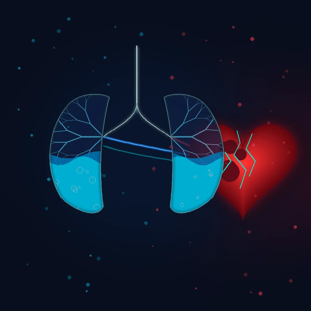
The failing ventricle cannot generate an adequate output at an acceptable filling pressure, so the filling pressure rises, and the pressure is transmitted back to the pulmonary capillaries. When the hydrostatic pressure exceeds the oncotic pressure, fluid floods the interstitium and then the alveoli, producing ventilation–perfusion mismatch and shunt, and so hypoxia. The hypoxia itself depresses the myocardium, and the sympathetic and renin–angiotensin activation that follows raises the afterload and closes the vicious circle. This is the rationale for the two most powerful emergency manoeuvres: non-invasive ventilation, which raises the intrathoracic pressure and so reduces both the venous return (preload) and the left-ventricular transmural pressure (afterload) while it recruits alveoli; and the nitrates, which reduce the preload and afterload directly.[2]
Clinical presentation
The classic presentation is acute respiratory distress: dyspnoea, orthopnoea, paroxysmal nocturnal dyspnoea, a productive cough with frothy or pink sputum, and wheeze (the cardiac asthma of bronchial-wall oedema). The patient is anxious, pale, sweaty and clammy, and sits upright fighting for breath. The examination shows tachypnoea and hypoxia, a raised jugular venous pressure, bibasal inspiratory crackles, a third heart sound, and oedema; the peripheries distinguish the warm (well-perfused) from the cold (hypoperfused) phenotype. In the elderly the presentation may be atypical — confusion and fatigue rather than breathlessness. A rising respiratory rate with fatigue and accessory-muscle use signals exhaustion and impending arrest. [1]
Differential diagnosis
The breathless, hypoxic patient has a differential, and the natriuretic peptide, the chest radiograph and the echo sort it out. [1]
Cardiogenic pulmonary oedema
- Raised JVP, S3, bibasal crackles; cardiomegaly
- CXR: Kerley B lines, bat-wing alveolar oedema, effusions
- BNP/NT-proBNP raised; echo shows a failing/left-heart cause
- Hypoxia with respiratory alkalosis (early)
COPD exacerbation
- Known COPD; wheeze, hyperinflation, pursed-lip breathing
- Hypercapnia on the gas; BNP normal/near-normal
- CXR: hyperinflation, no cardiomegaly
- Treat with bronchodilators, steroids, ± NIV for type 2 failure
Pneumonia
- Fever, purulent sputum, focal consolidation
- Septic features; leukocytosis
- CXR: focal consolidation; BNP may be mildly raised
- Antibiotics; oxygen
Pulmonary embolism / ARDS
- PE: pleuritic pain, syncope, DVT; ARDS: non-cardiogenic
- BNP normal/low (non-cardiogenic); echo may show RV strain (PE)
- CXR in ARDS: bilateral infiltrates without cardiomegaly
- Distinct pathway — not a nitrate/diuretic disease
Bedside assessment
Assess the airway and the breathing first — exhaustion and a falling respiratory rate are pre-arrest signs. Determine the phenotype: feel the peripheries for warmth (well perfused) or coldness (hypoperfused), and assess the congestion (raised JVP, crackles, oedema) against the dry state. Look for the precipitant: an ischaemic ECG, an irregularly irregular pulse of atrial fibrillation, a fever, the stigmata of chronic lung disease. [1]
Investigations and the targets
The ECG seeks the cause (an acute coronary syndrome, atrial fibrillation, left-ventricular hypertrophy). The chest radiograph shows cardiomegaly, Kerley B lines (the interstitial phase), the perihilar bat-wing alveolar oedema, and pleural effusions. The natriuretic peptide — BNP or NT-proBNP — is the single most useful single test: a low value largely excludes heart failure, while a high one supports it (BNP below about 100 is a strong rule-out, above 400 supports the diagnosis; interpret with the picture, as sepsis, renal failure and obesity distort it). The troponin identifies an ischaemic cause, the bedside echo distinguishes a reduced from a preserved ejection fraction and seeks a valve lesion, a regional wall-motion abnormality or a pericardial effusion, and the blood gas quantifies the hypoxia and detects the rising carbon dioxide of the tiring patient. Urea, creatinine and electrolytes guide the diuretic and the cause. [1]
Immediate management — the acute-oedema bundle, phenotype-led
Resuscitation and specific therapy begin together. Sit the patient upright and give high-flow oxygen to a saturation target of 94 to 98 per cent (88 to 92 in the chronic type-2 respiratory failure of COPD). Then the bundle, modified by the phenotype: [1]
[1]Phenotype → therapy

Non-invasive ventilation is first-line for respiratory distress or persistent hypoxia. The 3CPO trial showed no mortality benefit but a real reduction in breathlessness and in the need for intubation, so NIV is applied early for symptom relief and respiratory support rather than delayed in the hope of a survival difference.[2] Morphine (2.5 to 5 mg intravenously) is no longer routine — registry data link it to respiratory depression, ICU admission and worse outcomes — and is reserved for genuine distress or ischaemic pain. Treat the precipitant throughout: an acute coronary syndrome is reperfused, atrial fibrillation is rate- or rhythm-controlled, an infection is treated.
The diuretic strategy — dose, bolus, infusion
Loop diuretics are the cornerstone of decongestion. Give the first dose intravenously because gut-wall oedema impairs absorption. The standard initial dose is furosemide 40 to 80 mg IV for a patient not on chronic diuretics; for a patient already taking oral loop diuretics, give 1 to 2.5 times the total daily oral dose intravenously, because chronic use induces a rightward dose–response shift (the "braking phenomenon"). The DOSE-AHF trial randomised patients to high-dose (2.5× the home dose) versus low-dose (1×) furosemide and to bolus every 8 hours versus continuous infusion: high-dose produced faster fluid loss and greater dyspnoea relief with no significant difference in the composite primary endpoint or in renal dysfunction, and bolus and continuous infusion were equivalent.[3] The ROSE-AHF trial showed that neither low-dose dopamine nor low-dose nesiritide augmented decongestion or protected renal function in acute heart failure with renal dysfunction — so routine renal-dose dopamine is not supported.[4]
Diuretic and renal-augmentation trials in ADHF
DOSE-AHF (Felker, NEJM 2011)[3] — High-dose (2.5× home) vs low-dose (1×) furosemide; bolus q8h vs continuous infusion. Result: high-dose → more diuresis and dyspnoea relief; bolus = infusion; no difference in WRF or 60-day outcomes. Take-home: a higher initial IV dose is reasonable in the diuretic-experienced patient; choose bolus or infusion by convenience. ROSE-AHF (Chen, JAMA 2013)[4] — Low-dose dopamine or low-dose nesiritide vs placebo in ADHF with renal dysfunction. Result: neither augmented decongestion (72-hour cumulative urine volume) nor improved cystatin C. Take-home: renal-dose dopamine has no role in routine ADHF. Cotter (Lancet 1998)[5] — High-dose isosorbide dinitrate + low-dose furosemide vs high-dose furosemide + low-dose isosorbide in severe pulmonary oedema. Result: the nitrate-heavy arm had fewer MI, fewer needs for ventilation, and fewer deaths. Take-home: in hypertensive pulmonary oedema, the nitrate does the heavy lifting — the diuretic is the adjunct.
IV furosemide dosing at a glance
Vasodilators — when and how
Nitrates are added to the warm–wet, hypertensive patient (systolic above about 110 mmHg). Glyceryl trinitrate is started at 5–20 micrograms per minute and titrated up every few minutes (to 200 micrograms per minute) against the blood pressure and the dyspnoea. The mechanism is venodilation (preload reduction) and, at higher doses, arteriolar dilation (afterload reduction), which together drop the pulmonary-capillary wedge pressure faster than a diuretic alone. Sublingual GTN (0.4–0.8 mg) is a useful bridge while the infusion is set up. The Cotter trial established that a high-dose nitrate, low-dose diuretic strategy outperforms the reverse in hypertensive pulmonary oedema.[5] Sodium nitroprusside is reserved for the hypertensive emergency with severe afterload or aortic/mitral regurgitation, and requires arterial-line monitoring. Nesiritide (BNP analogue) showed no mortality or readmission benefit and is not routine. Serelaxin, tested in RELAX-AHF, improved dyspnoea but did not reduce cardiovascular mortality at 180 days in the definitive trial, and is not universally approved.[9]
[1]Inotropes and vasopressors for the cold phenotype
The cold–wet patient (cardiogenic shock) has low cardiac output with congestion and hypotension, and needs an inotrope rather than a venodilator. The choice is guided by the dominant problem — low output versus low pressure — and by whether the patient is also vasodilated. A bedside echo showing a small, underfilled, poorly contracting left ventricle helps; a pulmonary-artery catheter is justified when the haemodynamics remain opaque. [1]
Dobutamine
- β1 > β2 agonist; ↑ inotropy + mild vasodilation
- Dose 2–20 micrograms/kg/min
- Best for the low-output, warm-ish shock needing a push
- Tachyarrhythmia; ↑ myocardial oxygen demand; may drop BP (vasodilation)
Milrinone
- PDE-3 inhibitor; inotrope + lusitropy + vasodilation
- Dose 0.125–0.75 micrograms/kg/min
- Less tachyarrhythmia than dobutamine; useful in β-blocked patients
- Renal clearance — reduce dose in CKD; hypotension from vasodilation
Noradrenaline (norepinephrine)
- α > β agonist; the preferred first vasopressor for the cold + vasodilated
- Dose 0.05–1 micrograms/kg/min to MAP ≥ 65
- Restores perfusion pressure; add an inotrope alongside if output still low
- Excessive vasoconstriction ↑ afterload; central line only
Adrenaline (epinephrine)
- α + β agonist; inotrope + vasopressor in one
- Reserve for the refractory shock or arrest setting
- Lactic acidosis at higher doses; arrhythmia
- LevSimendan/omecamtiv mecarcinil — selective inotropes under investigation
The acute-oedema bundle as a sequence
Acute cardiogenic pulmonary oedema — first 30 minutes
- POSITION AND ASSESS — Sit upright. Assess airway, breathing, perfusion. Read the phenotype in 60 seconds: feel the peripheries (warm/cold), assess congestion (JVP, crackles, oedema), check the pulse pressure and the SBP. A cold, hypotensive patient takes the shock pathway, not this bundle
- OXYGEN — High-flow oxygen (non-rebreather 15 L/min) to SpO₂ 94–98% (88–92% in chronic CO₂ retainers). If distressed or hypoxia persists → NIV immediately
- NON-INVASIVE VENTILATION — CPAP 5–10 cm H₂O (or BiPAP IPAP 10–15 / EPAP 5–8 if hypercapnic). Reduces preload + afterload, recruits alveoli, relieves dyspnoea, and reduces intubation (3CPO; Cochrane)[2][8]
- VASODILATOR (if SBP > 110) — GTN infusion 5–20 micrograms/min, titrate up to 200; or sublingual GTN 0.4–0.8 mg as a bridge. Skip if hypotensive (cold–wet)
- LOOP DIURETIC — Furosemide 40–80 mg IV bolus (1–2.5× the oral dose if already on diuretics). Assess response at 2 h; double if inadequate
- IDENTIFY AND TREAT THE PRECIPITANT — ECG (ACS → reperfusion), rate/rhythm control for AF, sepsis workup, stop NSAIDs and negative inotropes
- REASSESS AND ESCALATE — If failing NIV or cold/hypotensive: intubate; inotrope/vasopressor ± MCS. Reassess the phenotype after every intervention
Subtypes and special scenarios
The hypertensive acute decompensated heart failure (warm–wet with a high pressure) responds best to a nitrate and a diuretic and has the best prognosis. Cardiogenic shock (wet–cold) is the other extreme and needs inotropes and mechanical circulatory support as a bridge to definitive treatment. Right-heart failure (cor pulmonale) shows a raised JVP with clear lungs and oedema. Peripartum cardiomyopathy presents in late pregnancy or the early postpartum period in a young woman and adds bromocriptine to the therapy. [1]
Complications and pitfalls
The complications are cardiogenic shock, acute kidney injury (from low output and the diuretic), respiratory failure and arrest, and arrhythmia. The pitfalls are the dangerous inverse of the bundle: giving a nitrate to the hypotensive patient; withholding NIV when it is indicated; over-diuresing the dry patient; using morphine routinely; missing the precipitant (an infarction that needs reperfusion); and mistaking a pulmonary embolism or ARDS for cardiogenic oedema. [1]
Prognosis and disposition
In-hospital mortality is around 4 to 10 per cent, much higher with cardiogenic shock; the 30-day readmission rate approaches 25 per cent. Every patient is admitted to cardiology or a high-dependency bed, the precipitant is treated, and guideline therapy is optimised before discharge — for the reduced-ejection-fraction patient, the four pillars of a beta-blocker, an ACE inhibitor or ARNI, a mineralocorticoid antagonist, and an SGLT2 inhibitor. [1]
Special populations
The elderly present atypically and have renal impairment that complicates the diuretic. Chronic kidney disease causes diuretic resistance and distorts the natriuretic peptide, so the clinical picture and the echo weigh more. COPD overlap is common; the natriuretic peptide and the chest radiograph distinguish a heart failure exacerbation from a COPD exacerbation. Peripartum cardiomyopathy is a young-woman presentation with its own therapy. [1]
Evidence and regional guidelines
The contemporary framework is the 2021 ESC heart-failure guideline with the 2023 focused update.[1] The non-invasive-ventilation evidence is the 3CPO trial.[2] The bundle and the four-pillar long-term therapy are global; the local cardiology and heart-failure pathways govern the agent choices and the disposition.
ANZ practice note. The acute-oedema bundle and the phenotype-led approach follow the ESC heart-failure guideline via local cardiology pathways; NIV is applied early for symptom relief and to avoid intubation, and the four-pillar reduced-ejection-fraction therapy is initiated before discharge. [1]
Discharge criteria and transition to oral therapy
The goal of the admission is not merely symptom relief but a euvolaemic, haemodynamically stable patient on optimised oral guideline therapy before discharge — because the 30-day readmission rate approaches 25 per cent and most readmissions are driven by residual congestion or incomplete uptitration. The transition from intravenous to oral diuretic is made once the oral route is reliably absorbed and the patient is clinically euvolaemic; the oral dose is typically half to two-thirds of the IV dose that achieved decongestion, and the weight, the JVP and the renal function are watched daily. [1]
Discharge readiness in acute heart failure
- EUVOLAEMIC — Resolution of dyspnoea at rest; no orthopnoea; JVP not raised; no new crackles; stable weight for 24–48 h off IV diuretic (or on a stable oral dose). Residual congestion is the commonest driver of early readmission — do not discharge a wet patient
- STABLE RENAL FUNCTION — Creatinine no more than 0.3 mg/dL (≈ 26 µmol/L) above baseline and stable for 24 h; potassium and electrolytes replete. The diuretic and the renin–angiotensin blocker have been held or adjusted to the new baseline
- HAEMODYNAMICALLY STABLE — Blood pressure stable without inotrope or vasopressor for at least 24 h; no symptomatic orthostatic drop; heart rate controlled; no new arrhythmia
- OPTIMISED ORAL GDMT — For HFrEF the four pillars are initiated or continued: beta-blocker, ACE inhibitor or ARNI, mineralocorticoid antagonist, and SGLT2 inhibitor — uptitrated as tolerated, not stopped for mild asymptomatic hypotension. Document the EF, the cause, and the plan
- PRECIPTANT ADDRESSED AND EDUCATION GIVEN — The trigger (ACS, AF, infection, drug) treated; NSAIDs stopped; weight-based self-monitoring and a written action plan explained; follow-up booked within 7–14 days; cardiology and heart-failure nurse referral made
Exam practice
SAQ — Flash pulmonary oedema in the hypertensive heart failure patient on BiPAP
10 minutes · 10 marks
A 72-year-old man with known HFrEF (ejection fraction 28 per cent, ischaemic cause) on furosemide 80 mg twice daily, bisoprolol 5 mg, ramipril 5 mg, spironolactone 25 mg and dapagliflozin 10 mg presents at 03:00 with acute severe breathlessness that woke him from sleep, coughing up pink frothy sputum. He missed his frusemide for three days while visiting family. On arrival he is sitting upright, distressed and sweaty: BP 198/108, HR 124 in new rapid atrial fibrillation, RR 34, SpO₂ 84 per cent on room air, temperature 36.4°C. The JVP is raised to the earlobes, there are loud bibasal inspiratory crackles to the apices, and a third heart sound is audible. The chest radiograph shows bat-wing perihilar alveolar oedema and Kerley B lines. The venous gas shows pH 7.32, lactate 2.1. BiPAP has just been applied at IPAP 12 / EPAP 6.
SAQ — Cardiogenic shock complicating an anterior STEMI
10 minutes · 10 marks
A 65-year-old woman presents 6 hours after the onset of crushing central chest pain. The ECG shows ST elevation in V1 to V4 with reciprocal ST depression in the inferior leads — an anterior STEMI — and she has been given aspirin 300 mg, ticagrelor 180 mg and 5000 units of intravenous unfractionated heparin. As the cath-lab is being prepared she deteriorates: she is pale, clammy and mottled from the knees down, BP 76/50, HR 128 in sinus tachycardia, RR 30, SpO₂ 90 per cent on 15 L oxygen, and she has passed only 15 mL of urine in the last hour. The JVP is raised, there are bibasal crackles, and the bedside echo shows a poorly contracting, mildly dilated left ventricle with anterior and apical akinesia and an estimated ejection fraction of 20 per cent. The venous lactate is 5.8 mmol/L.
Exam pearls
- Sit up, oxygen, NIV, GTN (if the pressure holds), furosemide — and read the phenotype first.
- NIV reduces preload and afterload and recruits alveoli; 3CPO showed symptom and intubation benefit, not a mortality difference — so use it early.
- Nitrates only if the blood pressure is adequate — the cold, hypotensive (wet–cold) phenotype has cardiogenic shock and wants an inotrope or a vasopressor and mechanical support.
- No routine morphine — respiratory depression, worse outcomes.
- BNP rules out heart failure; the chest radiograph shows Kerley B lines and bat-wing oedema.
- Find the precipitant — an acute coronary syndrome needs reperfusion, atrial fibrillation needs rate control.
- Forrester and Stevenson at the bedside — congestion (wet/dry) × perfusion (warm/cold) guides everything; the wet–warm is the bundle patient, the wet–cold is the shock patient.[6][7]
- Diuretic dosing — 40–80 mg IV if naïve; 1–2.5× the oral dose if already on a loop (DOSE-AHF); bolus and continuous infusion are equivalent.[3]
- A narrow pulse pressure (< 25% of systolic) marks a low stroke volume — a cold phenotype even before the blood pressure falls.
- The cotter principle — in hypertensive pulmonary oedema the nitrate does the heavy lifting; high-dose isosorbide/GTN + low-dose diuretic beats high-dose diuretic + low-dose nitrate.[5]
- Renal-dose dopamine has no role — ROSE-AHF showed no augmentation of decongestion or renal protection.[4]
- Atrial fibrillation is the classic arrhythmic precipitant — loss of the atrial kick plus a fast rate tips the failing ventricle; rate (or rhythm) control is part of the resuscitation.
- NSAIDs cause fluid and salt retention — a top iatrogenic precipitant; stop them on admission and counsel against them at discharge.
- An S3 gallop is the auscultatory hallmark of volume overload and a failing ventricle — together with a raised JVP it is highly specific for cardiogenic oedema.
- BiPAP over CPAP when there is hypercapnia (CO₂ retention, the tiring patient, COPD overlap) — otherwise CPAP is simpler and equally effective for pure cardiogenic oedema.
- Discharge only the euvolaemic patient — stable weight 24–48 h off IV diuretic, normalising renal function, and the four HFrEF pillars initiated; residual congestion drives the 25% 30-day readmission rate.
- Cardiac asthma — wheeze from bronchial-wall oedema can mimic COPD; the raised JVP, S3 and BNP distinguish the failing heart.
- Cotter, DOSE, ROSE, RELAX — the four trials an examiner expects: nitrates win, high-dose diuretic speeds decongestion, dopamine/nesiritide add nothing, serelaxin relieves dyspnoea but not mortality.[3][4][5][9]
Red flags
[1]References
- [1]McDonagh TA, Metra M, Adamo M, et al. [2023 Focused update of the 2021 ESC Guidelines for the diagnosis and treatment of acute and chronic heart failure] G Ital Cardiol (Rome), 2024.PMID 38410903
- [2]Gray A, Goodacre S, Newby DE, et al. Noninvasive ventilation in acute cardiogenic pulmonary edema N Engl J Med, 2008.PMID 18614781
- [3]Felker GM, Lee KL, Bull DA, et al. Diuretic strategies in patients with acute decompensated heart failure N Engl J Med, 2011.PMID 21366472
- [4]Chen HH, Anstrom KJ, Givertz MM, et al. Low-dose dopamine or low-dose nesiritide in acute heart failure with renal dysfunction: the ROSE acute heart failure randomized trial JAMA, 2013.PMID 24247300
- [5]Cotter G, Metzkor E, Kaluski E, et al. Randomised trial of high-dose isosorbide dinitrate plus low-dose furosemide versus high-dose furosemide plus low-dose isosorbide dinitrate in severe pulmonary oedema Lancet, 1998.PMID 9482291
- [6]Forrester JS, Diamond G, Chatterjee K, et al. Medical therapy of acute myocardial infarction by application of hemodynamic subsets (second of two parts) N Engl J Med, 1976.PMID 790194
- [7]Nohria A, Mielniczuk LM, Stevenson LW Evaluation and monitoring of patients with acute heart failure syndromes Am J Cardiol, 2005.PMID 16181821
- [8]Berbenetz N, Wang Y, Brown J, et al. Non-invasive positive pressure ventilation (CPAP or bilevel NPPV) for cardiogenic pulmonary oedema Cochrane Database Syst Rev, 2019.PMID 30950507
- [9]Teerlink JR, Cotter G, Davison BA, et al. Serelaxin, recombinant human relaxin-2, for treatment of acute heart failure (RELAX-AHF): a randomised, placebo-controlled trial Lancet, 2013.PMID 23141816