EM · Myocarditis & pericarditis
Myocarditis and pericarditis (the ACS mimic and the positional chest pain)
Also known as Myopericarditis · Acute pericarditis · Viral pericarditis · Myocarditis
Myocarditis and pericarditis — the viral or the autoimmune inflammation of the heart muscle and the pericardium, the clinical presentation (the positional and the pleuritic chest pain of the pericarditis; the ACS-mimic and the arrhythmia of the myocarditis), the ECG pattern (the diffuse ST elevation and the PR depression of the pericarditis), the troponin elevation (in both), the management (the NSAID and the colchicine for the pericarditis; the supportive and the heart-failure therapy for the myocarditis), and the fulminant myocarditis needing the mechanical support. ACEM-primary, globally tagged.
On this page & tools
Your progress
Saved locally on this device.
10 MCQs with explanations
Target exams
Red flags
Myocarditis and pericarditis are the inflammations of the heart muscle and the pericardium, most commonly from a viral infection, and they share a clinical space with the acute coronary syndrome — the chest pain, the ECG changes and the troponin rise that can mimic an infarct exactly. The Fellowship candidate must distinguish the pericarditis (the positional, the pleuritic pain, the diffuse ECG changes, the NSAID-responsive course) from the myocarditis (the ACS-mimic, the arrhythmia, the heart failure, the fulminant shock), because the management and the prognosis are very different.[1][2]
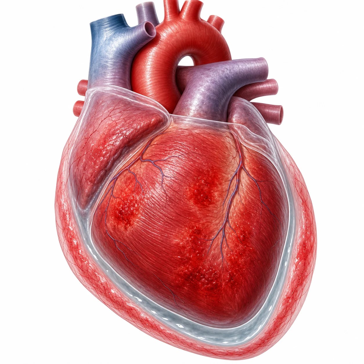
Definition and classification
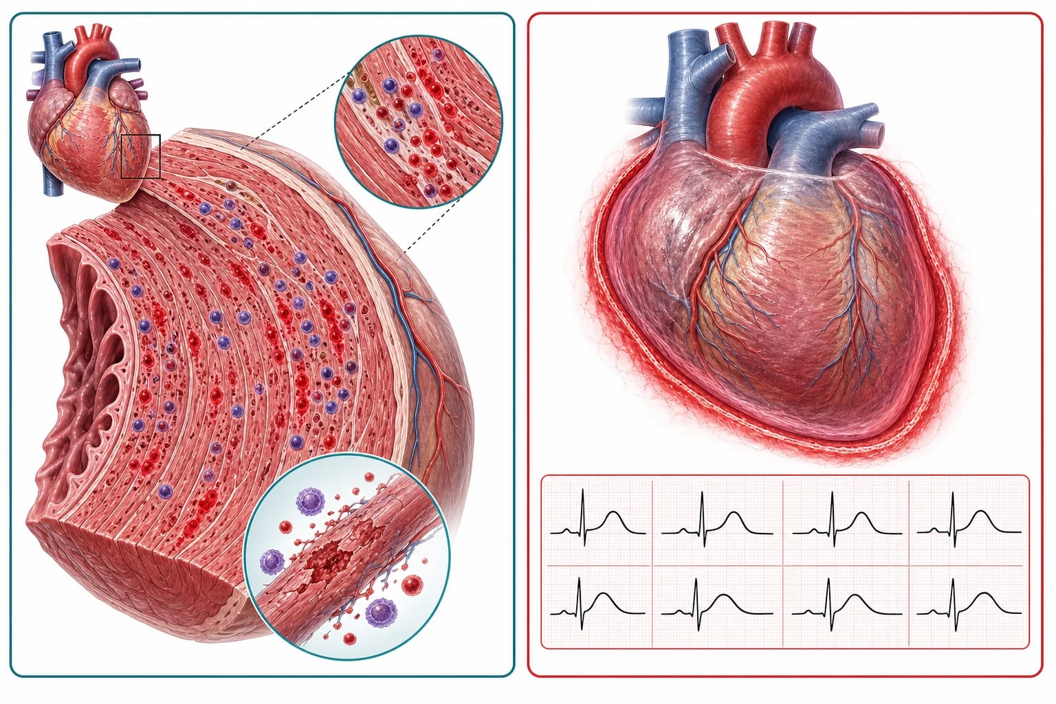
Pericarditis is the inflammation of the pericardium, usually from a viral infection (the coxsackie, the echovirus, the adenovirus, the influenza, the SARS-CoV-2), but also from the bacterial infection, the autoimmune disease (the systemic lupus, the rheumatoid, the vasculitis), the uraemia, the malignancy, the radiation, the post-MI (the Dressler syndrome) and the post-pericardiotomy. Myocarditis is the inflammation of the myocardium, most commonly viral (the same spectrum), but also from the autoimmune disease, the drug (the anthracycline, the clozapine), the heavy metal, the radiation and the post-vaccine (the rare mRNA COVID-vaccine myocarditis in the young male). The two conditions often co-exist — the myopericarditis — because the pericardium and the epicardial myocardium are anatomically contiguous. [1]
Pathophysiology — why the troponin rises and the ECG changes
The pericardial inflammation produces an exudate (the pericardial effusion) and the characteristic ECG changes — the diffuse ST elevation (from the epicardial injury, unlike the focal ST elevation of the STEMI) and the PR depression (from the atrial involvement). The troponin rises when the inflammation extends into the myocardium (the myopericarditis), which is common. The myocarditis in isolation produces a myocyte injury that raises the troponin and may produce a regional wall motion abnormality on the echo (mimicking a STEMI), a heart failure or an arrhythmia. The fulminant myocarditis — a massive, rapidly progressive inflammation of the myocardium — produces a cardiogenic shock within days of a viral prodrome and may need a mechanical circulatory support or an extracorporeal membrane oxygenation as a bridge to recovery. [1]
Differential diagnosis — the ACS mimic and the chest pain
The differential of the pericarditis and the myocarditis is the acute coronary syndrome, and the distinction is the emergency department's task. [1]
Pericarditis
- Positional, pleuritic pain; relieved by sitting forward
- Diffuse ST elevation + PR depression; no reciprocal changes
- Viral prodrome; fever; a pericardial rub
- NSAID + colchicine; the troponin may be mildly raised
Myocarditis (ACS mimic)
- Chest pain, troponin rise, ECG changes — mimics the STEMI
- A young patient; a viral prodrome; no coronary risk factors
- Echo: regional wall motion abnormality; MRI: late gadolinium enhancement
- Coronary angiography is normal — the diagnosis
ACS (STEMI/NSTEMI)
- Crescendo pressure pain; coronary risk factors
- Territorial ST elevation; reciprocal changes; dynamic
- Troponin with the rise-and-fall pattern
- Coronary angiography shows the occlusion
Fulminant myocarditis
- A young, previously well patient with a viral prodrome
- A rapid cardiogenic shock or a ventricular arrhythmia
- Echo: a globally reduced EF; a normal coronaries on the angio
- Mechanical circulatory support; the ICU; may recover fully
Investigations — the ECG, the troponin and the echo
The ECG of the pericarditis shows the diffuse, concave ST elevation in multiple leads (not the focal, convex elevation of the STEMI), the PR depression (in the limb and the precordial leads), and, later, the T-wave inversion. The ECG of the myocarditis may be non-specific, may mimic the STEMI (the regional ST elevation), or may show an arrhythmia (the ventricular ectopics, the VT, the heart block). The troponin is raised in both (higher and more dynamic in the myocarditis; mildly raised in the pure pericarditis). The CRP and the ESR are raised (the inflammatory markers). The echocardiography shows a pericardial effusion (in the pericarditis) and a regional or a global wall motion abnormality (in the myocarditis) with a reduced ejection fraction in the severe case. The cardiac MRI is the gold standard for the myocarditis — it shows the late gadolinium enhancement in the mid-wall (not the subendocardial pattern of the infarct), the T1 and the T2 mapping (the oedema), and the biventricular function. The coronary angiography is the definitive test when the ACS cannot be excluded — the normal coronaries with the elevated troponin and the wall motion abnormality confirm the myocarditis. [1]
Immediate management — the pericarditis and the myocarditis
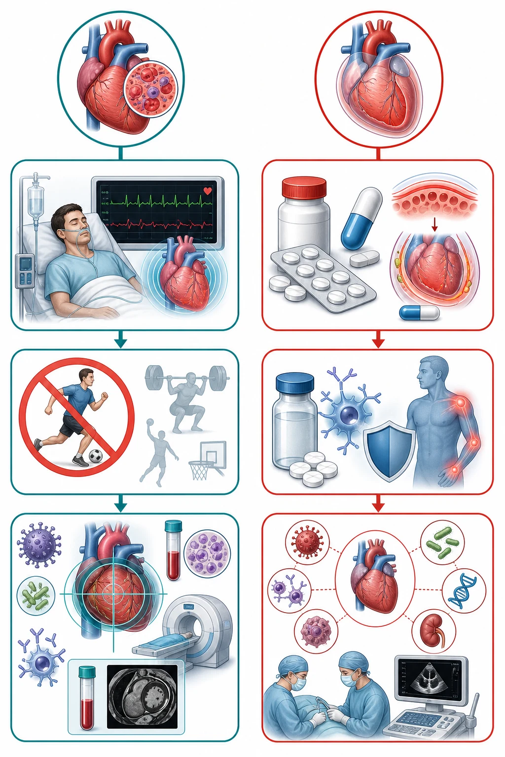
The management differs by the condition, and the distinction from the ACS is the prerequisite. [1]
[1]The targets and the doses
Complications and pitfalls
The complications of the pericarditis are the recurrence (30 per cent after the first episode — the colchicine halves it), the constrictive pericarditis (a late, chronic constriction from the organised pericardial thickening — needs the pericardiectomy), and the tamponade (from a large effusion — the serial echo monitors for it). The complications of the myocarditis are the heart failure (the dilated cardiomyopathy), the arrhythmia (the atrial and the ventricular, the heart block, the sudden cardiac death), and the fulminant course (the cardiogenic shock needing the mechanical support). The pitfalls are: giving the NSAIDs to the myocarditis (they may worsen the injury); giving the corticosteroids to the first-episode pericarditis (they increase the recurrence); not monitoring the effusion for the tamponade; not restricting the sport after the myocarditis; and missing the fulminant myocarditis behind a "viral illness with a chest pain." [1]
Prognosis and disposition
The pericarditis is a benign, self-limiting condition in most cases — the symptoms resolve over days to weeks with the NSAID and the colchicine, and the prognosis is excellent (apart from the recurrence). The myocarditis has a variable prognosis — the mild case recovers fully, the severe case may leave a residual cardiomyopathy, and the fulminant case has a mortality of up to 20 per cent but may recover completely with the mechanical support. The patient is admitted for the pericarditis if a large effusion or a high-risk feature is present; the myocarditis is admitted for the troponin rise, the arrhythmia, the heart failure, or the diagnostic uncertainty (the ACS exclusion). The follow-up includes the serial echo, the ECG, the troponin, and the sport-medicine assessment before the return to the exercise. [1]
Aetiology of myocarditis — the viral, the autoimmune and the drug
The myocarditis is the inflammation of the myocardium with the necrosis and the degeneration of the myocyte, and the aetiology is best held in four buckets — the infectious, the immune-mediated, the toxin and the drug, and the systemic.[4][11] The infectious causes dominate: the viruses are the commonest in the developed world (the coxsackie B, the parvovirus B19, the adenovirus, the echovirus, the influenza, the Epstein–Barr, the cytomegalovirus, the HIV, the SARS-CoV-2), followed by the bacterial (the staphylococcus, the streptococcus, the Borrelia of the Lyme disease, the Mycoplasma), the protozoal (the Trypanosoma cruzi — the Chagas disease, the commonest cause of the myocarditis worldwide), the rickettsial, and the fungal. The immune-mediated causes are the giant-cell myocarditis (a rare, rapidly fatal autoimmune disease of the young adult — a medical emergency), the sarcoid, the eosinophilic (the Churg–Strauss, the drug-hypersensitivity), and the collagen-vascular (the systemic lupus, the rheumatoid). The toxin and the drug causes are the anthracycline (the doxorubicin — the dose-dependent cumulative cardiotoxicity), the clozapine, the trastuzumab, the alcohol, the cocaine, the heavy metals, the snake and the scorpion venom, and the immune-checkpoint inhibitors (the anti-PD-1/PD-L1 — an increasingly recognised cause). The systemic causes include the peri-partum cardiomyopathy and the severe sepsis.
Infectious (the commonest)
- Viral #1: coxsackie B, parvovirus B19, adenovirus, SARS-CoV-2
- Bacterial: staph/strep, Borrelia (Lyme), Mycoplasma
- Protozoal: Trypanosoma cruzi (Chagas) — commonest worldwide
- Fungal and rickettsial in the immunocompromised host
Immune-mediated
- Giant-cell myocarditis — young adult, rapidly fatal, biopsy + MCS
- Cardiac sarcoidosis — conduction disease, bilateral hilar LN
- Eosinophilic (DRESS, Churg-Strauss, drug hypersensitivity)
- Systemic lupus, rheumatoid, Kawasaki
Drug / toxin
- Anthracycline (doxorubicin) — cumulative dose-dependent
- Clozapine — within 8 weeks of starting; check troponin
- Immune-checkpoint inhibitors (anti-PD1) — emerging cause
- Cocaine, alcohol, heavy metals, snake/scorpion venom
Systemic / other
- Peri-partum cardiomyopathy (last month to 5 months post-partum)
- Severe sepsis — the cytokine myocardial injury
- Radiation — years after mediastinal radiotherapy
- Vaccine-associated (rare, mRNA COVID, young male)
Aetiology of pericarditis — the spectrum
The pericarditis is most often idiopathic or viral (together over 80 per cent of the cases in the developed world — the same viral spectrum as the myocarditis, with the coxsackie, the echovirus, the adenovirus, the influenza, and the SARS-CoV-2).[3] The non-viral causes are best remembered by the system: metabolic (the uraemia of the renal failure, the hypothyroid and the myxoedema), autoimmune (the systemic lupus, the rheumatoid, the Dressler syndrome — 2 to 10 weeks after the MI; the post-cardiotomy syndrome), neoplastic (the lung, the breast, the melanoma; the mesothelioma), infectious (the tuberculosis — the commonest cause worldwide; the purulent bacterial from the contiguous pneumonia or the empyema), radiation, and the drug-induced (the procainamide, the hydralazine, the isoniazid — the drug-induced lupus). The tuberculous pericarditis is a high-yield aetiology — suspect it in the endemic area, the HIV-positive patient, or the chronic effusion with the fever and the weight loss, and treat with the anti-tuberculous therapy plus the adjunctive corticosteroid (it reduces the progression to the constriction).
The four ECG stages of acute pericarditis
The ECG of the acute pericarditis evolves through four stages over days to weeks, and recognising the stage is a classic Fellowship question. [1]
Stage 1 (first hours–days)
- Diffuse concave ST elevation in all leads except aVR and V1
- PR depression in the limb and precordial leads (PR elevation in aVR)
- Reciprocal ST depression only in aVR/V1 — the key to the STEMI distinction
- The stage most often seen in the ED
Stage 2 (days later)
- ST elevation and the PR depression normalise
- The J point returns to the baseline
- A transition stage — easily missed as a normal tracing
Stage 3 (late first–second week)
- T-wave inversion develops (the ST has returned to baseline)
- The T-wave inversion is diffuse (not territorial like the STEMI)
- May persist for weeks
Stage 4 (weeks later)
- The T waves normalise; the ECG returns to the baseline
- May be incomplete — persistent T-wave inversion
- A normal ECG does not exclude the pericarditis in the late presentation
The cardiac MRI — the Lake Louise criteria
The cardiac magnetic resonance is the non-invasive gold standard for the myocarditis, and the diagnosis rests on the Lake Louise criteria (updated in 2018).[5][6] The diagnosis requires at least TWO of the three classical criteria — combined, the sensitivity is about 67 per cent and the specificity about 91 per cent, rising further with the parametric T1 and T2 mapping in the 2018 update:
The Lake Louise criteria (2018) for the myocarditis
The endomyocardial biopsy and the Dallas criteria
The endomyocardial biopsy is the historical gold standard for the myocarditis, but it is invasive, the inter-observer variability is high, and the sensitivity is low (under 50 per cent because of the patchy disease — the sampling error).[4][11] The Dallas criteria (1987) classify the biopsy as the active myocarditis (the inflammatory infiltrate with the myocyte necrosis), the borderline myocarditis (the inflammatory infiltrate without the myocyte necrosis), or the no myocarditis.[12] The biopsy is now reserved for the clinically suspected fulminant or giant-cell myocarditis (where the histology changes the management — the immunosuppression or the transplant), the refractory cardiogenic shock of the unclear cause, and the suspected cardiac sarcoid or the infiltrative disease. For the routine case, the cardiac MRI has replaced the biopsy.
Fulminant myocarditis — recognition and the resuscitation
The fulminant myocarditis is the distinct, severe phenotype — a sudden, diffuse myocardial inflammation presenting as the cardiogenic shock within hours to days of a viral prodrome — that the emergency physician must not miss behind a flu with the chest pain and the breathlessness.[7][10] The McCarthy cohort established the paradox: the fulminant myocarditis has a HIGHER in-hospital mortality than the non-fulminant acute myocarditis, but a BETTER long-term survival in the survivors (the recovery of the ventricular function is near-complete in those who survive the acute episode).[10]
The fulminant myocarditis — the ED and the ICU pathway
1 — Recognise
A young, previously well patient with a recent viral prodrome (the fever, the myalgia, the gastrointestinal upset) presenting with the cardiogenic shock (the hypotension, the cool peripheries, the oliguria, the confusion), the pulmonary oedema, or the ventricular arrhythmia. The echo shows a globally reduced ejection fraction (often under 30 per cent) WITHOUT the regional wall motion abnormality of the infarct. The troponin is markedly raised.
2 — Secure the airway and the circulation
Early intubation and the mechanical ventilation for the respiratory failure and the cardiogenic pulmonary oedema. Establish the arterial line and the central access. The bedside ultrasound confirms the global hypokinesis, the lack of the pericardial effusion, and the normal RV (to exclude the massive PE). The transient pulseless electrical activity or the VT/VF is common — be ready to resuscitate.
3 — Exclude the acute coronary syndrome
An urgent coronary angiography (or the CT coronary if the patient is too unstable for the catheter) to exclude the infarct — the normal coronaries confirm the myocarditis. Do NOT send the unstable patient for the angiography without the inotrope and the mechanical support plan in place.
4 — The mechanical circulatory support — the bridge to recovery
Start the inotropes (the dobutamine, the milrinone) and the vasopressors (the noradrenaline) cautiously — they increase the myocardial oxygen demand. Move EARLY to the mechanical support: the VA-ECMO (the first-line for the biventricular failure and the severe shock), the Impella (the axial flow for the LV support), or the IABP (the second-line, less effective). The early MCS as a bridge to decision or recovery improves the survival.
5 — The targeted therapy and the disposition
Treat the heart failure (the diuretic once the perfusion is secured, the ACE inhibitor when stable). The endomyocardial biopsy if the giant-cell or the eosinophilic myocarditis is suspected (the immunosuppression is life-saving). The IVIG and the high-dose corticosteroid are CONTROVERSIAL for the viral myocarditis but may be used in the giant-cell or the immune-mediated phenotype. Transfer to the tertiary centre with the advanced heart-failure and the transplant service. Avoid the NSAIDs.
Drug-induced myocarditis — the high-yield agents
Recurrent pericarditis — the colchicine, the steroid and the anakinra
The recurrent pericarditis — the symptomatic recurrence after a symptom-free interval of at least 4 to 6 weeks — affects about 30 per cent after the first episode, and the recurrence rate climbs with each subsequent episode.[3] The colchicine is the evidence-based prevention: the ICAP trial (the New England Journal 2013) showed that the colchicine 0.5 mg twice daily for 3 months halved the recurrence at 18 months (from 37 to 16 per cent).[9] The management of the recurrence is the same as the first episode (the NSAID plus the colchicine); the corticosteroid is reserved for the failure of the NSAID or the autoimmune cause (it INCREASES the recurrence when used in the first episode). The refractory recurrent pericarditis responds to the anakinra (the recombinant IL-1 receptor antagonist) — an emerging and effective therapy — and the pericardiectomy is the last resort.
Constrictive pericarditis vs restrictive cardiomyopathy
Both present with the right heart failure (the elevated JVP, the hepatomegaly, the ascites, the peripheral oedema) and are notoriously hard to distinguish — the distinction is high-yield because the constriction is curable (the pericardiectomy) and the restriction is not. [1]
Constrictive pericarditis
- A prior pericarditis, the radiation, or the TB; the pericardium is thickened/calcified
- The Kussmaul sign (the JVP RISES on inspiration); the pericardial knock
- Echo: the septal bounce, the respirophasic variation; CT/MRI: the thickened pericardium
- Haemodynamics: the equalisation of the diastolic pressures; the dip-and-plateau (square-root) sign
- Cured by the pericardiectomy
Restrictive cardiomyopathy
- The amyloid, the sarcoid, the eosinophilic, the radiation, the storage disease
- The JVP may fall on inspiration (no Kussmaul); the S3 (not the knock)
- Echo: the biatrial enlargement, the thickened myocardium, the sparkling (amyloid); normal pericardium
- Haemodynamics: the LVEDP exceeds the RVEDP by over 5 mmHg; the pulmonary hypertension is more severe
- Treat the underlying cause; the diuretic; not curable by the surgery
The trial evidence — the landmark studies
Imazio — the ICAP trial, colchicine for the acute pericarditis (NEJM 2013)
New England Journal of Medicine
PMID 23992557
Key finding
A multicentre randomised double-blind placebo-controlled trial of 240 patients with the first episode of acute pericarditis, comparing the colchicine 0.5 to 1.0 mg daily for 3 months plus the conventional NSAID against the placebo plus the NSAID. The primary endpoint (the recurrent pericarditis at 18 months) occurred in 16.7 per cent of the colchicine group vs 37.5 per cent of the placebo — a 55 per cent relative reduction. The symptom persistence at 72 hours was also halved.
Practice change
Established the colchicine as the standard adjunct for the first episode of the acute pericarditis — the 3-month course halves the recurrence and accelerates the symptom resolution. The backbone of the modern ESC guideline.
Imazio — the COPE trial, colchicine for the acute pericarditis (Circulation 2005)
Circulation
PMID 16186437
Key finding
The first randomised open-label trial of 120 patients with the first episode of acute pericarditis, showing that the addition of colchicine to the aspirin reduced the treatment failure at 72 hours and the recurrence at 18 months (from 32 to 11 per cent). The therapy was well tolerated, the diarrhoea being the main adverse effect.
Practice change
The pioneering evidence that the colchicine reduces both the treatment failure and the recurrence in the acute pericarditis — the precursor to the ICAP and the basis for the guideline recommendation.
McCarthy — fulminant vs acute (non-fulminant) myocarditis (NEJM 2000)
New England Journal of Medicine
PMID 10706898
Key finding
The Johns Hopkins cohort of 147 patients with the biopsy-proven myocarditis, comparing the 15 with the fulminant myocarditis (the severe acute presentation needing the inotrope/MCS) against the 132 with the acute non-fulminant myocarditis. The in-hospital mortality was higher in the fulminant group, but the long-term transplant-free survival was dramatically BETTER in the fulminant survivors (93 per cent at 11 years vs 45 per cent).
Practice change
Established the defining paradox of the fulminant myocarditis — the worse acute course but the better long-term recovery — and the imperative to support the fulminant patient aggressively through the acute episode. The basis of the modern MCS-bridge-to-recovery strategy.
Imazio — the COPPS trial, colchicine for the post-pericardiotomy syndrome (Eur Heart J 2010)
European Heart Journal
PMID 20805112
Key finding
A multicentre randomised double-blind placebo-controlled trial of 360 patients after the cardiac surgery, given the colchicine 1 mg daily from post-op day 3 for one month. The colchicine reduced the post-pericardiotomy syndrome (from 18.9 to 8.9 per cent) and the post-operative atrial fibrillation (from 22 to 13 per cent).
Practice change
Extended the role of the colchicine beyond the idiopathic pericarditis to the post-cardiotomy syndrome and the post-operative atrial fibrillation prevention — a high-yield perioperative point.
Friedrich — the Lake Louise criteria for the CMR in the myocarditis (JACC 2009)
Journal of the American College of Cardiology
PMID 19389557
Key finding
The international consensus white paper that defined the Lake Louise criteria for the non-invasive CMR diagnosis of the myocarditis — the combination of the T2-based oedema imaging, the early gadolinium enhancement (the hyperaemia and the capillary leak), and the late gadolinium enhancement (the fibrosis and the necrosis). A diagnosis required two of the three. The criteria achieved a sensitivity of about 67 per cent and a specificity of about 91 per cent.
Practice change
Established the non-invasive gold standard for the myocarditis, replacing the endomyocardial biopsy for the routine diagnosis. Updated in 2018 (Ferreira et al) with the parametric T1 and T2 mapping.
Oster — the myocarditis after the mRNA COVID-19 vaccine (JAMA 2022)
JAMA
PMID 35076665
Key finding
The US Vaccine Adverse Event Reporting System review (December 2020 to August 2021) identifying 1626 reports of the myocarditis (1055 of the pericarditis) after the mRNA vaccination — predominantly in the young male, after the second dose, within a week, and with a mild, self-limiting course (the troponin rise, the chest pain, the rapid resolution). The reporting rate was highest in the males aged 12 to 17 after the second dose (about 100 per million doses).
Practice change
Quantified the rare but real association between the mRNA COVID-19 vaccine and the myocarditis — overwhelmingly mild, with a benefit-to-risk that overwhelmingly favours the vaccination. A high-yield modern Fellowship topic.
The disposition — who is admitted and who is discharged
The disposition follows the risk stratification, not the diagnosis. Admit the pericarditis with ANY of: a fever over 38 °C, a subacute onset (over a week), a large pericardial effusion (over 2 cm in diastole), a tamponade physiology, an immunosuppression, an oral anticoagulation, a trauma, a myopericarditis (the troponin raised), or the failure of the first-line therapy. The low-risk pericarditis (the classic viral presentation, a small or no effusion, a negative troponin) may be discharged on the NSAID plus the colchicine with the 24 to 48 hour review and the warning about the red flags. Admit ALL the myocarditis — the troponin rise, the arrhythmia, the heart failure, and the diagnostic uncertainty (the ACS exclusion) all warrant the admission and the monitoring. The fulminant myocarditis goes to the ICU or the HDU and the tertiary centre. [1]
High-yield one-liners — the aetiology and the diagnosis
- The pericardial rub is a triphasic (the systolic, the diastolic, the pre-systolic) scratch best heard at the left sternal border with the patient leaning forward in full expiration — its presence does not exclude a large effusion, and its absence does not exclude the pericarditis.
- The PR depression is the single best ECG discriminator of the pericarditis from the STEMI — and it may be the ONLY early ECG sign.
- The troponin is raised in BOTH the pericarditis and the myocarditis (the myopericarditis) — a raised troponin does NOT distinguish them, and does NOT, by itself, mandate the angiography if the ECG and the echo are typical for the pericarditis.
- The NSAIDs are AVOIDED in the acute myocarditis (they impair the healing, they increase the viral replication in the animal models, and they worsen the myocyte injury) — give the heart-failure therapy and the rest, NOT the ibuprofen.
- The corticosteroids INCREASE the recurrence of the pericarditis when given for the first episode — reserve them for the autoimmune cause, the uraemia, the failure of the NSAID, or the contraindication to the NSAID.
- The colchicine is the ONLY drug proven to reduce the recurrence (the ICAP and the COPE trials) — give it for 3 months.
- The Dressler syndrome is the pericarditis 2 to 10 weeks post-MI — the immune-mediated; treat with the aspirin (avoid the NSAID in the first week post-MI).
- The giant-cell myocarditis is the young adult with the rapidly progressive heart failure and the VT — the biopsy (the giant cells), the immunosuppression, and the transplant.
- The cardiac MRI Lake Louise criteria — TWO of the three: the T2 oedema, the early gadolinium, the late gadolinium (the mid-wall or the subepicardial, NOT the subendocardial).
- The fulminant myocarditis — support with the MCS (the VA-ECMO) as the bridge to recovery; the survivors recover the ventricular function.
- The recurrent pericarditis — the anakinra (the IL-1 receptor antagonist) for the refractory case. [1]
The bedside red flags — the clinical triggers
[1]Special populations
The athlete with the myocarditis is at a high risk of the sudden cardiac death — the return to the sport is restricted for 3 to 6 months, and the clearance requires a normal echo, a normal ECG, a normal exercise test, and no biomarker evidence of the ongoing inflammation. The post-COVID-vaccine myocarditis (the rare mRNA-vaccine-associated myocarditis in the young male, typically within a week of the second dose) is usually mild and self-limiting. The pregnant patient with the pericarditis is managed with the aspirin (the ibuprofen is avoided in the third trimester — the premature closure of the ductus). The anticoagulated patient is managed with caution (the NSAID increases the bleeding risk). [1]
Evidence and regional guidelines
The contemporary framework is the ESC 2015 pericardial-disease guideline (the NSAID + colchicine for the pericarditis)[2] and the contemporary myocarditis literature (the MRI diagnosis, the sport restriction, the mechanical support for the fulminant).[1] The drug choices and the sport-restriction criteria follow the local cardiology and the sports-medicine protocol.
ANZ practice note. The pericarditis is managed with the ibuprofen (or the aspirin) and the colchicine for 3 months; the myocarditis is managed supportively with the heart-failure therapy and the sport restriction for 3 to 6 months; the fulminant case is transferred to a centre with the mechanical circulatory support (the Impella or the VA-ECMO). The return to the sport after the myocarditis follows the sports-cardiology protocol. [1]
SAQs
SAQ — Acute myocarditis mimicking STEMI in a young adult after a viral illness
10 minutes · 10 marks
A 28-year-old previously well male presents to the emergency department with 12 hours of central chest pain, breathlessness and palpitations. He describes a flu-like illness with fever, myalgia and diarrhoea one week earlier. He has no coronary risk factors, does not smoke, and is a competitive amateur footballer. On examination he is febrile at 38.1 °C, heart rate 122 bpm in sinus tachycardia, blood pressure 104/64 mmHg, respiratory rate 24, SpO2 95 per cent on room air, with bilateral basal crackles and a raised JVP at 6 cm. The ECG shows 2 mm concave ST elevation in leads I, aVL and V3 to V6 with PR-segment depression in the limb leads. The high-sensitivity troponin is 1850 ng/L (upper reference limit 14). The chest X-ray shows pulmonary venous congestion with a normal cardiac silhouette and a normal mediastinum.
SAQ — Acute pericarditis: the management, the colchicine evidence and the recurrence
10 minutes · 10 marks
A 45-year-old man presents with three days of sharp, pleuritic, positional central chest pain that eases on sitting forward and worsens on lying flat. He had an upper respiratory tract infection two weeks earlier. He is afebrile, heart rate 96 bpm, blood pressure 128/78 mmHg, SpO2 98 per cent on room air. A triphasic pericardial rub is audible at the left sternal border. The ECG shows diffuse concave ST elevation in I, II, III, aVF and V3 to V6, with PR-segment depression in the limb leads and PR elevation in aVR; there is reciprocal ST depression only in aVR and V1. The high-sensitivity troponin is 60 ng/L (upper reference limit 14). The chest X-ray is normal. The bedside echocardiogram shows a 0.6 cm posterior pericardial effusion with no tamponade physiology and a normal biventricular function.
Exam pearls
- Pericarditis: positional, pleuritic pain + diffuse ST elevation + PR depression → NSAID + colchicine 0.5 mg bid for 3 months.
- Myocarditis: ACS mimic (the troponin, the ECG, the wall motion) in a young patient with a viral prodrome → supportive + sport restriction 3-6 months.
- The troponin is raised in BOTH (the myopericarditis) — it does not distinguish them.
- The coronary angiography (the normal coronaries) excludes the ACS and confirms the myocarditis in the unclear case.
- The colchicine halves the recurrence rate (from 30 to 15 per cent).
- The fulminant myocarditis → mechanical circulatory support (Impella/VA-ECMO).
- NSAIDs are AVOIDED in the myocarditis (they worsen the myocyte injury); corticosteroids are avoided in the first-episode pericarditis. [1]
Red flags
[1]References
- [1]Coaxum L, Jones P, Baliga BS, et al. Myopericarditis in an emergency department patient presenting with chest pain and ECG changes: a case report Egypt Heart J, 2026.PMID 41945202
- [2]Charron P, Imazio M, Adler Y. 'Ten Commandments' of 2015 ESC Guidelines for diagnosis and management of pericardial diseases Eur Heart J, 2015.PMID 26866075
- [3]Adler Y, Charron P, Imazio M, et al. 2015 ESC Guidelines for the diagnosis and management of pericardial diseases: The Task Force for the Diagnosis and Management of Pericardial Diseases of the European Society of Cardiology (ESC)Endorsed by: The European Association for Cardio-Thoracic Surgery (EACTS) Eur Heart J, 2015.PMID 26320112
- [4]Caforio AL, Pankuweit S, Arbustini E, et al. Current state of knowledge on aetiology, diagnosis, management, and therapy of myocarditis: a position statement of the European Society of Cardiology Working Group on Myocardial and Pericardial Diseases Eur Heart J, 2013.PMID 23824828
- [5]Friedrich MG, Sechtem U, Schulz-Menger J, et al. Cardiovascular magnetic resonance in myocarditis: A JACC White Paper J Am Coll Cardiol, 2009.PMID 19389557
- [6]Ferreira VM, Schulz-Menger J, Holmvang G, et al. Cardiovascular Magnetic Resonance in Nonischemic Myocardial Inflammation: Expert Recommendations J Am Coll Cardiol, 2018.PMID 30545455
- [7]Kociol RD, Cooper LT, Fang JC, et al. Recognition and Initial Management of Fulminant Myocarditis: A Scientific Statement From the American Heart Association Circulation, 2020.PMID 31902242
- [8]Imazio M, Bobbio M, Cecchi E, et al. Colchicine in addition to conventional therapy for acute pericarditis: results of the COlchicine for acute PEricarditis (COPE) trial Circulation, 2005.PMID 16186437
- [9]Imazio M, Brucato A, Cemin R, et al. A randomized trial of colchicine for acute pericarditis N Engl J Med, 2013.PMID 23992557
- [10]McCarthy RE 3rd, Boehmer JP, Hruban RH, et al. Long-term outcome of fulminant myocarditis as compared with acute (nonfulminant) myocarditis N Engl J Med, 2000.PMID 10706898
- [11]Magnani JW, Dec GW. Myocarditis: current trends in diagnosis and treatment Circulation, 2006.PMID 16476862
- [12]Aretz HT. Myocarditis: the Dallas criteria Hum Pathol, 1987.PMID 3297992
- [13]Oster ME, Shay DK, Su JR, et al. Myocarditis Cases Reported After mRNA-Based COVID-19 Vaccination in the US From December 2020 to August 2021 JAMA, 2022.PMID 35076665
- [14]Imazio M, Trinchero R, Brucato A, et al. COlchicine for the Prevention of the Post-pericardiotomy Syndrome (COPPS): a multicentre, randomized, double-blind, placebo-controlled trial Eur Heart J, 2010.PMID 20805112