EM · Pericardial tamponade
Pericardial tamponade
Also known as Cardiac tamponade · Pericardial effusion with tamponade
Pericardial tamponade — the Beck triad of hypotension, a raised jugular venous pressure and muffled heart sounds, the preload-limited diastolic pathophysiology (why a nitrate or a diuretic collapses the output and a fluid bolus temporises), the bedside echocardiogram as the diagnostic test, the drainage decision (echo-guided pericardiocentesis for the medical tamponade, a resuscitative thoracotomy for the traumatic clotted tamponade, surgery for the aortic-dissection tamponade), and the pulsus paradoxus. ACEM-primary, globally tagged.
On this page & tools
Your progress
Saved locally on this device.
Target exams
Red flags
Pericardial tamponade is the haemodynamically significant compression of the heart by fluid (or clot) in the pericardial space, and it is one of the few causes of shock in which the venous pressure is high rather than low. The Fellowship candidate must recognise the Beck triad — hypotension, a raised jugular venous pressure and muffled heart sounds — confirm the diagnosis at the bedside with the echocardiogram, and choose the right drainage for the cause: a needle for the medical effusion, a thoracotomy for the trauma, and surgery for the dissection.[1][2]
Definition and classification
Pericardial tamponade is the compression of the heart by an accumulation within the pericardial sac, sufficient to limit the diastolic filling and lower the cardiac output. It is classified by its tempo. An acute tamponade develops over minutes to hours from a small but rapidly accumulating volume — the haemorrhage of a penetrating injury, a cardiac procedure, or an aortic dissection — and the pericardium has no time to stretch, so a small volume is lethal. A subacute tamponade accumulates over days to weeks from a larger, more compliant effusion — a malignancy, a uraemia, an infection or an idiopathic pericarditis — and presents more insidiously. [1]
Pathophysiology — why a fluid bolus helps and a nitrate harms
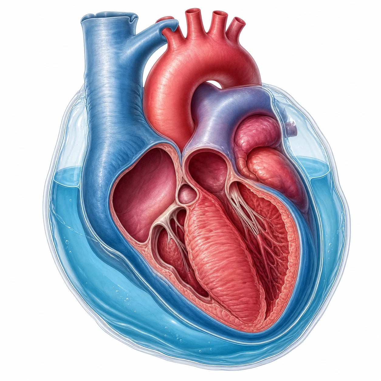
The pericardium is a relatively non-compliant sac, and once the intrapericardial volume exceeds the small reserve capacity, a further small rise in volume produces a steep rise in the intrapericardial pressure. The raised pressure compresses the cardiac chambers in diastole, limits the filling, and lowers the stroke volume and the cardiac output; the heart compensates initially with a tachycardia and a vasoconstriction, and decompensates when these are exhausted. The crucial consequence is that tamponade is a preload-limited state — the heart depends on a high venous return to fill against the raised intrapericardial pressure. This is why a fluid bolus temporises (it raises the venous return) and why a nitrate, a diuretic, or a deep anaesthesia collapses the cardiac output by dropping the preload. The pulsus paradoxus — an exaggerated inspiratory fall in the systolic blood pressure of more than 10 mmHg — reflects the enhanced interventricular dependence and is a classic sign. [1]
The pericardial pressure–volume curve and the three phases
The behaviour of tamponade is governed by the pericardial pressure–volume curve, which has a long shallow initial limb and then a steep, near-vertical terminal limb once the pericardial reserve is exhausted. Three haemodynamic phases follow. In phase 1 the intrapericardial pressure is still below the right-atrial and right-ventricular diastolic pressures, so filling is preserved and the patient is asymptomatic. In phase 2 the intrapericardial pressure rises to equal and then exceed the right-sided filling pressures; the right atrium and right ventricle begin to collapse in diastole and the cardiac output falls, but the systemic venous pressure rises to defend the filling. In phase 3 the intrapericardial pressure equals or exceeds all four chamber diastolic pressures — equalisation of diastolic pressures — and the heart is effectively empty in diastole; the stroke volume collapses and shock supervenes. The clinical implication is that removal of even a small volume of fluid (50 to 100 mL) can drop the intrapericardial pressure off the steep part of the curve and produce a dramatic improvement, while the addition of a similarly small volume can be lethal.[3]
The compensatory tachycardia and the vicious circle
As the stroke volume falls the sympathetic nervous system drives a tachycardia and a systemic vasoconstriction to preserve the cardiac output and the blood pressure; this compensation masks the underlying fall in the stroke volume and explains why the tachycardia may be the only early sign. The compensation fails when the heart rate can no longer rise, when the venous return cannot be raised further, or when a vasopressor or an afterload stress is added; the result is a precipitous circulatory collapse, frequently an arrest in pulseless electrical activity. A patient who is "compensating" on the tachycardia alone is one bolus of a vasodilator, one dose of positive-pressure ventilation, or one deepening of sedation away from cardiovascular collapse. [1]
[1]Interventricular dependence and pulsus paradoxus
In normal inspiration the intrathoracic pressure falls, the systemic venous return to the right heart rises, and the right ventricle expands; because the two ventricles share the interventricular septum and the pericardial constraint, the left ventricular filling falls slightly and the systolic blood pressure drops by up to 10 mmHg. In tamponade the heart is constrained within a fixed pericardial volume, so the inspiratory augmentation of the right-sided filling crowds the left side far more than normal, producing an exaggerated inspiratory fall in the systolic blood pressure of more than 10 mmHg — the pulsus paradoxus. It is measured with a sphygmomanometer by slowly deflating the cuff and noting the two pressures at which the Korotkoff sounds are heard only in expiration (first appearance) and then throughout the cycle (the true systolic). The absolute tamponade threshold is debated, but a fall of more than 10 mmHg is the classical cut-off, and a fall of more than 20 mmHg suggests severe tamponade. Pulsus paradoxus is absent in a left-ventricular dysfunction (the stiff ventricle does not receive the inspiratory volume), in a tamponade with an atrial septal defect (the two atria share the inflow and the septal shift is abolished), in severe aortic regurgitation, and in regional (loculated) post-surgical tamponade — its absence does not exclude the diagnosis. [1]
The Kussmaul sign
The Kussmaul sign — a paradoxical rise in the jugular venous pressure on inspiration — is classically associated with a constrictive pericarditis or a restrictive cardiomyopathy and a right-ventricular infarction, in which the stiff right heart cannot accommodate the inspiratory venous return. It is uncommon in a classical effusive tamponade (in which the venous pressure is high but fixed, and may even fall slightly with inspiration) but can appear in a low-pressure or a regional/loculated tamponade and in the post-operative tamponade with a concomitant constriction (an effusive-constrictive picture). The Fellowship point is that Kussmaul points away from a simple tamponade and toward a constriction or a restrictive physiology, and that the two can coexist. [1]
Why low-pressure tamponade is missed
Low-pressure tamponade[3] is the haemodynamically significant compression that occurs at a normal or only mildly raised venous pressure, typically in a hypovolaemic or a heavily diuresed patient with a moderate effusion. The classical Beck triad is absent (the venous pressure is not high), the pulsus paradoxus may be subtle, and the diagnosis rests on the echo (chamber collapse with a plethoric but smaller IVC, and the resolution with a fluid challenge). It is the tamponade of the dialysis patient, the over-diuresed heart-failure patient, and the cancer patient with cachexia, and the first step in its management is often a fluid challenge to raise the venous pressure and unmask — or treat — the compression. A fluid bolus that improves a shocked patient with an effusion is the clue to a low-pressure tamponade.
Causes
The causes span trauma and medicine. The acute causes are a penetrating chest injury, a cardiac procedure (a catheter, a pacemaker, an ablation), a cardiac surgery, and an aortic dissection. The subacute causes are a malignancy (lung, breast, lymphoma), a uraemia, an infection (viral, tuberculous, bacterial), an autoimmune or an idiopathic pericarditis, a radiation, and a free-wall rupture of a myocardial infarction. Identifying the cause is essential because it chooses the drainage and the definitive treatment. [1]
A free-wall rupture of an infarcted ventricle, typically three to seven days after a large transmural infarction, is a catastrophic cause that presents as a sudden tamponade with an electromechanical dissociation and is managed by an emergency surgical repair; it is one of the mechanical complications a Fellowship candidate must consider when a post-infarction patient suddenly collapses. A bleeding diathesis or a recent anticoagulation raises the risk of an iatrogenic or a spontaneous haemopericardium, and the anticoagulated patient with a falling blood pressure and a rising venous pressure has a tamponade until the echo proves otherwise. [1]
The aetiology at a glance
The Fellowship candidate should know the relative frequency of the causes, because it shapes the differential and the definitive management. The contemporary series place malignancy as the single commonest cause of a medical tamponade (lung, breast, lymphoma, leukaemia, melanoma), followed by uraemia (especially in dialysis-dependent or oliguric patients), viral and idiopathic pericarditis, tuberculosis (the dominant cause in endemic regions), post-myocardial-infarction (an early pericarditis, a Dressler syndrome, or a catastrophic free-wall rupture), post-cardiac-surgery and post-procedural (catheter, pacemaker, ablation, biopsy), aortic dissection with rupture into the pericardium, blunt or penetrating trauma, and radiation (typically a late effect, years after mediastinal radiotherapy, often with a superimposed constriction).[1][7][11]
Malignancy (#1 medical)
- Lung, breast, lymphoma, leukaemia, melanoma
- Often large, painless, subacute effusion
- May need a pericardial window + systemic therapy
- Cytology positive in >80%
Uraemia
- Dialysis or AKI patients; haemorrhagic effusion
- Treat with intensive dialysis + drainage
- Often coagulopathic — correct before procedure
- High recurrence; consider window
Viral / idiopathic
- Coxsackie, echovirus, influenza; mostly idiopathic
- Preceded by a viral prodrome and pleuritic chest pain
- Colchicine ± NSAID; drain if tamponaded
- Best prognosis of the medical causes
Tuberculosis
- Endemic regions; large bloody or serosanguinous effusion
- Send fluid for AFB, GeneXpert, adenosine deaminase
- Anti-TB therapy; window if recurrent
- May evolve into constrictive pericarditis
Post-MI
- Early pericarditis (1–3 days), Dressler (2–10 weeks)
- Free-wall rupture (3–7 days) = tamponade + EMD arrest
- Rupture needs emergency surgery; pericarditis = colchicine
- Avoid anticoagulation in early post-MI pericarditis
Post-surgery / iatrogenic
- Catheter, pacemaker, ablation, biopsy, valve/CABG
- Post-cardiac-surgery = loculated, often clotted, asymmetric
- Echo from multiple windows; needs surgical re-exploration
- COPPS-2: colchicine reduces post-pericardiotomy syndrome
Aortic dissection
- Type A rupture into pericardium = haemopericardium
- NEVER drain unless arresting — re-bleeds; surgery only
- Look for wide mediastinum, BP differential, tearing pain
- CT angio if stable; echo if not
Trauma
- Penetrating cardiac injury = clotted haemopericardium
- Needle will NOT aspirate clot — resuscitative thoracotomy
- Blunt — rare; myocardial contusion or rupture
- Beck triad in a stab wound = thoracotomy, not needle
Radiation
- Years after mediastinal radiotherapy (Hodgkin, breast)
- Often combined effusive-constrictive physiology
- Refractory to drainage — needs pericardiectomy
- Screen for concomitant coronary and valve disease
Iatrogenic and procedural causes — the rising modern threat
The iatrogenic tamponade is now one of the commonest acute presentations, driven by the volume of percutaneous cardiac procedures. A coronary catheterisation, a structural-heart intervention (TAVR, mitral clip, PFO/ASD closure), an electrophysiological ablation, a pacemaker or defibrillator lead, an endomyocardial biopsy, and a central venous catheter can all perforate the heart or a great vessel and produce a rapid, life-threatening haemopericardium. The Mayo Clinic experience established that an echo-guided rescue pericardiocentesis is the definitive management for the catheter-perforation tamponade and is life-saving in the majority.[4] The clinical clue is any haemodynamic deterioration during or soon after a cardiac procedure — tachycardia, hypotension, a raised venous pressure, or a chest pain — and the response is an immediate bedside echo, not a transfer to the CT scanner.
Clinical presentation
The classic presentation is Beck's triad — hypotension, a distended jugular venous pulse, and muffled or quiet heart sounds — together with a tachycardia, a dyspnoea and a hypoxia, and a pulsus paradoxus. The patient is anxious, pale and collapsed, with a high venous pressure that distinguishes tamponade from the more common low-pressure shock. A subacute tamponade may present more subtly, as fatigue, effort intolerance, and a rising dyspnoea over weeks, before a precipitous deterioration. [1]
The full clinical picture — signs beyond Beck
The classical Beck triad (hypotension, a raised JVP, muffled heart sounds) is a late and insensitive sign; it is present in only a minority of tamponades, and the modern candidate should regard it as a "when you see all three, the patient is about to arrest" sign rather than a screening tool. The earlier and more reliable findings are a tachycardia, a raised jugular venous pressure with a prominent x descent and an absent y descent (the right atrium cannot relax and fill in early diastole because it is compressed), a pulsus paradoxus, a dyspnoea and tachypnoea, and the constellation of shock with clear lung fields. The muffled heart sounds are best heard with the diaphragm at the left sternal edge with the patient sitting forward, and reflect the dampening of sound by the fluid; their absence does not exclude tamponade. A pericardial friction rub, when present, suggests an inflammatory or a viral/idiopathic cause rather than a malignancy, and it may disappear as the effusion enlarges. [1]
The jugular venous waveform
In tamponade the right atrium is compressed throughout the cardiac cycle except during ventricular systole, so the venous waveform shows a prominent x descent (the systolic fall, when the tricuspid valve is pulled down and the atrium briefly decompresses) and an attenuated or absent y descent (the early-diastolic fall is abolished because the atrium cannot empty into the compressed ventricle). This x-descent-dominant pattern, with a fixed and high venous pressure, is the haemodynamic signature of tamponade and distinguishes it from constriction (in which a steep x and steep y descent are seen). A Fellowship candidate asked to draw the JVP waveform in tamponade should show a high mean pressure, a preserved x descent, and a blunted y descent. [1]
Differential diagnosis
The combination of shock with a raised venous pressure has a short, dangerous differential, and the bedside ultrasound and the clinical signs resolve it. [1]
Pericardial tamponade
- Shock + raised JVP + clear lungs + muffled/quiet heart
- Echo: chamber collapse, plethoric IVC, swinging heart
- Pulsus paradoxus >10 mmHg
- Drainage: pericardiocentesis / surgery
Tension pneumothorax
- Shock + raised JVP + absent breath sounds + hyperresonance
- Tracheal deviation; clinical diagnosis
- Echo: no effusion; may show RV collapse from pressure
- Needle decompression, not drainage of the heart
Massive PE
- Shock + raised JVP; DVT, risk factors
- Echo: RV strain (McConnell), no pericardial fluid
- CXR normal; CTPA if time
- Thrombolysis / embolectomy
Constrictive pericarditis
- Chronic, not acute; raised JVP + Kussmaul + pericardial knock
- Calcified pericardium (chronic)
- A chronic filling-limit disease
- Surgical pericardiectomy
Bedside assessment
Look for the Beck triad and the pulsus paradoxus; feel for a high venous pressure with clear lung fields and a quiet precordium; and obtain the bedside echocardiogram at once — it is the diagnostic test, showing a right-atrial and a right-ventricular diastolic collapse, a plethoric (non-collapsing) inferior vena cava, and, in the effusive tamponade, a swinging heart. The cause is sought from the history (the trauma, the procedure, the cancer, the dialysis, the recent infarct). [1]
Echo assessment of tamponade — the protocol
The focused bedside echocardiogram is both the screening and the confirmatory test, and it should be obtained within minutes of the suspicion. The sequence below is the protocol a Fellowship candidate should be able to perform and describe.[8]
Bedside echo assessment of tamponade
Subcostal long-axis view — the fastest window in a shocked patient; look for the effusion as an echo-free space around the right ventricle and the right atrium
Subcostal IVC view — measure the IVC for plethora (>2.1 cm) and the absence of the inspiratory collapse (>50% collapse is normal); a plethoric fixed IVC reflects the raised right-atrial pressure
Parasternal long-axis — quantify the effusion size (small under 1 cm, moderate 1–2 cm, large over 2 cm at end-diastole) and look for the right-ventricular outflow collapse in early diastole
Parasternal short-axis and apical four-chamber — look for the right-atrial collapse in late diastole (the earliest sign, >one-third of the cardiac cycle is specific) and the right-ventricular free-wall collapse in early diastole
Look for the swinging heart — the heart oscillating within a large effusion, which produces the electrical alternans on the ECG
Assess the left ventricle — a small, hyperdynamic, underfilled LV; exclude a concomitant LV failure or a pulmonary hypertension
Doppler (if available) — an exaggerated inspiratory variation of the mitral (>25%) and the tricuspid (>40%) inflow velocities reflects the enhanced interventricular dependence
Agitated-saline contrast — inject through a peripheral line to confirm the intrapericardial needle position during pericardiocentesis
Investigations and the targets
The bedside echocardiogram is the diagnostic test in the unstable patient and shows the effusion, the chamber collapse, the plethoric inferior vena cava and the swinging heart. The ECG may show a low voltage and an electrical alternans (a beat-to-beat variation in the QRS amplitude from the swinging heart), but these are non-specific. The chest radiograph shows an enlarged globular cardiac silhouette in the subacute effusive tamponade and is normal in the acute haemorrhagic one. The arterial blood gas shows the hypoxia and the metabolic or lactic acidosis of the shocked state. The Beck triad and the pulsus-paradoxus threshold of 10 mmHg are reproduced because they are examined. [1]
The ECG in tamponade — three patterns
The ECG is non-diagnostic but supports the clinical picture. Electrical alternans — a beat-to-beat variation in the QRS amplitude and, less commonly, the P-wave and the ST-segment — is produced by the heart swinging within a large effusion; it is highly specific but insensitive (seen in perhaps 20% of tamponades, mostly the large malignant effusions) and resolves immediately after drainage.[10] Low QRS voltage (limb leads <5 mm, precordial <10 mm) reflects the dampening of the electrical signal by the surrounding fluid and is also non-specific. PR-segment depression and diffuse concave ST elevation point to the underlying pericarditis rather than to the tamponade itself. A sinus tachycardia is near-universal; a bradycardia in a tamponade suggests a pre-existing conduction disease, a drug effect, or a terminal agonal rhythm. Pulseless electrical activity (PEA) in a patient with an effusion is a pre-arrest tamponade until proven otherwise, and the treatment is an emergency drainage, not a drug round.
[1]Immediate management — temporise, then drain
Stabilise the airway and the breathing with oxygen, give a cautious 250 to 500 mL bolus of a balanced crystalloid to raise the venous return against the compressed heart (a temporising measure), and avoid any preload-lowering drug. Use a vasopressor (noradrenaline 0.05 to 0.5 micrograms per kilogram per minute) as a bridge to drainage while the team is mobilised. [1]
Vasopressor and the ventilated patient
A pure alpha agonist such as noradrenaline (0.05–0.5 micrograms/kg/min) is the vasopressor of choice as a bridge to drainage, because it raises the systemic vascular resistance and the venous return (it is a venoconstrictor) without the vasodilatory beta-2 effect of adrenaline. Adrenaline (1–10 micrograms/min, or 0.05–0.5 micrograms/kg/min) is used in the arresting or the profoundly shocked patient. Vasopressin is an alternative that preserves the coronary and the renal perfusion. Avoid the pure vasodilators (glyceryl trinitrate, sodium nitroprusside), the venodilating opiates in large doses, and the deepening of sedation, all of which drop the preload and collapse the output. In the intubated patient, use the lowest possible mean airway pressure, avoid high PEEP, and accept a permissive hypercapnia — the positive-pressure ventilation is itself a preload-lowering intervention. [1]
[1]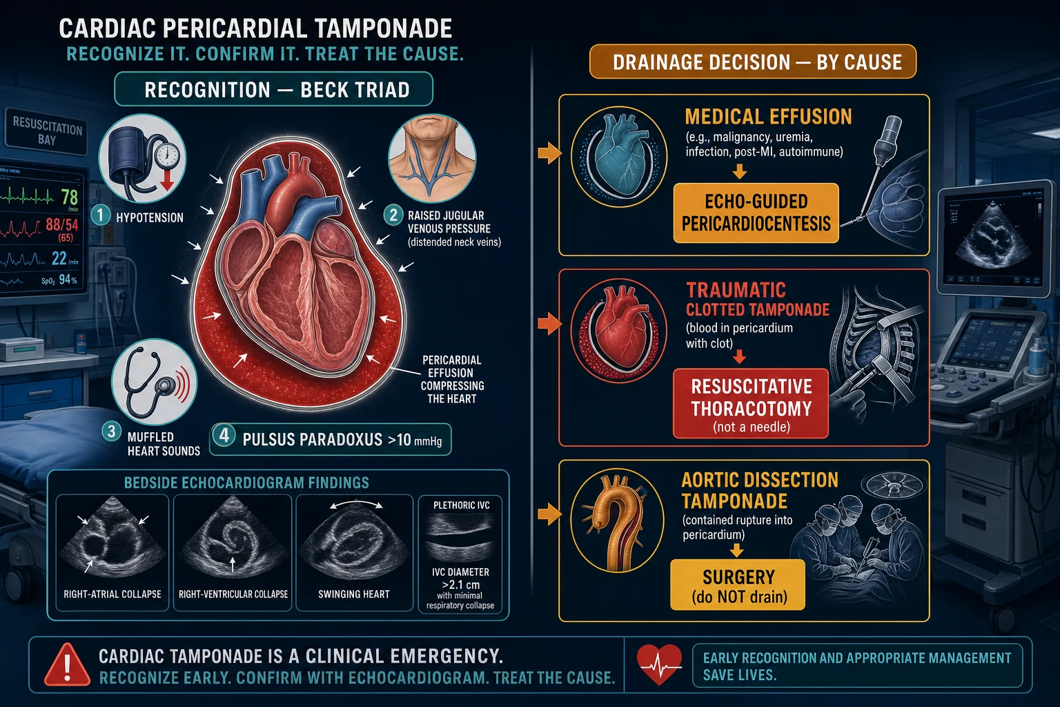
Tamponade signs and the drainage
The echo-guided pericardiocentesis is performed under strict asepsis with local anaesthesia — lidocaine 1 per cent, with adrenaline 1:200,000 to a maximum of about 3 mg per kilogram, or 7 mg per kilogram without adrenaline — infiltrated along the subxiphoid track. The needle is advanced from just below and to the left of the xiphisternum, aimed toward the left shoulder at a shallow angle of about 30 degrees, under continuous echocardiographic guidance (with agitated-saline contrast to confirm the intrapericardial position if needed); on reaching the effusion a guidewire is passed, a multi-side-hole catheter is inserted over the wire by a Seldinger technique, and the effusion is aspirated to dry. A drain is left in situ for ongoing drainage and to monitor a re-accumulation. The fluid is sent for the cytology, the microbiology and the biochemistry, which often reveal the underlying malignancy or the infection. The complications are a puncture of the right ventricle or a coronary vessel, a pneumothorax, and a re-accumulation; the echo guidance and the left-in drain reduce these. A surgical pericardial window is offered for the recurrent or the loculated effusion (often a malignancy) and provides tissue for the diagnosis. Over-aspiration of a large chronic effusion is avoided in the first instance, because a too-rapid removal can provoke an acute pulmonary oedema of the suddenly decompressed heart — analogous to the re-expansion oedema of a drained pneumothorax. [1]
Echo-guided pericardiocentesis — the procedure
The technique has evolved from a blind "blind-stick" to a real-time echo-monitored procedure, which has reduced the major complication rate from over 20% to under 2% and made outpatient drainage feasible for the stable, selected effusion.[8][9][5] The Fellowship candidate should know the steps and the local-anaesthetic doses.
Echo-guided subxiphoid pericardiocentesis
Position the patient semi-recumbent at 30–45°; attach full monitoring (ECG, BP, pulse oximetry); ensure a defibrillator and a crash team are available
Identify the largest, closest pocket of fluid from the subcostal window and mark the entry site and the angle (often subxiphoid, or parasternal/apical where the pocket is largest)
Prepare a sterile field; infiltrate 1% lidocaine with adrenaline 1:200,000 (max 3 mg/kg, or 7 mg/kg plain) along the planned track down to the pericardial border
Advance an 18-gauge (or longer) needle under continuous echo monitoring, aiming from just below and left of the xiphisternum toward the left shoulder at ~30°; on entering the effusion, aspirate gently
Confirm intrapericardial position with agitated-saline contrast (microbubbles opacify the pericardial space, not the ventricle) or by passing a guidewire under echo
Pass a guidewire (Seldinger); dilate the track; insert a multi-side-hole pigtail catheter over the wire
Aspirate the effusion to dry (send for biochemistry, cell count, cytology, microbiology including AFB/GeneXpert); leave the catheter on free drainage
Reassess the haemodynamics and the echo immediately — the blood pressure should rise and the venous pressure fall as the intrapericardial pressure drops off the steep curve
Secure the drain; arrange a pericardial window or definitive management for the underlying cause; remove the drain when the output is under 50 mL/24 h
Tsang et al. — Mayo Clinic rescue pericardiocentesis
PMID 9809946
Retrospective case series, tertiary centre
Population: Cardiac perforation complicating catheter-based procedures
Key finding
Echo-guided pericardiocentesis was life-saving in the majority; surgical rescue needed in a minority. Established echo-guidance as the standard for iatrogenic perforation.
Practice change
Echo-guided pericardiocentesis is the first-line rescue for catheter-induced tamponade; reserve surgery for failure or anatomic unsuitability.
Imazio et al. — COPPS-2 trial
PMID 25172965
Randomised, placebo-controlled, multicentre
Population: Patients undergoing cardiac surgery (CABG, valve)
Key finding
Colchicine (1 mg/day) starting post-op day 3 reduced the post-pericardiotomy syndrome (relative reduction ~40%) and post-operative atrial fibrillation. GI intolerance was the main side effect.
Practice change
Colchicine prophylaxis reduces the inflammatory post-surgical effusion — a common precursor of a post-cardiac-surgery tamponade.
Sagristà-Sauleda et al. — Low-pressure tamponade
PMID 16923755
Prospective haemodynamic series
Population: Patients with tamponade and a normal/slightly raised venous pressure
Key finding
Defined low-pressure tamponade as the haemodynamically significant compression with a right-atrial pressure under 10 mmHg; classically in hypovolaemic/dialysis patients. Responds to a fluid challenge and drainage.
Practice change
A normal venous pressure does not exclude tamponade — suspect it in the cachectic, dialysis, or over-diuresed patient with an effusion and unexplained shock.
Complications and pitfalls
The complication of the untreated tamponade is a cardiac arrest; the complications of the drainage are the procedural injury and the re-accumulation. The pitfalls are the inverse of the management: draining an aortic-dissection tamponade (re-bleed); attempting pericardiocentesis on a traumatic clotted tamponade (ineffective — needs a thoracotomy); giving a nitrate or a diuretic (collapses the preload); waiting for a chest radiograph or a CT in the unstable patient; and missing the underlying cause, which chooses the definitive treatment. [1]
Prognosis and disposition
Untreated, a tamponade is fatal; the drainage is curative of the immediate threat, and the survival after an effective drainage is high. The patient is admitted to the intensive care or the cardiology unit, or directly to the theatre for the surgical cases; the underlying cause is treated (the malignancy, the infection, the uraemia, the dissection, the free-wall rupture); and a pericardial window is offered for the recurrence. The long-term outlook turns on that underlying cause — a benign idiopathic effusion does well, whereas a malignant effusion carries a poor prognosis despite the successful drainage. [1]
Special populations
The cancer patient with a malignant effusion often needs a pericardial window and an oncological management. The uraemic patient on dialysis is managed with dialysis and a drainage. The trauma patient with a penetrating injury and a clotted tamponade is taken to a resuscitative thoracotomy. The post-cardiac-surgery patient has a loculated, often-clotted tamponade that needs surgical reopening. [1]
Post-cardiac-surgery and regional tamponade
The post-cardiac-surgery tamponade is the great mimic of the intensive-care unit. It is frequently loculated (clotted behind the right atrium or the left atrium, rather than circumferential), the Beck triad is absent, the pulsus paradoxus is absent (the localised compression does not produce the global interventricular dependence), and the classical circumferential effusion on the echo may be missed. The diagnosis rests on a high index of suspicion — a rising lactate, a falling urine output, a rising filling pressure, a low cardiac output, or an unexplained tachycardia — in any patient in the first post-operative week. The echo must be taken from multiple windows (subcostal, apical, parasternal) and may require a transoesophageal echo; the management is a surgical re-exploration, not a percutaneous tap, because the clot will not aspirate.[6]
[1]The cancer patient and the malignant effusion
A malignant pericardial effusion is the commonest medical cause of tamponade in the developed world; lung cancer, breast cancer, lymphoma and leukaemia predominate. The effusion is typically large, painless and subacute, and may be the presenting feature of the malignancy. The cytology of the pericardial fluid is positive in over 80%, and the histology of a pericardial window confirms the diagnosis. The management is a drainage (pericardiocentesis or a window) followed by the systemic oncological treatment; a pericardial window is preferred for the recurrent malignant effusion because it reduces the re-accumulation and provides tissue.[11] The prognosis is guarded and turns on the underlying tumour — a hormone-responsive breast cancer may do well, whereas a metastatic lung cancer carries a median survival of months.
Pregnancy and tamponade
Tamponade in pregnancy is rare but carries a high maternal and foetal mortality if missed. The causes are the same as in the non-pregnant patient, with an increased representation of peripartum cardiomyopathy complications, pre-eclampsia-related haemorrhage, and iatrogenic procedural causes. The physiological tachycardia and the elevated venous pressure of the third trimester can mask the early signs, and the supine hypotensive syndrome can be confused with the tamponade. The principle is to drain early and drain before delivery if the mother is unstable; a pericardiocentesis under echo guidance is safe in pregnancy, and the foetal distress usually resolves once the maternal circulation is restored. A multidisciplinary approach (cardiology, obstetrics, anaesthetics, neonatology) is essential. [1]
Paediatric tamponade
In children the causes are viral or idiopathic pericarditis, a post-cardiac-surgery effusion (especially after congenital repair), a central-line perforation, a malignancy (lymphoma), and a Kawasaki or an autoimmune disease. The presentation is non-specific — irritability, tachycardia, tachypnoea, a hepatomegaly, and a poor perfusion — and the Beck triad is rarely present. The pulsus paradoxus is harder to elicit. The bedside echo is the diagnostic test, and the pericardiocentesis is performed under echo guidance, usually by a paediatric cardiologist. The doses of the local anaesthetic must be weight-adjusted (lidocaine max 3 mg/kg with adrenaline, 7 mg/kg plain) to avoid toxicity in the small child. [1]
Evidence and regional guidelines
The contemporary framework is the 2015 ESC pericardial-disease guideline[1] and the focused reviews of the complex pericardial case.[2] — supplemented by recent reviews of the inflammatory pericardial diseases that contextualise the viral and the idiopathic effusions.[12] The drainage approach is global; the pericardiocentesis-vs-surgery and the resuscitative-thoracotomy criteria are governed by the local cardiology and the trauma protocols.
ANZ practice note. The drainage approach follows the ESC framework via the local cardiology and cardiothoracic service; a non-traumatic effusion is echo-guided pericardiocentesis, a traumatic clotted tamponade is a resuscitative thoracotomy per the ATLS/ANZ trauma protocol, and an aortic-dissection tamponade goes to cardiothoracic surgery. [1]
Exam practice
SAQ — Cardiac tamponade from a malignant pericardial effusion
12 minutes · 10 marks
A 62-year-old woman with metastatic lung cancer presents with five days of progressive dyspnoea, orthopnoea and dizziness. She is diaphoretic, breathless and unable to lie flat. T 37.0, HR 128, BP 86/58 with a pulsus paradoxus of 18 mmHg, JVP distended to the angle of the jaw at 45 degrees, muffled heart sounds, clear lung fields, cool peripheries. SpO2 92 per cent on room air. GCS 15 but increasingly drowsy. Bedside echocardiography shows a large circumferential effusion with right atrial collapse in late diastole, RV diastolic collapse, a plethoric fixed IVC and a swinging heart.
SAQ — Traumatic cardiac tamponade and the drainage decision
10 minutes · 10 marks
A 24-year-old man is brought to the emergency department after a stab wound to the left chest, 2 cm from the sternal edge at the fourth intercostal space. He is agitated, pale and diaphoretic. T 36.4, HR 132, BP 78/50 with a pulsus paradoxus of 15 mmHg, JVP distended, muffled heart sounds. Bedside focused echo shows a pericardial effusion with clotted blood, RV diastolic collapse and a plethoric IVC.
Exam pearls
- Beck's triad: hypotension, raised JVP, muffled heart sounds — a late, insensitive triad; the modern candidate recognises the earlier signs (tachycardia, raised JVP with prominent x and absent y descent, pulsus paradoxus, shock with clear lungs).
- Pulsus paradoxus = an inspiratory fall in systolic BP >10 mmHg; >20 mmHg suggests severe tamponade. Absent in LV failure, ASD, severe aortic regurgitation, and regional/loculated tamponade — its absence does not exclude the diagnosis.
- The JVP waveform: high mean pressure, a prominent x descent and an absent/attenuated y descent (the atrium cannot empty into the compressed ventricle). Distinguishes tamponade from constriction (steep x and y).
- Kussmaul sign (a paradoxical inspiratory rise in JVP) points away from a simple tamponade and toward a constriction or a restrictive physiology — but can appear in low-pressure or effusive-constrictive disease.
- The bedside echo is the diagnostic test: chamber collapse (RA late diastole >⅓ cycle = most specific; RV diastolic = most sensitive), a plethoric fixed IVC, and the swinging heart. Do not wait for a CXR or a CT in the unstable patient.
- Electrical alternans on the ECG (beat-to-beat variation in QRS amplitude from the swinging heart) is specific but insensitive (~20%, mostly large malignant effusions); low voltage is non-specific. PEA + effusion = drain now.
- A fluid bolus temporises (preload-limited); never a nitrate, a diuretic, or a deep sedation — they collapse the output. Noradrenaline is the vasopressor of choice (venoconstrictor + alpha agonist).
- Equalisation of diastolic pressures is the haemodynamic definition of severe tamponade — the intrapericardial pressure exceeds all four chamber pressures.
- Malignancy is the #1 medical cause (lung, breast, lymphoma); others are uraemia, viral/idiopathic, TB, post-MI, post-surgery/procedural, aortic dissection, trauma, radiation.
- The drainage turns on the cause: medical effusion → echo-guided pericardiocentesis; traumatic clotted → resuscitative thoracotomy (clot won't aspirate); aortic dissection → surgery (do not drain unless arresting — re-bleeds); post-surgical loculated → surgical re-exploration.
- Drain before you intubate — positive-pressure ventilation drops the preload and can precipitate an arrest in a borderline tamponade.
- Pericardiocentesis technique: subxiphoid, echo-monitored, 1% lidocaine with adrenaline 1:200,000 (max 3 mg/kg, or 7 mg/kg plain), 18-gauge needle toward the left shoulder at ~30°, guidewire Seldinger, pigtail catheter, aspirate to dry, agitated-saline to confirm position.
- Low-pressure tamponade (normal/mildly raised JVP in a hypovolaemic/dialysis/cachectic patient) is easily missed — the Beck triad is absent; suspect it, give a fluid challenge, and look for chamber collapse on the echo.
- Surgical pericardial window for the recurrent (malignant/uraemic), loculated, or tissue-diagnosis effusion; it reduces recurrence and provides histology.
- Post-cardiac-surgery tamponade is the great mimic — loculated, no Beck triad, no pulsus paradoxus; suspect it in the post-op patient who fails to wean from inotropes or re-develops a low-output state; needs a transoesophageal echo and a surgical re-exploration. [1]
Red flags
[1] [1]References
- [1]Charron P, Adler Y. 'Ten Commandments' of 2015 ESC Guidelines for diagnosis and management of pericardial diseases Eur Heart J, 2015.PMID 26866075
- [2]Marchetta M, Adamo L, Bonaventura A, et al. ABCDE of complex pericarditis cases Heart, 2026.PMID 42045051
- [3]Sagristà-Sauleda J, Angel J, Sambola A, Permanyer-Miralda G. Low-pressure cardiac tamponade: clinical and hemodynamic profile Circulation, 2006.PMID 16923755
- [4]Tsang TS, Freeman WK, Barnes ME, et al. Rescue echocardiographically guided pericardiocentesis for cardiac perforation complicating catheter-based procedures. The Mayo Clinic experience J Am Coll Cardiol, 1998.PMID 9809946
- [5]Drummond JB, Seward JB, Tsang TS, et al. Outpatient two-dimensional echocardiography-guided pericardiocentesis J Am Soc Echocardiogr, 1998.PMID 9619614
- [6]Imazio M, Brucato A, Ferrazzi P, et al. Colchicine for prevention of postpericardiotomy syndrome and postoperative atrial fibrillation: the COPPS-2 randomized clinical trial JAMA, 2014.PMID 25172965
- [7]Imazio M, Lazaros G, Valenti A, et al. Outcomes of idiopathic chronic large pericardial effusion Heart, 2019.PMID 30274986
- [8]Maggiolini S, Gentile G, Farina A, et al. Safety, Efficacy, and Complications of Pericardiocentesis by Real-Time Echo-Monitored Procedure Am J Cardiol, 2016.PMID 26956635
- [9]Maggiolini S, De Carlini CC, Imazio M. Evolution of the pericardiocentesis technique J Cardiovasc Med (Hagerstown), 2018.PMID 29553993
- [10]Khalid N, Chhabra L, Spodick DH. Response to Role of a 12-Lead Electrocardiogram in the Diagnosis of Cardiac Tamponade as Diagnosed by Transthoracic Echocardiography in Patients With Malignant Pericardial Effusion Clin Cardiol, 2015.PMID 26216007
- [11]Avondo S, Andreis A, Casula M, et al. Update on diagnosis and management of neoplastic pericardial disease Expert Rev Cardiovasc Ther, 2020.PMID 32797759
- [12]Bizzi E, Picchi C, Mastrangelo G, et al. Recent advances in pericarditis Eur J Intern Med, 2022.PMID 34556390