EM · Pneumothorax
Pneumothorax (including tension pneumothorax)
Also known as Spontaneous pneumothorax · Primary pneumothorax · Tension pneumothorax
Pneumothorax — primary and secondary spontaneous, traumatic, iatrogenic, and tension. The one-way-valve pathophysiology of tension and why it kills, the clinical (not radiological) diagnosis of tension, the 5th-intercostal-space decompression site, the size-based management of a spontaneous pneumothorax (observe, aspirate, drain), the chest-drain landmarks and the underwater seal, the re-expansion-oedema pitfall, and the bulla-mimic trap. ACEM-primary, globally tagged.
On this page & tools
Your progress
Saved locally on this device.
Target exams
Red flags
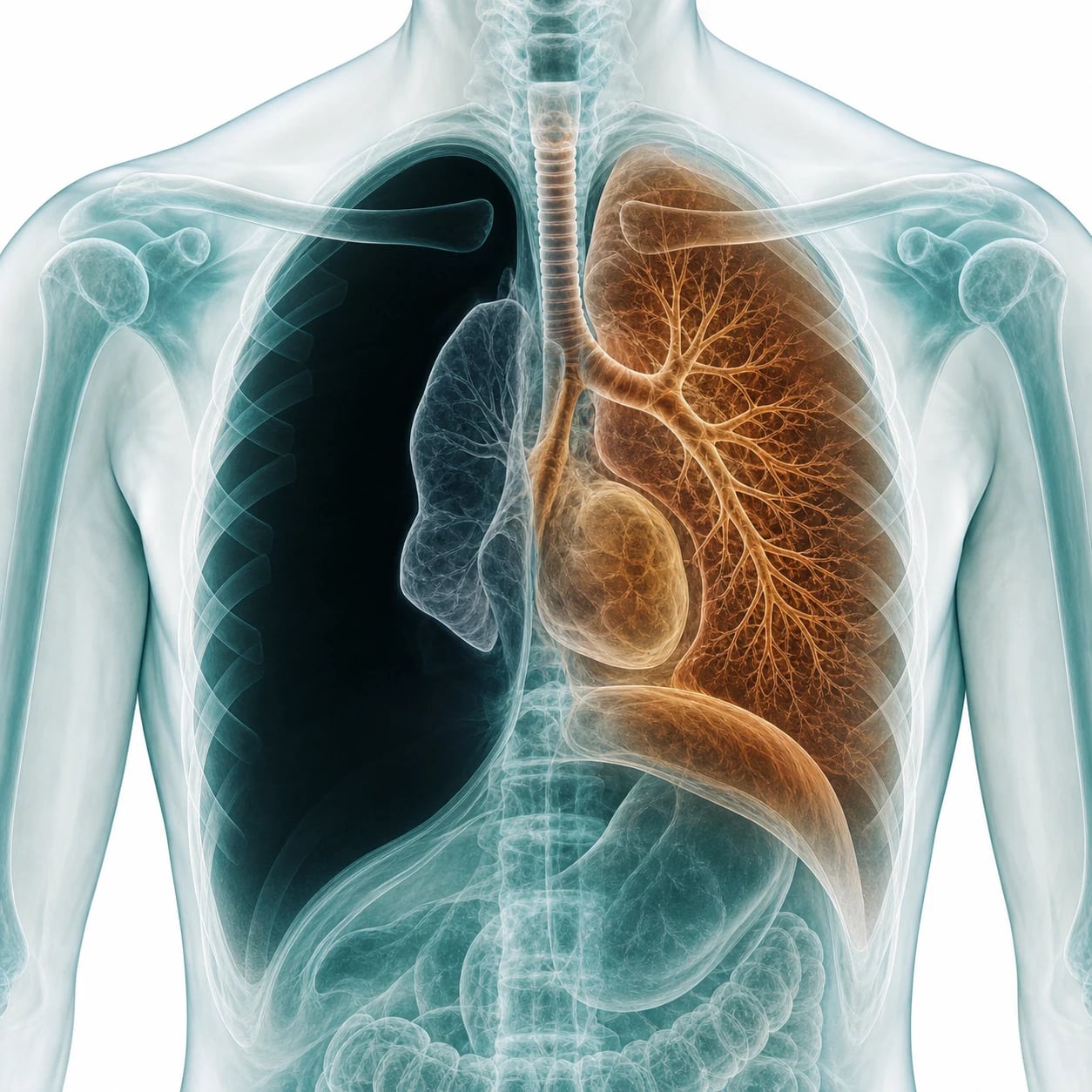
A pneumothorax is air in the pleural space, and although most are simple, one form — tension pneumothorax — is among the few genuinely immediately fatal emergencies in medicine, killed by the one-way-valve physiology that traps air in the chest until it obstructs venous return and the circulation arrests. The Fellowship candidate must recognise tension as a clinical diagnosis and decompress before any imaging, then apply the size-based management to the spontaneous pneumothorax and the correct landmarks to the chest drain.[1]
Definition and classification
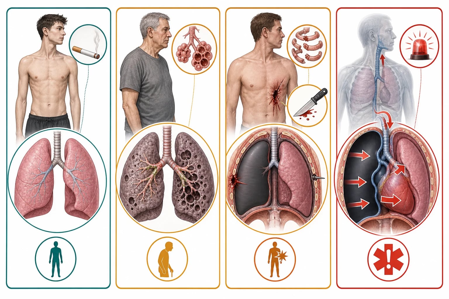
A pneumothorax is air in the pleural space between the visceral and parietal pleura, which collapses the lung toward the hilum. It is classified by cause. A primary spontaneous pneumothorax arises in a patient with no underlying lung disease — typically a tall, thin, young, male smoker, occasionally with a family history or a connective-tissue disorder such as Marfan syndrome. A secondary spontaneous pneumothorax complicates underlying lung disease, most commonly chronic obstructive pulmonary disease, but also asthma, cystic fibrosis, interstitial lung disease, lymphangioleiomyomatosis, and Pneumocystis pneumonia. A traumatic pneumothorax follows blunt or penetrating injury and an iatrogenic one follows a procedure (a central venous catheter, a biopsy, positive-pressure ventilation). The lethal form is tension pneumothorax, in which a one-way valve traps air on each inspiration. [1]
The four broad categories have distinct demographics, mechanisms, and risks, and the table below frames the comparison the examiner expects. [1]
Primary spontaneous (PSP)
- No underlying lung disease; tall, thin, young male (M:F ≈ 3:1), 15–34 yr, smoker (relative risk ×9)
- Ruptured apical subpleural bleb/bulla; ± Marfan, homocystinuria, α₁-antitrypsin, Birt-Hogg-Dubé, family history
- Sudden pleuritic pain ± dyspnoea; usually haemodynamically stable
- Manage by size: observe / aspirate / drain; consider VATS after 2nd event
Secondary spontaneous (SSP)
- Underlying lung disease: COPD (commonest), asthma, CF, ILD, LAM, PCP (PJP), TB, lung abscess
- Older patient, less reserve; even a small pneumothorax is dangerous
- Often drained outright — aspiration less likely to succeed
- Higher mortality; recurrence-prevention offered earlier
Traumatic / iatrogenic
- Blunt (rib fracture, alveolar tear) or penetrating injury; positive-pressure ventilation
- Iatrogenic: CVC insertion, transthoracic biopsy, thoracentesis, IPPV, CPR
- Often coexists with haemothorax → haemopneumothorax
- Large-bore (24–32 Fr) chest tube; trauma protocol
Tension
- Any pneumothorax + one-way valve; highest risk in ventilated patient
- Tracheal deviation (away), hypotension, raised JVP, absent breath sounds, hyperresonance
- CLINICAL diagnosis — do NOT wait for CXR
- Immediate needle decompression 5th ICS mid-axillary, then chest tube
Risk factors and aetiology in detail
The strongest modifiable risk factor for a primary spontaneous pneumothorax is cigarette smoking — the relative risk rises roughly in proportion to cumulative dose, and the lifetime incidence in smokers is about twelve per cent versus under one per cent in non-smokers. The classic phenotype is the tall, thin, young male whose greater pleural pressure gradient at the apex predisposes the subpleural bleb to rupture. A family history and the connective-tissue disorders — Marfan syndrome, homocystinuria, Loeys-Dietz, α₁-antitrypsin deficiency, and Birt-Hogg-Dubé syndrome — each carry an elevated baseline risk and should be named in the viva. Catamenial pneumothorax, occurring within 72 hours of menses in women aged 30–40 with thoracic endometriosis, is a recurring secondary cause worth naming. For the secondary pneumothorax the underlying disease is the driver: COPD dominates, but cystic fibrosis, pulmonary Langerhans-cell histiocytosis, lymphangioleiomyomatosis (almost exclusively young women), idiopathic pulmonary fibrosis, tuberculosis, lung abscess, and Pneumocystis jirovecii pneumonia (classically pneumocystis in an HIV-positive patient on prophylactic trimethoprim-sulfamethoxazole, often bilateral and recurrent) are all high-yield. Iatrogenic causes cluster around the procedures that breach the pleura — central venous catheterisation (subclavian more than internal jugular), transthoracic needle biopsy, thoracentesis, transbronchial biopsy, barotrauma from positive-pressure ventilation, and cardiopulmonary resuscitation.[5]
Pathophysiology — why tension kills
A pleural defect lets air enter the pleural space, and the elastic recoil of the lung collapses it. In tension pneumothorax a one-way valve admits air on inspiration but prevents its escape, so the intrapleural pressure climbs with each breath. The rising pressure shifts the mediastinum to the contralateral side, kinks and compresses the great veins and the contralateral lung, and obstructs the venous return to the heart. The result is obstructive shock and, untreated, cardiac arrest. The single intervention that reverses this — releasing the trapped air by a needle or a drain — is therefore both diagnostic and therapeutic, and it must not wait for a radiograph. [1]
Clinical presentation
A spontaneous pneumothorax presents with sudden pleuritic chest pain and breathlessness. A small primary pneumothorax may be only minimally symptomatic; a secondary one, in a patient with little respiratory reserve, can be life-threatening from the outset. Tension pneumothorax presents with severe respiratory distress and the obstructive-shock constellation: hypoxia, hypotension, a distended jugular venous pulse, a deviated trachea (a late sign), reduced air entry and a hyperresonant percussion note on the affected side, and tachycardia. In the ventilated patient the clues are a rising peak airway pressure, hypoxia, and hypotension, and a tension must be assumed in any ventilated patient who deteriorates with high airway pressures. [1]
Differential diagnosis
The sudden pleuritic pain and breathlessness have a differential, and the chest radiograph (or the bedside ultrasound) resolves it — except in tension, which is clinical. [1]
Pneumothorax
- Sudden pleuritic pain, breathlessness; reduced air entry, hyperresonance
- CXR: visceral pleural line, absent lung markings beyond
- USS: absent lung sliding
- Tension: shock + mediastinal shift (clinical)
Pulmonary embolism
- Sudden pleuritic pain, dyspnoea, syncope; hypoxia
- DVT signs; no hyperresonance; CXR usually normal
- Wells/D-dimer/CTPA pathway
- Right-heart strain on echo/ECG
Pneumonia / pleurisy
- Fever, purulent sputum, progressive (not sudden) onset
- Focal consolidation; septic features
- CXR: consolidation; not a pleural line
- Antibiotics
Giant bulla (mimic)
- CXR can mimic a pneumothorax
- No discrete visceral pleural line; curved wall
- Confirm with CT before draining
- Draining a bulla makes a bronchopleural fistula
Investigations and the targets
For the stable patient the erect chest radiograph is the standard: it shows the visceral pleural line with absent lung markings beyond it, and the size is graded by the interpleural distance at the hilum (a small pneumothorax has a rim under 2 cm, a large one 2 cm or more).[1] The bedside ultrasound (absent lung sliding, the lung-point sign) is rapid and is used in the trauma and the undifferentiated-dyspnoea patient. A computed-tomography scan is reserved for the complex, loculated or small pneumothorax, for the secondary case, and — critically — to distinguish a giant bulla from a pneumothorax before any drainage. An arterial blood gas quantifies the hypoxia in the unwell patient. In suspected tension, none of these is performed first — the diagnosis is clinical and the treatment is immediate decompression.
Immediate management — tension first
Tension pneumothorax is decompressed at once. The preferred site is the fifth intercostal space, anterior to the mid-axillary line, with a device long enough to reach the pleura, because the chest wall at this site is on average thinner than at the classical second-intercostal mid-clavicular site and a standard short intravenous cannula often fails to enter the pleural space.[2] Decompression is followed at once by a formal chest drain (the needle is a bridge, not a treatment). High-flow oxygen is given throughout.
[1]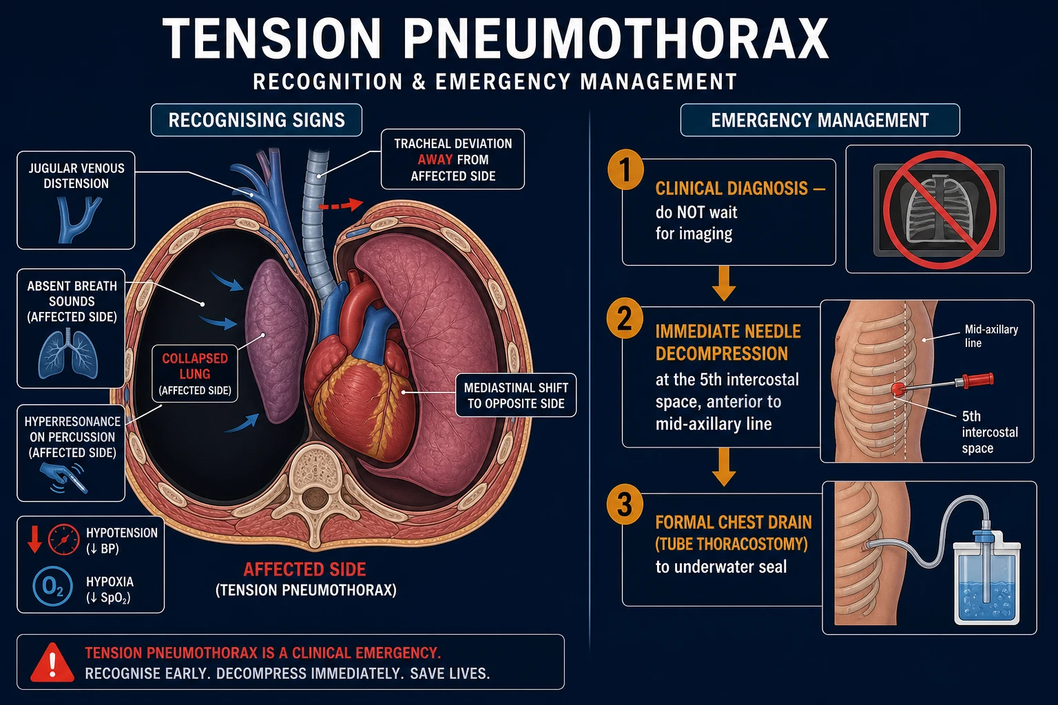
Needle decompression — step by step (FlowSteps)
Tension pneumothorax — emergency decompression
Recognise the CLINICAL picture: hypoxia, hypotension, raised JVP, absent breath sounds, hyperresonance on the affected side — do NOT wait for imaging
Call for help; give high-flow oxygen 15 L/min via non-rebreather mask
Identify the affected side (hyperresonant, silent hemithorax); confirm landmarks — 5th intercostal space, anterior to mid-axillary line
Select a LONG device (≥5 cm cannula / dedicated decompression needle / 14-gauge) — a standard short IV cannula frequently fails to reach the pleura
Clean rapidly (sterility is secondary to speed); insert perpendicular to skin over the UPPER border of the 6th rib, aiming up into the 5th space
A rush of air and an improvement in BP/oxygenation confirm the diagnosis and the relief — leave the cannula in place and secure it
Insert a definitive intercostal drain (large-bore) in the safe triangle — the needle is a bridge, not the treatment
Reassess: chest radiograph AFTER decompression; reassess ventilation and circulation; arrange thoracic surgical referral if an air leak persists
Why the 5th intercostal space mid-axillary displaced the 2nd space mid-clavicular
The classical teaching placed the decompression cannula at the second intercostal space, mid-clavicular line. Anatomical and clinical evidence has shifted the recommended site to the fifth intercostal space, anterior to the mid-axillary line.[2][4] The reasons are shown below.
2nd ICS mid-clavicular (classical)
- Historical site; thinner chest wall in theory
- High failure: mean chest wall here often >5 cm; standard 14G/16G cannula too short
- Risks to great vessels and heart on left
- Largely abandoned in modern trauma guidelines
5th ICS mid-axillary (current)
- Thinner mean chest-wall thickness; higher success rate entering pleura
- Within the "safe triangle" of chest drainage
- Avoids great vessels; consistent with drain site
- Use a LONG device (≥5 cm) or dedicated decompression needle / finger thoracostomy
Chest-wall thickness and decompression failure
Injury 2016
PMID 26724173
A systematic review and meta-analysis of chest-wall thickness at the candidate decompression sites. The 5th intercostal space, anterior/mid-axillary line, has a thinner mean chest wall than the 2nd intercostal mid-clavicular site, and a substantial proportion of patients at both sites have a wall thicker than a standard 14–16-gauge cannula (commonly 4.5 cm). The finding underpins the move to a longer decompression device at the 5th space and explains the historical failure rate of the classical 2nd-space needle.
Definitive management — the spontaneous pneumothorax by size
The spontaneous pneumothorax is managed by size and symptoms.[1]
[1]The chest drain (tube thoracostomy) is inserted by blunt dissection (or a Seldinger technique for a small-bore drain) in the safe triangle at the fourth or fifth intercostal space, anterior axillary line, directed toward the apex, and connected to an underwater seal. The British Thoracic Society accepts a small-bore (8 to 14 French pigtail) drain for most spontaneous pneumothoraces, reserving a large-bore (24 to 32 French) drain for the traumatic, the haemothorax, and the persistent air leak. A Heimlich flutter valve is used for transfer. [1]
The insertion is performed under strict asepsis with local anaesthesia — lidocaine 1 per cent, with adrenaline 1:200,000 to a maximum of about 3 mg per kilogram, or 7 mg per kilogram without adrenaline — infiltrated generously down to and across the pleura at the chosen site, with conscious sedation (midazolam 1 to 2 mg intravenously, or fentanyl 50 to 100 micrograms) for the larger drain if the patient is stable. The steps confirm the safe triangle (bordered by the anterior edge of latissimus dorsi, the lateral border of pectoralis major, and a line above the fifth intercostal space, below the nipple and the axilla); a 2 to 3 cm incision is made along the upper border of the rib to spare the intercostal nerve and vessels that run in the subcostal groove; a blunt clamp dissects through the intercostal muscles to and through the pleura; a gloved finger confirms entry and sweeps adhesions; the drain is advanced toward the apex; the tube is connected to the underwater seal; and it is secured and dressed. The underwater seal allows air and fluid to leave but not to return, and swinging (tidalling) of the water column with respiration confirms the intrapleural position. Aftercare is analgesia, a confirmatory radiograph, and observation for a persistent air leak or a re-expansion oedema. A persistent air leak beyond four to five days, or a failure of the lung to re-expand, prompts a thoracic-surgical referral for a suspected bronchopleural fistula and definitive closure, since a chronic leak will not resolve with continued drainage alone. [1]
Pneumothorax size and action
Simple aspiration — step by step (FlowSteps)
Simple aspiration for a large/symptomatic primary pneumothorax
Confirm a large (≥2 cm) or symptomatic PSP; explain and consent; position semi-recumbent
Landmark the 2nd ICS mid-clavicular line OR the safe triangle (4th/5th ICS mid-axillary); clean and drape
Infiltrate 1% lidocaine with adrenaline down to pleura; confirm aspirating air first with a green needle
Insert a 16-gauge cannula connected to a 3-way tap and 50 mL syringe; aspirate up to 2.5 L
Stop if resistance, cough, or pain; re-expansion is the endpoint — success ≈ 50–70% for first PSP
If successful and stable: discharge with 2–4 week respiratory review and return precautions
If aspiration fails or pneumothorax recurs: proceed to small-bore (8–14 Fr) Seldinger chest drain
Simple aspiration (16G)
- First-line for large/symptomatic PSP per BTS; bedside, low discomfort
- Up to 2.5 L aspirated via 2nd ICS mid-clavicular or 5th ICS mid-axillary
- Success ~50–70% first PSP; lower in SSP — not first-line there
- Failure or recurrence → chest drain
Small-bore Seldinger (8–14 Fr pigtail)
- Preferred drain for most spontaneous pneumothoraces (BTS 2010/2023)
- Less painful, easier to insert, comparable re-expansion to large-bore
- Suitable for persistent air leak; may kink or block
- Connect to underwater seal / Heimlich flutter valve
Large-bore (24–32 Fr)
- Reserved for trauma, haemothorax, haemopneumothorax, large/persistent air leak
- Blunt-dissection technique; more painful; higher complication rate
- Required when fast air/fluid egress expected
- Same safe-triangle site, upper border of rib
Heimlich flutter valve
- One-way valve allowing air egress only; ambulatory and transfer use
- Used for stable PSP and inter-hospital transfer
- May fail with large leaks; not for haemothorax
- Bridges to definitive care
BTS Guideline for pleural disease (2023 update)
Thorax 2023
PMID 37553157
The 2023 British Thoracic Society pleural disease guideline. Reaffirms the 2-cm interpleural-distance cut-off defining a "large" pneumothorax, endorses small-bore Seldinger (8–14 Fr) drains as first-line for most spontaneous pneumothoraces, supports ambulatory (Heimlich-valve) management in selected stable PSP, and consolidates the shift of tension decompression to the 5th intercostal space, anterior/mid-axillary line, with a device long enough to reach the pleura.
Recurrence is common — about 30 per cent after a first primary pneumothorax — so recurrence-prevention (medical or surgical pleurodesis, or VATS) is offered after a second event or in high-risk patients such as aviators and divers. [1]
Complications and pitfalls
The complications include infection and empyema, a persistent air leak or bronchopleural fistula, pain, and re-expansion pulmonary oedema, which follows the too-rapid re-expansion of a large, long-standing pneumothorax and is avoided by draining slowly or incrementally in that setting. The pitfalls are the dangerous inverse of the management: waiting for a radiograph in a suspected tension; using a standard short cannula for decompression; mistaking a giant bulla for a pneumothorax and draining it (creating a fistula); draining a chronic pneumothorax too quickly; and not warning the patient about recurrence and the aviation and diving restrictions. [1]
[1] [1] [1] [1] [1] [1] [1] [1]Prognosis and disposition
Most primary spontaneous pneumothoraces resolve with observation, aspiration, or a drain; recurrence is around 30 per cent, lower after pleurodesis. A patient with a drain is admitted; a small primary pneumothorax managed by observation may be discharged with early follow-up and clear return precautions. Every patient receives explicit advice about the lifetime aviation and diving restrictions after a pneumothorax. [1]
Special populations
Chronic obstructive pulmonary disease is the commonest cause of a secondary pneumothorax and the commonest source of a bulla-mimic — so a CT is sought before drainage when the radiograph is uncertain. Pregnancy modifies the drainage approach. Aviators and divers are grounded for life after a pneumothorax because a recurrence at altitude or depth is catastrophic. Cystic fibrosis and lymphangioleiomyomatosis are high-risk secondary causes. [1]
Evidence and regional guidelines
The contemporary framework is the British Thoracic Society pleural disease guideline[1]; the decompression-site and device-choice evidence, which has moved away from the classical second-intercostal mid-clavicular site toward the fifth-intercostal anterior-axillary site with a longer device, is summarised in recent meta-analysis.[2] The drug and procedural choices are broadly global; the local trauma protocol governs the decompression device and the drain size.
ANZ practice note. The size-based management follows the BTS guideline via local respiratory and trauma pathways; tension decompression uses the fifth-intercostal anterior-axillary site with a dedicated longer device, and the lifetime aviation and diving restrictions are emphasised at discharge. [1]
Exam practice
SAQ — Tension pneumothorax in the trauma patient
10 minutes · 10 marks
A 28-year-old man is brought to the emergency department after a high-speed motor vehicle collision. He is agitated and distressed. T 36.2, HR 132, BP 76/48, RR 30, SpO2 90 per cent on high-flow oxygen. Examination reveals absent breath sounds and hyperresonance to percussion on the right, distended neck veins, and tracheal deviation to the left. He is being prepared for intubation.
SAQ — Primary spontaneous pneumothorax: aspiration versus intercostal drain
10 minutes · 10 marks
A 23-year-old tall thin man presents to the emergency department with two hours of sudden right-sided pleuritic chest pain and breathlessness while at rest. He is a non-smoker with no lung disease. RR 22, SpO2 96 per cent on room air, BP 118/72, HR 92. Chest examination reveals reduced breath sounds and reduced vocal resonance on the right. A chest radiograph confirms a 3 cm right primary spontaneous pneumothorax with no mediastinal shift.
Exam pearls
- Tension is clinical — hypotension, raised JVP, absent breath sounds, hyperresonance — decompress before any imaging.
- Decompress at the fifth intercostal space, anterior to mid-axillary, with a device long enough (≥5 cm) to reach the pleura; the 2nd ICS mid-clavicular site has largely been abandoned.
- PSP small (<2 cm) and asymptomatic: observe ± oxygen; large (≥2 cm) or symptomatic: aspirate first (16G, ≤2.5 L), then drain if it fails; SSP: usually drain outright.
- High-flow oxygen accelerates nitrogen resorption ~4-fold — but avoid in CO₂-retaining COPD.
- Confirm a suspected bulla with CT before draining — draining a bulla creates a bronchopleural fistula.
- Safe triangle, fourth or fifth intercostal space, anterior axillary line, upper border of the rib, for a chest drain.
- Beware re-expansion oedema — drain a long-standing collapse slowly; aspirate ≤2 L; stop for chest tightness/cough.
- Aviation and diving: lifetime grounding after a pneumothorax — a recurrence at altitude or depth is catastrophic.
- Recurrence ≈30% after a first PSP — offer pleurodesis/VATS after a second event or for high-risk occupations.
- The underwater seal should swing — no swing = blocked, kinked, malpositioned, or fully re-expanded; continuous bubbling = significant air leak.
- Never clamp a bubbling chest drain in the ED — it recreates a tension; clamp only on respiratory-surgical instruction.
- Aspirate up to 2.5 litres — sudden chest tightness, cough, or re-expansion pain means stop.
- The ventilated patient who deteriorates with rising peak airway pressure has a tension until proven otherwise — decompress on suspicion.
- Subclavian CVC > internal jugular for iatrogenic pneumothorax — post-procedure erect CXR is mandatory.
- PJP (Pneumocystis) in HIV causes bilateral, recurrent, refractory pneumothoraces — early surgical referral.
- Catamenial pneumothorax: recurrent right-sided PSP in a woman within 72 h of menses — think thoracic endometriosis. [1]
Red flags
[1]References
- [1]MacDuff A, Arnold A, Harvey J, et al. Management of spontaneous pneumothorax: British Thoracic Society Pleural Disease Guideline 2010 Thorax, 2010.PMID 20696690
- [2]Ahmad SJS, Clark ATM, Momin R, et al. Meta-analysis of the optimal needle length and decompression site for tension pneumothorax and consensus recommendations on current ATLS and ETC guidelines World J Emerg Surg, 2025.PMID 40383767
- [3]Roberts ME, Rahman NM, Maskell NA, et al. British Thoracic Society Guideline for pleural disease Thorax, 2023.PMID 37553157
- [4]Laan DV, Vu TD, Thiels CA, et al. Chest wall thickness and decompression failure: A systematic review and meta-analysis comparing anatomic locations in needle thoracostomy Injury, 2016.PMID 26724173
- [5]Tschopp JM, Bintcliffe O, Astoul P, et al. ERS task force statement: diagnosis and treatment of primary spontaneous pneumothorax Eur Respir J, 2015.PMID 26113675