Nephrology · Nephrology
IgA Nephropathy
Also known as Berger disease · Berger's nephropathy · IgAN · mesangial IgA nephropathy
IgA nephropathy (Berger disease) is the commonest primary glomerulonephritis worldwide, defined by mesangial deposition of galactose-deficient IgA1 immune complexes. Classically presents with synpharyngitic gross haematuria 1 to 2 days after a mucosal infection. Diagnosis is renal biopsy showing dominant mesangial IgA on immunofluorescence. Foundation of management is RAAS blockade plus an SGLT2 inhibitor; steroids, targeted-release budesonide and complement-directed therapy are reserved for high-risk progressive disease.
On this page & tools
Your progress
Saved locally on this device.
Practise this topic
Exam tags
Red flags
Related topics

Overview & Definition
IgA nephropathy (IgAN), also called Berger disease (after Jean Berger, who described the immunofluorescence pattern in 1968), is an immune-complex-mediated glomerulonephritis defined pathologically by dominant or co-dominant deposition of IgA (predominantly IgA1) in the glomerular mesangium.[1] It is the commonest primary glomerulonephritis worldwide and a leading glomerular cause of end-stage kidney disease (ESKD). The deposited IgA is abnormally glycosylated — specifically galactose-deficient IgA1 (Gd-IgA1) — and the disease is now understood as an autoimmune process in which autoantibodies against Gd-IgA1 form pathogenic circulating immune complexes that lodge in the mesangium and trigger inflammation, proliferation and scarring.[2]
Clinically, IgA nephropathy is a great imitator: it can present with episodic gross haematuria after a mucosal infection (the classic Berger pattern), asymptomatic microscopic haematuria found on screening, slowly progressive chronic kidney disease (CKD) with hypertension and proteinuria, rapidly progressive crescentic glomerulonephritis with acute kidney injury (AKI), or — less commonly — overt nephrotic syndrome. This protean presentation, combined with a histology-based diagnosis and a strikingly variable prognosis (from lifelong stable renal function to ESKD within decades), makes IgA nephropathy a high-yield examination topic at every level. Approximately 20 to 40 percent of patients progress to ESKD over 20 to 30 years, and because it strikes young adults in their productive decades, the public-health burden is disproportionate. [1]
[1]The term Berger disease should not be confused with Buerger disease (thromboangiitis obliterans, a vasculitis of peripheral vessels in smokers) — a perennial MCQ trap. IgA nephropathy is also distinct from IgA vasculitis (Henoch-Schönlein purpura), which shares the same mesangial IgA lesion but adds systemic small-vessel vasculitis (palpable purpura, abdominal pain, arthritis); IgA nephropathy is now regarded as the renal-limited end of the same disease spectrum. [1]
Classification
IgA nephropathy is classified along three axes — clinical syndrome, primary versus secondary, and the histological Oxford MEST-C lesion score. [1]
Primary (idiopathic) IgAN
- Berger disease in the strict sense — no identifiable systemic cause
- Driven by the multi-hit Gd-IgA1 autoimmune pathway
- Diagnosis requires exclusion of secondary causes
Secondary IgAN
- Same mesangial IgA lesion triggered by a systemic disease
- Liver cirrhosis (hepatic IgA clearance falls) — 'hepatic glomerulosclerosis'
- Coeliac disease, inflammatory bowel disease, psoriasis, ankylosing spondylitis
- Infections: HIV, hepatitis B; mucosal malignancy
IgA vasculitis (HSP)
- Systemic small-vessel IgA vasculitis — the same mesangial IgA lesion
- Plus palpable purpura (lower limbs/buttocks), abdominal pain, arthritis
- Often paediatric; IgA nephropathy is regarded as the renal-limited form of the same spectrum

The Oxford MEST-C classification
The Oxford classification (2009, updated 2016/2025) scores the renal biopsy on five lesions and is the universally adopted histological language for IgA nephropathy.[8] Each letter predicts prognosis and informs the need for immunosuppression:
| Letter | Lesion | Score | Prognostic / therapeutic implication |
|---|---|---|---|
| M | Mesangial hypercellularity | M0 / M1 (over 50% of glomeruli) | M1 = more proliferative; predicts steroid responsiveness |
| E | Endocapillary hypercellularity | E0 / E1 | E1 predicts better response to immunosuppression |
| S | Segmental sclerosis | S0 / S1 | S1 = worse prognosis (chronic scarring) |
| T | Tubular atrophy / interstitial fibrosis | T0 (0 to 25%) / T1 (26 to 50%) / T2 (over 50%) | T is the strongest histological predictor of progression |
| C | Cellular / fibrocellular crescents | C0 / C1 (present) / C2 (over 25% of glomeruli) | C2 = rapidly progressive course; predicts need for urgent immunosuppression |
MEST-C is reported as a string (e.g. M1 E0 S1 T1 C0). Note that T and C are the dominant prognostic drivers: a high T means irreversible chronic damage; a high C means active, potentially treatable crescentic disease. The score is interpreted with clinical data (proteinuria, eGFR, blood pressure) — never in isolation. The MEST-C score has been externally validated in large multinational cohorts including the European VALIGA study and is incorporated into KDIGO guidance as the standard histological descriptor.[7]
Clinical-syndrome spectrum
The same histological lesion can produce five overlapping syndromes: [1]
- Episodic (synpharyngitic) gross haematuria — the classic Berger presentation.
- Asymptomatic microscopic haematuria ± mild proteinuria — found on screening (school, occupational, insurance, antenatal).
- Chronic glomerulonephritis — slowly progressive CKD with hypertension and increasing proteinuria; the commonest mode of presentation in adults.
- Rapidly progressive (crescentic) IgA nephropathy — AKI with active sediment and crescents on biopsy (an RPGN mimic).
- Nephrotic syndrome — uncommon; usually reflects superimposed minimal-change disease (IgA-MCD overlap). [1]
Epidemiology & Risk Factors
IgA nephropathy — key numbers
- Frequency. IgA nephropathy is the commonest primary glomerulonephritis worldwide and accounts for an appreciable fraction of biopsy-proven glomerulonephritis in most registries. Lifetime biopsy prevalence is highest in East Asia (Japan, China, Korea) — reflecting both genuine incidence and a tradition of aggressive urinalysis screening and biopsy. In Japan, mandatory school urinalysis screening detects large numbers of asymptomatic cases; in China up to 45 percent of native-kidney biopsies show IgA nephropathy.[1]
- Age. Peak presentation in the second and third decades (15 to 35 years), although the disease is recognised from childhood into the eighth decade.
- Sex. Male predominance of approximately 2 to 1; in Caucasian populations the ratio approaches 6 to 1 in some series. The female-to-male label is a common distractor — the correct statement is male predominance.
- Ethnicity and geography. Highest incidence in East Asian populations; lower in Africans and African Americans (in whom the disease is under-represented relative to other glomerular diseases). This geographic gradient is partly genetic — multiple GWAS loci differ in frequency across populations.
- Genetics. IgA nephropathy has a strong heritable component (twin concordance, familial clustering). Genome-wide association studies have identified more than 15 susceptibility loci, most prominently in the HLA region (HLA-DRB1/HLA-DQB1), the IGSF1 region, and loci involved in IgA production and mucosal immunity (e.g. DEFA defensin locus, ITGAM-ITGAX, ST6GAL1 and C1GALT1 — the gene encoding the galactosyltransferase central to the multi-hit hypothesis).[2] Most loci have small individual effects, consistent with a polygenic model; monogenic familial IgA nephropathy is rare.
- Risk factors for progression (the predictors examiners reward): persistent proteinuria (the strongest modifiable risk factor), hypertension, reduced eGFR at diagnosis, male sex, persistent microscopic haematuria, and adverse Oxford MEST-C features (especially T, S and C lesions). Obesity and smoking are additional accelerants of CKD progression.
Pathophysiology
IgA nephropathy is the textbook example of an immune-complex disease, and the modern understanding is captured by the multi-hit (four-hit) hypothesis.[2] The four hits are sequential and each is a therapeutic target.
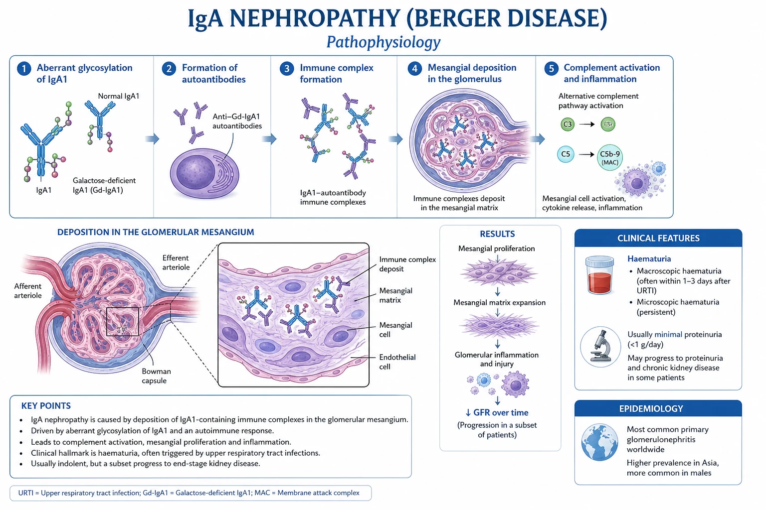
Hit 1 — Over-production of galactose-deficient IgA1 (Gd-IgA1)
IgA1 hinge-region O-glycans are normally galactosylated. In IgA nephropathy, the enzyme C1GalT1 (core-1 beta-1,3-galactosyltransferase, encoded by C1GALT1) is under-active relative to its molecular chaperone Cosmc (encoded by C1GALT1C1), leaving the hinge-region O-glycans galactose-deficient. The result is Gd-IgA1 — an abnormally glycosylated, poorly cleared IgA1 (often in polymeric form) that exposes neoepitopes to the immune system. Gd-IgA1 production is heritable (explaining familial aggregation and GWAS signals at C1GALT1) and up-regulated by mucosal infection — explaining why respiratory or gastrointestinal infections trigger flares.[2]
Hit 2 — Formation of anti-Gd-IgA1 autoantibodies
The exposed hinge-region glycans act as neoepitopes. IgG and IgA autoantibodies (anti-glycan antibodies, including anti-Gd-IgA1 IgG and anti-Gd-IgA1 IgA) are produced, predominantly by mucosal B-cell compartments. These autoantibodies are the second pathogenic hit and are themselves measurable biomarkers of disease activity — elevated anti-Gd-IgA1 IgG titres correlate with disease activity and progression, and form the basis of emerging diagnostic serology. [1]
Hit 3 — Circulating immune complex formation
Anti-Gd-IgA1 autoantibodies bind Gd-IgA1 (especially its polymeric forms) to form large circulating immune complexes. These complexes are too large for normal hepatic clearance (which removes properly glycosylated monomeric IgA1 via the asialoglycoprotein receptor on hepatocytes) and so persist in the circulation. In secondary IgA nephropathy of cirrhosis, hepatic clearance fails by a different mechanism (impaired asialoglycoprotein receptor function), producing the same net effect — circulating immune complexes accumulate and deposit in the mesangium. Their formation is the third hit. [1]
Hit 4 — Mesangial deposition and glomerular injury
The circulating immune complexes lodge in the glomerular mesangium, where they bind mesangial cell receptors (notably the transferrin receptor CD71, which is over-expressed in IgA nephropathy) and activate mesangial cells. The result is:
- Mesangial cell proliferation and matrix expansion (the M and E lesions).
- Release of inflammatory mediators, cytokines and growth factors.
- Complement activation — predominantly via the lectin pathway (mannose-binding lectin binds the exposed galactose-deficient hinge glycans) and the alternative pathway; this explains C3 co-deposition on immunofluorescence and provides a therapeutic rationale for complement inhibitors.
- Podocyte injury (contributing to proteinuria) and, over time, segmental sclerosis and tubulointerstitial fibrosis (the S and T lesions) — the irreversible scarring that drives progression to ESKD.
- Crescent formation (extracapillary proliferation in Bowman's space from fibrin extravasation and parietal epithelial cell activation) in aggressive disease — the C lesion and the hallmark of rapidly progressive IgA nephropathy. [1]
Why mucosal infection matters — the mucosa–bone marrow axis
The mucosa–bone marrow axis is central. Mucosal B cells stimulated by respiratory or gastrointestinal infection produce polymeric IgA1 — and in IgA nephropathy this output is preferentially galactose-deficient. A surge of Gd-IgA1 during a mucosal infection floods the system, forms fresh immune complexes, deposits in the mesangium and produces visible haematuria within 1 to 2 days — the synpharyngitic hallmark. This also explains why B-cell-directed therapies (e.g. anti-BAFF/APRIL strategies such as sibeprenlimab and atacicept, in development) and complement-directed therapies (iptacopan, ravulizumab) work — they interrupt the upstream production or downstream effector arms of the same cascade. [1]
Histological correlate
- Light microscopy: mesangial hypercellularity and matrix expansion; variable segmental sclerosis, endocapillary hypercellularity, tubular atrophy/interstitial fibrosis and crescents (the MEST-C features).
- Immunofluorescence (the defining modality): dominant or co-dominant mesangial IgA (IgA1, not IgA2) deposition, often with C3; IgG and IgM may co-deposit but are not dominant. C1q deposition is typically absent — a useful discriminator from lupus nephritis (where "full-house" immunofluorescence including C1q is characteristic).
- Electron microscopy: mesangial electron-dense immune-type deposits corresponding to the immune complexes; in the IgA-MCD overlap, diffuse foot-process effacement is additionally seen.[1]
Clinical Presentation
The clinical presentation is heterogeneous and a candidate should be able to recognise every pattern. [1]
Classic (synpharyngitic) gross haematuria
A young adult (often male) develops painless visible (macroscopic) haematuria within 1 to 2 days of a sore throat (or gastrointestinal infection). The haematuria is synpharyngitic — concurrent with the infection, not after a latent interval. This is the textbook presentation and the single most testable fact about IgA nephropathy. Episodes are typically self-limiting over days, may recur with subsequent infections, and may be accompanied by loin or flank discomfort (clot colic from tubular obstruction by red cells). Transient AKI can accompany a severe episode (the gross haematuria–AKI syndrome of tubular obstruction and injury by red cells — usually recovers with supportive care over days to weeks, rarely requiring temporary dialysis). [1]
Asymptomatic microscopic haematuria with mild proteinuria
The commonest presentation in screened populations (e.g. Japan, where school urinalysis screening is universal). Persistent dysmorphic microscopic haematuria with mild proteinuria (under 1 g/day), normal renal function and often normal blood pressure. Found on school, occupational, insurance or antenatal screening. [1]
Chronic glomerulonephritis
The commonest presentation in adult nephrology clinics: slowly progressive CKD with hypertension, persistent proteinuria (typically 1 to 3 g/day) and a gradually falling eGFR. Microscopic haematuria is usually present. This is the presentation that most often reaches ESKD, because the diagnosis is made late and the disease has already accumulated chronic (T and S) damage. [1]
Rapidly progressive (crescentic) IgA nephropathy
Acute kidney injury with active urinary sediment (dysmorphic RBCs, red-cell casts) and crescents on biopsy (C1/C2 in MEST-C). This is the IgA nephropathy presentation that mimics ANCA-associated vasculitis, anti-GBM disease or lupus nephritis and requires urgent serology and immunosuppression. It is uncommon but is the presentation where delay costs the kidney. [1]
Nephrotic syndrome
Uncommon. When it occurs, consider IgA nephropathy with superimposed minimal-change disease (IgA-MCD overlap) — the biopsy shows mesangial IgA plus diffuse foot-process effacement on electron microscopy, and the patient is typically steroid-responsive as for minimal-change disease. [1]
Timing — the discriminator from post-streptococcal GN
IgA nephropathy
- Haematuria within 1 to 2 days of infection — synpharyngitic
- Serum complement NORMAL
- ASO titre normal
- Recurrent episodes with each infection
Post-streptococcal GN
- Haematuria 1 to 3 WEEKS after a streptococcal infection (latent period)
- Serum C3 LOW (C4 normal)
- ASO / anti-DNase B titre raised
- Usually single episode; resolves spontaneously
Atypical presentations
- Elderly: more likely to present with hypertension and progressive CKD rather than gross haematuria; higher cardiovascular comorbidity; immunosuppression risk is higher, and secondary causes (including malignancy-associated) must be excluded.
- Diabetic patients: mesangial IgA can co-exist with diabetic nephropathy — atypical features (active urinary sediment, sudden proteinuria, haematuria disproportionate to diabetic change) warrant biopsy.
- Pregnancy: may be first detected as new hypertension/proteinuria; pre-existing IgA nephropathy worsens fetal and maternal outcomes if hypertension or renal impairment is uncontrolled.
- Immunocompromised (HIV): IgA nephropathy is a recognised HIV-associated glomerular lesion, often with a more rapidly progressive course.
- Children: the classic synpharyngitic presentation predominates; tonsillectomy combined with steroid pulses is more commonly used in East Asian paediatric practice than in international guidelines. [1]
Differential Diagnosis
The differential depends on the presenting syndrome. The reasoning is built around timing, complement level and serology, and the urine sediment pattern.[1]
Glomerular haematuria — the differential
HAS-PIC
Synpharyngitic (1 to 2 days) leads to IgAN; latent (1 to 3 weeks) leads to post-strep GN
Raised in post-strep; normal in IgAN, thin basement membrane, Alport
Low C3 in post-strep GN, lupus, infective endocarditis; normal in IgAN, AAV, anti-GBM
Palpable purpura of lower limbs indicates IgA vasculitis (HSP)
Family history + deafness/eye signs indicates Alport; isolated familial haematuria indicates thin basement membrane nephropathy
RPGN picture indicates AAV, anti-GBM, lupus — send ANCA, anti-GBM, ANA/dsDNA
Distinguished one by one
-
Post-streptococcal glomerulonephritis. Distinguished by timing (1 to 3 weeks after pharyngitis or impetigo, not synpharyngitic), low serum C3 (C4 normal), raised ASO and anti-DNase B, and a self-limiting course. Biopsy shows subepithelial "humps" on electron microscopy and granular IgG/C3 deposition. [1]
-
Thin basement membrane nephropathy. Persistent isolated microscopic haematuria, family history of haematuria, normal renal function and blood pressure, minimal proteinuria. Renal biopsy shows diffusely thinned glomerular basement membrane on electron microscopy with no immune deposits. Benign course (formerly "benign familial haematuria"). [1]
-
Alport syndrome (hereditary nephritis). X-linked (in 85 percent), sensorineural deafness, ocular signs (anterior lenticonus, macular flecks), progressive renal failure in affected males. Biopsy shows GBM splitting and lamellation ("basket-weave") on electron microscopy. Family history is the cardinal clue. [1]
-
IgA vasculitis (Henoch-Schönlein purpura). The same mesangial IgA lesion plus palpable purpura (typically lower limbs and buttocks), abdominal pain, arthritis and (in some) gastrointestinal bleeding. Predominantly affects children. IgA nephropathy is regarded as the renal-limited end of the same spectrum. [1]
-
ANCA-associated vasculitis, anti-GBM disease and lupus nephritis — when IgA nephropathy presents as rapidly progressive GN. Distinguished by serology (ANCA, anti-GBM, ANA/dsDNA, complement) and by biopsy features (pauci-immune crescentic GN, linear IgG along the GBM, or full-house immunofluorescence with C1q). [1]
-
Urological causes of haematuria — stones, transitional cell carcinoma, renal cell carcinoma, UTI. Excluded by urine microscopy (dysmorphic RBCs and red-cell casts indicate a glomerular source; isomorphic RBCs without proteinuria suggest a urological source) and by imaging plus cystoscopy in older patients with painless visible haematuria. [1]
-
Membranous nephropathy, minimal-change disease, FSGS — when IgA nephropathy presents with nephrotic-range proteinuria. Biopsy resolves it. [1]
-
Secondary IgA nephropathy — cirrhosis, coeliac disease, inflammatory bowel disease, psoriasis, ankylosing spondylitis, HIV, hepatitis B. The history and examination (stigmata of chronic liver disease, malabsorption, sacroiliitis, dermatitis herpetiformis) reveal the underlying trigger. [1]
Clinical & Bedside Assessment
A focused bedside assessment establishes the syndrome, severity, complications and clues to secondary or alternative diagnoses. [1]
- History. Capture the timing of haematuria relative to mucosal infection — the synpharyngitic clue is the single most diagnostic historical feature. Ask about preceding sore throat or gastrointestinal illness, episodes of visible blood (cola-coloured or smoky urine), abdominal or loin pain, oedema, foamy urine (proteinuria), and family history of renal disease, deafness or haematuria. Record drug and NSAID history (NSAIDs can trigger AKI and interstitial nephritis), and symptoms of systemic disease (rash, arthralgia, sinusitis, haemoptysis, fever, weight loss).
- Examination — fluid status and blood pressure. Hypertension is a key prognostic marker and treatment target. Measure blood pressure carefully and repeatedly. Look for oedema, basal crackles, raised JVP (volume overload), and signs of advanced CKD (pallor of anaemia, uraemic flavour, scratch marks of uraemic pruritus).
- Examination — secondary IgAN clues. Stigmata of chronic liver disease (hepatic IgA clearance failure — spider naevi, palmar erythema, jaundice, ascites), coeliac disease (dermatitis herpetiformis, malabsorption, short stature), psoriasis, inflammatory bowel disease, and ankylosing spondylitis (sacroiliitis, reduced spinal mobility).
- Examination — alternative systemic diagnoses. Palpable purpura (IgA vasculitis), malar rash and arthritis (lupus), sinus disease and lung signs (ANCA vasculitis, granulomatosis with polyangiitis), oral and nasal ulcers.
- Bedside complications. Volume overload (basal crackles, raised JVP, oedema), severe hypertension, signs of AKI, and thromboembolism in nephrotic-range disease. [1]
Investigations
Investigations confirm a glomerular source, stage the disease, exclude mimics, and prepare for biopsy.[1][7]
Urine tests
- Urinalysis (dipstick). Blood and (often) protein. Glucose negative unless diabetic.
- Urine microscopy. Dysmorphic red blood cells (acanthocytes) and red-cell casts confirm a glomerular source — distinguishing IgA nephropathy from urological bleeding.
- Proteinuria quantification. Spot urine protein-to-creatinine ratio (PCR) or albumin-to-creatinine ratio (ACR) on a first-morning sample. PCR over 0.5 to 1 g/day (ACR over 30 mg/mmol) is the trigger for nephrology referral and biopsy consideration. Twenty-four-hour urine protein is rarely needed in routine practice but remains the reference standard. [1]
Blood tests
- Serum creatinine and eGFR — to stage CKD and trend over time.
- Serum albumin, total protein, cholesterol — when nephrotic-range proteinuria suspected.
- Serum IgA level — raised in up to half of patients but non-specific and non-diagnostic; a normal level does not exclude IgA nephropathy, and an elevated level does not confirm it. Newer assays for Gd-IgA1 and anti-Gd-IgA1 autoantibodies (the multi-hit biomarkers) are entering clinical use and may eventually replace total IgA, but are not yet routine.
- Complement C3 and C4 — typically NORMAL in IgA nephropathy; C3 may be mildly reduced in a minority (reflecting lectin and alternative pathway activation), but a frankly low C3 redirects the diagnosis to post-streptococcal GN (low C3, normal C4), lupus nephritis (low C3 and C4) or infective endocarditis-associated GN (low C3). A normal complement is a key discriminator and is tested at every level. [1]
Serology to exclude mimics
- ANA, anti-dsDNA, complements — lupus nephritis.
- ANCA (MPO, PR3) — ANCA-associated vasculitis.
- Anti-GBM antibody — anti-GBM (Goodpasture) disease.
- Hepatitis B surface antigen, anti-hepatitis C, HIV serology — secondary IgA nephropathy and other glomerular diseases.
- ASO and anti-DNase B titre — post-streptococcal GN (raised).
- Serum electrophoresis, serum free light chains — exclude myeloma in older patients with renal impairment. [1]
Renal biopsy — the diagnostic test
The diagnosis of IgA nephropathy requires a renal biopsy.[7]
- Immunofluorescence (the defining finding): dominant or co-dominant mesangial IgA deposition (IgA1), often with C3; IgG and IgM may co-deposit but are not dominant. C1q is typically absent — a useful discriminator from lupus nephritis.
- Light microscopy: mesangial hypercellularity and matrix expansion, with the Oxford MEST-C features (see Classification).
- Electron microscopy: mesangial electron-dense immune-type deposits. [1]
When to biopsy. Indicated when IgA nephropathy is suspected AND any of: proteinuria above 0.5 to 1 g/day, rising or elevated creatinine, hypertension, atypical features (active sediment suggesting RPGN, nephrotic syndrome, suspected systemic disease). Isolated microscopic haematuria with normal renal function and minimal proteinuria (under 0.5 g/day) may be monitored without biopsy — but persistent or increasing proteinuria shifts the balance to biopsy. [1]
Imaging
- Renal ultrasound — to exclude obstruction, assess kidney size and cortical thickness (small, scarred kidneys argue against biopsy yield and against aggressive immunosuppression), and to screen for urological causes of haematuria where the sediment is equivocal. [1]
Risk stratification — the International IgAN Prediction Tool
The International IgA Nephropathy Prediction Tool integrates the MEST-C score with clinical variables at biopsy (eGFR, mean arterial pressure, proteinuria, use of RAAS blockade, prior immunosuppression) to estimate the risk of a 50 percent decline in eGFR or ESKD at 5 and 10 years. It outperforms MEST-C alone and is recommended by KDIGO to inform the decision to start immunosuppression. The tool quantifies what every clinician weighs implicitly: a young patient with preserved eGFR, well-controlled blood pressure, and proteinuria under 0.5 g/day has a very low 5-year risk and does not need immunosuppression, whereas a patient with proteinuria over 1 g/day, falling eGFR and a high MEST-C score has a substantial risk and is a candidate for escalation. The tool is freely available online and is a viva-friendly answer to "how do you decide whom to immunosuppress?"[7]
Monitoring schedule
Stable patients are monitored every 3 to 6 months (more frequently after starting or changing therapy): blood pressure, serum creatinine and eGFR, PCR or ACR, and urinalysis. The goal is to detect rising proteinuria or falling eGFR early — the triggers to intensify therapy. Annual cardiovascular risk assessment (lipids, HbA1c if diabetic) and vaccination review are part of long-term CKD care. [1]
Management — Resuscitation & Supportive Care
There is no specific cure for IgA nephropathy. The foundation for every patient is optimised supportive care, which should be in place before any consideration of immunosuppression.[4][7]
General measures for ALL patients
- Blood pressure control. Target under 130/80 mmHg (and under 125/75 mmHg if proteinuric), using a RAAS blocker first-line. The RAAS blocker reduces intraglomerular pressure and proteinuria independent of systemic blood pressure.
- Proteinuria reduction. The strongest modifiable predictor of progression — sustained proteinuria reduction is the central goal of therapy.
- Lifestyle. Smoking cessation (smoking accelerates CKD progression), salt restriction (under 5 to 6 g sodium per day — supports blood pressure and RAAS blockade), weight optimisation, regular exercise, and avoidance of nephrotoxins (NSAIDs, herbal remedies, iodinated contrast without cover).
- Vaccination and infection prevention. Prompt treatment of mucosal infections; tonsillectomy is NOT routinely recommended internationally (controversial — see Regional Differences).
- Cardiovascular risk reduction. IgA nephropathy is a cardiovascular risk equivalent in CKD — statin per QRISK or CKD guidance, glycaemic control if diabetic, antiplatelet as indicated for established vascular disease. [1]
Managing an acute gross haematuria episode
- Usually self-limiting; reassure the patient.
- Ensure adequate hydration, monitor renal function; most episodes resolve within days.
- Transient AKI during severe gross haematuria (the gross-haematuria–AKI syndrome of tubular obstruction and injury by red cells) usually recovers with supportive care — rarely requires temporary dialysis.
- If AKI is severe or prolonged, repeat biopsy may be needed to exclude superimposed crescentic change or acute interstitial nephritis. [1]
When to refer to nephrology
- Proteinuria over 0.5 to 1 g/day (ACR over 30 mg/mmol).
- Falling eGFR (sustained decline or rapid rise).
- Hypertension uncontrolled on first-line therapy.
- Active urinary sediment (red-cell casts, dysmorphic RBCs) suggesting RPGN.
- Nephrotic-range proteinuria or nephrotic syndrome. [1]
The cardinal principle: supportive care is the foundation before any immunosuppression. The STOP-IgAN trial proved that adding immunosuppression without optimised supportive care does not help.[4]
Management — Definitive & Stepwise
The escalation pathway follows a clear logic: everyone gets supportive care; high-risk patients with persistent proteinuria escalate stepwise; crescentic disease bypasses the ladder to urgent immunosuppression. The decision is anchored on the International IgAN Prediction Tool and the proteinuria response to optimised supportive care over at least 90 days.[7]
IgA nephropathy — escalation algorithm
Optimised supportive care — ALL patients
ACE inhibitor or ARB (ramipril 2.5 to 10 mg/day, or losartan 50 to 100 mg/day) plus SGLT2 inhibitor (dapagliflozin or empagliflozin 10 mg/day); BP target under 130/80 mmHg; lifestyle, smoking cessation, salt restriction, statin
Reassess at 90 days
Recheck proteinuria (PCR/ACR), eGFR, BP. If proteinuria over 0.75 to 1 g/day persists despite optimised supportive care, escalate
Targeted-release budesonide (Nefecon)
16 mg orally once daily for 9 months, then taper to 12 mg then 8 mg — for high-risk disease with persistent proteinuria (NefIgArd)
Systemic corticosteroids (selective)
Modified TESTING regimen: methylprednisolone 0.4 to 0.6 mg/kg/day for 6 to 8 months, with PJP prophylaxis, bone and GI protection — for high-risk patients with preserved GFR (over 30 to 50 mL/min)
Crescentic / RPGN course
Urgent cyclophosphamide plus glucocorticoid induction: pulse methylprednisolone 0.5 to 1 g IV daily x 3, then prednisolone 1 mg/kg/day plus cyclophosphamide 2 mg/kg/day for 3 to 6 months
ESKD
Dialysis access planning and transplantation; counsel on 20 to 50 percent graft recurrence risk
Step 1 — Optimised supportive care (every patient)
The cornerstone of modern management and the non-negotiable foundation before any escalation. [1]
- ACE inhibitor or ARB, for all patients with proteinuria above 0.5 to 1 g/day — titrated to blood pressure, proteinuria and potassium.
- Example: ramipril 2.5 mg orally once daily, titrated by increments to 10 mg daily (max 10 mg/day); or losartan 50 mg orally once daily, titrated to 100 mg daily.
- Rationale: lowers intraglomerular pressure, reduces proteinuria, slows CKD progression independent of blood pressure.
- Monitoring: creatinine and potassium within 1 to 2 weeks of starting or dose change; acceptable rise in creatinine up to 30 percent; do not stop for asymptomatic hyperkalaemia under 6.0 mmol/L (treat with dietary measures, potassium binders).
- Contraindications: bilateral renal artery stenosis, pregnancy (switch before conception), history of angioedema (ACEi).
- SGLT2 inhibitor — dapagliflozin 10 mg orally once daily or empagliflozin 10 mg orally once daily, for proteinuric CKD regardless of diabetes.[6]
- Evidence: EMPA-KIDNEY (2023) showed empagliflozin slowed CKD progression across primary kidney diseases including glomerular disease, with consistent benefit in IgA nephropathy.
- Monitoring: volume status, glycaemic control (in diabetics — risk of euglycaemic ketoacidosis), genital mycotic infection. Avoid in primary extreme volume depletion.
- Stop in pregnancy and around acute illness or surgery.
- Blood pressure target under 130/80 mmHg (under 125/75 mmHg if proteinuric); add dihydropyridine calcium-channel blocker (e.g. amlodipine 5 to 10 mg orally once daily) or diuretic as needed.
- Lifestyle — smoking cessation, salt restriction, weight, exercise.
Step 2 — Targeted-release budesonide (Nefecon) for high-risk disease
For patients at risk of progression with persistent proteinuria (over 0.75 to 1 g/day) despite 90 days or more of optimised supportive care: [1]
- Targeted-release budesonide 16 mg orally once daily for 9 months, then a tapering course to 12 mg then 8 mg (per NefIgArd regimen).
- Rationale: delivering budesonide to the distal ileum (the Peyer's-patch-rich region where Gd-IgA1-producing mucosal B cells reside) down-regulates the upstream autoimmune process. The NefIgArd trial (Lancet 2023, 2-year results) showed proteinuria reduction and slowed eGFR decline in high-risk IgA nephropathy.[5]
- Cautions: glucocorticoid effects (BP, glucose, bone — despite high first-pass inactivation, systemic absorption occurs); not a substitute for optimised supportive care.
Step 3 — Systemic corticosteroids (selective use)
Reserved for high-risk patients with persistent proteinuria (over 1 g/day, particularly over 2 g/day) despite optimised supportive care and an adequate GFR, and informed by trial evidence: [1]
- TESTING trial (Lv, JAMA 2017; long-term follow-up 2022): oral methylprednisolone 0.6 to 0.8 mg/kg/day, tapering over 6 to 8 months, reduced the risk of kidney failure but with a significant increase in serious infections (including a stopped-early high-dose arm at 0.8 to 1 mg/kg/day). The modified regimen (0.4 to 0.6 mg/kg/day with Pneumocystis prophylaxis and bone and GI protection) is used in practice; the long-term follow-up confirmed durable renal benefit with the lower-dose modified protocol.[3][9]
- STOP-IgAN trial (Rauen, NEJM 2015): adding immunosuppression to optimised supportive care gave no added renal benefit at 3 years — tempering routine steroid use.[4]
Net position (KDIGO 2021 / 2025): a short course of systemic steroids may be considered in selected high-risk patients with preserved GFR (usually over 30 to 50 mL/min/1.73 m squared) after informed discussion of infection risk; they are NOT routine.[7]
- Monitoring: glucose, BP, bone density, Pneumocystis jirovecii prophylaxis (e.g. co-trimoxazole 480 mg orally once daily or three times weekly) for prolonged courses, GI protection, infection surveillance. [1]
Step 4 — Sparsentan and emerging complement-directed and B-cell therapies
For high-risk disease with persistent proteinuria despite the above, the following are available or emerging options: [1]
- Sparsentan — dual endothelin-A and angiotensin receptor antagonist; the PROTECT trial (Lancet 2023, 2-year results) showed greater proteinuria reduction than irbesartan in IgA nephropathy.[10]
- Iptacopan — oral factor B inhibitor of the alternative complement pathway; proteinuria reduction in phase III (APPLAUSE-IgAN).
- Ravulizumab, crovalimab — anti-C5 monoclonal antibodies (terminal complement blockade).
- B-cell depletion and BAFF-APRIL inhibition (e.g. sibeprenlimab, atacicept) — targeting upstream Gd-IgA1 autoantibody production.
- Mycophenolate mofetil — NOT effective in IgA nephropathy; the evidence base does not support its use, and KDIGO recommends against it as monotherapy for proteinuria reduction. This negative is a common exam point.
Step 5 — Rapidly progressive (crescentic) IgA nephropathy
Treat as an RPGN: urgent immunosuppression with cyclophosphamide plus glucocorticoids (as for ANCA-associated vasculitis). [1]
- Example induction: pulse methylprednisolone 0.5 to 1 g IV daily for 3 days, then oral prednisolone 1 mg/kg/day (max 60 to 80 mg/day) tapering, plus cyclophosphamide 2 mg/kg/day orally (or IV pulse 15 mg/kg every 2 weeks, age- and GFR-adjusted) for 3 to 6 months; then maintenance with azathioprine or mycophenolate mofetil plus low-dose prednisolone.
- Plasma exchange has a limited role — reserved for selected severe cases with active crescents and rapid GFR decline.
- Crescents on biopsy (C1, C2) are the trigger to escalate from supportive care to this regimen. [1]
Step 6 — Renal replacement therapy and transplantation
For patients reaching ESKD: [1]
- Dialysis — haemodialysis or peritoneal dialysis; plan access early.
- Kidney transplantation — preferred RRT for fit patients. IgA nephropathy recurs in the graft in approximately 20 to 50 percent of recipients (on protocol biopsy the recurrence rate is higher still); recurrence is usually mild (subclinical mesangial IgA) but can be aggressive and is a cause of graft loss in a minority. Living-donor transplant offers the best outcomes; counsel the donor and recipient about recurrence risk. [1]
Adjuncts
- Fish oil (omega-3 fatty acids) — modest and controversial proteinuria reduction; may be considered as adjunct; evidence heterogeneous (US Mayo Clinic trial positive; others negative). Dose approximately 3 to 4 g/day of combined EPA and DHA.
- Tonsillectomy — for recurrent macroscopic haematuria, combined with steroid pulses; common practice in East Asia (notably Japan) but not recommended internationally — evidence is inconsistent and selection-biased.
- Anticoagulation — for nephrotic-range proteinuria and documented thromboembolism. [1]
Specific Subtypes & Scenarios
- Synpharyngitic gross haematuria presentation. Recognise classic IgA nephropathy; biopsy if persistent proteinuria or renal impairment; supportive care for the acute episode.
- Asymptomatic microscopic haematuria with mild proteinuria (screening-detected case). Quantify proteinuria, check renal function and complement, exclude mimics; biopsy if proteinuria over 0.5 to 1 g/day or atypical features. Isolated microscopic haematuria with minimal proteinuria may be monitored.
- Rapidly progressive (crescentic) IgA nephropathy. AKI, active sediment, crescents on biopsy (C1/C2); treat as RPGN with cyclophosphamide plus glucocorticoids.
- IgA nephropathy with nephrotic syndrome (IgA-MCD overlap). Superimposed minimal-change disease with foot-process effacement; biopsy to confirm; typically steroid-responsive as for minimal-change disease.
- Secondary IgA nephropathy. Treat the underlying disease (manage cirrhosis, gluten-free diet for coeliac disease, IBD therapy, psoriasis treatment); the mesangial IgA lesion is shared and may improve with control of the trigger.
- Recurrent IgA nephropathy after transplant. Recurrence is seen in 20 to 50 percent of grafts over time; subclinical mesangial IgA recurrence is common and usually mild; recurrence with proteinuria warrants RAAS blockade and, for severe recurrence, the same escalation pathway as native-kidney disease. [1]
Complications & Pitfalls
Disease complications
- Progression to CKD and ESKD (20 to 40% over 20 to 30 years)
- Hypertension — a key modifiable risk
- Persistent proteinuria — the strongest predictor of progression
- Acute kidney injury during severe gross haematuria (tubular injury)
- Thromboembolism in nephrotic-range disease
- Cardiovascular disease — CKD-equivalent risk
Treatment-related complications
- Glucocorticoid toxicity (infection, diabetes, osteoporosis, hypertension) — TESTING infection signal
- RAAS hyperkalaemia and creatinine rise
- SGLT2 euglycaemic ketoacidosis, volume depletion, genital mycotic infection
- Cyclophosphamide cytopenias, infertility, infection, malignancy
- Budesonide glucocorticoid effects despite gut targeting
Pitfalls to avoid:
- Failing to biopsy when proteinuria rises or when an RPGN course emerges.
- Over-using immunosuppression — ignoring the TESTING infection risk and the STOP-IgAN negative result; immunosuppression is selective, not routine. Mycophenolate mofetil is not effective in IgA nephropathy and should not be used as monotherapy for proteinuria reduction.
- Forgetting SGLT2 inhibitor and RAAS blockade as the foundation — these are the interventions with the strongest evidence base.
- Missing a rapidly progressive (crescentic) course — check the C score and treat as RPGN.
- Confusing IgA nephropathy with post-streptococcal GN on timing — the synpharyngitic versus latent distinction is fundamental.
- Confusing IgA nephropathy with IgA vasculitis (HSP) — same mesangial IgA lesion, but HSP has the systemic features (purpura, abdominal pain, arthritis).
- Missing secondary IgA nephropathy — examine for liver disease, coeliac disease, IBD, psoriasis. [1]
Prognosis & Disposition
- Overall. Approximately 20 to 40 percent of patients progress to ESKD over 20 to 30 years; the remainder have stable or slowly progressive disease.
- Predictors of poor renal outcome: persistent proteinuria (the strongest modifiable predictor), hypertension, reduced GFR at presentation, male sex, persistent microscopic haematuria, and adverse Oxford MEST-C features (especially T and S lesions and crescents (C)).
- Targets that change prognosis. Sustained remission of proteinuria (under 0.5 g/day, ideally under 0.3 g/day) with controlled blood pressure (under 130/80 mmHg) markedly improves prognosis.
- Disposition. Long-term nephrology follow-up with regular monitoring of proteinuria (PCR/ACR), blood pressure and renal function; intensify therapy as proteinuria rises or eGFR falls; plan dialysis access and transplantation as ESKD approaches. [1]
Special Populations
- Children and young adults. Classic synpharyngitic presentation; weight-based dosing of steroids and RAAS blockers (e.g. enalapril 0.1 mg/kg/day starting); biopsy for atypical features, significant proteinuria (over 0.5 to 1 g/day) or declining function. Tonsillectomy combined with steroid pulses is more commonly used in East Asian paediatric practice than in international guidelines.
- Elderly. More likely to present with hypertension and progressive CKD; higher cardiovascular comorbidity; balance the infection risk of immunosuppression against the benefit. Nephrotic-range proteinuria in the elderly should prompt exclusion of secondary causes (malignancy-associated).
- Pregnancy. Pre-existing proteinuria, hypertension or renal impairment worsens fetal (pre-eclampsia, fetal growth restriction, prematurity) and maternal outcomes. Switch ACE inhibitor/ARB to pregnancy-safe alternatives (labetalol, nifedipine, methyldopa) before conception; stop SGLT2 inhibitors pre-conception; monitor closely throughout pregnancy; resume RAAS blockade post-partum if breastfeeding allows. New-onset hypertension and proteinuria in pregnancy requires distinguishing IgA nephropathy from pre-eclampsia (urine microscopy for dysmorphic RBCs/casts, pre-pregnancy urinalysis history).
- Patients approaching ESKD. Plan dialysis access (fistula, peritoneal dialysis catheter) and transplantation early; counsel on recurrence risk in the graft (20 to 50 percent); pre-emptive living-donor transplant offers best outcome.
- Secondary IgA nephropathy. Manage the underlying systemic disease (cirrhosis, coeliac disease, IBD, psoriasis) — control of the trigger can ameliorate the renal lesion.
- Patients with diabetes. Distinguish IgA nephropathy from diabetic nephropathy; atypical features (active sediment, abrupt proteinuria) warrant biopsy. [1]
Evidence, Guidelines & Regional Differences
[1] [1]The current international standard is the KDIGO 2021 Glomerular Diseases guideline (with an IgA nephropathy-specific update in 2025).[7]
Landmark trials
| Trial | Reference | Intervention | Result |
|---|---|---|---|
| TESTING | Lv, JAMA 2017; 2022[3][9] | Oral methylprednisolone 0.6 to 0.8 mg/kg/day | Reduced kidney failure; significant increase in serious infections (high-dose arm stopped early); long-term follow-up confirmed durable benefit at modified lower dose |
| STOP-IgAN | Rauen, NEJM 2015[4] | Immunosuppression added to optimised supportive care | No added renal benefit at 3 years |
| NefIgArd | Lafayette, Lancet 2023[5] | Targeted-release budesonide 16 mg/day | Reduced proteinuria and slowed eGFR decline in high-risk IgAN |
| EMPA-KIDNEY | NEJM 2023[6] | Empagliflozin in CKD | Slowed CKD progression across primary kidney diseases including IgAN |
| PROTECT | Rovin, Lancet 2023[10] | Sparsentan versus irbesartan | Greater proteinuria reduction with sparsentan |
| Oxford classification | Cattran, Kidney Int 2009[8] | MEST-C score | Reproduced the universal histological language for IgAN |
| VALIGA | Coppo et al., European cohort | MEST-C validation in 1147 patients | Confirmed T and S as the dominant prognostic drivers across European populations |
Regional practice differences
- Tonsillectomy. Combined with steroid pulses, this is common practice in East Asia (notably Japan) on the rationale of reducing the mucosal source of Gd-IgA1, but is not recommended internationally — evidence is inconsistent and selection-biased.
- Biopsy practice. In Japan, mass school urinalysis screening and biopsy for isolated microscopic haematuria detects IgA nephropathy early and frequently; in resource-limited settings (including much of South Asia and Africa), biopsy is reserved for proteinuria or declining function, so only advanced disease is seen.
- Steroid practice. Indian Society of Nephrology and ERA-EDTA consensus statements emphasise optimised supportive care first, given infection risks in tropical practice and resource constraints; the TESTING modified lower-dose regimen is preferred where steroids are used.
- VALIGA. The European VALIGA cohort validated the Oxford classification in a predominantly Caucasian population and confirmed the prognostic weight of the T and S lesions, underpinning KDIGO's adoption of MEST-C worldwide. [1]
Exam Pearls
- IgA nephropathy equals Berger disease equals the commonest primary glomerulonephritis worldwide — mesangial deposition of galactose-deficient IgA1 (Gd-IgA1) immune complexes.[1]
- Hallmark timing: synpharyngitic gross haematuria (1 to 2 days after mucosal infection) — versus post-streptococcal GN (1 to 3 weeks latent).
- Diagnosis: renal biopsy — dominant mesangial IgA on immunofluorescence; Oxford MEST-C score; serum complement is NORMAL (low C3 leads to post-strep GN, lupus, endocarditis).
- Prognosis: 20 to 40 percent progress to ESKD over 20 to 30 years; strongest predictor is sustained proteinuria.
- Foundation of treatment: ACE inhibitor/ARB plus SGLT2 inhibitor to reduce proteinuria and slow progression; BP under 130/80 mmHg.
- Steroids: shaped by TESTING (benefit but infection risk — modified lower-dose 0.4 to 0.6 mg/kg/day) and STOP-IgAN (no added benefit when added to optimised supportive care) — selective use only. Targeted-release budesonide (Nefecon, NefIgArd) for high-risk disease. Mycophenolate mofetil is NOT effective in IgA nephropathy.
- Rapidly progressive / crescentic IgA nephropathy (C1/C2): cyclophosphamide plus glucocorticoids, as for ANCA-associated vasculitis.
- Distinguish IgA nephropathy from IgA vasculitis (HSP): same mesangial IgA lesion, but HSP has palpable purpura, abdominal pain, arthritis.
- Multi-hit (four-hit) hypothesis: Gd-IgA1 production, anti-Gd-IgA1 autoantibody, circulating immune complex, mesangial deposition and injury.
- Raised serum IgA in up to half (non-specific); normal serum complement (a discriminator).
- Berger disease is not Buerger disease (the latter is thromboangiitis obliterans, a smoker's vasculitis) — perennial trap.
- Recurrence after transplant in 20 to 50 percent of grafts but usually mild — mesangial IgA on protocol biopsy.
- MEST-C: M, E, S, T (0/1/2 by percent interstitial fibrosis), C — T is the strongest histological prognosticator; C2 drives urgent immunosuppression.
Exam application bank (NEET-PG / INICET)
One-line answer
IgA nephropathy (Berger disease) is the commonest primary glomerulonephritis worldwide, defined by mesangial deposition of galactose-deficient IgA1 immune complexes. Classically presents with synpharyngitic gross haematuria 1 to 2 days after a mucosal infection. Diagnosis is renal biopsy showing dominant mesangial IgA on immunofluorescence. Foundation of management is RAAS blockade plus an SGLT2 inhibitor; steroids, targeted-release budesonide and complement-directed therapy are reserved for high-risk progressive disease.
Worked stems (answer without another resource)
Stem 1 — Classic presentation. Map symptoms to mechanism; name the first investigation and first treatment step with dose/route if drug therapy is standard. [1]
Stem 2 — Unstable / complicated. List red flags that force immediate resuscitation, theatre, ICU, antidote, or reperfusion — and what you do in the first 15 minutes. [1]
Stem 3 — Atypical group. Elderly, pregnancy, child, or immunocompromised: how presentation and thresholds change. [1]
Stem 4 — Differential trap. Name the three closest mimics and one discriminator for each. [1]
Stem 5 — Disposition. Who goes home with safety-netting, who is admitted, who needs HDU/ICU/theatre, and what follow-up is mandatory. [1]
Rapid viva checklist
- Definition + classification
- Pathophysiology chain
- Bedside signs / criteria
- Score with exact components (if any)
- Emergency bundle
- Definitive therapy with doses
- Complications of disease and of treatment
- Special populations
- Guideline/trial name if classic
- Three exam traps
Coverage self-check
If you cannot answer any stem above from this page alone, re-read the matching section — the page is intended to be self-sufficient for final-prof and NEET-PG/INICET questions on IgA Nephropathy.
[1] [1]References
- [1]Roberts IS. Pathology of IgA nephropathy. Nature Reviews Nephrology, 2014.PMID 24861083
- [2]Cheung CK, Alexander S, Reich HN, et al. The pathogenesis of IgA nephropathy and implications for treatment. Nature Reviews Nephrology, 2025.PMID 39232245
- [3]Lv J, Zhang H, Wong MG, et al. Effect of oral methylprednisolone on clinical outcomes in patients with IgA nephropathy: the TESTING randomized clinical trial. JAMA, 2017.PMID 28763548
- [4]Rauen T, Eitner F, Fitzner C, et al. Intensive supportive care plus immunosuppression in IgA nephropathy. New England Journal of Medicine, 2015.PMID 26630142
- [5]Lafayette RA, Canetta PA, Rovin BH, et al. Efficacy and safety of a targeted-release formulation of budesonide in patients with primary IgA nephropathy (NefIgArd): 2-year results from a randomised phase 3 trial. The Lancet, 2023.PMID 37591292
- [6]Empa-Kidney Collaborative Group. Empagliflozin in patients with chronic kidney disease. New England Journal of Medicine, 2023.PMID 36331190
- [7]Kidney Disease: Improving Global Outcomes (KDIGO) Glomerular Diseases Work Group. KDIGO 2021 clinical practice guideline for the management of glomerular diseases. Kidney International, 2021.PMID 34556256
- [8]Cattran DC, Coppo R, Cook HT, et al. The Oxford classification of IgA nephropathy: pathology definitions, correlations, and reproducibility. Kidney International, 2009.PMID 19571790
- [9]Lv J, Wong MG, Hladunewich MA, et al. Effect of Oral Methylprednisolone on Decline in Kidney Function or Kidney Failure in Patients With IgA Nephropathy: The TESTING Randomized Clinical Trial. JAMA, 2022.PMID 35579642
- [10]Rovin BH, Barratt J, Heerspink HJL, et al. Efficacy and safety of sparsentan versus irbesartan in patients with IgA nephropathy (PROTECT): 2-year results from a randomised, active-controlled phase 3 trial. The Lancet, 2023.PMID 37931634