obstetrics-gynaecology · obstetrics-gynaecology
Uterine Fibroids
Also known as Uterine fibroids · Leiomyoma · Leiomyomata · Myoma · Fibromyoma · Uterine fibroid embolisation target lesion
Uterine fibroids (leiomyomas) are benign, monoclonal, oestrogen- and progesterone-responsive smooth-muscle tumours of the myometrium — the commonest benign tumour of women (up to 70 to 80 per cent by age 50). Most are asymptomatic; when symptomatic the leading complaint is abnormal uterine bleeding (heavy/prolonged menses), followed by bulk/pressure symptoms (pelvic heaviness, urinary frequency, constipation) and infertility (predominantly submucosal). Classified by location (FIGO 0 to 8): submucosal cause bleeding and subfertility, intramural are commonest, subserosal cause pressure. Diagnosis is clinical plus transvaginal ultrasound; MRI maps submucosal extent and excludes sarcoma. Management is goal- and fertility-directed: observe if asymptomatic; medical (tranexamic acid, NSAIDs, LNG-IUS, GnRH agonist with add-back, GnRH antagonist); surgery (myomectomy for uterus/fertility preservation, hysterectomy definitive cure for completed family); and uterus-sparing radiological options (uterine artery embolisation, MRgFUS, radiofrequency ablation).
On this page & tools
Your progress
Saved locally on this device.
Practise this topic
Exam tags
Red flags
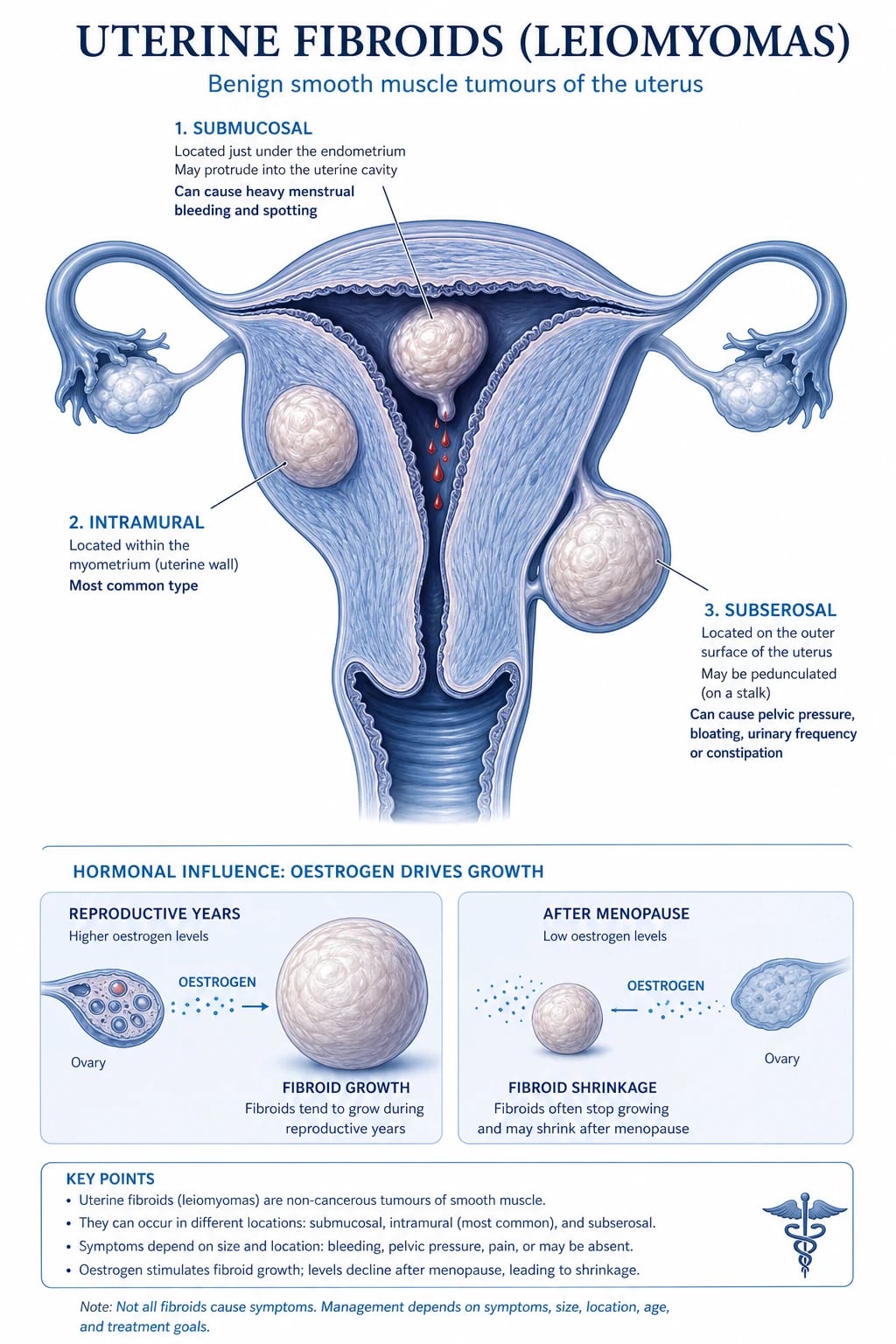
Overview & Definition
Uterine leiomyomas (commonly called fibroids, myomas, fibromyomas or leiomyomata) are benign, hormone-responsive, monoclonal neoplasms arising from the smooth-muscle cell of the myometrium, each surrounded by a fibrous extracellular matrix and a false (compressive, not anatomical) capsule. They are the single commonest benign tumour of the female genital tract and the commonest pelvic tumour overall.[1]
A leiomyoma must be distinguished from adenomyosis (ectopic endometrial glands and stroma scattered within the myometrium, producing a diffusely enlarged, boggy, tender uterus with menorrhagia and secondary dysmenorrhoea — no discrete mass), from an endometrial polyp (a focal outgrowth of endometrial glands and stroma projecting into the cavity), and — critically — from a leiomyosarcoma (a malignant smooth-muscle tumour with mitotic activity, nuclear atypia and coagulative tumour-cell necrosis).[1][2]
The clinical skill in fibroids is not the diagnosis (it is usually ultrasound-evident) but deciding whom to treat, with what, and when — balancing symptom severity, fertility desires, age, fibroid location, and the patient's tolerance of medical versus surgical and uterus-sparing versus definitive options. The single biggest process lever is to classify each fibroid anatomically (FIGO type) before choosing therapy: a submucosal fibroid causing infertility or heavy bleeding warrants hysteroscopic resection; a subserosal bulk lesion is suitable for embolisation or myomectomy; and definitive hysterectomy is reserved for the woman who has completed her family and wants a cure.[2][6]
Classification
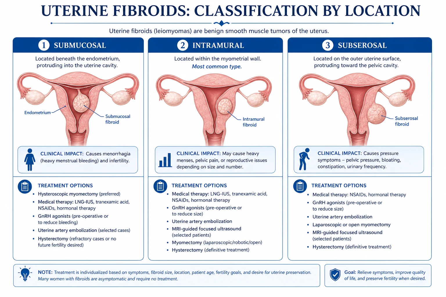
Fibroids are classified by anatomical location relative to the uterine wall (submucosal, intramural, subserosal), and more precisely by the FIGO PALM-COEIN-aligned leiomyoma subclassification system (FIGO types 0 to 8) introduced by the International Federation of Gynecology and Obstetrics (Munro, 2011) and now the universal pre-operative mapping standard.[6]
Three classical anatomical types (high-yield and the exam favourite):[6]
- Submucosal — beneath the endometrium, projecting into the uterine cavity. The least common anatomical type but the most symptom-producing per gram of tumour: heavy, prolonged, irregular menstrual bleeding, infertility (distorts the cavity, impairs implantation) and recurrent miscarriage. Treat with hysteroscopic myomectomy.[8]
- Intramural — the commonest type (around 70 per cent). Confined to the myometrial wall. Produces menorrhagia, an enlarged uterus and, when large, bulk/pressure symptoms.[1]
- Subserosal — beneath the serosal surface, growing outward. May be pedunculated (attached by a stalk) and undergo torsion. Produces pressure symptoms (bladder, bowel, ureter) rather than heavy bleeding; can grow very large and present as an abdominal mass.[1]
FIGO leiomyoma subclassification (0 to 8) — reproduced:[6]
| FIGO type | Location |
|---|---|
| 0 | Pedunculated submucosal (intracavitary, attached by stalk) |
| 1 | Submucosal, less than 50 per cent intramural |
| 2 | Submucosal, 50 per cent or more intramural |
| 3 | Contacts the endometrium; 100 per cent intramural (abuts cavity but no surface component) |
| 4 | Intramural (entirely within wall, no contact with endometrium or serosa) |
| 5 | Subserosal, at least 50 per cent intramural |
| 6 | Subserosal, less than 50 per cent intramural |
| 7 | Subserosal, pedunculated (attached by stalk to the serosa) |
| 8 | Other (cervical, parasitic, broad-ligament / intraligamentary) |
Submucosal
- Beneath endometrium; FIGO 0 to 3; the most symptom-producing per gram
- Heavy, prolonged, irregular AUB; infertility, recurrent miscarriage
- Hysteroscopic myomectomy is first-line surgical treatment
- Saline-infusion sonography / hysteroscopy map cavity distortion
Intramural
- Commonest type (around 70%); FIGO 4 (or 3/5 abutting)
- Menorrhagia; uniformly enlarged, firm, mobile, non-tender uterus
- Medical (LNG-IUS, GnRH) or surgical myomectomy / hysterectomy
- Laparoscopic or open myomectomy when fertility-sparing
Subserosal
- Beneath serosa; FIGO 5 to 7; grows outward, may be pedunculated
- Bulk/pressure: urinary frequency, constipation, hydronephrosis
- Pedunculated can tort (acute pain) or parasitise the omentum
- Embolisation, myomectomy, or MRgFUS suitable
Cervical / parasitic
- FIGO type 8; rare but high-yield
- Cervical: acute urinary retention, ureteric compression, hard to remove
- Parasitic: detaches from uterus, draws blood supply from omentum
- Broad-ligament: ureter at risk at surgery; pre-op MRI essential
Special morphological variants: [1]
- Parasitic fibroid — a subserosal/pedunculated fibroid that detaches from the uterus and derives a new blood supply from the omentum or mesentery; rare, more common after prior uterine surgery or uterine artery embolisation.
- Broad-ligament (intraligamentary) fibroid — grows between the leaves of the broad ligament; the ureter is at risk during surgery — always image pre-operatively.
- Cervical fibroid — arises from the cervix; can impact in the pelvis and cause acute urinary retention and ureteric compression; often requires laparotomy and careful ureteric identification.
- Lipoleiomyoma — a degenerated fibroid with fatty change; benign, often in postmenopausal women.
- Diffuse uterine leiomyomatosis — innumerable small fibroids replacing the myometrium; rare, hormonal-driven, recurs after surgery. [1]
Patterns of benign degeneration (each fibroid can undergo one or more): hyaline (commonest, homogenous eosinophilic), cystic, calcific (commonest in postmenopausal fibroids; 'withered' and radio-opaque), myxoid, red (carneous) (acute haemorrhagic infarction, classically in pregnancy), fatty, and hydropic. Sarcomatous change is not a degeneration pattern — it is malignant transformation (see Prognosis). [1]
Epidemiology & Risk Factors
Fibroids are present in 70 to 80 per cent of women by age 50 at autopsy and ultrasound screening, but only 25 to 30 per cent are symptomatic. They are the commonest indication for hysterectomy worldwide.[1][1]
Demographic and reproductive risk factors (the unifying theme is prolonged oestrogen exposure / cumulative menstrual cycles):[1]
| Risk factor | Mechanism / magnitude |
|---|---|
| Age | Peak in the fourth and fifth decades (35 to 49); rare before puberty; shrink after menopause |
| African ancestry | 2 to 3 times the prevalence of White women; earlier onset, larger, more numerous, more symptomatic |
| Nulliparity | More ovulatory cycles; pregnancy and lactation are protective |
| Early menarche | More lifetime oestrogen exposure |
| Obesity | Peripheral aromatisation of androgens to oestrogens in adipose tissue |
| Family history | First-degree relative — relative risk around 3; monozygotic twin concordance high |
| Tamoxifen (postmenopausal) | Partial oestrogen agonist on the uterus |
| Polycystic ovary syndrome / anovulatory infertility | Unopposed oestrogen |
| Hypertension | Independent association (shared smooth-muscle pathobiology) |
| Diet (red meat, low fruit/vegetable, vitamin D deficiency) | Plausible mechanistic links; vitamin D inhibits fibroid cell proliferation |
Protective factors (anti-oestrogenic): multiparity, prolonged lactation, combined oral contraceptive use (especially long-term), smoking (anti-oestrogenic but obviously not recommended), and late menarche.[1]
Heritability and genetics: fibroids are clonal — each tumour arises from a single myometrial smooth-muscle cell. Around 40 per cent carry a driver cytogenetic abnormality; the commonest are translocation t(12;14) (involving HMGA2), trisomy 12, and deletions of chromosome 7 (involving the candidate tumour suppressor LOC157273). Somatic mutations of MED12 (mediator complex subunit 12, on the X chromosome) are found in up to 70 per cent of fibroids — the single commonest driver, and a high-yield molecular fact. Twin and family studies estimate heritability of 30 to 50 per cent. A familial syndrome, hereditary leiomyomatosis and renal cell carcinoma (HLRCC, Reed's syndrome) due to germline fumarate hydratase mutation causes cutaneous and uterine leiomyomas with aggressive papillary renal cancer — worth knowing.[1][9]
Pathophysiology
Each fibroid is monoclonal (a single myometrial smooth-muscle progenitor — likely a medi-MSC, a Stro-1+/CD44+ mesenchymal stem-like cell) that has acquired a driver mutation (most often MED12, otherwise HMGA2 overexpression) and proliferated under the permissive influence of oestrogen and progesterone, surrounded by a copious extracellular matrix (ECM) rich in collagen, proteoglycans and growth factors.[1][9]
Hormonal sensitivity — the cornerstone (and the therapeutic target): [1]
- Oestrogen promotes fibroid growth by upregulating progesterone receptors and stimulating local aromatase activity (fibroids overexpress aromatase and so synthesise their own oestradiol in situ). Oestrogen also drives IGF-1, EGF and TGF-beta expression.
- Progesterone is now recognised as the dominant mitogen: it drives cell proliferation (Bcl-2 anti-apoptosis, EGF, cyclin D1). This is the rationale for progesterone receptor modulators (SPRMs — ulipristal acetate) and is why fibroids regress during GnRH-induced hypo-oestrogenism. [1]
The why (the chain that explains symptoms):[1][2]
- Oestrogen and progesterone → proliferation of myometrial smooth-muscle cells and extracellular matrix deposition → expanding, well-circumscribed, whorled mass with a compressive pseudocapsule and dilated venous plexus.
- A fibroid beneath or distorting the endometrium → increased endometrial surface area, abnormal vascular maturation (dysregulated venules), and impaired myometrial contractility → heavy, prolonged, irregular menstrual bleeding and anaemia.
- A fibroid distorting the cavity → impairs implantation, gamete transport and decidualisation → infertility and recurrent pregnancy loss (especially submucosal, FIGO 0 to 2).[8]
- A large fibroid → mass effect on bladder, ureter, rectum and pelvic veins → frequency, retention, hydronephrosis, constipation, venous stasis, lower-limb oedema, and (rarely, with very large fibroids) DVT/pulmonary embolism.
- Pregnancy → high oestrogen and rapid growth outstripping blood supply → acute haemorrhagic infarction (red degeneration) with pain.
- Menopause → falling oestrogen → involution and calcification of fibroids.
Molecular hallmarks — the four examiners reward:[1][9]
- MED12 mutation — found in up to 70 per cent of fibroids; disrupts the mediator complex and RNA polymerase II transcription of cell-cycle genes.
- Oestrogen and progesterone dependence — the rationale for all medical therapy (GnRH analogues/antagonists, SPRMs, progestogens).
- Aromatase overexpression — local in-situ oestradiol synthesis means fibroids make their own oestrogen; basis for aromatase inhibitors (letrozole) off-label use.
- Extracellular matrix (ECM) abundance — stiffens the tissue, contributes to bulk symptoms, and is itself mitogenic (mechanotransduction via integrin signalling); also drives fibroid-associated pain. [1]
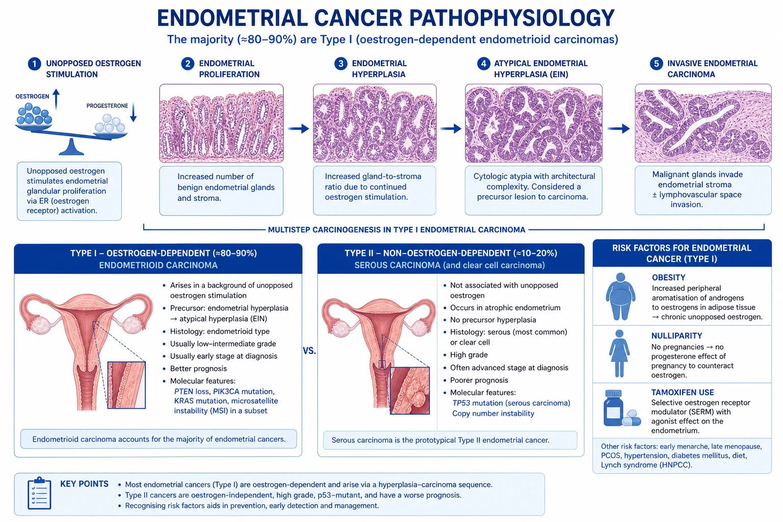
Clinical Presentation
The majority of fibroids are asymptomatic and are incidental findings on ultrasound or at laparoscopy. Symptoms are determined by number, size and — above all — location.[1][6]
Symptom-by-symptom (the classical picture): [1]
- Abnormal uterine bleeding — the commonest symptom (around 30 per cent of symptomatic women). Classically heavy menstrual bleeding (menorrhagia) — regular, cyclical, but heavy and prolonged; less commonly intermenstrual bleeding (especially submucosal). Bleeding is rarely acyclical/postmenopausal — that finding mandates endometrial sampling to exclude malignancy.
- Bulk/pressure symptoms. Pelvic heaviness, pressure, bloating, increasing abdominal girth; a palpable abdominal mass. Urinary frequency, urgency and nocturia (bladder compression); acute urinary retention (cervical fibroid impacting in pelvis — a high-yield presentation); ureteric compression with hydronephrosis (broad-ligament or large cervical fibroid); constipation and tenesmus (rectal compression); lower-limb oedema and venous stasis (iliac-vein compression).
- Pain. Fibroids are usually painless; pain suggests a complication: red (carneous) degeneration in pregnancy, torsion of a pedunculated subserosal fibroid, sarcomatous change (especially postmenopause), or expulsion of a submucosal fibroid through the cervix (labour-like cramps). Some women report deep dyspareunia or chronic pelvic ache with large fibroids.
- Reproductive symptoms. Infertility (submucosal fibroids distort the cavity; large intramural fibroids impair implantation and tubal motility), recurrent first-trimester miscarriage, and — in pregnancy — complications including malpresentation, antepartum haemorrhage, preterm labour, and obstructed labour.[8]
On examination: [1]
- Abdominal examination — an irregularly enlarged, firm, smooth, mobile, non-tender uterus arising from the pelvis. The irregular contour (bosselated) distinguishes fibroids from pregnancy (a uniformly enlarged, soft, symmetrical uterus) and from a large ovarian cyst (cystic, separate from the uterus).
- Bimanual vaginal examination — confirms the uterine origin (the mass moves with the cervix/uterus), assesses size, mobility and adnexa. The cervix may be displaced by a cervical fibroid.
- Speculum examination — usually normal; excludes a cervical cause of bleeding. [1]
Atypical presentations (high-yield): [1]
- The young woman with infertility and a single submucosal fibroid — may have no menstrual symptoms at all; the fibroid is found on fertility work-up. Hysteroscopic resection restores fertility.
- The postmenopausal woman with a 'growing fibroid' or postmenopausal bleeding — do not assume a benign fibroid; rule out leiomyosarcoma and endometrial cancer (endometrial sampling, MRI). Fibroids should shrink after menopause.
- The pregnant woman with acute abdominal pain — red degeneration (carneous degeneration): acute, well-localised pain over a fibroid, low-grade fever, mild tachycardia, a tender uterine mass; managed conservatively with analgesia.[6]
- The woman on tamoxifen for breast cancer — fibroids may grow despite menopause due to partial oestrogen agonism.
- The woman with acute urinary retention — feel for a cervical fibroid impacted behind the pubic symphysis before assuming a neurological cause.
- The woman with severe anaemia from heavy menses — iron-deficiency anaemia may be the presenting feature of an otherwise silent submucosal fibroid.
Differential Diagnosis
An enlarged, irregular uterus or pelvic mass is not always a fibroid. Distinguish each by its distinguishing features:[1][2]
Adenomyosis
- Endometrial glands AND stroma WITHIN myometrium (diffuse, not a discrete mass)
- Diffusely enlarged, BOGGY, TENDER uterus; menorrhagia + secondary dysmenorrhoea
- TVUS: junctional zone over 12 mm, myometrial cysts, asymmetric walls; MRI: junctional zone thickening
- Often coexists with fibroids; does NOT shrink with GnRH as dramatically
Pregnancy
- Reproductive-age woman with amenorrhoea and a soft, symmetrically enlarged uterus
- ALWAYS do a beta-hCG first in any reproductive-age woman with pelvic mass/AUB
- Uterus soft and globular (vs firm irregular fibroid); morning sickness, breast changes
- Ectopic must be excluded if pain or bleeding
Ovarian mass / neoplasm
- Mass SEPARATE from the uterus on bimanual exam — the key discriminator
- TVUS: cystic, solid, papillary projections, ascites (malignancy features)
- CA-125 + RMI scoring; dermoid shows fat/fluid level and calcification
- Risk of torsion/rupture; do NOT assume a fibroid without confirming uterine origin
Endometrial cancer
- POSTMENOPAUSAL (or perimenopausal) bleeding — the cardinal red flag
- Risk: obesity, nulliparity, unopposed oestrogen, diabetes, tamoxifen, Lynch syndrome
- Endometrial sampling (pipelle/hysteroscopy D&C) is diagnostic; thickened endometrium on TVUS
- Can coexist with fibroids; never assume AUB is 'just the fibroid' in high-risk women
Endometrial polyp
- Focal outgrowth of endometrium into cavity; intermenstrual or postcoital bleeding
- TVUS: focal echogenic thickening; SIS better defines; hysteroscopy diagnostic and therapeutic
- Usually smaller, no myometrial mass; can be malignant in postmenopausal women
- Hysteroscopic polypectomy
Leiomyosarcoma
- RAPID growth, especially POSTMENOPAUSE or not exposed to oestrogen; pain, weight loss
- Pre-op suspicion: LDH and LDH isoenzymes raised; MRI DWI restricted diffusion
- Definitive: histology — atypia, mitoses over 10 per 10 HPF, coagulative tumour-cell necrosis
- NEVER morcellate — spreads intra-abdominally; en bloc hysterectomy
For heavy menstrual bleeding specifically (PALM-COEIN differential): structural causes — Polyp, Adenomyosis, Leiomyoma, Malignancy/hyperplasia (the PALM group, seen on imaging/histology); non-structural — Coagulopathy, Ovulatory dysfunction, Endometrial, Iatrogenic, Not yet classified (the COEIN group). Fibroids are the L of PALM and are the commonest structural cause of HMB.[3]
Always exclude pregnancy (beta-hCG) in any reproductive-age woman, and exclude malignancy (endometrial sampling in postmenopausal / high-risk women) before attributing bleeding to a fibroid. [1]
Clinical & Bedside Assessment
The diagnosis of fibroids is clinical plus transvaginal ultrasound — but the bedside examination is what frames the differential and the urgency.[6]
Focused abdominal and pelvic examination findings in fibroids: [1]
- Irregularly enlarged, firm, smooth-surfaced, mobile, non-tender uterus — the cardinal finding. The uterus is firm (vs soft in pregnancy, cystic in ovarian mass), irregular/bosselated (vs symmetrical in pregnancy or adenomyosis), and mobile (vs fixed in endometriosis, advanced malignancy or PID).
- Cervix — usually normal; a cervical fibroid displaces the cervix and may be palpable as a firm mass in the vaginal fornix; the external os may be above the pubic symphysis when a cervical fibroid impacts in the pelvis.
- Bimanual examination — confirms uterine origin (the mass moves with the cervix), assesses size (in weeks of gestation), mobility, adnexa, and pelvic mass effect (low-lying fibroid).
- Speculum — excludes cervical polyp, ectropion or malignancy as a cause of bleeding. [1]
Named signs / clinical manoeuvres (high-yield): [1]
- Size expressed in 'weeks of gestation' — universal convention: a 12-week-size fibroid uterus reaches the pubic symphysis; a 20-week-size reaches the umbilicus. Use a tape to document fundal height.
- ** ballotability** — a mobile fibroid can be balloted between the abdominal and vaginal hands; an ovarian cyst is separately balloted from the uterus.
- Assess for anaemia — conjunctival pallor, tachycardia, flow murmur in severe iron deficiency; any woman with heavy menses needs a full blood count. [1]
Bedside work-up at first presentation: a structured menstrual history (cycles, days of bleeding, flooding, clots, dysmenorrhoea, impact on life), a reproductive history (fertility desires, parity, pregnancy plans — these drive every management decision), a family history (fibroids, HLRCC), assessment of bulk/pressure symptoms (urinary, bowel, venous), a drug history (tamoxifen, hormonal contraception), and screening for red flags (postmenopausal bleeding, rapid growth, severe acute pain, urinary retention). [1]
Investigations
The diagnostic pathway is stepwise: clinical suspicion then transvaginal ultrasound first-line, saline-infusion sonography for submucosal mapping, MRI for surgical planning and sarcoma suspicion, and hysteroscopy with biopsy for cavity lesions.[1][6]
Baseline bloods (always): [1]
- Full blood count — to detect and grade iron-deficiency anaemia (microcytic, hypochromic; low ferritin) from chronic menorrhagia — the commonest systemic consequence of fibroids.
- Beta-hCG — always before imaging in any reproductive-age woman to exclude pregnancy.
- Thyroid function, coagulation screen (if heavy bleeding since menarche or family history — exclude von Willebrand disease, the commonest inherited bleeding disorder in women with HMB).
- Iron studies, ferritin — confirms iron deficiency. [1]
Transvaginal ultrasound (TVUS) — first-line imaging. Inexpensive, widely available, no radiation. Fibroids appear as well-defined, rounded, hypoechoic, heterogeneous masses within the myometrium, with acoustic shadowing and (sometimes) calcification (postmenopausal). TVUS defines number, size, location (FIGO type) and effect on the cavity. Saline-infusion sonography (SIS / sonohysterography) improves accuracy for submucosal fibroids and intracavitary component — it is the test of choice to distinguish a submucosal fibroid from an endometrial polyp.[6]
Pelvic MRI — second-line for mapping and surgical planning. MRI is the most accurate modality for fibroid mapping: it defines the FIGO type, depth of myometrial invasion, proximity to the serosa and endometrium, and detects degeneration (T2-hyperintense cystic/myxoid change; T1-hyperintense haemorrhagic/red degeneration; low-signal calcification). Indications: before complex myomectomy (especially multiple, large, or broad-ligament fibroids where ureter at risk); before uterine artery embolisation (to confirm vascularity and exclude pedunculated subserosal fibroids that may detach); when TVUS is non-diagnostic or uterus is too large; and — critically — to raise or lower suspicion of leiomyosarcoma (diffusion-weighted imaging DWI with restricted diffusion, LDH/LDH isoenzymes). MRI is not needed for routine diagnosis.[1]
Hysteroscopy — direct visualisation and biopsy of cavity lesions. Gold standard for submucosal fibroids (FIGO 0 to 2): it allows direct visualisation, assessment of intramural extension, and simultaneous resection (hysteroscopic myomectomy). Combined with endometrial biopsy / dilatation and curettage to exclude malignancy in any woman with postmenopausal bleeding, persistent intermenstrual bleeding, age over 40 to 45 with risk factors (obesity, nulliparity, unopposed oestrogen, tamoxifen, Lynch syndrome), or atypical glandular cells on cervical cytology.[3]
Investigations for the suspected sarcoma (high-yield):[1]
- LDH and LDH isoenzymes — a raised LDH and its isoenzyme 3 is the most promising non-invasive marker (no single test is definitive).
- MRI with diffusion-weighted imaging (DWI) — restricted diffusion (low ADC value) and T2 heterogeneity favour sarcoma over a benign degenerating fibroid.
- Histology is definitive — mitoses over 10 per 10 high-power fields, diffuse moderate-to-severe nuclear atypia, and coagulative tumour-cell necrosis (the three Stanford criteria).
- NEVER transabdominal needle biopsy of a suspected fibroid if morcellation/sarcoma is a concern — risks intra-abdominal spread. [1]
Severity / impact scoring: there is no universally applied numeric score for fibroid severity (unlike CURB-65 in CAP). The clinically relevant 'grading' is the FIGO type plus uterine size in weeks plus symptom severity (pictorial blood-loss assessment chart, PBAC, with a score over 100 defining menorrhagia). The Uterine Fibroid Symptom and Quality-of-Life (UFS-QOL) questionnaire is the validated disease-specific instrument used in trials and clinical practice to assess symptom burden and treatment response.[1]
Management — Resuscitation
Fibroids are usually chronic, but severe acute heavy menstrual bleeding is a medical emergency.[3][6]
Acute severe AUB (flooding, haemodynamic compromise, Hb below 70 g/L or symptomatic): [1]
- ABCDE — IV access, fluids (balanced crystalloid), group and save / crossmatch.
- Exclude pregnancy (beta-hCG) before any drug or procedure.
- Tranexamic acid 1 g IV three times daily (or 1 to 1.5 g oral three times daily during acute bleed) — antifibrinolytic; inhibits plasminogen activation, stabilises clot. Avoid in active thromboembolic disease or intrinsic thrombotic risk.
- High-dose IV conjugated equine oestrogen (Premarin) 25 mg IV every 4 to 6 hours (up to 24 hours) — the rapid-acting medical haemostatic for acute severe AUB; raises clotting factors and promotes endometrial proliferation. Avoid in active breast cancer, prior VTE, active liver disease, undiagnosed vaginal bleeding.
- Blood transfusion if Hb below 70 g/L or symptomatic anaemia; IV iron (ferric carboxymaltose) if iron-deficient and Hb 70 to 100 g/L (faster repletion than oral, better tolerated).
- Foley catheter balloon tamponade (30 mL balloon inflated in the uterine cavity for 12 to 24 hours) — mechanical haemostasis for refractory bleeding when drugs fail.
- Emergency dilatation and curettage / hysteroscopic resection of a bleeding submucosal fibroid — definitive if a cavity lesion is the source.
- GnRH agonist (goserelin 3.6 mg SC) — to induce amenorrhoea while planning definitive surgery.
- Emergency hysterectomy — last resort for life-threatening haemorrhage unresponsive to all measures (often perimenopausal / completed family). [1]
Management — Definitive & Stepwise
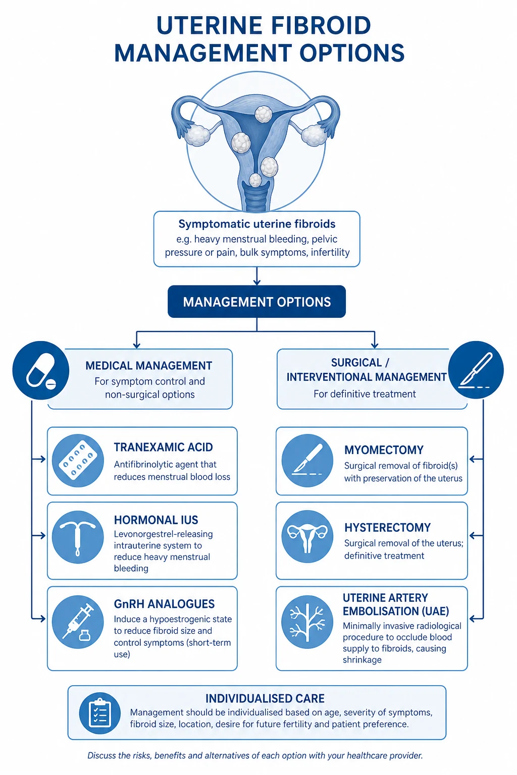
Treatment is goal-directed — define first the primary goal: (1) control of bleeding, (2) control of bulk/pressure symptoms, (3) fertility preservation / restoration, or (4) definitive cure. The choice then depends on fertility desires, age, fibroid FIGO type, size and number, severity of symptoms, prior treatments, and patient preference.[1][2][6]
Principles
- Asymptomatic fibroids do NOT require treatment — observe. The exception is a fibroid that is rapidly growing, causes ureteric obstruction/hydronephrosis, or is in a postmenopausal woman (sarcoma concern).[6]
- Fertility desires are the master variable. Myomectomy (uterus-sparing) for women wanting future fertility; hysterectomy (definitive) only for completed family.
- Location drives modality. Submucosal (FIGO 0 to 2) → hysteroscopic myomectomy; intramural/subserosal (FIGO 3 to 7) → medical, laparoscopic/abdominal myomectomy, embolisation, or hysterectomy; cervical/broad-ligament (FIGO 8) → open surgery with ureteric care.
Medical management — drug, dose, route, rationale, monitoring
1. Symptomatic control of heavy menstrual bleeding (non-hormonal, first-line):[3]
- Tranexamic acid 1 g oral three times daily for up to 4 days during menses (max 4 g/day). Rationale: antifibrinolytic, inhibits plasminogen activation at the endometrial surface, reduces menstrual loss by 40 to 50 per cent. Monitoring: avoid in active thromboembolism, acquired colour-vision disturbance. Does not treat bulk symptoms or shrink fibroids. Often combined with an NSAID.
- NSAIDs — mefenamic acid 500 mg oral three times daily during menses (or naproxen 500 mg BD, ibuprofen 400 mg TDS). Rationale: inhibit prostaglandin synthesis, reduce menstrual loss by around 30 per cent and relieve dysmenorrhoea. Avoid in peptic ulcer, renal impairment, asthma, women trying to conceive. [1]
2. Levonorgestrel-releasing intrauterine system (LNG-IUS, Mirena) — the first-line hormonal treatment for HMB in a woman with a fibroid uterus under 12 weeks size:[3]
- LNG-IUS 52 mg levonorgestrel intrauterine, releasing 20 micrograms/day over 5 years. Rationale: local progestogenic effect atrophies the endometrium → reduces menstrual loss by 70 to 95 per cent, induces amenorrhoea in many, and provides contraception. Cautions: higher expulsion rate with a distorted cavity (especially submucosal fibroids or uterus over 12 weeks size) — consider hysteroscopic resection of submucosal fibroids first. Does not significantly shrink fibroids, but treats the symptom (bleeding) that matters most. Monitoring: check threads at 6 weeks; risk of perforation at insertion. [1]
3. Combined hormonal contraception — useful when contraception is also desired:[3]
- Combined oral contraceptive pill (e.g. ethinylestradiol 30 micrograms + levonorgestrel 150 micrograms daily, continuous or cyclic). Rationale: suppresses ovulation, regularises and lightens menses, provides contraception. Avoid in migraine with aura, prior VTE, smoking over age 35, uncontrolled hypertension, breast cancer. [1]
4. Progestogens — oral or injectable:[3]
- Norethisterone acetate 5 mg oral two to three times daily from day 5 to 26 (or medroxyprogesterone acetate 10 mg daily for 14 days per cycle for a luteal-phase regimen). Rationale: atrophies endometrium, reduces bleeding. Injectable depot medroxyprogesterone acetate (DMPA) 150 mg IM every 12 weeks induces amenorrhoea in many — but bone density concern with long-term use. Side effects: irregular bleeding, weight gain, mood change. [1]
5. Gonadotropin-releasing hormone (GnRH) agonists — 'pseudo-menopause' (second-line, short-term):[1][2]
- Goserelin 3.6 mg SC every 4 weeks (or 10.8 mg SC every 12 weeks); leuprorelin 3.75 mg IM every 4 weeks (or 11.25 mg IM every 12 weeks); triptorelin 3.75 mg IM every 4 weeks. Rationale: initial flare then down-regulation of pituitary GnRH receptors → reversible hypo-oestrogenism → fibroids shrink by 30 to 50 per cent and amenorrhoea ensues, with correction of anaemia. Indications: pre-operative shrinkage for 3 to 6 months before myomectomy, hysterectomy or hysteroscopic resection (reduces blood loss, converts some laparotomies to laparoscopy, corrects anaemia) and as a perimenopausal bridge to natural menopause. Limit to 3 to 6 months without add-back therapy because of bone density loss and menopausal side effects (hot flushes, vaginal dryness, mood). Add-back therapy — tibolone 2.5 mg OD or norethisterone 5 mg OD — mitigates bone loss and symptoms, allowing slightly longer use. Fibroids re-grow after stopping — so GnRH agonists are NOT a standalone curative therapy.[2]
6. GnRH antagonists — the modern oral option:[5]
- Relugolix combination (Ryeqo) — relugolix 40 mg + estradiol 1 mg + norethindrone acetate 0.5 mg, one tablet oral once daily. Rationale: immediate (no flare) dose-dependent suppression of gonadotrophins and ovarian oestrogen, with built-in add-back to limit bone loss. Supported by the LIBERTY 1 and 2 phase 3 trials, which showed significant reduction in heavy menstrual bleeding and fibroid-associated pain versus placebo over 24 weeks, with sustained response to 52 weeks. Suitable for long-term use (unlike GnRH agonists). Monitoring: contraindicated in pregnancy, known osteoporosis, active VTE; caution with prior VTE. [1]
7. Selective progesterone receptor modulators (SPRMs):[2][7]
- Ulipristal acetate (Esmya) 5 mg oral once daily, for up to 3 courses of 3 months each, with treatment breaks. Rationale: a progesterone receptor modulator that inhibits ovulation and induces apoptosis of fibroid cells without inducing hypo-oestrogenism — fibroids shrink and bleeding stops, without the menopausal side effects of GnRH. Supported by the PEARL I to IV trials. CRITICAL SAFETY UPDATE — EMA PRAC 2018 and 2020: ulipristal acetate is associated with rare but serious drug-induced liver injury, including liver failure requiring transplant. It is now contraindicated in any hepatic impairment, requires liver function tests before, during and after each course, and is suspended or restricted in many countries — verify local availability. Vilaprisan (another SPRM) was similarly suspended in trials for hepatotoxicity.[7]
8. Other (off-label / emerging): [1]
- Aromatase inhibitors — letrozole 2.5 mg oral once daily — block local oestrogen synthesis; off-label for symptomatic fibroids, useful in infertile women (preserves ovulation unlike GnRH).
- Raloxifene (selective oestrogen receptor modulator) — studied but not standard. [1]
Surgical management
Indications for surgery:[2][6]
- Symptomatic fibroids failing or unsuitable for medical therapy (heavy bleeding with anaemia, severe bulk/pressure symptoms, ureteric obstruction, infertility with cavity distortion).
- Submucosal fibroid (FIGO 0 to 2) causing bleeding or infertility → hysteroscopic myomectomy (first-line surgical).
- Rapid growth, postmenopausal growth, or imaging suggestive of sarcoma → hysterectomy (do not morcellate). [1]
Myomectomy — uterus-sparing, for women desiring fertility or uterus preservation: [1]
- Hysteroscopic myomectomy — for submucosal fibroids FIGO 0, 1 and 2. Resectoscope with monopolar or bipolar energy shaves the fibroid under direct vision; cold-loop (Bigatti shaver) reduces thermal injury. Limit total operative time to under 90 minutes and use hypotonic distension media (glycine) judiciously — risk of fluid overload, hyponatraemia (TURP syndrome). Pretreatment with GnRH agonist for FIGO 0 to 1 may reduce size. Recurrence around 15 to 30 per cent.
- Laparoscopic myomectomy — for intramural and subserosal fibroids (FIGO 3 to 7), especially under 10 cm and fewer than 4. Multilayer uterine closure essential; counsel on risk of uterine rupture in future pregnancy — these women should usually deliver by elective caesarean if the cavity was entered.
- Abdominal (open) myomectomy — for very large (over 10 cm), numerous, cervical or broad-ligament fibroids where laparoscopy is not feasible; minilaparotomy is an alternative. Blood loss is higher; preoperative GnRH agonist or preoperative uterine artery occlusion reduces bleeding. [1]
Hysterectomy — the definitive cure, for women who have completed their family:[6]
- Total hysterectomy (with conservation or removal of tubes and ovaries depending on age and ovarian-cancer risk) cures the disease — no recurrence. Routes: vaginal, laparoscopic, robotic, abdominal. Choose the least invasive route feasible. Consider concurrent BSO in postmenopausal women or those at high ovarian-cancer risk; counsel on surgical menopause.
- Never perform hysterectomy in a woman desiring future fertility without exhaustive counselling. [1]
Uterus-sparing radiological / minimally invasive options
Uterine artery embolisation (UAE):[4]
- Interventional radiology — bilateral uterine artery catheterisation via femoral artery with injection of polyvinyl alcohol (PVA) particles (500 to 700 micrometres) or gelatin-coated tris-acryl microspheres to embolise the fibroid's arterial supply → ischaemic infarction and shrinkage (around 40 to 60 per cent volume reduction at 6 months). Symptom relief in 85 to 90 per cent of women. Supported by the REST (NEJM 2007) and EMMY trials showing UAE is less invasive than surgery with comparable medium-term symptom control but higher reintervention rates than hysterectomy/myomectomy.[4] Not suitable for pedunculated subserosal fibroids (FIGO 7) (risk of detachment/torsion) or where infection is present. Post-embolisation syndrome (pain, low-grade fever, nausea) common for a few days. Pregnancy after UAE is possible but associated with higher rates of preterm delivery, miscarriage, malpresentation and postpartum haemorrhage — counsel women wanting future fertility.
MRI-guided focused ultrasound surgery (MRgFUS / HIFU): [1]
- High-intensity focused ultrasound under real-time MRI thermometry ablates fibroid tissue non-invasively through intact skin — no incision, day-case. Suitable for non-calcified, accessible fibroids (typically FIGO 2 to 6, not submucosal-pedunculated). Symptom relief around 70 per cent; lower reintervention risk if well-selected. Limited by depth, bowel interposition, and scar tissue. [1]
Radiofrequency ablation (Acessa, Sonata): [1]
- Laparoscopic or transvaginal ultrasound-guided radiofrequency ablation of fibroid tissue (laparoscopic Acessa; transcervical Sonata). Minimally invasive; suitable for intramural and subserosal fibroids; preserves uterus. Symptom relief around 70 to 80 per cent. [1]
Endometrial ablation — only for small submucosal fibroids with completed family and no fertility desire; must be combined with sterilisation or reliable contraception as future pregnancy is high-risk. [1]
Specific Subtypes & Scenarios
Submucosal (FIGO 0-2)
- Heavy, prolonged, irregular AUB; infertility; recurrent miscarriage
- Hysteroscopic myomectomy is first-line surgical; map with SIS
- Watch for TURP syndrome with hypotonic distension media
- Pretreat with GnRH agonist for FIGO 0-1 to reduce size and vascularity
Intramural (FIGO 4)
- Commonest type; menorrhagia; enlarged irregular firm uterus
- LNG-IUS first-line medical; laparoscopic or open myomectomy
- Risk of uterine rupture after myomectomy — counsel on delivery mode
- Pre-op GnRH reduces blood loss and converts some to laparoscopic
Subserosal (FIGO 5-7)
- Bulk/pressure: urinary, bowel, ureter, venous
- Pedunculated (FIGO 7) can tort — acute pain; not for UAE (detachment)
- Myomectomy, UAE (FIGO 5-6), or MRgFUS suitable
- Parasitic variant draws omental supply — more common post-UAE
Cervical / broad-ligament (FIGO 8)
- Cervical: acute urinary retention, ureteric compression
- Broad-ligament: ureter at risk — pre-op MRI, ureteric stent
- Often require laparotomy; not suitable for hysteroscopic resection
- May displace the cervix and complicate vaginal approach
Pregnancy with fibroids
- RED (carneous) DEGENERATION: acute pain, low-grade fever, tender fibroid
- Conservative: rest, paracetamol, NSAIDs short course, opiate if severe
- Avoid surgery in pregnancy unless torsion/rupture
- Complications: malpresentation, abruption, preterm labour, PPH, obstructed labour
Postmenopausal fibroid
- Should SHRINK — growth is a red flag for leiomyosarcoma
- New/postmenopausal bleeding: endometrial sampling to exclude cancer
- Calcified fibroid is benign and typical
- Avoid unopposed oestrogen HRT — use combined; or avoid HRT
- Submucosal fibroid (the high-yield subtype): the most symptom-producing per gram. Causes heavy bleeding, infertility and recurrent miscarriage. Hysteroscopic resection (FIGO 0 to 2) restores fertility and controls bleeding. Pretreat with GnRH agonist for 2 to 3 months for FIGO 0 to 1 to reduce size and bleeding. Stepwise enucleation (cold or thermal); respect the 90-minute fluid-window to avoid TURP syndrome.
- Pedunculated subserosal fibroid (FIGO 7): can torsion (acute pain, mimics ovarian torsion) — emergency laparoscopic myomectomy. Not suitable for UAE (risk of detachment and parasitic migration).
- Red (carneous) degeneration in pregnancy: the fibroid outgrows its blood supply under high pregnancy oestrogen, undergoes acute haemorrhagic infarction. Presents with acute focal pain over a fibroid, low-grade fever, mild tachycardia, nausea, a tender uterine mass in pregnancy. Diagnosed clinically (after excluding ectopic, abruption, appendicitis); ultrasound may show a heterogeneous fibroid. Management is conservative — rest, paracetamol, NSAIDs (short course, avoid after 30 weeks), opiate if severe. Surgery is reserved for torsion or rupture. Usually self-limiting; fibroid may calcify afterwards. Leiomyoma in pregnancy rarely interferes with delivery unless low-lying/cervical.
- Cervical fibroid: a true surgical challenge — acute urinary retention from impaction behind the symphysis, ureteric compression, and displacement of the cervix making vaginal hysterectomy impossible. Open surgery, identify and protect ureters, may need ureteric stents.
- Parasitic fibroid: a detached fibroid surviving on omental/mesenteric blood supply; more common after prior uterine surgery or UAE. Surgical resection; recurrence common.
- The infertile woman with fibroids: submucosal fibroids (FIGO 0 to 2) and large intramural fibroids distorting the cavity clearly impair fertility and warrant myomectomy before IVF. The role of removing non-cavity-distorting intramural fibroids to improve IVF success is debated. Cavity evaluation by SIS or hysteroscopy is part of the fertility work-up.[8]
Complications & Pitfalls
- Iron-deficiency anaemia from chronic menorrhagia — the commonest systemic complication; can be severe and require transfusion or IV iron.
- Infertility and recurrent miscarriage — predominantly from submucosal and cavity-distorting intramural fibroids.
- Urinary complications — frequency, urgency, acute urinary retention (cervical fibroid), hydronephrosis and renal impairment (ureteric compression by large cervical or broad-ligament fibroid).
- Bowel complications — constipation, tenesmus.
- Venous stasis — lower-limb oedema; DVT/pulmonary embolism rarely with very large fibroids (over 20 weeks size).
- Pregnancy complications — red degeneration (acute pain), malpresentation (fibroid occupying the lower segment), antepartum haemorrhage (low-lying placenta more common), preterm labour, obstructed labour (cervical or lower-segment fibroid), postpartum haemorrhage (poor uterine retraction), and retained placenta.
- Torsion of a pedunculated subserosal fibroid — acute abdomen.
- Sarcomatous transformation — see Prognosis. [1]
Treatment-related complications:[1][4]
- Hysterectomy: bleeding, infection, visceral injury (bladder, ureter, bowel), vault haematoma, premature ovarian failure if BSO, VTE.
- Myomectomy: blood loss (often requiring transfusion), post-operative adhesions (especially open), uterine rupture in future pregnancy (after laparoscopic/abdominal myomectomy that entered the cavity — these women should deliver by elective caesarean), recurrence (15 to 30 per cent), Asherman syndrome (after hysteroscopic resection with over-aggressive resection of intramural component), TURP syndrome (hyponatraemia from glycine absorption during hysteroscopic resection).
- UAE: post-embolisation syndrome (pain, fever, nausea, leucocytosis for up to a week), infection (occasionally requiring emergency hysterectomy), premature ovarian failure (especially over age 45 — embolisation of ovarian branches), passage of fibroid tissue (submucosal), future pregnancy risks (preterm delivery, miscarriage, malpresentation, PPH).
- GnRH agonists: bone density loss, menopausal symptoms (limit to 6 months without add-back), initial flare (transient worsening in first 1 to 2 weeks).
- Ulipristal acetate: drug-induced liver injury, including liver failure requiring transplant (EMA 2018/2020) — LFT monitoring mandatory; contraindicated in hepatic impairment.[7]
- Power morcellation of an unsuspected leiomyosarcoma at hysterectomy/myomectomy spreads tumour intra-abdominally, dramatically worsening prognosis — the FDA warned against power morcellation in 2014; use contained (in-bag) morcellation where morcellation is unavoidable.[6]
- Failing to exclude pregnancy in a reproductive-age woman with a pelvic mass or AUB.
- Assuming postmenopausal bleeding or a growing postmenopausal fibroid is benign — always exclude leiomyosarcoma and endometrial cancer.
- Treating asymptomatic fibroids — observe; do not subject women to surgical risk for incidental findings.
- Placing an LNG-IUS in a severely distorted cavity without first resecting the submucosal fibroid — high expulsion rate.
- Morcellating a suspected fibroid without excluding sarcoma.
- Not counselling on uterine rupture risk after myomectomy, and on delivery mode.
- Missing TURP syndrome during prolonged hysteroscopic resection.
- Prescribing ulipristal acetate without LFT monitoring or in hepatic impairment.
- Dismissing severe anaemia as 'just heavy periods' without investigating the cause. [1]
Prognosis & Disposition
Fibroids are benign and most are asymptomatic; symptomatic disease significantly impairs quality of life, work and fertility. The natural history is governed by hormonal status.[1][6]
Natural history by hormonal status: [1]
- Reproductive years — fibroids grow slowly (around 1 to 2 cm per year in symptomatic women), accelerated by pregnancy.
- Pregnancy — growth and risk of red degeneration; usually no obstruction unless low-lying/cervical.
- Menopause — fibroids shrink (volume reduction of 20 to 50 per cent) as oestrogen falls; symptoms typically resolve. Growth after menopause is a red flag for sarcoma. Postmenopausal fibroids often calcify.
- Combined HRT with systemic oestrogen may prevent involution or cause growth — use combined (oestrogen + progestogen) and the lowest effective dose; LNG-IUS provides endometrial protection and is a good choice. [1]
Recurrence: [1]
- After myomectomy, recurrence is 15 to 30 per cent at 5 years; higher in younger women, multiple fibroids, and incomplete resection. Hysterectomy is the only treatment with zero recurrence.
- After UAE, recurrent symptoms requiring reintervention occur in around 20 to 30 per cent at 5 years (higher than hysterectomy). [1]
Leiomyosarcoma and malignant transformation:[1][6]
- The risk of a presumed fibroid being a leiomyosarcoma is around 1 in 200 to 1 in 400 hysterectomies or myomectomies performed for presumed fibroids (older literature cited 0.1 to 0.5 per cent). The risk is higher in postmenopausal women and those with rapid growth.
- Definite malignant transformation of a benign fibroid is debated — many leiomyosarcomas may arise de novo rather than from a pre-existing fibroid. The exam answer remains 'under 0.5 per cent'.
- Poor prognostic features: advanced stage, mitotic count over 10 per 10 HPF, tumour size over 10 cm, coagulative tumour-cell necrosis. Five-year survival for stage I leiomyosarcoma is around 50 to 60 per cent; metastatic disease has a poor prognosis. [1]
Disposition: most women with fibroids are managed ambulatory / outpatient by gynaecology with a multidisciplinary team (radiology, fertility, anaesthesia). Admit only for acute severe bleeding, urinary retention, torsion, sarcoma suspicion requiring urgent surgery, or for elective surgery. Referral to a specialist fibroid / minimally invasive centre is appropriate for complex, large, or fertility-sparing cases. [1]
Special Populations
Adolescents:[6]
- Fibroids are rare before menarche. In adolescents, suspect an associated syndrome — HLRCC (Reed's syndrome, fumarate hydratase mutation) with cutaneous leiomyomas, or diethylstilboestrol-exposure history (older generation). Fertility-sparing management preferred; medical first-line. [1]
Women desiring fertility:[8]
- Submucosal fibroids (FIGO 0 to 2) and cavity-distorting intramural fibroids should be resected before fertility treatment — they impair implantation and increase miscarriage.
- Laparoscopic/abdominal myomectomy for large intramural/subserosal fibroids in infertile women; counsel on uterine rupture risk and delivery by elective caesarean if the cavity was entered.
- Avoid UAE as first-line in women who want future pregnancy (higher pregnancy complications); if used, counsel carefully.
- Medical therapy does NOT improve natural fertility (it suppresses ovulation); it is symptomatic only, except as a bridge before surgery. [1]
Pregnancy:[6]
- Fibroids grow under high oestrogen; red degeneration (acute focal pain) managed conservatively.
- Pregnancy complications: malpresentation (especially lower-segment fibroids), antepartum haemorrhage (placenta praevia and abruption more common), preterm labour, obstructed labour (cervical or low-segment fibroid), postpartum haemorrhage (poor retraction), retained placenta.
- Caesarean myomectomy is generally avoided at the time of caesarean section (high blood loss) unless the fibroid is in the incision line or pedunculated; myomectomies can be done postpartum once involution completes.
- Discontinue medical therapy (GnRH, SPRMs, combined OCP) before conception. [1]
Perimenopausal and menopausal:[1]
- Fibroids shrink after menopause; symptoms usually resolve. Growth or new symptoms postmenopause mandate exclusion of sarcoma and endometrial cancer.
- GnRH agonists as a bridge to natural menopause can avoid surgery entirely in perimenopausal women.
- HRT may prevent involution — use combined, lowest dose, or LNG-IUS for the progestogen component. [1]
Women on tamoxifen (breast cancer):[6]
- Tamoxifen's partial oestrogen agonism can stimulate fibroid growth despite menopause. Monitor; consider switching to an aromatase inhibitor in postmenopausal women if symptomatic. LNG-IUS provides both endometrial protection and bleeding control. [1]
Immunocompromised / on anticoagulation: no specific change in fibroid behaviour, but heavy bleeding risk is compounded by anticoagulation — tranexamic acid (avoid with active VTE) and LNG-IUS are particularly useful; plan surgery carefully with anticoagulation bridging. [1]
Evidence, Guidelines & Regional Differences
NICE NG88 (Heavy menstrual bleeding: assessment and management, 2018, updated 2024) — key recommendations (anchored in the ECLIPSE primary-care RCT of LNG-IUS vs standard medical therapy):[3]
- LNG-IUS (Mirena) is first-line pharmacological treatment for HMB in women with no pathology, small fibroids (under 3 cm), or no cavity distortion; consider hysteroscopic resection of submucosal fibroids first if cavity distortion.
- If LNG-IUS fails or is unsuitable: non-hormonal (tranexamic acid and/or NSAIDs) OR hormonal (combined OCP, oral or injectable long-acting progestogen).
- Investigations: FBC, TFTs, coagulation screen if HMB since menarche or family history; TVUS first-line for structural pathology; hysteroscopy + biopsy if persistent intermenstrual/postmenopausal bleeding or non-diagnostic ultrasound.
- Second-line / refractory: GnRH agonist (with add-back) for short-term symptom control before definitive surgery.
- Surgical: endometrial ablation (small fibroids, completed family), myomectomy (fertility-sparing), hysterectomy (definitive). Choose the least invasive feasible route. [1]
ACOG Practice Bulletin 228 (Uterine Leiomyomas, 2021) — key recommendations:[6]
- Asymptomatic fibroids do not require treatment.
- Diagnosis by TVUS; MRI for surgical mapping and equivocal cases.
- Hysteroscopic myomectomy for FIGO 0 to 2; laparoscopic or abdominal myomectomy for FIGO 3 to 7; hysterectomy definitive.
- UAE and MRgFUS are uterus-sparing alternatives in well-selected women.
- Counsel on uterine rupture risk after myomectomy and on delivery mode; contained (in-bag) morcellation where morcellation is unavoidable. [1]
Key randomised trials: [1]
- REST (Edwards et al., NEJM 2007): UAE versus surgery (hysterectomy or myomectomy) for symptomatic fibroids — UAE was less invasive with shorter hospital stay, and similar symptom control at 1 year, but higher reintervention rate than surgery.[4]
- EMMY trial: UAE versus hysterectomy — UAE had shorter recovery and lower complication rates, but higher reintervention rates.
- PEARL I to IV (Donnez et al., NEJM): ulipristal acetate controlled bleeding and shrank fibroids pre-operatively — the basis for SPRM use; subsequently complicated by hepatotoxicity (EMA PRAC 2018/2020).[2][7]
- LIBERTY 1 and 2 (Schlaff et al., NEJM 2021): relugolix combination (relugolix 40 mg + estradiol 1 mg + norethindrone acetate 0.5 mg OD) significantly reduced heavy menstrual bleeding and pain from fibroids versus placebo, with sustained response at 52 weeks — the modern oral long-term therapy.[5]
- Ulipristal acetate and liver injury — the EMA PRAC restricted use in 2018 and again in 2020; some countries have withdrawn it; LFT monitoring is mandatory where still licensed.
- Power morcellation and occult sarcoma — FDA 2014 warning against uncontained power morcellation in hysterectomy/myomectomy for presumed fibroids due to the risk of intra-abdominal spread of an unsuspected leiomyosarcoma; contained in-bag morcellation is the modern standard where morcellation is needed.
- UAE in women wanting future pregnancy — possible but associated with higher pregnancy complication rates; not first-line.
- Myomectomy for non-cavity-distorting intramural fibroids to improve IVF outcomes — evidence debated; individualise. [1]
Regional differences: [1]
- Access to interventional radiology (UAE, MRgFUS) varies — urban tertiary centres versus rural; many low- and middle-income countries rely on open surgery.
- Ulipristal acetate availability varies — suspended in some EU countries, restricted in others, available with LFT monitoring in some, never licensed in others.
- Hysterectomy rates are higher in the United States and many developing countries than in Western Europe — reflecting fertility, cultural, and access differences. [1]
Exam Pearls
- Fibroids (leiomyomas) are the COMMONEST BENIGN tumour of women (up to 70 to 80 per cent by age 50); the commonest anatomical type is intramural; the most symptom-producing per gram is submucosal.[1]
- Oestrogen- and progesterone-responsive — grow in reproductive years (and pregnancy), shrink after menopause. More common in women of African descent. [1]
- Submucosal = bleeding + infertility. Intramural = commonest. Subserosal = pressure. Cervical = urinary retention.[8]
- Most common symptom = heavy menstrual bleeding (menorrhagia). Always do beta-hCG first in any reproductive-age woman with AUB or pelvic mass.
- Diagnosis: TVUS (well-defined hypoechoic masses). MRI for surgical mapping and sarcoma suspicion. SIS / hysteroscopy for submucosal.[6]
- Treatment ladder — observe (asymptomatic), medical (tranexamic acid + NSAIDs, LNG-IUS, GnRH agonist with add-back), surgery (myomectomy for fertility, hysterectomy for completed family), uterus-sparing radiology (UAE, MRgFUS, RFA).[3][6]
- Red (carneous) degeneration — acute pain in pregnancy over a fibroid, conservative management with analgesia.[6]
- Leiomyosarcoma — under 0.5 per cent of presumed fibroids; suspect with rapid growth, postmenopausal growth, or imaging features (DWI restricted diffusion, raised LDH). Never morcellate.[1]
- Acute severe AUB — IV tranexamic acid 1 g TDS plus high-dose IV conjugated oestrogen 25 mg every 4 to 6 hours.
- GnRH agonists (goserelin 3.6 mg SC 4-weekly) shrink fibroids by 30 to 50 per cent and induce amenorrhoea — limit to 6 months without add-back (bone loss). Fibroids re-grow after stopping.[2]
- Relugolix combination (relugolix 40 mg + E2 1 mg + NETA 0.5 mg OD) — modern oral GnRH antagonist with built-in add-back; long-term use (LIBERTY 1 and 2).[5]
- Ulipristal acetate (Esmya) — SPRM, 5 mg OD — effective but EMA-restricted for hepatotoxicity (2018/2020); LFT monitoring mandatory.[7]
- FIGO 0 to 8 classification — submucosal 0 to 3, intramural 4 (and 3/5 abutting), subserosal 5 to 7, other 8. Hysteroscopic resection for FIGO 0 to 2.[6]
- MED12 mutation in up to 70 per cent of fibroids; t(12;14) (HMGA2); HLRCC (fumarate hydratase) syndrome.[9]
- After myomectomy that entered the cavity, plan elective caesarean for future delivery (uterine rupture risk).[6]
- PALM-COEIN — fibroids are the L (Leiomyoma) of structural causes of AUB.[3]
Fibroid locations — mnemonic
FIBROID
Bedside: firm, bosselated, mobile, non-tender — vs soft, symmetric pregnancy
Around 70 per cent; within myometrial wall
Submucosal = heavy bleeding + infertility — the most symptom-producing per gram
In pregnancy: acute focal pain, conservative analgesia
Grow in reproductive years, shrink after menopause; treat with GnRH/SPRM
Submucosal and cavity-distorting intramural impair implantation — resect
Myomectomy for fertility, hysterectomy for completed family, UAE uterus-sparing
Uterine fibroids — key numbers
Exam application bank (NEET-PG / INICET)
One-line answer
Uterine fibroids (leiomyomas) are benign, monoclonal, oestrogen- and progesterone-responsive smooth-muscle tumours of the myometrium — the commonest benign tumour of women (up to 70 to 80 per cent by age 50). Most are asymptomatic; when symptomatic the leading complaint is abnormal uterine bleeding (heavy/prolonged menses), followed by bulk/pressure symptoms (pelvic heaviness, urinary frequency, constipation) and infertility (predominantly submucosal). Classified by location (FIGO 0 to 8): submucosal cause bleeding and subfertility, intramural are commonest, subserosal cause pressure. Diagnosis is clinical plus transvaginal ultrasound; MRI maps submucosal extent and excludes sarcoma. Management is goal- and fertility-directed: observe if asymptomatic; medical (tranexamic acid, NSAIDs, LNG-IUS, GnRH agonist with add-back, GnRH antagonist); surgery (myomectomy for uterus/fertility preserva
Worked stems (answer without another resource)
Stem 1 — Classic presentation. Map symptoms to mechanism; name the first investigation and first treatment step with dose/route if drug therapy is standard. [1]
Stem 2 — Unstable / complicated. List red flags that force immediate resuscitation, theatre, ICU, antidote, or reperfusion — and what you do in the first 15 minutes. [1]
Stem 3 — Atypical group. Elderly, pregnancy, child, or immunocompromised: how presentation and thresholds change. [1]
Stem 4 — Differential trap. Name the three closest mimics and one discriminator for each. [1]
Stem 5 — Disposition. Who goes home with safety-netting, who is admitted, who needs HDU/ICU/theatre, and what follow-up is mandatory. [1]
Rapid viva checklist
- Definition + classification
- Pathophysiology chain
- Bedside signs / criteria
- Score with exact components (if any)
- Emergency bundle
- Definitive therapy with doses
- Complications of disease and of treatment
- Special populations
- Guideline/trial name if classic
- Three exam traps
Coverage self-check
If you cannot answer any stem above from this page alone, re-read the matching section — the page is intended to be self-sufficient for final-prof and NEET-PG/INICET questions on Uterine Fibroids.
References
- [1]Stewart EA, Cookson CL, Gandolfo RA, Schulze-Rath R. Epidemiology of uterine fibroids: a systematic review BJOG, 2017.PMID 28296146
- [2]Donnez J, Dolmans MM. Uterine fibroid management: from the present to the future Hum Reprod Update, 2016.PMID 27466209
- [3]Gupta JK, Daniels JP, Middleton LJ, Pattison HM, Prileszky G, Roberts TE, Sanghera S, Rose E, Carroll G, Kai J, Lumsden MA; ECLIPSE Collaborative Group. A randomised controlled trial of the clinical effectiveness and cost-effectiveness of the levonorgestrel-releasing intrauterine system in primary care against standard treatment for menorrhagia: the ECLIPSE trial Health Technol Assess, 2015.PMID 26507206
- [4]Edwards RD, Moss JG, Lumsden MA, et al.; REST trial collaborators. Uterine-artery embolization versus surgery for symptomatic uterine fibroids N Engl J Med, 2007.PMID 17251532
- [5]Al-Hendy A, Lukes AS, Stewart EA, et al. Treatment of Uterine Fibroid Symptoms with Relugolix Combination Therapy N Engl J Med, 2021.PMID 33596357
- [6]American College of Obstetricians and Gynecologists (ACOG) Management of Symptomatic Uterine Leiomyomas: ACOG Practice Bulletin, Number 228 Obstet Gynecol, 2021.PMID 34011888
- [7]Liver Injury with Ulipristal Acetate: Exploring the Underlying Pharmacological Basis Drug Saf, 2020.PMID 32748236
- [8]Puri K, Famuyide AO, Erwin PJ, Stewart EA, Laughlin-Tommaso SK. Submucosal fibroids and the relation to heavy menstrual bleeding and anemia Am J Obstet Gynecol, 2014.PMID 24080304
- [9]Islam MS, Protic O, Giannubilo SR, et al. Uterine leiomyoma: available medical treatments and new possible therapeutic options J Clin Endocrinol Metab, 2013.PMID 23393173