EM · Abdominal aortic aneurysm
Abdominal aortic aneurysm (ruptured and intact)
Also known as AAA · Ruptured AAA · Triple A
The abdominal aortic aneurysm — the triad of sudden abdominal or back pain, hypotension and a pulsatile mass in a ruptured AAA, the permissive-hypotension resuscitation principle (do not disrupt the retroperitoneal tamponade), the clinical (not radiological) diagnosis of the unstable rupture, the immediate repair (open or endovascular), and the massive haemorrhage protocol. ACEM-primary, globally tagged.
On this page & tools
Your progress
Saved locally on this device.
Target exams
Red flags
A ruptured abdominal aortic aneurysm is among the most lethal emergencies in medicine, and the Fellowship candidate must recognise it at the bedside, resuscitate with permissive hypotension (not a full blood pressure), and route the patient to the theatre or the catheter-laboratory without a CT scan. The intact aneurysm, by contrast, is silent, found on screening, and managed electively at the 5.5-cm threshold. The two share a pathology but not an urgency.[1][2]
Definition and classification
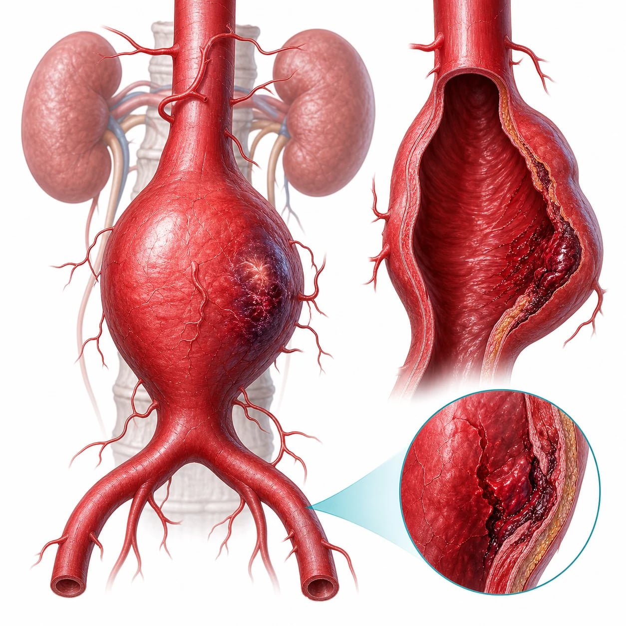
An abdominal aortic aneurysm is a permanent, localised dilation of the abdominal aorta to 3 cm or more (or 1.5 times the normal diameter), most commonly infrarenal. It is classified by its clinical state: an intact aneurysm is asymptomatic and found on screening or incidentally; a symptomatic (expanding or tender) aneurysm is a pre-rupture warning that needs urgent repair; and a ruptured aneurysm is the lethal emergency. The rupture is usually into the retroperitoneum (contained by the tissue pressure — a tamponade) and less commonly into the peritoneum (free, and fatal within minutes). [1]
Pathophysiology — Laplace's law and permissive hypotension
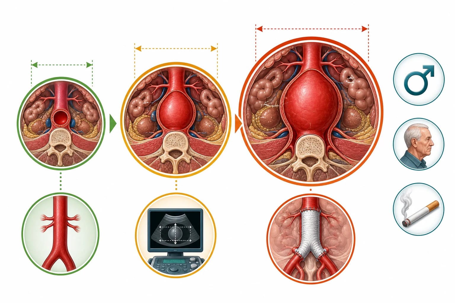
The wall tension of a vessel is proportional to the pressure and the radius (Laplace's law), so the larger the aneurysm, the higher the wall stress, the faster it grows, and the greater the risk of rupture — an exponential spiral. The wall thins and the structural elastin and collagen degrade with age, smoking and inflammation. When the aneurysm ruptures into the retroperitoneum, the extravasated blood is contained by the tissue pressure, forming a tamponade that limits the ongoing bleeding — this is the physiology behind permissive hypotension. A full resuscitation to a normal blood pressure raises the transmural pressure and disrupts the tamponade, causing a catastrophic re-bleed; a lower blood pressure (a systolic of 80 to 100) is enough for consciousness and urine output and preserves the tamponade.[1]
Epidemiology and risk factors
AAA is predominantly a disease of the older male smoker. The prevalence is 4 to 8 per cent in men over 65 (the population that screening programmes target), and roughly 1 to 2 per cent in women of the same age.[1] Smoking is the single strongest modifiable risk factor — it triples the risk of aneurysm formation and quadruples the risk of rupture — and smoking cessation slows the growth. The other risk factors are age (prevalence rises steeply after 65), male sex (a four-to-six-fold excess), hypertension, atherosclerosis, and a family history of an AAA in a first-degree relative (which roughly doubles the prevalence and warrants earlier screening, often from age 55 in men with an affected relative). The connective-tissue disorders (Marfan syndrome, vascular-type Ehlers-Danlos, Loeys-Dietz) and the vasculitides (Takayasu, giant-cell arteritis) produce aneurysms at a younger age but are uncommon. Women rupture at smaller diameters and present later, which is why the elective-repair threshold is lower for them.
Smoking
dominant modifiable risk
- Triples formation risk; quadruples rupture risk
- The number-one risk factor to name in a viva
- Cessation slows growth but does not reverse it
- Drives the screening target — the older male smoker
Age and male sex
demographic risk
- Prevalence 4–8% in men over 65
- Male-to-female ratio ~4–6:1
- Women rupture smaller and present later
- Repair threshold lower in women (5.0 cm)
Family history
first-degree relative
- ~2× prevalence with an affected first-degree relative
- Screening considered from age 50–55 in at-risk families
- Suggests a heritable connective-tissue trait
- Adds to risk even without a named syndrome
Connective-tissue disease
younger, rarer
- Marfan, vascular-type Ehlers-Danlos, Loeys-Dietz
- Produces aneurysms at a younger age
- Often thoracic and abdominal; more diffuse
- Low prevalence but high exam yield
Rupture risk by aneurysm diameter
The rupture risk is the single statistic that sets the elective-repair threshold, and it rises exponentially — not linearly — with diameter. The widely cited population estimates are: under 4 cm, less than 1 per cent per year; 4 to 5.5 cm, between 1 and 11 per cent per year; over 5.5 cm, over 30 per cent per year; and over 6 cm, the high-risk zone where rupture becomes a near-term probability rather than a distant possibility.[1] The growth rate itself accelerates with size (a 3-cm aneurysm grows roughly 0.1 to 0.2 cm a year; a 5-cm aneurysm grows faster), reinforcing the exponential spiral that Laplace's law predicts. Because the risk is non-linear, the surveillance interval shortens as the aneurysm enlarges — a 3 to 4 cm aneurysm is rescanned every two to three years, a 4 to 4.5 cm aneurysm yearly, and a 4.5 to 5.5 cm aneurysm every six months until it crosses the operative threshold.
Under 4 cm
- Rupture risk under 1% per year
- Surveillance ultrasound every 2–3 years
- Below operative consideration
- Reassure; manage cardiovascular risk and smoking
4 – 5.5 cm
- Rupture risk 1–11% per year
- Surveillance ultrasound every 6–12 months
- Growth rate accelerates with size
- Repair when crossing 5.5 cm (men) / 5.0 cm (women)
Over 5.5 cm
- Rupture risk over 30% per year
- Elective repair indicated
- Operative mortality 2–5% (elective)
- Far safer than the ~50% operative mortality of rupture
Over 6 cm
- High-risk zone — near-term rupture
- Repair strongly indicated; symptomatic even more so
- Rapid growth on serial scans is itself an indication
- Consider urgent rather than purely elective timing
Population screening
A one-time ultrasound of the abdominal aorta in men aged 65 to 74 reduces aneurysm-related mortality and is cost-effective, on the strength of the four randomised screening trials (MASS, Chichester, Viborg, Western Australia).[7] MASS, the largest, showed a sustained mortality benefit out to 10 years and remains the evidence cornerstone. Screening is not routinely recommended for women (the prevalence is low and the benefit marginal), for men under 65 (the prevalence is too low), or as a repeated lifelong test (a normal scan at 65 makes a later rupture very unlikely). The incidental finding of an AAA on an ultrasound, CT, or MRI performed for another reason — the back-pain scan, the renal-colic CT, the prostate workup — is a common route to diagnosis and must always be followed up with a measured diameter and a surveillance or referral plan.
[1]The rupture — presentation and the triad
The classic presentation is the triad (present in only about half the patients): sudden, severe abdominal, back or flank pain, hypotension, and a pulsatile abdominal mass. The atypical presentations are dangerous: a syncope or a collapse alone (from the acute blood loss), a groin pain that mimics renal colic (the older patient with "a stone" is always an AAA until proven otherwise), an acute limb ischaemia (a thromboembolism from the sac), and a gastrointestinal bleed (an aorto-enteric fistula — a "herald" bleed that precedes a massive, fatal haemorrhage). The key is a high index of suspicion in the older male smoker with abdominal or back symptoms. [1]
The triad is the headline, but the dangerous presentations are the ones that violate it. Each atypical rupture carries a classic trap that the Fellowship candidate must be able to name aloud. [1]
Syncope alone
- Sudden collapse from acute blood loss, no pain recalled
- The retroperitoneal tamponade hides the bleed
- An unexplained faint in an older patient is AAA until excluded
- Bedside ultrasound on every unexplained elderly collapse
Renal-colic mimic
- Flank/groin pain with haematuria from retroperitoneal irritation
- The older first-time "stone" patient is AAA first
- Pulsatile mass and a bedside aortic measure are the defence
- A CT KUB will also show the aneurysm — but check it
Acute limb ischaemia
- Thromboembolism from mural thrombus in the aneurysm sac
- May precede or accompany the rupture
- Examine the abdomen of every acutely ischaemic limb
- A cold leg plus a pulsatile mass is a vascular emergency
Aorto-enteric fistula
- A herald GI bleed hours-to-days before a fatal massive bleed
- Prior AAA repair or a known aneurysm is the clue
- The duodenum (third/fourth part) is the commonest site
- Endoscopy may miss it; CT angiography is the test
Differential diagnosis
The abdominal or back pain of a ruptured AAA has a differential, and the danger is treating it as something less lethal. [1]
Ruptured AAA
- Sudden abdominal/back pain, hypotension, pulsatile mass
- Older male smoker; the triad in ~50%
- Unstable → theatre, not CT; permissive hypotension
- Mortality ~80–90% overall
Renal colic
- Flank pain, haematuria; younger, no mass
- No hypotension (unless septic from obstruction)
- CT KUB confirms the stone
- AAA MUST be excluded in the older patient
Pancreatitis / perforation
- Epigastric pain, raised lipase (pancreatitis)
- Perforation: sudden, rigid abdomen, pneumoperitoneum
- No pulsatile mass
- CT confirms
Aorto-enteric fistula
- Herald GI bleed then massive; prior AAA repair or a known aneurysm
- Needs urgent surgery and an endovascular bridge
- A known graft → fistula until proven otherwise
- Not a "simple" GI bleed
Investigations and the stability decision
The single most important decision is the stability, because it determines the imaging. The unstable patient with a clinically suspected rupture is diagnosed at the bedside — a pulsatile mass and a bedside ultrasound that confirms the aneurysm's diameter — and goes straight to the theatre or the catheter-laboratory without a CT scan, because the delay is lethal. The stable or the equivocal patient (a possible leak, an unclear cause) gets a rapid CT angiography that confirms the aneurysm, the rupture site, the contrast extravasation, and the suitability for an endovascular repair. Blood is drawn for a group and crossmatch (6 to 10 units), a full blood count, the coagulation, the electrolytes and the lactate, and the massive haemorrhage protocol is activated. An ECG excludes a myocardial infarction as a differential or a precipitant. [1]
Bedside ultrasound in the unstable patient
In the haemodynamically unstable patient with a suspected rupture, point-of-care ultrasound is the only imaging that should happen. The operator images the aorta in the transverse plane from the xiphisternum caudally to the bifurcation, measuring the maximal outer-wall to outer-wall diameter; an aortic diameter of 3 cm or more confirms the aneurysm, and in the right clinical setting (an unstable older patient with abdominal or back pain) that confirmation is sufficient to take the patient to theatre. Critically, the ultrasound confirms the aneurysm but not the rupture — there is no free fluid to see because the blood is contained retroperitoneally, and a normal-looking aorta does not exclude a para-anastomotic or iliac rupture — so a clinical suspicion with a confirmatory aneurysm goes to the operating room regardless. The FAST (focused assessment with sonography in trauma) sweep may show a thin sliver of intraperitoneal fluid in the free intraperitoneal rupture, but its absence is meaningless. [1]
[1]Suspected ruptured AAA — the ED first 30 minutes
Triage to the resuscitation bay; high-flow oxygen if hypoxic; full monitoring (three-lead ECG, continuous SpO2, non-invasive BP cycled every 3 minutes); two large-bore (14–16 G) cannulae. Do NOT delay for a third line.
Focused history: sudden abdominal/back/flank pain, syncope, prior AAA repair or known aneurysm, smoking and cardiovascular risk. Targeted exam: palpate for a pulsatile mass, feel all pulses, exclude a rigid abdomen (perforation) and check for melaena (aorto-enteric fistula).
Apply permissive hypotension: target a systolic of 80–100 mmHg in the conscious patient. Give small aliquots of blood or a balanced crystalloid only to maintain consciousness, never to chase a normal pressure.
Perform bedside ultrasound to confirm the aneurysm diameter (aorta 3 cm or more). Do NOT send the unstable patient to CT.
Activate the massive haemorrhage protocol: blood, FFP and platelets in a 1:1:1 ratio; crossmatch 6 to 10 units; give 10 mL of 10% calcium chloride after every four units of red cells; warm the patient and the fluids.
Send bloods (group and crossmatch, full blood count, coagulation, electrolytes, lactate, troponin) and a 12-lead ECG, but do NOT wait for results to act.
Notify the vascular surgeon, the anaesthetist, the theatre or the endovascular suite, and the blood bank at once — the activation is part of the resuscitation, not a step after it. Mobilise the endovascular-first pathway where available.
If collapse or arrest supervenes before control, consider resuscitative endovascular balloon occlusion of the aorta (REBOA) at zone 1 as a bridge to definitive repair.
Immediate management — permissive hypotension and the protocol
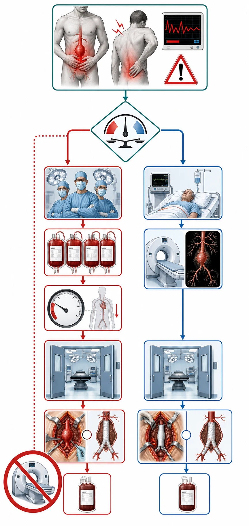
Stabilise the airway and the breathing, give oxygen, and apply the permissive-hypotension principle. [1]
[1] [1]The AAA thresholds and the protocol
The massive haemorrhage protocol delivers the blood products in packs of red cells, fresh-frozen plasma and platelets in a 1:1:1 ratio, and includes calcium replacement (10 mL of 10 per cent calcium chloride intravenously after every four units of red cells) to counteract the citrate-induced hypocalcaemia of the massive transfusion. The resuscitation fluid is blood, not crystalloid: a 250 mL aliquot of a balanced crystalloid is used to maintain the conscious level between the blood-product packs, and a larger crystalloid volume is avoided because it dilutes the coagulation factors and worsens the bleeding. A vasopressor is avoided in principle, but a noradrenaline infusion at 0.05 to 0.5 micrograms per kilogram per minute is reserved for the profoundly unconscious patient (a systolic under 70) where the permissive-hypotension target cannot be met and the brain is at risk. The patient is kept warm with a forced-air warmer and a fluid warmer, because a core temperature below 35 degrees Celsius produces a coagulopathy that worsens the bleeding, and the coagulation is monitored with a point-of-care viscoelastic test (a ROTEM or a TEG) and corrected with cryoprecipitate and fibrinogen as the products are transfused. The anaesthetist is warned of the risk of an induction hypotension and a rapid blood loss on the cross-clamp. The early notification of the vascular surgeon, the anaesthetist, the theatre and the blood bank is the single most important system step — it is the call that buys the minutes the patient has. [1]
Definitive management — the repair
The ruptured AAA is repaired immediately by an open surgical repair (a laparotomy, a supracoeliac cross-clamp, and a tube or a bifurcation graft) or an endovascular aneurysm repair (EVAR) (a stent-graft delivered from the femoral access, if the anatomy is suitable and the centre is equipped). The IMPROVE trial showed that an endovascular-first strategy for a suspected rupture reduced the 30-day mortality in suitable patients.[2] The choice depends on the haemodynamic stability, the computed-tomographic anatomy (if available), and the centre's capability. The symptomatic non-ruptured aneurysm is repaired urgently within 24 hours. The asymptomatic aneurysm is repaired electively at or above 5.5 cm in men and 5.0 cm in women, and surveilled with a yearly ultrasound below the threshold.[1]
Open versus endovascular repair — the anatomy decides
EVAR is only possible if the anatomy fits, and the eligibility rests on four features of the preoperative CT: a proximal aneurysm neck (a length of normal aorta below the renal arteries for the stent-graft to grip) of at least 10 to 15 mm, a neck diameter under about 28 mm, a neck angulation under 60 degrees, and non-diseased access vessels (common femoral and iliac arteries of adequate calibre to admit the delivery sheath). Only about half of all ruptures meet these criteria on anatomy alone. Open repair has no anatomical constraint but carries the cost of a laparotomy, a supracoeliac cross-clamp (with its renal, mesenteric and spinal-perfusion insult), and a higher physiological burden; it remains the default where EVAR anatomy is unfavourable or the centre is not equipped for an emergency endovascular repair. [1]
EVAR
endovascular aneurysm repair
- Anatomy-dependent (neck length, angulation, access)
- Lower perioperative mortality in elective and selected ruptures
- Faster recovery; no cross-clamp insult
- Needs lifelong surveillance for endoleak; re-interventions common
Open repair
- No anatomical constraint — the universal option
- Supracoeliac clamp; higher physiological insult
- Longer stay and recovery; renal and mesenteric risk
- Durable; no endoleak surveillance burden
The endoleak — the EVAR-specific late problem
An endoleak is persistent blood flow into the aneurysm sac after EVAR, and it is the reason every EVAR patient needs lifelong CT surveillance. The five types matter for the viva: type 1 (an incomplete seal at the proximal or distal attachment — high pressure, needs repair), type 2 (retrograde flow from a lumbar or inferior mesenteric artery — low pressure, the commonest, often observed), type 3 (graft fabric tear or component separation — high pressure, needs repair), type 4 (graft porosity, perioperative, self-limiting), and type 5 (continued sac expansion with no identifiable leak — "endotension"). Types 1 and 3 threaten rupture and need re-intervention; type 2 is usually watched unless the sac grows. [1]
Type 1 endoleak
- Incomplete seal at the attachment site (proximal or distal)
- High-pressure — threatens rupture
- Needs prompt repair (cuff, extension, re-grafting)
- A technical failure of the seal
Type 2 endoleak
- Retrograde flow from a lumbar or IMA branch
- Low pressure; the commonest type
- Usually observed unless the sac grows
- Intervention only if sac expands on surveillance
Type 3 endoleak
- Graft fabric tear or modular-component separation
- High pressure — threatens rupture
- Needs repair (relining or component exchange)
- A structural graft failure
Type 4 / 5
- Type 4: graft-wall porosity, perioperative, self-limiting
- Type 5: sac growth with no identifiable leak ("endotension")
- Both managed conservatively initially
- Sac growth drives escalation in either
REBOA — the bridge to definitive control
Resuscitative endovascular balloon occlusion of the aorta (REBOA) inflates a balloon in the descending thoracic aorta (zone 1, above the coeliac) or the infrarenal aorta (zone 3) to arrest non-compressible torso haemorrhage. In the ruptured AAA it is deployed as a bridge — to stabilise the blood pressure and reduce ongoing loss while the patient is moved to theatre or the endovascular suite — not as definitive treatment. Its risks are ischaemia distal to the balloon (renal, mesenteric, spinal, limb), and the occlusion time must be kept short and reassessed continuously. In the arresting or peri-arrest patient it can replace an emergency resuscitative thoracotomy with aortic cross-clamping as the initial control manoeuvre. [1]
[1]Elective repair thresholds and surveillance
The asymptomatic aneurysm is managed by diameter against a surveillance schedule, and the operative threshold balances the annual rupture risk against the operative mortality.[1] In men, elective repair is offered at 5.5 cm; in women, at 5.0 cm (because women rupture at smaller diameters). A rapid growth rate (over 0.5 cm in six months, or over 1 cm a year) is an independent indication to repair even below the diameter threshold, as is a symptomatic aneurysm (tenderness or back pain), which is treated as a pre-rupture and repaired urgently. Below the threshold, surveillance follows the growth: 3 to 4 cm every two to three years; 4 to 4.5 cm yearly; 4.5 to 5.5 cm every six months.
Elective thresholds and surveillance
The trials in brief — open, EVAR, and the ruptured-aneurysm question
The elective EVAR evidence rests on three randomised trials (EVAR-1, DREAM, OVER) that each showed a short-term survival advantage for EVAR that narrows with long-term follow-up as late ruptures and re-interventions accrue. The ruptured-aneurysm evidence is the IMPROVE trial and its longer-term follow-up, plus the Sweeting individual-patient-data meta-analysis that pooled the rupture trials.[2][3][4][5][6][8]
IMPROVE — endovascular-first strategy for ruptured AAA (BMJ 2014)
BMJ
PMID 24418950
Key finding
A pragmatic randomised trial of 613 patients with a clinical diagnosis of ruptured AAA, comparing an endovascular-first strategy (angiography and EVAR if anatomy suited) against open repair. The 30-day mortality was not significantly different overall (35.7% endovascular versus 36.4% open), but among women and the anatomically suitable patients the endovascular strategy showed a clear mortality benefit, and the strategy reduced ICU and hospital stay.
Practice change
Established that an endovascular-first pathway is safe and standard for a suspected ruptured AAA where the anatomy and the centre permit it — the protocol that drives the ED activation.<Cite id='2' />
IMPROVE — 1.3-year comparative effectiveness and cost-effectiveness (BMJ 2017)
BMJ
PMID 29138135
Key finding
The longer-term follow-up of IMPROVE reporting clinical effectiveness and cost-effectiveness of the endovascular strategy versus open repair for ruptured AAA. The mortality advantage in women persisted, and the endovascular strategy was cost-effective on the strength of shorter critical-care and hospital stays and a better quality of life in survivors.
Practice change
Confirmed that the endovascular-first strategy is not only safe and effective but cost-effective, solidifying it as the default pathway in equipped centres.<Cite id='3' />
EVAR trial 1 — EVAR vs open for large AAA (Lancet 2004)
Lancet
PMID 15351191
Key finding
A randomised trial of 1,082 patients with an AAA of at least 5.5 cm who were fit for open repair, comparing EVAR with open repair. EVAR reduced the 30-day operative mortality from 4.7% to 1.7%, an absolute benefit that persisted over the intermediate term, though graft-related complications and re-interventions were common and the long-term mortality advantage narrowed.
Practice change
Established EVAR as a legitimate alternative to open repair for the anatomically suitable elective AAA, with an early survival advantage offset by a surveillance and re-intervention burden.<Cite id='4' />
DREAM — EVAR vs open for AAA (NEJM 2004)
New England Journal of Medicine
PMID 15483279
Key finding
A multicentre randomised trial of 345 patients with an AAA of at least 5 cm and suitable anatomy, comparing endovascular with open repair. The cumulative operative mortality at 30 days was 1.2% after EVAR versus 4.6% after open repair, a significant early advantage that, as in EVAR-1, narrowed over the longer term.
Practice change
Independently corroborated EVAR-1: EVAR carries a lower early operative mortality in fit patients with suitable anatomy, at the cost of later re-intervention.<Cite id='5' />
OVER — endovascular vs open repair in veterans (JAMA 2009)
JAMA
PMID 19826022
Key finding
A randomised trial of 881 veterans with an AAA fit for either repair. EVAR again reduced perioperative mortality (0.5% versus 3.0%), but the survival curves converged by two years and there was no significant long-term survival difference; EVAR carried more subsequent procedures.
Practice change
The third and largest of the elective EVAR trials: the early mortality advantage is real but transient, and the long-term outcomes are equivalent — emphasising that EVAR is an alternative, not a superior, strategy, and that lifelong surveillance is essential.<Cite id='6' />
MASS — screening men for AAA (Ann Intern Med 2007)
Annals of Internal Medicine
PMID 17502630
Key finding
The long-term follow-up of the Multicentre Aneurysm Screening Study, a randomised trial of a one-time invitation to an abdominal aortic ultrasound in men aged 65 to 74. Screening produced a sustained reduction in aneurysm-related mortality that persisted out to 10 years, and the benefit was cost-effective.
Practice change
The evidence cornerstone for the one-time ultrasound screening programme in older men — the basis of population screening recommendations worldwide.<Cite id='7' />
Sweeting IPD meta-analysis — ruptured AAA 1-year mortality (EJVES 2015)
European Journal of Vascular and Endovascular Surgery
PMID 25981698
Key finding
An individual-patient-data meta-analysis of the three randomised ruptured-AAA trials (IMPROVE, AJAX, ECAR) examining 1-year mortality after endovascular-first versus open strategies. The pooled analysis showed no significant overall difference in 1-year mortality, but the endovascular strategy appeared superior in women and in those haemodynamically unstable at presentation.
Practice change
Synthesised the ruptured-AAA randomised evidence: the endovascular-first strategy is at least as good as open repair overall and may benefit the highest-risk subgroups — supporting a default endovascular-first pathway in equipped centres.<Cite id='8' />
Aorto-enteric fistula — the herald bleed
An aorto-enteric fistula is a communication between the aorta (native or, far more commonly, a graft) and the bowel, almost always the third or fourth part of the duodenum. The classic history is a herald (sentinel) bleed — a small, self-limiting episode of upper gastrointestinal bleeding — followed hours to days later by a massive, exsanguinating haemorrhage as the fistula opens fully. The single most important risk factor is a prior AAA repair (open or endovascular): any patient with a known aortic graft who presents with any gastrointestinal bleeding has an aorto-enteric fistula until proven otherwise. Endoscopy may miss it (the fistula is distal, in the duodenum, and easy to overlook); CT angiography is the diagnostic test of choice, and the management is urgent surgery, often with an endovascular bridge to control the bleeding first. A primary aorto-enteric fistula (in a native, ungrafted aneurysm) is rarer but follows the same rule. The trap is to manage the herald bleed as a simple upper GI bleed and discharge the patient, who then returns with the fatal bleed. [1]
[1]Perioperative and anaesthetic pitfalls
The anaesthetic induction in the ruptured AAA is itself a high-risk moment: the sympathectomy of induction on a hypovolaemic, vasoconstricted patient precipitates a profound drop in blood pressure, and the loss of the compensatory tone can unmask the full extent of the bleed. The vascular and anaesthetic teams therefore prepare for an induction hypotension with blood already running and the surgeon scrubbed and ready to cross-clamp or balloon-occlude the moment the patient is anaesthetised. The supracoeliac cross-clamp controls the bleeding but at the cost of mesenteric, renal and spinal ischaemia; it is released to an infrarenal position as soon as the proximal control is secured. The lethal triad of hypothermia, acidosis and coagulopathy is the on-table enemy, and damage-control resuscitation — warmed blood in a 1:1:1 ratio, calcium, viscoelastic-guided factor replacement, and prompt definitive control — is aimed at breaking the triad before it becomes irreversible. The patient who survives the operation is admitted to the intensive care for the reperfusion, the renal, the respiratory and the abdominal-compartment consequences of the insult. [1]
[1] [1]Complications and pitfalls
The complication of the untreated rupture is a massive haemorrhage and death; the complications of the repair are the renal failure (from hypoperfusion and the contrast), the colonic ischaemia, the spinal-cord ischaemia, the lower-limb ischaemia, the graft infection, and the late aorto-enteric fistula. The pitfalls are the inverse of the protocol: over-resuscitation disrupting the tamponade; waiting for a CT in the unstable patient; missing the diagnosis by treating the patient as a renal colic or a pancreatitis; not activating the massive haemorrhage protocol; and delaying the surgical or the endovascular activation. [1]
Prognosis and disposition
The overall mortality of a ruptured AAA is 80 to 90 per cent, including the pre-hospital deaths; of those who reach the theatre alive, about half survive. The symptomatic non-ruptured aneurysm has a much better prognosis with an urgent repair, and the elective repair carries a mortality of 2 to 5 per cent. The patient with a rupture is taken directly to the theatre or the catheter-laboratory and then to the intensive care. The long-term endovascular patient is surveilled for an endoleak. [1]
The survival is also determined by the time from the rupture to the cross-clamp or the balloon occlusion — every minute of delay increases the blood loss, the coagulopathy and the multi-organ failure that follows, which is why the pre-hospital notification and the theatre-activation pathway are as important as the operative technique itself. [1]
The elderly male smoker is the classic patient, and the combination of smoking and age accounts for most of the population risk. Women have a lower prevalence but a higher rupture risk at a smaller diameter, so the elective-repair threshold is lower (5.0 cm) and the diagnosis is more often missed because it is less expected. A family history of an AAA in a first-degree relative lowers the screening age and raises the prevalence. The cardiac comorbidity (ischaemic heart disease, heart failure) raises the perioperative risk of both the open and the endovascular repair and may determine the repair strategy — the patient who cannot tolerate a cross-clamp may be better served by an endovascular approach. [1]
Evidence and regional guidelines
The contemporary framework is the 2022 ACC/AHA aortic disease guideline.[1] The endovascular-first strategy evidence is the IMPROVE trial.[2] The repair approach (open vs EVAR) and the screening programme follow the local vascular-society pathway — in ANZ there is no universal screening programme, and the repair choice is governed by the centre's vascular service.
ANZ practice note. The permissive-hypotension protocol and the clinical diagnosis of the unstable rupture follow the vascular-society pathways; the patient is routed to the nearest vascular service for an open or an endovascular repair, and the screening of the at-risk older male is opportunistic rather than programme-based. [1]
A basket of high-yield pearls
[1] [1] [1] [1] [1] [1] [1] [1] [1] [1] [1] [1] [1] [1] [1] [1] [1]Additional red flags
[1]Exam pearls
- Triad: sudden abdominal/back pain + hypotension + pulsatile mass — present in only ~50 per cent.
- Unstable → clinical diagnosis → theatre, NOT a CT scanner.
- Permissive hypotension: systolic 80 to 100 — do not disrupt the retroperitoneal tamponade.
- Activate the massive haemorrhage protocol: blood : FFP : platelets 1:1:1, crossmatch 6 to 10 units.
- Always consider AAA in the older patient with apparent renal colic.
- 5.5 cm (men) / 5.0 cm (women) = the elective-repair threshold.
- Mortality ~80 to 90 per cent overall; ~50 per cent of those who reach the theatre. [1]
Exam practice
SAQ — Ruptured AAA in an elderly male smoker
10 minutes · 10 marks
A 78-year-old male smoker is brought to the emergency department 25 minutes after the sudden onset of severe central abdominal and lower-back pain at home, followed by a syncopal episode. In the resuscitation bay he is pale, diaphoretic and drowsy (GCS 14). BP 76/48, HR 128, SpO2 95 per cent on 15 L oxygen via a non-rebreather mask. Abdominal examination reveals a tender, pulsatile epigastric mass. Point-of-care ultrasound shows an infrarenal aortic diameter of 7.2 cm with no free intraperitoneal fluid. Lactate 6.4 mmol/L, haemoglobin 92 g/L. The vascular surgeon is 40 minutes away.
SAQ — Symptomatic non-ruptured AAA with perioperative optimisation
10 minutes · 10 marks
A 68-year-old male is referred to the emergency department by his general practitioner. He has a known infrarenal AAA measured at 5.4 cm on ultrasound six months ago under surveillance. Over the last 48 hours he has developed new, constant, dull central abdominal and back pain with tenderness directly over a palpable, pulsatile epigastric mass. He is alert and haemodynamically stable: BP 138/82, HR 84, SpO2 98 per cent on room air, lactate 1.1 mmol/L, haemoglobin 141 g/L. He is a current 40-pack-year smoker with hypertension, dyslipidaemia and a prior NSTEMI three years ago. CT angiography confirms a 6.1 cm infrarenal AAA with a favourable proximal neck (length 18 mm, diameter 26 mm, angulation 35 degrees) and patent iliac arteries.
Red flags
[1]References
- [1]Isselbacher EM, Preventza O, Hamilton Black J, et al. 2022 ACC/AHA guideline for the diagnosis and management of aortic disease: A report of the American Heart Association/American College of Cardiology Joint Committee on Clinical Practice Guidelines J Thorac Cardiovasc Surg, 2023.PMID 37389507
- [2]IMPROVE Trial Investigators. Endovascular or open repair strategy for ruptured abdominal aortic aneurysm: 30 day outcomes from IMPROVE randomised trial BMJ, 2014.PMID 24418950
- [3]IMPROVE Trial Investigators. Comparative clinical effectiveness and cost effectiveness of endovascular strategy v open repair for ruptured abdominal aortic aneurysm: three year results of the IMPROVE randomised trial BMJ, 2017.PMID 29138135
- [4]Greenhalgh RM, Brown LC, Kwong GP, et al. Comparison of endovascular aneurysm repair with open repair in patients with abdominal aortic aneurysm (EVAR trial 1), 30-day operative mortality results: randomised controlled trial Lancet, 2004.PMID 15351191
- [5]Prinssen M, Verhoeven EL, Buth J, et al. A randomized trial comparing conventional and endovascular repair of abdominal aortic aneurysms N Engl J Med, 2004.PMID 15483279
- [6]Lederle FA, Freischlag JA, Kyriakides TC, et al. Outcomes following endovascular vs open repair of abdominal aortic aneurysm: a randomized trial JAMA, 2009.PMID 19826022
- [7]Kim LG, Scott RA, Ashton HA, et al. A sustained mortality benefit from screening for abdominal aortic aneurysm Ann Intern Med, 2007.PMID 17502630
- [8]Sweeting MJ, Bown MJ, Roy-Choudhury P, et al. Ruptured Aneurysm Trials: The Importance of Longer-term Outcomes and Meta-analysis for 1-year Mortality Eur J Vasc Endovasc Surg, 2015.PMID 25981698