EM · Procedural & diagnostic ED skills
Wound assessment and management
Also known as Wound care · Laceration repair · Wound closure · Traumatic wound management · Bite wound management · Tetanus prophylaxis · Wound irrigation
Wound assessment and management in the ED — the wound classes (clean, clean-contaminated, contaminated, dirty-infected) mapped to the mechanism classes (laceration, crush, bite, puncture, avulsion), the structured MEAT assessment (mechanism, examination of neurovascular-tendon-bone-depth-contamination-foreign body, age of wound, type), the irrigation standard (normal saline under 7 to 15 psi pressure via splash-guard syringe, large volume), debridement of devitalised tissue, the tetanus prophylaxis schedule (diphtheria-tetanus-acellular-pertussis vaccine 0.5 mL IM plus tetanus immune globulin 250 IU IM per the ACIP and Australian schedule by wound type and vaccination history), the antibiotic decision (amoxicillin-clavulanate 875/125 mg BD for mammalian bites covering Pasteurella), and the closure decision (primary versus delayed primary versus secondary). ACEM-primary, globally tagged.
On this page & tools
Your progress
Saved locally on this device.
8 MCQs with explanations
Target exams
Red flags
Related topics
- Local anaesthesia and topical agents
- Procedural sedation in the emergency department
- Consent, capacity and the medico-legal framework in the emergency department
- Major trauma resuscitation — the team-based systematic approach
- Secondary survey
- Burn management in the emergency department
- Paediatric trauma — the modified approach
- Acute limb ischaemia (the emergency of the 6 Ps)
Wound assessment and management is one of the highest-volume procedural skills in emergency medicine — lacerations, abrasions, bites and punctures together account for a third or more of injury presentations to Australasian EDs. The Fellowship candidate must master three decisions that the examiner will press in any wound stem: how to assess the wound systematically (mechanism, depth, contamination, neurovascular, tendon, bone, foreign body), how to clean and debride it to the irrigation standard, and how to decide between primary, delayed primary, and secondary closure — because the wrong closure on a contaminated wound converts a low-risk laceration into a deep infection, a missed tendon injury, or a tetanus case. Two further obligations ride on every wound: tetanus prophylaxis decided by wound type and vaccination history, and antibiotic prophylaxis reserved for the high-risk wound — the bite, the heavily contaminated laceration, the immunocompromised host, and the hand wound.[3][4]
Wound classification — the two frameworks
Two classification systems run in parallel and the candidate must hold both. The first is the surgical wound-class system, originally describing operative contamination risk and adapted to traumatic wounds, which stratifies by the expected bacterial load and predicts infection risk. The second is the mechanism class, which describes how the wound was produced and predicts the pattern of tissue injury, the likely contaminant, and the closure decision. [1]
The mechanism class maps onto these and onto the management plan, because each mechanism carries a signature pattern of injury and contamination. A laceration is a sharp-edge injury (knife, glass, metal) producing clean edges and low contamination — the wound most often suitable for primary closure. A crush or abrasion is a blunt-force injury producing irregular, devitalised edges, a higher infection risk, and often underlying fracture or compartment injury. A bite — dog, cat, human or other mammal — inoculates the wound with oral flora and is the prototypical wound that is cleaned but not closed. A puncture drives a small skin breach deep into tissue, carrying surface bacteria and material (rubber, fabric) with it, and is the mechanism behind the high-risk plantar puncture through a shoe. An avulsion tears tissue away from its blood supply, producing devitalised flaps that require debridement. A burn is a coagulative injury with its own management pathway.[5]
Indications and the structured MEAT assessment
Every traumatic wound is assessed against a fixed sequence so that no high-risk feature is missed under time pressure. The candidate must be able to deliver the assessment out loud in an OSCE: Mechanism, Examination (depth, contamination, foreign body, neurovascular, tendon, bone), Age of the wound, Type of contamination and host. [1]
The structured wound assessment
MEAT
Laceration (sharp, clean edges), crush (devitalised), bite (inoculated), puncture (deep narrow tract), avulsion (flap), burn. Mechanism predicts injury pattern and contamination.
Depth through skin layers, contamination with soil/saliva/water, retained foreign body (explore, image), neurovascular distal to the wound (pulse, capillary refill, two-point discrimination), tendon tested through full active range of motion against resistance, underlying bone or joint involvement.
Time since injury. The historical "golden period" of 6 hours is contested — modern evidence supports primary closure of low-risk lacerations up to 18 to 24 hours, but heavily contaminated and bite wounds are never closed primarily regardless of age.
Type of contaminant (soil, saliva, fresh or salt water, organic material) and the host (diabetes, immunosuppression, peripheral vascular disease, anticoagulation, prosthetic valve or joint) — both lower the threshold for antibiotics and delayed closure.
The neurovascular examination is the non-negotiable step: document the distal pulse, capillary refill, and light-touch and two-point discrimination distal to the wound BEFORE injecting local anaesthetic, because anaesthesia abolishes the chance to re-test. On a digit, two-point discrimination is tested at 5 to 8 mm on the pulp; an asymmetry greater than 2 mm compared with the contralateral digit indicates a digital nerve injury. The tendon examination is performed by asking the patient to move the relevant joint through full active range of motion against resistance while the examiner watches the tendon glide through the wound bed — a partial tendon laceration can be missed if the test is not done against resistance, because the intact fibres hold the tendon in apposition. The foreign body examination rests on exploration under good lighting and local anaesthesia, supplemented by imaging: glass and metal are radio-opaque and seen on plain radiograph; wood and plastic are radiolucent and require ultrasound. A retained foreign body is a leading cause of delayed infection and a frequent medicolegal claim.[3]
Contraindications to primary closure
The decision to close a wound primarily is the decision the examiner most often tests, because the wrong closure is worse than no closure. Primary closure is contraindicated whenever the wound is too contaminated, too old, or too devitalised to heal by first intention, or whenever closing it would trap inoculated bacteria in a deep space. [1]
Bite wounds (mammalian and human)
- Inoculated with oral flora — Pasteurella multocida (cat, dog), Capnocytophaga canimorsus (dog), Streptococcus, Staphylococcus, anaerobes; human bites add Eikenella corrodens
- Infection rate 10 to 50 per cent if closed; the prototypical wound to clean and leave open or close by delayed primary intention
- Exceptions: facial bites (high cosmetic priority, low infection rate if seen early) may be closed primarily by plastics after copious irrigation; clean early bites of the face seen within 6 to 12 hours
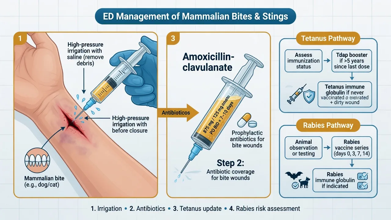
- Give amoxicillin-clavulanate 875/125 mg PO BD for 5 to 7 days to ALL mammalian bites regardless of closure decision
Puncture wounds (especially plantar through footwear)
- Small skin breach, deep tract — do not enlarge or close; irrigation cannot reach the depth of the tract
- Plantar puncture through a rubber-soled shoe carries Pseudomonas aeruginosa and osteomyelitis risk; image for retained shoe material, do not probe blindly
- Antibiotic coverage if deep, proximal, immunocompromised, or through footwear — ciprofloxacin covers Pseudomonas
- Warn the patient of the late osteomyelitis risk and arrange early review
Crush and heavily contaminated wounds
- Irregular, devitalised edges and gross contamination (soil, organic material, lake or sea water)
- Requires surgical debridement of non-viable tissue and copious irrigation before any closure decision
- Delay closure (delayed primary at day 4 to 5) if contamination is heavy or the host is high-risk
- Tetanus prophylaxis is especially important — soil and organic material are the classic vectors for Clostridium tetani spores
Old wounds (over 6 to 24 hours, depending on site)
- The historical "golden period" of 6 hours is contested — Quinn showed no infection difference for low-risk lacerations closed up to 18 to 24 hours
- However, infection risk rises with age, contamination and high-tension sites (extremities); face has low infection risk and heals fast — close facial lacerations up to 24 hours
- The decision integrates age, contamination, site, mechanism and host, not a single clock threshold
Wounds with established infection
- Already clinically infected (pus, cellulitis, systemic signs) — never close; open, debride, culture if deep, antibiotics, heal by secondary intention
- Treat as an abscess if a cavity is present — incise and drain
The discriminating question on every wound is: does closing this wound trap bacteria in a space I cannot irrigate? If yes, do not close.[4][5]
Differential diagnosis — what looks like a simple laceration but is not
The recogniser's task is to identify the wound that is more than a wound. Several high-risk entities present as a laceration, abrasion or puncture and demand a different pathway. [1]
Open fracture
- Any wound communicating with a fracture, or a wound over a fracture site with periosteal contamination
- Requires IV antibiotics (cephalosporin + aminoglycoside ± metronidazole per Gustilo grade), tetanus, orthopaedic theatre — not ED closure
- Suspect in crush mechanisms, high-energy wounds, wounds near a joint, or when bone is visible on exploration
Necrotising soft tissue infection
- Pain out of proportion, tensely oedematous, rapidly spreading induration, systemic toxicity, skin necrosis or bullae, crepitus
- Time-critical surgical emergency — do not close, do not wait for a swab; broad-spectrum antibiotics and urgent surgical debridement
- Risk factors: diabetes, immunosuppression, deep wound, sea water (Vibrio), contaminated wound
Retained foreign body
- Persistent pain, failure to heal, recurrent infection after a wound — the classic late presentation
- Explore under LA, image (radiograph for glass/metal, ultrasound for wood/plastic)
- Remove in ED if accessible; refer for theatre if deep, near a nerve or vessel, or in a child needing sedation
Tendon, nerve or vessel injury
- Test tendon through full active range of motion against resistance; test 2-point discrimination and light touch on every digit
- A partial tendon laceration is easily missed if not tested under load — refer to hand surgery for repair
- Document neurovascular status BEFORE local anaesthesia; a missed digital nerve is a functional and medicolegal injury
Infected bite with sepsis
- Dog or cat bite with rapidly spreading cellulitis, lymphangitis, fever — Capnocytophaga canimorsus can cause sepsis and DIC in the immunocompromised or asplenic patient
- IV antibiotics (amoxicillin-clavulanate or piperacillin-tazobactam), sepsis pathway, surgical review for deep infection
- Asplenic and immunocompromised patients with dog bites are high-risk and warrant admission
Equipment, patient preparation and consent
Wound repair needs a defined set: irrigation equipment (a 35 to 60 mL syringe, a 19-gauge needle or a splash-guard device, normal saline in large volume — 250 mL minimum, up to a litre for contaminated wounds), debridement instruments (iris scissors, forceps, a scalpel with a number 15 blade), exploration instruments (a probe, good lighting, sometimes loupes), closure materials (sutures of the chosen gauge, tissue adhesive, adhesive strips, staples), dressing materials (sterile gauze, non-adherent dressing, bandage), and local anaesthesia equipment per the local-anaesthesia topic (lidocaine 1 per cent with or without adrenaline, LET gel for children). Patient preparation covers the focused history — mechanism and time, allergies (especially to latex, adhesive, local anaesthetic preservative), tetanus status, comorbidity (diabetes, immunosuppression, vascular disease, anticoagulation), medications and last meal if sedation is contemplated. Consent is obtained with the procedure, the agent, and the risks explained (infection, dehiscence, scar, retained foreign body, neurovascular injury, tetanus). The single most important preparation step is the neurovascular and tendon check documented before anaesthesia — the step most often omitted and most often litigated.[3]
The irrigation standard
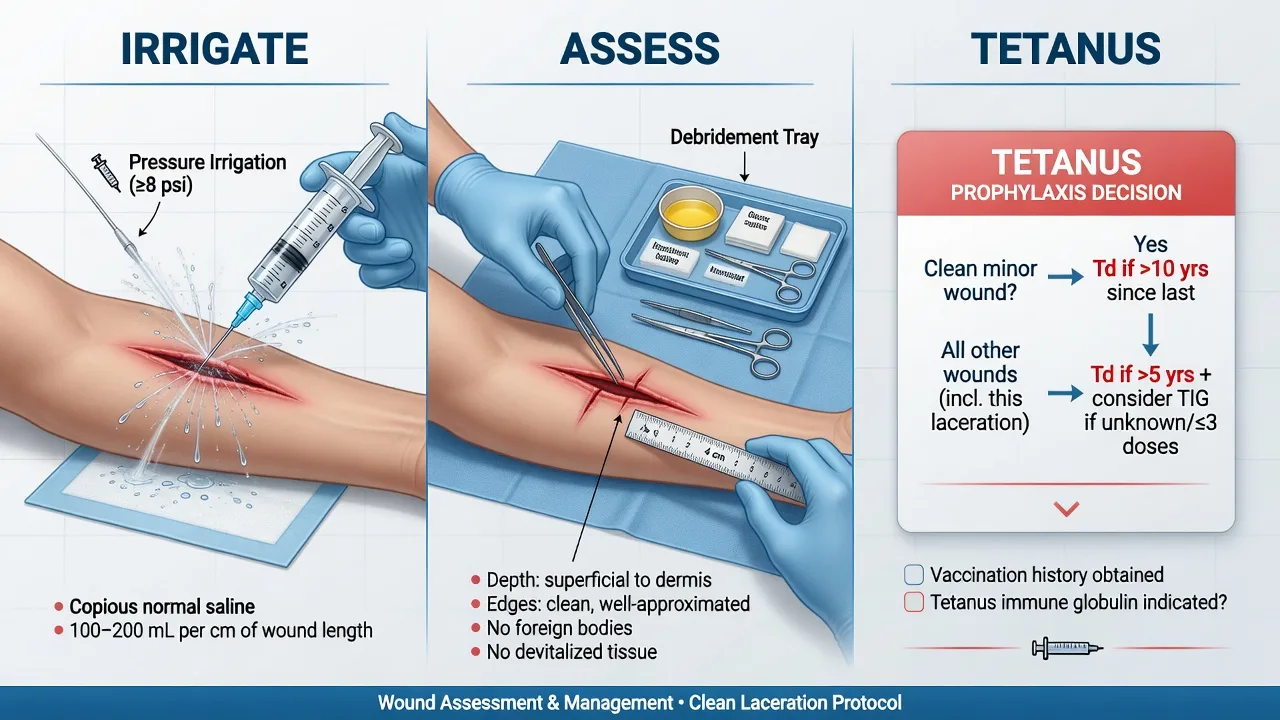
Irrigation is the single most effective intervention to reduce wound infection, more important than the choice of suture or the choice of antibiotic. The standard is normal saline delivered under pressure in large volume. The pressure target is 5 to 8 psi (some authorities accept up to 15 psi) — achieved practically with a 35 to 60 mL syringe fitted with a 19-gauge needle or a commercial splash-guard device, held close to the wound surface. Higher pressure (over 15 to 20 psi, as from a pressurised fluid bag or pulsatile lavage) is avoided for routine lacerations because it can drive bacteria deeper into tissue, though it is used selectively for heavily contaminated wounds. The volume is at least 50 to 100 mL per centimetre of wound length, or until the effluent runs clear — typically 250 mL for a small clean laceration and up to a litre for a contaminated wound. [1]
The irrigation parameters the examiner rewards
Two evidence points the examiner rewards: tap water is equivalent to sterile saline for wound irrigation in simple lacerations, demonstrated in a multicentre randomised trial and a systematic review — which means that in a resource-limited or paediatric setting, running tap water is an acceptable alternative to sterile saline and is less painful than syringe irrigation.[1][2] The second is that antiseptic solutions (povidone-iodine, chlorhexidine, hydrogen peroxide) are cytotoxic to fibroblasts in vitro and should not be instilled into the open wound — they are reserved for skin preparation around the wound, not for the wound bed itself.
Debridement and foreign body removal
Debridement removes devitalised tissue, foreign material and bacterial load — the substrate on which infection develops. The candidate must distinguish tissue that will heal (pink, bleeding, attached) from tissue that will not (grey, non-bleeding, detached) and excise the latter with iris scissors or a scalpel to a clean bleeding margin. Crush wounds demand the most aggressive debridement because the energy disperses over a wider zone than the visible skin breach, leaving devitalised fat and dermis that look intact. Bite wounds are debrided of obviously non-viable tissue but are not aggressively resected — the goal is to open and irrigate, not to create a larger wound. Foreign bodies are removed if accessible; a retained glass, wood, metal or rubber fragment is a leading cause of delayed infection and chronic pain, and is removed in ED if visible on exploration or imaging and accessible, or referred for theatre if deep, near a neurovascular structure, or in a child requiring sedation. The wound is re-explored under local anaesthesia with good lighting (loupes where available) before any closure, to confirm that no tendon, nerve or foreign body has been missed.[3][10]
Tetanus prophylaxis — the schedule reproduced
Tetanus prophylaxis is decided by two variables: the wound type and the patient's vaccination history. The schedule the candidate must reproduce is the ACIP / Australian Immunisation Handbook standard, built on the principle that a dirty wound in a patient with fewer than three documented tetanus doses is the highest-risk combination.[6][9]
The decision rule integrates the wound and the host: [1]
The ACIP tetanus prophylaxis decision rule
Dirty / contaminated / burn / bite / puncture wound (soil, faecal contamination, saliva, retained devitalised tissue, deep puncture):
- Fewer than 3 documented doses, OR unknown vaccination status: give BOTH Tdap/Td 0.5 mL IM AND TIG 250 IU IM at separate sites.
- 3 or more documented doses: give Tdap/Td 0.5 mL IM only if MORE THAN 5 YEARS since the last dose. No TIG needed. [1]
Clean / minor wound (sharp, low-contamination laceration):
- Fewer than 3 documented doses, OR unknown status: give Tdap/Td 0.5 mL IM only. No TIG.
- 3 or more documented doses: give Tdap/Td 0.5 mL IM only if MORE THAN 10 YEARS since the last dose. [1]
The unifying rule: a dirty wound shortens the booster interval to 5 years and adds TIG for the inadequately vaccinated patient; a clean wound uses a 10-year interval and never needs TIG in the previously vaccinated patient. [1]
In ANZ practice the products are the ADT (adsorbed diphtheria-tetanus) vaccine and dTpa (reduced-antigen diphtheria-tetanus-acellular-pertussis) vaccine, with TIG available as a separate vial; the principle and the 0.5 mL IM doses are identical. The candidate must always document the product, dose, route, site and the reasoning (wound class plus vaccination history) — tetanus is a notifiable disease and prophylaxis is a common source of complaint when missed.[6][9]
Antibiotic prophylaxis — when and what
Antibiotic prophylaxis is NOT routine for the uncomplicated clean laceration — it adds resistance, allergy and cost without reducing infection in low-risk wounds. It IS indicated for a defined set of high-risk wounds where the bacterial inoculum, the host, or the location makes infection likely or dangerous. [1]
The antibiotic decisions in wound management
The bite wound is the prototypical indication: amoxicillin-clavulanate 875/125 mg PO twice daily for 5 to 7 days is first-line for ALL mammalian bites, given regardless of whether the wound is closed, because Pasteurella (cat and dog) and Eikenella (human) are not covered by flucloxacillin or cephalexin alone, and because bite infection rates are 10 to 50 per cent without prophylaxis. The systematic review of dog-bite management confirms that prophylactic antibiotics reduce infection in high-risk bites, particularly cat bites and hand bites.[5][7] For the penicillin-allergic patient, doxycycline plus metronidazole, or moxifloxacin, are alternatives; clindamycin plus a fluoroquinolone for severe infection. The clenched-fist human bite over the metacarpophalangeal joint is a surgical emergency — the tooth inoculates the joint space with Eikenella, and these injuries are admitted for IV antibiotics and surgical washout, not managed as a simple laceration.
The closure decision — primary, delayed primary, secondary
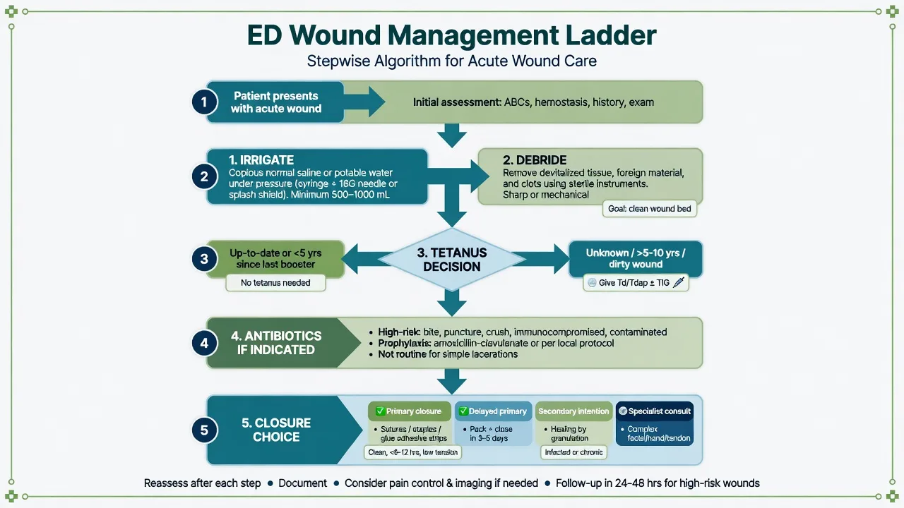
The three closure options are not interchangeable, and the candidate must justify the choice from the wound class, the mechanism, the age and the host. [1]
The mapping is: clean and clean-contaminated → primary; contaminated → delayed primary or secondary depending on debridement adequacy; dirty and infected → secondary. Bites are secondary or delayed primary, with the facial exception. Punctures are secondary — they are never closed because the tract cannot be irrigated. The historical golden period of 6 hours (after which a wound was "too old" to close) has been overturned by modern evidence — Quinn found no increase in infection for low-risk lacerations closed up to 18 to 24 hours after injury, and the decision now integrates age with contamination, site, mechanism and host, not a single clock threshold.[4] The suture technique, gauge and removal interval are covered in the wound-closure topic; for this topic the closure decision is the testable point.
ANZ practice note. In Australasian EDs the workhorse suture is non-absorbable nylon (Ethilon) for skin on the face and extremities and absorbable Vicryl for mucosa and deep layers; tissue adhesive (Histoacryl / Dermabond) and adhesive strips (Steri-Strip) are first-line for small low-tension lacerations in children. Tetanus prophylaxis uses dTpa (reduced-antigen diphtheria-tetanus-acellular-pertussis) 0.5 mL IM and TIG 250 IU IM per the Australian Immunisation Handbook, with the same dirty-wound 5-year and clean-wound 10-year booster intervals as the ACIP schedule. Bite prophylaxis is amoxicillin-clavulanate 875/125 mg PO BD for 5 to 7 days. Rabies is not endemic in Australia but the bat-handling or overseas-animal-bite patient requires post-exposure prophylaxis — notify public health. [1]
Complications and pitfalls
The complications fall into infection-related, repair-related and host-related groups. Infection is the commonest — wound infection rates are 1 to 5 per cent for clean lacerations, 5 to 15 per cent for contaminated, and 10 to 50 per cent for bites; the risk factors are contamination, age over 6 hours, location on the extremity (and especially the hand), diabetes, immunosuppression, and retained foreign body.[3] Dehiscence follows excessive tension, premature suture removal, or infection. Hypertrophic and keloid scars are more common in darker skin types and over the sternum, shoulders and upper arms; counsel the patient and consider silicone gel and early plastic review. Retained foreign body presents as recurrent infection or chronic pain weeks to months later — the classic delayed pitfall. Missed tendon or nerve injury is the medicolegal pitfall — prevented only by the documented pre-anaesthesia neurovascular and tendon examination. Tetanus is the rare but devastating complication of a missed prophylaxis decision. Necrotising fasciitis is the time-critical mimic — pain out of proportion, rapid spread, systemic toxicity — and is a surgical emergency, not an ED closure.[5]
The pitfalls invert the structure: closing a bite or puncture (the classic error); failing to give tetanus prophylaxis in the inadequately vaccinated dirty wound; using antiseptic in the wound bed (cytotoxic to fibroblasts); under-irrigating (the single biggest infection driver); missing a retained foreign body (not exploring, not imaging); missing a partial tendon laceration (not testing against resistance); omitting the documented pre-anaesthesia neurovascular check; prescribing antibiotics for the clean low-risk laceration (adds harm without benefit); and forgetting the safety-net — the discharge advice to return for spreading redness, increasing pain, fever, or a cold limb. [1]
Special populations
The diabetic and immunocompromised patient has a lower threshold for antibiotic prophylaxis and for delayed closure — the host defence is impaired and infection spreads faster; admit or review at 48 hours for any contaminated wound. The child with a laceration is best managed with topical anaesthesia (LET gel into the wound for 20 to 30 minutes), tissue adhesive or absorbable sutures where possible, and a calm, parent-present approach — refer to paediatric sedation for the distressed child with a complex repair. The anticoagulated patient bleeds more from the wound and from the local-anaesthetic injection; pressure and careful haemostasis replace dose modification, and deep blocks carry compartment risk. The elderly patient has fragile skin (skin tears), comorbidity, and slower healing — adhesive strips and tissue adhesive are often kinder than sutures for the thin-skinned avulsion. The pregnant patient is managed as for any adult — local anaesthesia, antibiotics and tetanus prophylaxis are safe in pregnancy. The patient with a prosthetic joint or valve merits antibiotic prophylaxis for any contaminated wound to prevent haematogenous seeding, though the evidence for routine prophylaxis is weak and the decision is individualised. [1]
Evidence and regional guidelines
The contemporary framework rests on several pillars. The irrigation evidence — Weiss 2013 and Moscati 2007 — established that tap water is equivalent to sterile saline for simple lacerations, displacing the dogma of saline-only irrigation and reducing cost and pain.[1][2] The infection-risk and golden-period evidence — Hollander 2001 on risk factors and Quinn 2014 on the contested golden period — reframed closure decisions from a rigid 6-hour clock to an integrated judgement of age, contamination, site and host.[3][4] The bite evidence — Ellis 2014 and Yoon 2025 — confirmed amoxicillin-clavulanate as first-line prophylaxis covering the Pasteurella, Capnocytophaga and Eikenella spectrum.[5][7] The tetanus schedule — Liang 2018 and Havers 2020 ACIP recommendations, mirrored by the Australian Immunisation Handbook — codified the dirty-wound 5-year and clean-wound 10-year booster intervals and the role of TIG 250 IU IM in the inadequately vaccinated.[6][9] The puncture-wound evidence — Eidelman 2003 — documented the Pseudomonas osteomyelitis risk of plantar puncture through footwear and the case for imaging and early review.[10] The suture-technique review (Elfar 2025) summarises the comparative evidence for sutures, tissue adhesive and staples across wound types.[8]
SAQ — Mammalian bite to the hand — assessment and management
10 minutes · 10 marks
A 34-year-old woman presents 2 hours after being bitten on the dorsum of her right hand by her own cat. Examination reveals two puncture wounds over the third and fourth metacarpophalangeal joints, with mild surrounding erythema and tenderness. She is fully immunised. No neurovascular or tendon compromise.
SAQ — Contaminated scalp laceration in the anticoagulated elderly patient
10 minutes · 10 marks
A 78-year-old man on warfarin for atrial fibrillation (INR 2.4) presents after a ground-level fall with a 5 cm contaminated scalp laceration over the occiput. He is on no antiplatelet. GCS 15, no focal deficit, no loss of consciousness. He has not had a tetanus booster in over 15 years.
Exam pearls
- MEAT for assessment — Mechanism, Examination (neurovascular, tendon, bone, depth, contamination, foreign body), Age, Type/host. Deliver it out loud in the OSCE.
- Irrigation is the intervention — normal saline under 5 to 8 psi pressure, 250 mL to a litre, before closure; tap water is equivalent for simple lacerations; antiseptics are cytotoxic to the wound bed.
- Never close a bite or a puncture — the wound that traps bacteria in a space you cannot irrigate. The facial bite is the exception, closed by plastics after copious irrigation.
- Tetanus is two variables — wound type (clean vs dirty) and vaccination history (3 doses vs fewer). Dirty + fewer than 3 doses = vaccine AND TIG. Dirty + 3 doses = booster if over 5 years. Clean + 3 doses = booster if over 10 years. State the product, dose and route.
- Amoxicillin-clavulanate 875/125 mg BD for all mammalian bites, 5 to 7 days, regardless of closure. Eikenella in the human clenched-fist injury; Pasteurella in cat and dog; the clenched-fist MCP bite is a surgical emergency.
- The golden period is dead — close low-risk lacerations up to 18 to 24 hours; integrate age with contamination, site, mechanism and host.
- Document the neurovascular and tendon exam before local anaesthesia — the most common omission and the most common claim.
- Safety-net every wound — return for spreading redness, increasing pain, fever, or a cold limb. [1]
Red flags
[1]References
- [1]Weiss EA, Oldham G, Lin M, Foster E, Quinn JV. Water is a safe and effective alternative to sterile normal saline for wound irrigation prior to suturing: a prospective, double-blind, randomised, controlled clinical trial BMJ Open, 2013.PMID 23325896
- [2]Moscati RM, Mayrose J, Fincher L, Jehle D. A multicenter comparison of tap water versus sterile saline for wound irrigation Acad Emerg Med, 2007.PMID 17456554
- [3]Hollander JE, Singer AJ, Valentine SM, Shofer FS. Risk factors for infection in patients with traumatic lacerations Acad Emerg Med, 2001.PMID 11435186
- [4]Quinn JV, Polevoi SK, Kohn MA. Traumatic lacerations: what are the risks for infection and has the 'golden period' of laceration care disappeared? Emerg Med J, 2014.PMID 23314208
- [5]Ellis R, Ellis C. Dog and cat bites Am Fam Physician, 2014.PMID 25250997
- [6]Havers FP, Moro PL, Hunter P, Hariri S, Bernstein H. Use of Tetanus Toxoid, Reduced Diphtheria Toxoid, and Acellular Pertussis Vaccines: Updated Recommendations of the Advisory Committee on Immunization Practices - United States, 2019 MMWR Morb Mortal Wkly Rep, 2020.PMID 31971933
- [7]Yoon Y, Kim H, Kim J, et al. Infection prevention and treatment following dog bites: a systematic review of randomized controlled trials J Trauma Inj, 2025.PMID 40175075
- [8]Elfar A, Mubarak A, Salem M, Elgamal M. Suture Techniques for Traumatic Wound Closure in the Emergency Department: A Systematic Review of Cosmetic, Functional, and Infection-Related Outcomes Cureus, 2025.PMID 40792329
- [9]Liang JL, Tiwari T, Moro P, et al. Prevention of Pertussis, Tetanus, and Diphtheria with Vaccines in the United States: Recommendations of the Advisory Committee on Immunization Practices (ACIP) MMWR Recomm Rep, 2018.PMID 29702631
- [10]Eidelman M, Bialik V, Miller Y, Kassis I. Plantar puncture wounds in children: analysis of 80 hospitalized patients and late sequelae Isr Med Assoc J, 2003.PMID 14509132