EM · Acute abdominal pain (approach)
Acute abdominal pain — the emergency department approach
Also known as Acute abdomen · Belly pain · Abdominal emergency · The surgical abdomen
The acute abdominal pain approach — the structured emergency department framework that separates the surgical abdomen from the medical, the time-critical from the benign; the three clinical axes (the surgical vs the medical, the upper vs the lower, the colicky vs the constant); the demographic sieve by the age and the sex (the child — the appendicitis, the intussusception; the woman of the reproductive age — the ectopic, the PID, the ovarian; the elderly — the AAA, the mesenteric ischaemia, the SBO, the perforation, the biliary); the location-specific pain patterns (the epigastric, the RUQ, the RLQ, the LLQ, the diffuse); the investigation ladder (the bloods, the urine, the beta-hCG, the lipase, the lactate, the X-ray, the ultrasound, the CT); and the resuscitation with the named drug doses (the morphine 5 mg IV, the ondansetron 4 mg IV, the ceftriaxone 2 g IV, the metronidazole 500 mg IV, the 500 mL fluid bolus). ACEM-primary, globally tagged.
On this page & tools
Your progress
Saved locally on this device.
7 MCQs with explanations
Target exams
Red flags
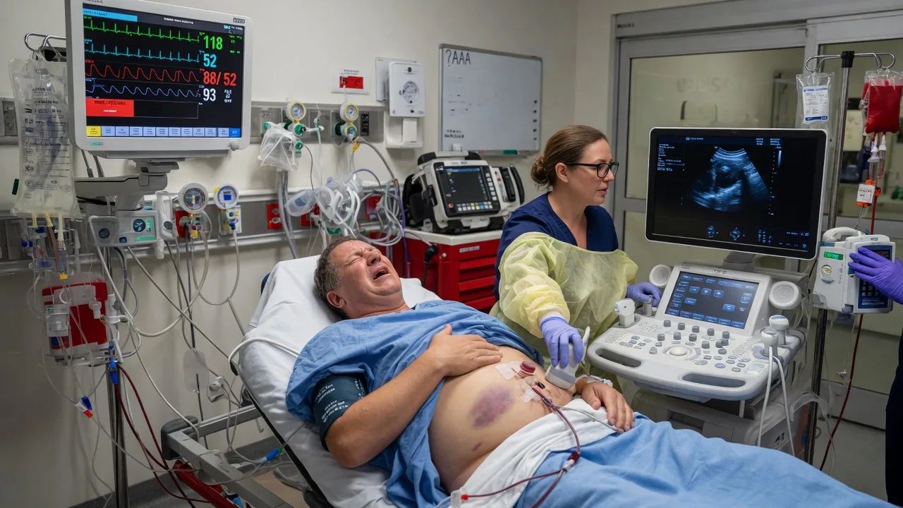
Acute abdominal pain is one of the commonest presentations to the emergency department, accounting for up to 10 per cent of the adult attendances, and it spans the entire spectrum from the self-limiting gastroenteritis to the time-critical ruptured aneurysm. The Fellowship candidate's task is not to make a single diagnosis on the first contact but to apply a structured framework that triages the abdomen safely: to separate the surgical abdomen from the medical, the time-critical from the benign, and the patient who goes to the theatre from the patient who goes home. The recurring error is not missing a rare diagnosis — it is anchoring on a common, benign explanation (the gastroenteritis, the gastritis, the constipation) in a patient who harbours a lethal mimic such as the mesenteric ischaemia, the ectopic pregnancy or the ruptured aneurysm.[1][2][3]
When and why to use the framework
The abdominal pain framework exists because the abdomen is diagnostically opaque — the symptoms are non-specific, the examination is confounded by the body habitus and the analgesia, and the differentials span every organ system. The framework forces the candidate to think in axes rather than in a single linear differential. The first decision is the sick versus the not-sick — the airway, the breathing, the circulation and the perfusion are assessed before any history, and the unstable patient is resuscitated while the workup proceeds. The second decision is the surgical versus the medical — the patient who needs the theatre is identified early, because the delay to the surgery is the modifiable mortality in the perforation, the ischaemia and the ruptured aneurysm. The third decision is the disposition — the admission, the observation, the theatre or the safe discharge with the safety-net. The framework prevents the two classic failures: the premature closure on a benign diagnosis, and the open-ended investigation of every abdomen with the indiscriminate CT. [1]
The three clinical axes
The Fellowship candidate dissects every abdominal pain presentation along three axes, and each axis narrows the differential. [1]
[1]The colicky-versus-constant distinction is the single most useful bedside discriminator. The patient who is writhing around the bed, doubling over and unable to find a position of comfort, is obstructing a hollow viscus — the ureteric colic, the biliary colic, the small-bowel obstruction. The patient who is lying dead still, splinting the abdomen, with the knees drawn up, and who winces on every movement or jolt of the ambulance trolley, has the peritoneal irritation or the bowel ischaemia. Confusing the two leads to the missed diagnosis: the mesenteric ischaemia is frequently labelled as the gastroenteritis because the early examination is soft, while the patient with the ureteric colic is over-investigated for the peritonitis that is not there. [1]
The demographic sieve — the age and the sex
The single most powerful diagnostic aid is the patient's demographic, because the incidence of the surgical causes clusters tightly by the age and the sex. The Fellowship candidate builds a different differential for the child, the woman of the reproductive age, and the elderly patient. [1]
The demographic sieve — the age and the sex
AGES
The appendicitis (most common over age 2), the intussusception (the 3-month to 3-year age with the redcurrant-jelly stool and the sausage-shaped mass), the volvulus, the Meckel diverticulum, the testicular torsion in the boy
The ectopic pregnancy, the pelvic inflammatory disease, the ovarian torsion, the ruptured ovarian cyst, the mittelschmerz — the beta-hCG is non-negotiable
The ruptured AAA, the mesenteric ischaemia, the SBO, the perforated viscus, the biliary sepsis, the diverticulitis — the atypical and the muted presentation, the high mortality
The woman — the ectopic, the PID, the torsion; the man — the testicular torsion (examine the scrotum in every male with the lower-abdominal pain); both — the aortic and the biliary disease in the elderly
The child presents with the appendicitis as the most common surgical emergency (the peak incidence between 10 and 20 years), but the younger child (under 5) is more likely to present atypically and to perforate before the diagnosis. The intussusception peaks between 3 months and 3 years and presents with the intermittent colicky pain, the drawing-up of the legs, the vomiting and the classic redcurrant-jelly stool (a late sign). The woman of the reproductive age carries the ectopic pregnancy, the pelvic inflammatory disease, the ovarian torsion and the ruptured corpus luteum cyst as the leading gynaecological diagnoses — every one of these patients receives a urinary or serum beta-hCG before any imaging, because the ectopic is the leading cause of the first-trimester maternal death. The elderly patient is the high-risk abdomen: the muted pain, the lower fever, the less pronounced leukocytosis, and the dangerous causes — the ruptured AAA, the mesenteric ischaemia, the strangulated hernia, the perforated diverticulum, the biliary sepsis. The threshold for the imaging, the admission and the surgical review is lower in the elderly. [1]
The pain patterns by location
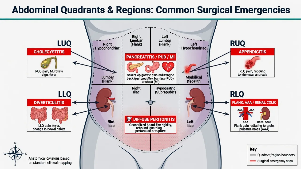
The location of the pain, combined with the migration, the radiation and the character, generates the location-specific shortlist. The Fellowship candidate knows the classic pattern for each abdominal quadrant. [1]
[1]The epigastric pain is the danger zone because it overlaps with the thorax. The myocardial infarction, the aortic dissection and the basal pneumonia all present with the epigastric or the upper-abdominal pain, and the missed cardiac or aortic cause is the high-frequency litigation. An ECG and a bedside ultrasound are part of every epigastric pain workup, particularly in the elderly, the diabetic and the hypertensive patient. The sudden-onset severe epigastric pain that radiates to the back raises the perforated ulcer, the pancreatitis and the aortic dissection together — the erect CXR (the free air), the lipase (the pancreatitis) and the CT angiogram (the dissection) are the discriminating tests. [1]
The right lower quadrant pain is the appendicitis until proven otherwise, but the differential is broad and the lethal mimics — the ectopic, the torsion, the mesenteric ischaemia — must be excluded by the beta-hCG, the ultrasound and the high index of suspicion. The diffuse abdominal pain is the peritonitis or the ischaemia until proven otherwise; the rigid abdomen with the guarding and the rebound is the surgical emergency, and the pain out of proportion to the examination is the mesenteric ischaemia until the CT angiogram proves otherwise. [1]
Surgical versus medical causes by quadrant
The Fellowship candidate maps every quadrant to a paired surgical and a medical shortlist, because the surgical diagnosis drives the theatre, the analgesia, the antibiotics and the disposition, while the medical diagnosis drives the ward pathway. The table below is the exam-exhaustive quadrant map; commit it to memory and rehearse the lethal surgical entry in each cell. [1]
Right upper quadrant (RUQ)
- Surgical: acute cholecystitis, ascending cholangitis, empyema or perforation of the gallbladder, choledocholithiasis with obstruction, hepatic abscess, perforated duodenal ulcer (posterior into the retroperitoneum)
- Medical: biliary colic (no infection, no obstruction — discharged after resolution), acute viral hepatitis, alcoholic hepatitis, hepatic congestion from right heart failure, Fitz-Hugh-Curtis syndrome (PID), herpes zoster (T5-T8 dermatome)
- Referred: right lower-lobe pneumonia, right basal pleurisy, renal/caliceal pathology
- Diagnostic: ultrasound first (stones, wall thickening over 3 mm, sonographic Murphy, pericholecystic fluid); LFTs, WCC, CRP; MRCP or CT for complications; Charcot triad and Reynolds pentad escalate to the septic cholangitis pathway
Epigastric
- Surgical: perforated peptic ulcer (sudden severe pain, free air), acute pancreatitis (severe, radiates to back), aortic dissection, ruptured AAA, mesenteric ischaemia
- Medical and cardiac: acute myocardial infarction (the ECG and the troponin in EVERY epigastric pain over 40), gastritis, oesophagitis, oesophageal rupture (Boerhaave), biliary colic
- Diagnostic: ECG mandatory; lipase over 3x the upper limit; erect CXR for free air; CT angiogram for the dissection, the AAA and the mesenteric ischaemia; troponin for the cardiac cause
- The epigastric region is the THORAX-ABDOMEN overlap — the missed MI, the missed dissection and the missed AAA are the high-frequency litigation here
Left upper quadrant (LUQ)
- Surgical: splenic rupture (trauma, infectious mononucleosis, post-splenectomy risk), splenic infarct (sickle cell, AF, embolic), perforated gastric ulcer, left subphrenic abscess, gastric volvulus
- Medical: splenomegaly (portal hypertension, haematological malignancy, infection), pancreatitis (tail), left lower-lobe pneumonia, splenic artery aneurysm, herpes zoster
- Referred: left basal pneumonia, left pleurisy, left renal pathology
- Diagnostic: ultrasound or CT for the splenic injury; Kehr sign (the referred left shoulder-tip pain from the diaphragmatic irritation) signals the splenic rupture or the free intraperitoneal blood
Right iliac fossa (RIF / RLQ)
- Surgical: acute appendicitis (the default diagnosis until excluded), ectopic pregnancy, ovarian torsion, Meckel diverticulitis, intussusception (the child), right-sided diverticulitis, caecal volvulus, incarcerated right inguinal hernia
- Medical: mesenteric adenitis, ureteric colic, testicular torsion (the REFERRED pain — examine the scrotum in every male), pelvic inflammatory disease, ruptured ovarian cyst, mittelschmerz, inflammatory bowel disease flare
- Diagnostic: beta-hCG NON-NEGOTIABLE; ultrasound in the child and the young woman; CT in the adult and the atypical; Alvarado score (the mnemonic MANTRELS) to stratify the appendicitis risk
Left iliac fossa (LIF / LLQ)
- Surgical: acute diverticulitis (the "left-sided appendicitis"), sigmoid volvulus (the coffee-bean sign), perforated diverticulum, incarcerated left inguinal hernia, ovarian torsion, ectopic pregnancy
- Medical: ureteric colic, inflammatory bowel disease, ischaemic colitis, ovarian cyst rupture, constipation/fecal impaction (the elderly), pelvic inflammatory disease
- Diagnostic: beta-hCG; CT for the diverticulitis (fat stranding, bowel-wall thickening, abscess); the modified Hinchey classification grades the severity and the need for the intervention
Suprapubic
- Surgical: urinary retention (the elderly male with the benign prostatic hypertrophy — palpable distended bladder, the emergency catheter), ruptured bladder (trauma), ovarian torsion, ectopic pregnancy, incarcerated femoral or inguinal hernia
- Medical: cystitis, pyelonephritis, interstitial cystitis, pelvic inflammatory disease, endometriosis, overactive bladder
- Diagnostic: bladder scan or ultrasound for the retention; urinalysis and culture; the retention with the overflow incontinence and the abdominal discomfort is the masked presentation in the elderly
Diffuse or generalised
- Surgical and time-critical: ruptured AAA (the elderly, the haemodynamic instability), acute mesenteric ischaemia (pain out of proportion), generalised peritonitis (the perforation, the pancreatitis), small or large bowel obstruction with strangulation
- Medical: diabetic ketoacidosis, uraemia, porphyria, sickle cell crisis, lead poisoning, gastroenteritis, Addisonian crisis, hereditary angioedema
- Diagnostic: the diffuse pain with the peritonism is the surgical emergency; the lactate, the VBG and the CT angiogram drive the time-critical pathway; the bedside ultrasound (the aorta, the FAST) precedes the CT in the unstable
Pain characteristics — the colicky, the constant-progressive, the burning
The character of the pain is the second great discriminator after the location, and it predicts the underlying mechanism — the obstruction, the ischaemia, the inflammation or the ulceration. The Fellowship candidate elicits the character explicitly and never settles for the generic "the pain came on". [1]
Colicky (waxing and waning)
- The mechanism: the intermittent obstruction of a hollow viscus with the smooth-muscle spasm against the blockage — the patient WRITHES, doubles over, paces, cannot find a position of comfort
- Ureteric colic: the sudden severe loin-to-groin pain, the writhing patient, the haematuria, the benign abdomen
- Biliary colic: the post-prandial RUQ pain lasting hours then resolving, the patient relatively well between the episodes
- Small-bowel obstruction: the central colicky pain with the distension, the vomiting and the absolute obstipation
- Renal and the biliary colic resolve; the colicky pain that BECOMES constant signals the ischaemia, the strangulation or the perforation — the surgical escalation
Constant and progressive
- The mechanism: the ongoing inflammation, the ischaemia, the necrosis or the perforation — the patient lies STILL, splints the abdomen, draws up the knees, winces on every movement
- Peritonitis: the constant severe pain worsened by movement, the rigid board-like abdomen, the guarding and the rebound
- Mesenteric ischaemia: the constant severe pain OUT OF ALL PROPORTION to the soft early examination — the lactate rises, the CT angiogram is time-critical
- Pancreatitis: the constant severe epigastric pain radiating to the back, the relief leaning forward
- The constant-progressive pain is the surgical or the time-critical until proven otherwise — the delay is the mortality
Burning or gnawing
- The mechanism: the mucosal inflammation or the acid-peptic injury — the peptic ulcer disease, the gastritis, the oesophagitis
- Peptic ulcer: the burning epigastric pain, the duodenal ulcer RELIEVED by food and WORSENED by hunger (the nocturnal waking), the gastric ulcer WORSENED by food
- The burning pain radiating to the back with the sudden worsening and the peritonism is the posterior perforation — the erect CXR for the free air
- The burning epigastric pain with the haematemesis or the melaena is the upper GI bleed — the group-and-save, the PPI infusion and the endoscopy
Tearing or ripping
- The mechanism: the acute dissection of the aortic wall or the rupture of the aneurysm — the SURGICAL and the VASCULAR emergency
- Aortic dissection: the sudden severe TEARING pain radiating to the back (the interscapular or the lumbar), the pulse or the blood-pressure differential between the arms
- Ruptured AAA: the elderly patient, the sudden severe back or abdominal pain, the syncope, the hypotension, the pulsatile mass
- The tearing pain gets the ECG, the bedside ultrasound (the aorta), and the CT angiogram — the time-critical vascular pathway
The ED approach — the structured ABCDE to the disposition
The Fellowship candidate applies a reproducible sequence to every acute abdomen, so that no high-risk diagnosis is missed under the cognitive load of the busy resuscitation bay. The sequence runs the resuscitation, the focused history and exam, the targeted investigations, and the disposition — in parallel, not in series. [1]
The structured ED approach to the acute abdomen
ABCDE and the sick-or-not-sick triage
The focused history (the SOCRATES)
The focused examination
The bedside ultrasound
The first-line investigations
The targeted imaging
The resuscitation in parallel
The disposition
Surgical emergency indicators — the triggers for the theatre
The Fellowship candidate identifies the surgical abdomen by a set of convergent signs, because the delay to the surgery is the modifiable mortality in the perforation, the ischaemia and the ruptured aneurysm. Any ONE of these indicators in the right clinical context escalates the patient to the surgical or the vascular pathway, and the formal imaging does not delay the call. [1]
The surgical emergency indicators
Differential diagnosis — the broad list distinguished
The acute abdomen has a long differential, and the Fellowship candidate must distinguish the time-critical surgical causes from the medical and the benign. The differentials below are the core list the examiner expects.[2][3]
Acute appendicitis
- The central pain migrating to the RLQ over 12 to 24 hours, the anorexia, the low-grade fever, the McBurney tenderness
- The raised WCC and CRP; the ultrasound (the child, the young woman) or the CT (the adult, the atypical)
- The analgesia, the ceftriaxone 2 g IV plus the metronidazole 500 mg IV, the surgical referral for the appendicectomy
- The most common acute surgical abdomen; the perforation risk rises with the duration of the symptoms
Acute pancreatitis
- The severe constant epigastric pain radiating to the back, the vomiting, the gallstones or the alcohol
- The lipase over three times the upper limit; the CT if the diagnosis is uncertain or for the complications
- The aggressive intravenous fluid, the analgesia, the early enteral nutrition; the antibiotics only for the infected necrosis
- The commonest GI cause of the admission; the 10 to 15 per cent severe course with the multi-organ failure
Biliary disease (colic, cholecystitis, cholangitis)
- The RUQ pain after the fatty food, the colicky-to-constant; the Charcot triad (the RUQ pain, the fever, the jaundice) in the cholangitis; the Murphy sign and the sonographic Murphy for the cholecystitis
- The deranged LFTs, the raised WCC; the ultrasound first; the MRCP or the CT for the complications; the Tokyo Guidelines 2018 grade the severity
- The analgesia, the fluids; the ceftriaxone and the metronidazole for the cholangitis; the early ERCP for the obstruction (the biliary drainage within 24 to 48 hours for the severe)
- The ascending cholangitis is the emergency — the Reynolds pentad adds the shock and the confusion; the Tokyo Guidelines direct the antibiotics and the timing
Bowel obstruction
- The colicky central abdominal pain, the distension, the vomiting, the constipation and the absolute obstipation
- The raised lactate in the ischaemia; the supine abdominal X-ray (the dilated loops, the step-pattern); the CT for the level and the cause
- The nasogastric tube, the intravenous fluid, the analgesia; the surgical referral; the strangulation or the closed-loop is the emergency
- The adhesive bands are the commonest cause in the adult; the hernia and the malignancy in the elderly
Mesenteric ischaemia
- The pain out of all proportion to the examination; the AF, the vascular disease, the hypoperfusion; the bloody diarrhoea in the late presentation
- The raised lactate, the metabolic acidosis, the raised amylase (a red herring); the CT angiography is the diagnostic test; a NORMAL lactate does NOT exclude it
- The aggressive resuscitation, the heparin for the thrombotic, the urgent surgical or the vascular review; the time-critical — the WSES 2022 guidelines
- The mortality is over 50 per cent — the diagnosis is missed because the early examination is soft
Ruptured AAA
- The elderly male with the sudden severe back or abdominal pain, the syncope, the hypotension; the pulsatile mass; the cardiovascular risk factors
- The bedside ultrasound shows the aneurysm and the free fluid; do NOT delay for the CT in the unstable; the aortic diameter over 5.5 cm is the repair threshold
- The permissive hypotension (the target SBP around 90), the immediate vascular and the surgical call, the theatre — the time-critical; the endovascular option for the stable
- The mortality rises with the delay; the bedside ultrasound and the call precede the CT — the screening prevalence falls with the modern population screening
Perforated viscus
- The sudden severe generalized pain, the rigid abdomen, the guarding and the rebound; the rigid board-like abdomen
- The erect CXR (the free subdiaphragmatic air in 70 per cent); the CT for the site and the cause
- The fluid resuscitation, the broad-spectrum antibiotics, the analgesia, the urgent surgery
- The perforated duodenal ulcer in the adult; the perforated diverticulum or the caecum in the elderly
Ectopic pregnancy
- The woman of reproductive age; the missed period, the vaginal bleeding, the shoulder-tip pain from the diaphragmatic irritation
- The positive beta-hCG; the transvaginal ultrasound (the empty uterus, the adnexal mass, the free fluid)
- The resuscitation, the group-and-save; the methotrexate for the stable unruptured; the laparoscopic salpingectomy for the ruptured
- The leading cause of the first-trimester maternal death — exclude with the beta-hCG in every woman
Ureteric colic
- The sudden severe colicky loin-to-groin pain, the writhing patient, the haematuria; the no-peritonism
- The urinalysis (the haematuria); the CT KUB shows the stone; the normal abdomen on examination
- The diclofenac 75 mg IM, the morphine 5 mg IV, the hydration, the alpha-blocker; the admission for the sepsis or the obstruction
- The pain is out of proportion to the benign examination — but exclude the AAA in the elderly first
Diverticulitis
- The LLQ pain, the fever, the altered bowel habit; the elderly patient; the Left-sided appendicitis
- The raised WCC and CRP; the CT shows the fat stranding and the bowel-wall thickening
- The analgesia, the fluids, the antibiotics (the co-amoxiclav or the ceftriaxone and the metronidazole) for the complicated
- The perforation, the abscess and the fistula are the complications; the surgery for the complicated
The medical and the extra-abdominal mimics complete the differential and must not be missed: the myocardial infarction (the epigastric pain in the elderly — the ECG), the diabetic ketoacidosis (the abdominal pain and the vomiting — the glucose and the ketones), the lower-lobe pneumonia (the referred upper-abdominal pain — the chest exam and the X-ray), the gastroenteritis (the vomiting and the diarrhoea preceding the pain), the sickle-cell crisis (the vaso-occlusive pain in the relevant population), the porphyria and the lead poisoning (the rare but the classic abdominal-pain-with-the-normal-examination presentations), the herpes zoster (the dermatomal pain preceding the rash), and the spider-bite envenomation (the redback in the ANZ setting). The testicular torsion presents with the referred lower-abdominal pain — the scrotum is examined in every male with the lower-abdominal pain. [1]
Bedside assessment — the history and the examination
The history uses the SOCRATES structure — the Site, the Onset, the Character, the Radiation, the Associations, the Time course, the Exacerbating and the relieving factors, the Severity. The onset is the high-yield element: the sudden-onset (seconds to minutes) pain raises the ruptured aneurysm, the perforated viscus, the ectopic and the dissection; the rapidly progressive (over hours) pain raises the appendicitis, the pancreatitis, the cholecystitis; the insidious (over days) pain raises the obstruction, the diverticulitis, the malignancy. The associated symptoms — the vomiting (the onset relative to the pain: the pain before the vomiting is the surgical abdomen; the vomiting before the pain is the medical), the fever, the rigors, the jaundice, the change in the bowel habit, the urinary symptoms, the vaginal bleeding — narrow the differential further. The past history — the previous surgery (the adhesions), the gallstones, the AAA, the vascular disease, the atrial fibrillation (the mesenteric ischaemia), the medications (the anticoagulation, the NSAIDs, the steroids) — is sought. [1]
The examination begins with the vital signs and the perfusion (the shock is the surgical emergency), then the inspection (the distension, the scars, the visible peristalsis, the hernial orifices), the auscultation (the tinkling or the absent bowel sounds of the obstruction; the hyperactive of the gastroenteritis), and finally the palpation. The palpation starts in the quadrant furthest from the pain, with the warm hands, and assesses the tenderness, the guarding (the voluntary, the involuntary), the rigidity, the rebound and the organomegaly. The named peritoneal signs (the McBurney point, the Rovsing, the psoas, the obturator, the Murphy) localise the pathology. The rectal and the pelvic examination are performed when indicated — the rectal for the mass, the blood and the prostate, the pelvic for the cervical motion tenderness and the adnexal masses. The scrotum is examined in every male with the lower-abdominal pain (the testicular torsion). The bedside ultrasound (the FAST for the free fluid, the aorta for the AAA, the gallbladder for the stones, the kidneys for the hydronephrosis) is part of the modern abdominal examination. [1]
Investigations — the bloods, the urine, the X-ray, the ultrasound, the CT
The investigation strategy runs in parallel with the resuscitation, not after it. The first-line bloods are the full blood count (the leukocytosis, the anaemia), the CRP (the inflammation), the urea and electrolytes (the dehydration, the renal function, the metabolic acidosis of the ischaemia), the liver function tests (the biliary obstruction, the hepatitis), the lipase (over three times the upper limit confirms the pancreatitis), the lactate (the tissue hypoperfusion — the mesenteric ischaemia, the sepsis, the shock; a raised lactate supports but a normal lactate does NOT exclude the mesenteric ischaemia), the venous or the arterial blood gas (the metabolic acidosis), the amylase (less specific than the lipase), and the beta-hCG in every woman of the reproductive age — the ectopic is the non-negotiable exclusion.[2][3][9][10][13]
The urinalysis screens for the haematuria (the ureteric colic, the malignancy), the leucocytes and the nitrites (the urinary tract infection, the pyelonephritis), the ketones (the DKA), and the glucose (the hyperglycaemia). The pregnancy test (the urinary beta-hCG, backed by the serum) is performed on every woman of the reproductive age. [1]
The erect chest X-ray (or the left lateral decubitus film in the patient who cannot stand) detects the free subdiaphragmatic air of the perforated viscus — sensitivity around 70 per cent, and a positive film precludes the need for further imaging before the surgery. The supine abdominal X-ray shows the dilated bowel loops and the step-pattern of the small-bowel obstruction, the coffee-bean sign of the sigmoid volvulus, and occasionally the calcified aneurysm or the radiopaque stones. The plain X-ray has a limited role in the modern era but remains the cheap, accessible, first-line screen in the obstruction and the perforation. [1]
The ultrasound is the first-line imaging in the child, the young woman and the pregnant patient — it avoids the ionising radiation and it evaluates the gallbladder (the stones, the wall thickening, the sonographic Murphy), the gynaecological organs (the ectopic, the torsion), the kidneys (the hydronephrosis) and the appendix. The bedside ultrasound by the emergency physician evaluates the aorta (the AAA), the free fluid (the FAST) and the gallbladder at the bedside. The CT of the abdomen and the pelvis with the intravenous contrast is the first-line investigation in the adult male, the elderly, and the atypical or the undifferentiated abdomen — it has the highest diagnostic accuracy, the sensitivity and the specificity over 90 per cent for most surgical causes, and it is the test of choice for the mesenteric ischaemia (the CT angiogram), the diverticulitis, the complicated appendicitis, the bowel obstruction and the intra-abdominal abscess. The imaging is matched to the patient — the ultrasound first in the young and the pregnant, the CT first in the older and the atypical — to balance the diagnostic yield against the radiation and the contrast risk.[1][2]
The investigation thresholds and the targets
Immediate management and resuscitation — the drug doses
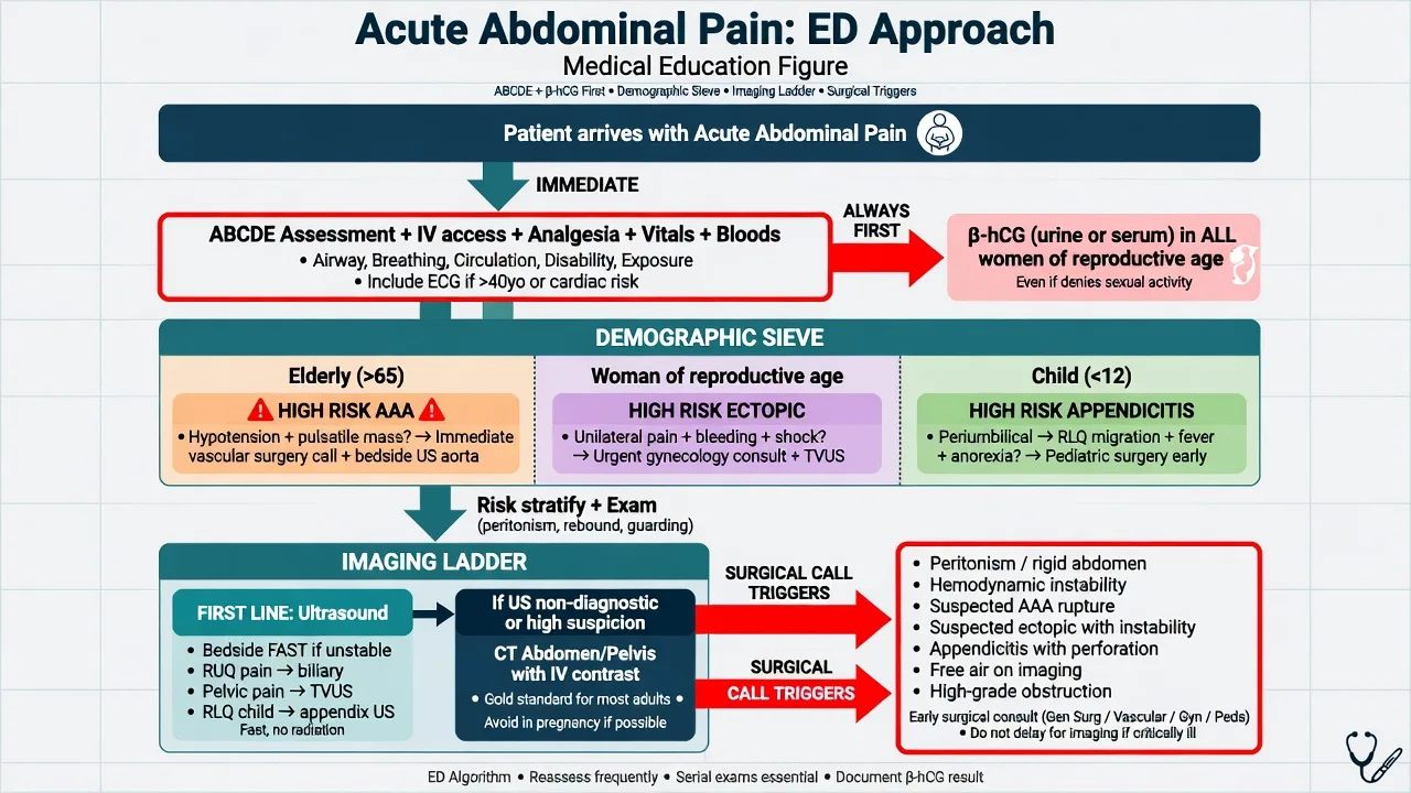
The management follows the ABCDE and the treat-the-cause, with the resuscitation running in parallel with the workup. The unstable patient — the hypotension, the tachycardia, the altered mental state, the metabolic acidosis — is resuscitated first and investigated second. The fluid resuscitation with the balanced crystalloid (the Hartmann or the normal saline) 500 mL intravenous bolus, repeated to the endpoint of the perfusion (the mean arterial pressure over 65, the urine output over 0.5 mL/kg/h, the normal mentation), is the first intervention. The analgesia is given early and adequately — the morphine 5 mg intravenously (titrated, every 5 to 10 minutes, to the pain relief) is the standard opioid; the modern evidence confirms the analgesia does not mask the abdominal signs or delay the diagnosis, and the systematic-review data support the early opioid in the abdominal pain of the child and the adult alike.[14] The withholding of the analgesia for the examination is the outdated and the harmful practice. The antiemetic — the ondansetron 4 mg intravenously — is given for the nausea and the vomiting, and it complements the analgesia. The broad-spectrum antibiotics — the ceftriaxone 2 g intravenously plus the metronidazole 500 mg intravenously — are started for the suspected sepsis, the perforation, the cholangitis, the complicated diverticulitis and the appendicitis, after the cultures (the blood, the urine) are taken.[2][3][7]
[1]The surgical referral is made early and explicitly — the emergency physician does not wait for the morning or the formal imaging in the patient with the peritonitis, the obstruction with the strangulation, the ruptured aneurysm or the perforation. The referral states the working diagnosis, the vitals, the resuscitation given, the relevant findings and the imaging — the structured ISBAR handover. The disposition follows the working diagnosis: the theatre for the surgical emergency, the ward or the HDU for the severe medical abdomen (the pancreatitis, the cholangitis), the ED observation unit for the undifferentiated but the stable abdomen, and the discharge with the safety-net for the clearly benign diagnosis (the gastroenteritis, the resolved biliary colic) with the explicit return precautions. [1]
Escalation triggers
The escalation triggers identify the patient who cannot wait. The haemodynamic instability (the systolic BP under 90, the tachycardia over 120, the altered mentation) in the abdominal pain is the surgical or the vascular emergency until proven otherwise — the bedside ultrasound (the aorta, the FAST), the simultaneous resuscitation and the immediate surgical and the vascular call. The peritonitis (the rigid abdomen, the guarding, the rebound) is the surgical emergency. The sepsis (the fever or the hypothermia, the raised lactate, the organ dysfunction) receives the broad-spectrum antibiotics within the hour and the source control. The abnormal lactate (over 2 mmol/L) with the severe pain, particularly in the AF or the vascular patient, escalates to the CT angiogram for the mesenteric ischaemia. The positive beta-hCG with the abdominal pain escalates to the transvaginal ultrasound for the ectopic. The beta-hCG with the haemodynamic instability is the ruptured ectopic until proven otherwise — the simultaneous resuscitation, the group-and-save and the gynaecology call, without the delay for the CT. [1]
Special populations
The elderly patient is the high-risk abdomen — the atypical presentation (the mild pain, the absent fever, the minimal tenderness), the dangerous causes (the AAA, the mesenteric ischaemia, the perforation, the biliary sepsis, the SBO), the comorbidities, and the high mortality. The threshold for the CT, the admission and the surgical review is lower; the conservative "gastroenteritis" label in the elderly is the classic missed-diagnosis error. The pregnant patient has the physiological changes (the gravid uterus displacing the appendix upwards and to the right in the third trimester, the mild leukocytosis and the nausea that are normal in the pregnancy) and the imaging constraints — the ultrasound first, the MRI second, the CT only if life-saving. The immunocompromised patient (the steroids, the chemotherapy, the transplant, the neutropenia) presents with the muted signs and the opportunistic pathology — the neutropenic enterocolitis (the typhlitis), the CMV colitis, the atypical infections. The child is approached with the paediatric dosing, the ultrasound-first imaging, and the awareness that the appendicitis is the most common surgical emergency and the intussusception is the classic under-3 diagnosis. The anticoagulated patient with the abdominal pain has the intra-abdominal bleeding (the retroperitoneal, the intramural haematoma) until proven otherwise — the CT and the coagulation reversal are considered. [1]
Common errors and pitfalls
The recurring errors are: the premature closure on the benign diagnosis (the gastroenteritis, the gastritis, the constipation) in the patient with the lethal mimic; the failure to take the beta-hCG in the woman of the reproductive age (the missed ectopic); the failure to examine the scrotum in the male with the lower-abdominal pain (the missed testicular torsion); the failure to perform the ECG in the elderly patient with the epigastric pain (the missed MI); the failure to check the lipase (the missed pancreatitis) and the lactate (the missed mesenteric ischaemia); the delay to the CT in the unstable patient with the suspected ruptured AAA (the bedside ultrasound and the surgical call precede the CT); the withholding of the analgesia for the examination (the outdated and the harmful practice); the over-reliance on a single investigation (the normal WCC and CRP in the early appendicitis; the soft abdomen in the early mesenteric ischaemia); and the anchoring on the common diagnosis in the elderly (the caecal carcinoma, the ischaemic bowel, the sigmoid volvulus). The recurring trap is the patient with the "soft abdomen" and the severe pain — the mesenteric ischaemia, the early appendicitis, the ureteric colic and the diabetic ketoacidosis all present with the disproportionate pain and the soft abdomen, and the discharge of these patients is the high-risk decision. [1]
Evidence and regional guidelines
The contemporary framework for the acute abdominal pain in the emergency department is built on the structured history and the examination, the targeted imaging, and the parallel resuscitation. The Laméris BMJ study (2009) established the diagnostic-accuracy hierarchy of the imaging strategies — the ultrasound first in the young and the pregnant, the CT first in the older and the atypical, with the MRI as the problem-solving modality in the pregnancy — and it underpins the modern imaging pathway.[1] The Cartwright American Family Physician reviews (2008, 2015) are the canonical primary-care and the emergency-department references for the evaluation and the imaging of the acute abdominal pain, and they emphasise the location-specific differential, the demographic sieve, and the role of the targeted CT.[2][3] The regional practice: the early analgesia is universal; the ultrasound-first in the child, the young woman and the pregnant patient is the ANZ and the UK standard; the CT-first in the adult male, the elderly and the atypical is the modern standard; and the surgical and the vascular referral thresholds are governed by the local protocols. The disease-specific guidelines refine the management further. The Tokyo Guidelines 2018 (the Yokoe diagnostic criteria and the severity grading, the Gomi antimicrobial recommendations, the Miura initial-management flowchart) standardise the diagnosis, the severity stratification and the antimicrobial therapy of the acute cholecystitis and the cholangitis, and they direct the timing of the early biliary drainage (the ERCP within 24 to 48 hours for the severe cholangitis).[6][7][8] The World Society of Emergency Surgery 2022 mesenteric ischaemia guidelines (Bala, Catena) emphasise the high clinical suspicion, the CT angiography as the diagnostic standard, the normal-lactate-does-not-exclude caveat, and the time-critical surgical or the endovascular revascularisation.[9][10] The ACOG Practice Bulletin on the tubal ectopic pregnancy (2018) codifies the transvaginal-ultrasound-and-beta-hCG diagnostic paradigm and the methotrexate-versus-surgery management thresholds.[13] The Alvarado score (1986) remains the most widely taught bedside stratification of the appendicitis, and the Snyder AFP review (2018) updates the efficient modern diagnosis and the management.[4][5] The Golledge European Heart Journal review (2023) summarises the contemporary pathogenesis, the screening and the management of the AAA.[12] The Rodrigues neutropenic-enterocolitis review (2017) describes the typhlitis of the immunocompromised.[11]
Laméris et al., BMJ 2009 — OPTIMA diagnostic-accuracy study
PMID 19561056
Multicentre prospective diagnostic-accuracy study (1020 patients with the acute abdominal pain)
Population: The adults presenting to the emergency department with the non-traumatic acute abdominal pain
Key finding
The ultrasound-first strategy was the most cost-effective; the immediate CT the most sensitive. The ultrasound followed by the CT (on the indication) was the optimal strategy — the ultrasound first in the young and the pregnant, the CT first in the older and the atypical.
Practice change
Establishes the modern imaging-strategy hierarchy for the acute abdomen: the ultrasound first, the CT on the indication, the MRI in the pregnancy.
Yokoe et al., Tokyo Guidelines 2018 — acute cholecystitis criteria
PMID 29032636
International consensus guideline (the revised Tokyo Guidelines 2018)
Population: The adults with the suspected acute cholecystitis or the cholangitis
Key finding
The standardised diagnostic criteria (the local signs, the systemic inflammation, the imaging), the severity grading (the mild, the moderate, the severe), and the antimicrobial recommendations that direct the empirical therapy and the early biliary drainage.
Practice change
The Tokyo Guidelines 2018 are the global standard for the diagnosis, the severity and the antimicrobial therapy of the biliary infection.
Bala, Catena et al., WSES 2022 — mesenteric ischaemia guidelines
PMID 36261857
Updated World Society of Emergency Surgery consensus guideline
Population: The adults with the suspected acute mesenteric ischaemia
Key finding
The high clinical suspicion, the CT angiography as the diagnostic standard, the caveat that the normal lactate does NOT exclude the ischaemia, and the time-critical surgical or the endovascular revascularisation. The mortality remains over 50 per cent.
Practice change
The contemporary standard for the time-critical diagnosis and the management of the acute mesenteric ischaemia.
ANZ practice note. The acute abdomen is approached with the ABCDE, the structured history (the SOCRATES, the onset, the migration), the demographic sieve, and the location-specific differential. The beta-hCG is taken in every woman of the reproductive age. The early analgesia (the morphine 5 mg IV, the ondansetron 4 mg IV) is the standard and it does not mask the signs. The ultrasound is the first-line imaging in the child, the young woman and the pregnant; the CT is the first-line in the adult male, the elderly and the atypical. The surgical and the vascular referral is early and explicit for the peritonitis, the obstruction with the strangulation, the ruptured AAA and the perforation. [1]
Model answer — the ED workup of the undifferentiated acute abdomen
The undifferentiated acute abdomen is assessed with the ABCDE first — the airway, the breathing, the circulation, the disability (the glucose), the exposure. The unstable patient is resuscitated with the balanced crystalloid 500 mL IV bolus, the morphine 5 mg IV for the pain, the ondansetron 4 mg IV for the nausea, and the broad-spectrum ceftriaxone 2 g IV plus the metronidazole 500 mg IV if the sepsis or the perforation is suspected. The bedside ultrasound evaluates the aorta (the AAA), the FAST (the free fluid), and the gallbladder. The bloods (the FBC, the CRP, the U and E, the LFTs, the lipase, the lactate) and the beta-hCG in every woman of the reproductive age are sent. The history and the examination localise the pain; the imaging is matched to the patient (the ultrasound in the young and the pregnant, the CT in the older and the atypical). The disposition: the theatre for the surgical emergency (the peritonitis, the perforation, the ruptured AAA, the ectopic), the ward or the HDU for the severe medical abdomen, the ED observation unit for the undifferentiated but the stable, and the discharge with the safety-net for the clearly benign.
Exam pearls
- The three axes: the surgical versus the medical, the upper versus the lower, the colicky versus the constant.
- The demographic sieve: the child (the appendicitis, the intussusception), the woman of the reproductive age (the ectopic, the PID, the torsion), the elderly (the AAA, the mesenteric ischaemia, the SBO, the perforation, the biliary).
- The beta-hCG in every woman of the reproductive age — non-negotiable; the ectopic is the lethal mimic.
- The epigastric pain gets the ECG — the MI is the high-frequency missed diagnosis in the elderly.
- The pain out of proportion to the examination is the mesenteric ischaemia — the lactate and the CT angiogram.
- The rigid, board-like abdomen is the perforation — the erect CXR for the free air, the resuscitation, the urgent surgery.
- The analgesia is given BEFORE the surgical review — the morphine 5 mg IV does not mask the signs or the diagnosis.
- The ultrasound first in the child, the young woman and the pregnant; the CT first in the adult male, the elderly and the atypical.
- Examine the scrotum in every male with the lower-abdominal pain — the testicular torsion.
- The onset is the highest-yield history element: the sudden (seconds) pain is the AAA, the perforation, the ectopic, the dissection; the progressive (hours) is the appendicitis, the pancreatitis, the cholecystitis; the insidious (days) is the obstruction, the diverticulitis.
- The pain-vomiting sequence: the PAIN before the vomiting is the surgical abdomen; the vomiting before the pain is the medical (the gastroenteritis, the gastritis).
- The colicky pain (the patient writhing) is the obstructed hollow viscus; the constant pain (the patient still) is the peritonitis or the ischaemia — the bedside behaviour is the discriminator.
- The burning epigastric pain: the duodenal ulcer is RELIEVED by food and the gastric ulcer is WORSENED by food — the nocturnal waking is the duodenal ulcer.
- The Alvarado score (the MANTRELS) stratifies the appendicitis; the score over 7 is the probable appendicitis — but the negative score does NOT exclude it.
- The Charcot triad (the RUQ pain, the fever, the jaundice) is the cholangitis; the Reynolds pentad adds the shock and the confusion — the severe cholangitis gets the ERCP within 24 to 48 hours.
- The lipase over three times the upper limit confirms the pancreatitis; the amylase is less specific and may be raised in the mesenteric ischaemia (a red herring).
- The elderly abdomen is the high-risk abdomen — the muted signs, the dangerous causes (the AAA always), and the lower threshold for the CT and the surgical review; the "gastroenteritis" label is the classic error.
- The immunocompromised or the neutropenic patient with the RIF pain has the neutropenic enterocolitis (the typhlitis) — the CT, the broad-spectrum antibiotics and the G-CSF. [1]
SAQ — The perforated viscus and the surgical abdomen
10 minutes · 10 marks
A 58-year-old man presents to the emergency department with the sudden severe generalized abdominal pain that woke him from sleep 6 hours ago. He has taken the ibuprofen 400 mg three times daily for the osteoarthritis for two years, and he smokes 20 cigarettes a day. On arrival: BP 96/62, HR 118, RR 24, temperature 38.4 degrees Celsius, SaO2 95 per cent on the room air. The abdomen is rigid and board-like with the involuntary guarding and the rebound tenderness generalized to all four quadrants. The erect chest X-ray shows the free subdiaphragmatic air.
SAQ — The acute mesenteric ischaemia in the atrial fibrillation
10 minutes · 10 marks
A 76-year-old woman presents with the severe constant central abdominal pain that began 4 hours ago. She has the chronic atrial fibrillation on the apixaban 5 mg twice daily and the ischaemic heart disease. On arrival: BP 108/70, HR 96 irregularly irregular, RR 22, afebrile. The abdomen is SOFT and non-tender with the active bowel sounds — the pain is out of all proportion to the examination. The venous blood gas: pH 7.28, lactate 4.2 mmol/L, bicarbonate 16. The WCC is 16.5 and the CRP is 12.
Red flags
[1]References
- [1]Laméris W, van Randen A, van Es HW, et al. Imaging strategies for detection of urgent conditions in patients with acute abdominal pain: diagnostic accuracy study BMJ, 2009.PMID 19561056
- [2]Cartwright SL, Knudson MP. Diagnostic imaging of acute abdominal pain in adults Am Fam Physician, 2015.PMID 25884745
- [3]Cartwright SL, Knudson MP. Evaluation of acute abdominal pain in adults Am Fam Physician, 2008.PMID 18441863
- [4]Alvarado A. A practical score for the early diagnosis of acute appendicitis Ann Emerg Med, 1986.PMID 3963537
- [5]Snyder MJ, Guthrie M, Cagle S. Acute Appendicitis: Efficient Diagnosis and Management Am Fam Physician, 2018.PMID 30215950
- [6]Yokoe M, Hata J, Takada T, et al. Tokyo Guidelines 2018: diagnostic criteria and severity grading of acute cholecystitis (with videos) J Hepatobiliary Pancreat Sci, 2018.PMID 29032636
- [7]Gomi H, Solomkin JS, Takada T, et al. Tokyo Guidelines 2018: antimicrobial therapy for acute cholangitis and cholecystitis J Hepatobiliary Pancreat Sci, 2018.PMID 29090866
- [8]Miura F, Okamoto K, Takada T, et al. Tokyo Guidelines 2018: initial management of acute biliary infection and flowchart for acute cholangitis J Hepatobiliary Pancreat Sci, 2018.PMID 28941329
- [9]Bala M, Catena F, Kashuk J, et al. Acute mesenteric ischemia: updated guidelines of the World Society of Emergency Surgery World J Emerg Surg, 2022.PMID 36261857
- [10]Isfordink CJ, Dekker D, Meijer RP, et al. Clinical value of serum lactate measurement in diagnosing acute mesenteric ischaemia Neth J Med, 2018.PMID 29515007
- [11]Rodrigues FG, Dasilva G, Wexner SD. Neutropenic enterocolitis World J Gastroenterol, 2017.PMID 28104979
- [12]Golledge J, Thanigaimani S. Pathogenesis and management of abdominal aortic aneurysm Eur Heart J, 2023.PMID 37387260
- [13]American College of Obstetricians and Gynecologists' Committee on Practice Bulletins—Gynecology. ACOG Practice Bulletin No. 193: Tubal Ectopic Pregnancy Obstet Gynecol, 2018.PMID 29470343
- [14]Poonai N, Paskar D, Koneru B, et al. Opioid analgesia for acute abdominal pain in children: A systematic review and meta-analysis Acad Emerg Med, 2014.PMID 25377394