EM · Chest trauma
Chest trauma (the immediately and potentially life-threatening injuries)
Also known as Thoracic trauma · Blunt chest injury · Penetrating chest injury · Flail chest
Chest trauma — the immediately life-threatening 'lethal six' (tension pneumothorax, open pneumothorax, massive haemothorax, flail chest with pulmonary contusion, cardiac tamponade, tracheobronchial injury) identified and managed in the primary survey, the potentially life-threatening 'hidden six' (blunt aortic injury, blunt cardiac injury, diaphragmatic rupture, oesophageal rupture, pulmonary contusion, simple pneumo/haemothorax) found on the secondary survey, the analgesia and the chest-drain management, and the CT-angiogram for the blunt aortic injury. ACEM-primary, globally tagged.
On this page & tools
Your progress
Saved locally on this device.
8 MCQs with explanations
Target exams
Red flags
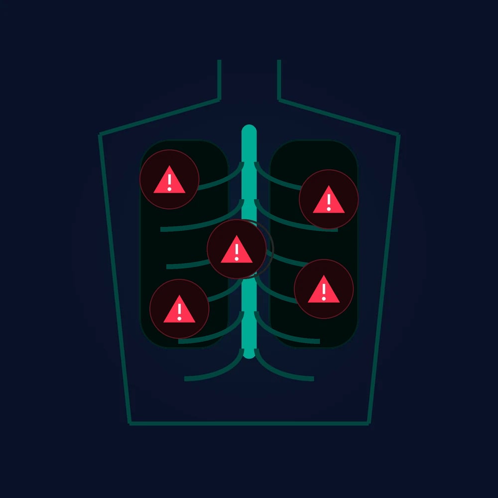
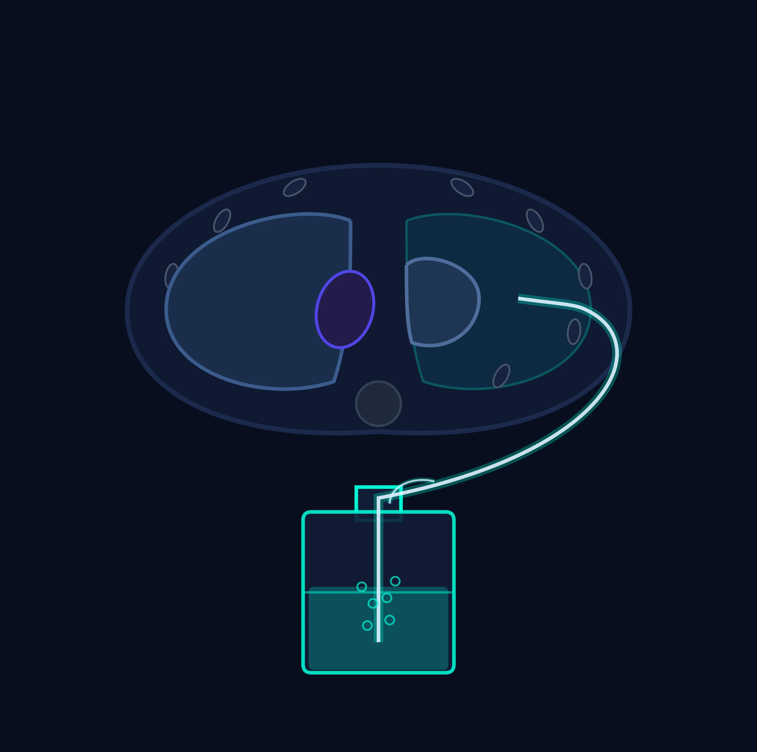
Chest trauma is managed by a structured search for the immediately life-threatening injuries during the primary survey (the "lethal six") and the potentially life-threatening injuries during the secondary survey (the "hidden six"). The Fellowship candidate must know each of the lethal six, recognise its clinical features, and deliver its immediate management without waiting for a CT scan, because the lethal six kill in minutes, not hours. The analgesia, the chest drain and the CT angiogram are the adjuncts that stabilise and then define the full injury.[1][2]
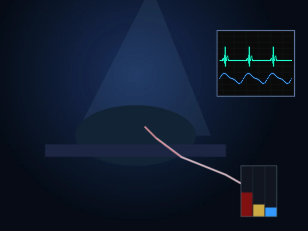
Differential diagnosis — the lethal six (the primary survey injuries)
The immediately life-threatening chest injuries are found and managed during the primary survey (the B and the C of the ABCDE), before any imaging. They are the following six, and each is a clinical diagnosis with a specific immediate management that does not wait for a CT scan. The Fellowship candidate must enumerate them, recognise their clinical features at the bedside, and deliver the immediate intervention for each one without delay. [1][2] The mnemonic and the framework are the ATLS standard, and the lethal six are the reason a trauma primary survey is performed in the order it is — the chest is assessed before the abdomen and the limbs because the chest kills first.
The blunt-aortic-injury mechanism and the high-speed deceleration are the indicators for the CT angiogram — a low threshold is the safe approach, and the penalty for missing the injury is a fatal rupture. [1]
Tension pneumothorax
- Shock, hypoxia, distended neck veins, absent breath sounds, hyperresonance
- CLINICAL diagnosis — decompress at the 5th ICS anterior axillary before imaging
- Needle decompression then a formal chest drain
- The commonest lethal chest injury
Open pneumothorax
- A communicating chest-wall wound; a "sucking chest wound"
- Seal with a three-sided dressing → chest drain (not through the wound)
- Surgical closure of the defect after the drain
- May progress to a tension
Massive haemothorax
- Shock, dull to percussion, absent breath sounds, hypoxia
- Chest drain; if >1500 mL immediately or >200 mL/h for 2–4 h → thoracotomy
- Resuscitate with blood; activate the massive-haemorrhage protocol
- Source: lung laceration, intercostal vessel, great vessel, mediastinum
Flail chest + contusion
- ≥3 ribs fractured in ≥2 places → paradoxical segment movement
- The pulmonary contusion is the killer (worsens over 24–48 h)
- Analgesia (epidural/paravertebral), oxygen, ± NIV/ventilation
- Monitor for respiratory failure
Cardiac tamponade
- Beck triad; muffled heart sounds, distended neck veins, hypotension
- FAST or echo confirms; pericardiocentesis if medical, thoracotomy if traumatic
- Traumatic (clotted) → resuscitative thoracotomy, not a needle
- Cross-link to the tamponade topic
Tracheobronchial injury
- Massive air leak, haemoptysis, subcutaneous emphysema, failed ventilation
- A persistent air leak despite a functioning chest drain
- Bronchoscopy and a surgical repair; one-lung ventilation
- Rare but lethal
The hidden six — the secondary survey injuries
The potentially life-threatening injuries are found during the secondary survey and on the imaging, and they may evolve over hours to days. The blunt aortic injury is screened by a CT angiogram in any high-speed-deceleration mechanism (a classic but insensitive CXR clue: a widened mediastinum, an apical cap, a deviated nasogastric tube, a depressed left main bronchus); the management is an endovascular or an open repair, and a beta-blocker to lower the dP/dt while the repair is arranged. The blunt cardiac injury ranges from a myocardial contusion (an ECG change, a troponin rise) to a commotio cordis (a sudden arrhythmic death from a precordial blow); the management is the ECG monitoring and the treatment of the arrhythmia. The diaphragmatic rupture (more common on the left, after a blunt high-pressure abdominal injury) is found on the CT (a herniated stomach or bowel into the chest) and repaired surgically. The oesophageal rupture (from a severe blunt or a penetrating injury) presents with a severe chest pain and a subcutaneous emphysema; it is a surgical emergency. The pulmonary contusion (a bruised lung that worsens over 24 to 48 hours, producing a hypoxia and a respiratory failure) is managed with the oxygen, the NIV or the ventilation, the fluid restriction, and the monitoring. The simple pneumothorax and the simple haemothorax are found on the imaging and drained. [1]
Immediate management — the primary survey and the adjuncts
The management follows the trauma primary survey (the ABCDE), with the chest as the priority organ for the B and the C. [1]
[1]The chest drain is inserted at the safe triangle (the fourth or fifth intercostal space, anterior axillary line), under a local anaesthesia (lidocaine 1 per cent up to 3 mg per kilogram), connected to an underwater seal. The chest-tube output and the swing are monitored. A FAST (the focused assessment with sonography in trauma) or a bedside echo screens for the pericardial fluid (the tamponade) and the intra-abdominal free fluid (the associated injury). A CT angiogram of the chest is performed in the stable patient for the full definition of the injuries (the aorta, the lungs, the bones, the mediastinum, the diaphragm).[1]
The chest-trauma thresholds
The analgesia and the flail chest
The rib fractures are painful, and the pain prevents the deep breathing and the coughing, producing an atelectasis and a pneumonia — the secondary complication that kills the elderly rib-fracture patient. The analgesia is the key management. The first-line is the multimodal oral analgesia (paracetamol, an NSAID, a weak opioid), escalated to the intravenous opioid (morphine 5 to 10 mg, titrated; or fentanyl 50 to 100 micrograms) for the severe pain. The regional anaesthesia (a thoracic epidural, a paravertebral block, or an ultrasound-guided serratus-anterior plane block) is the gold standard for the multiple-rib-fracture and the flail-chest patient, because it provides a profound analgesia without the respiratory depression of the systemic opioid.[2] The flail chest (three or more ribs fractured in two or more places, producing a free-floating segment that moves paradoxically) is managed with the analgesia, the oxygen, the pulmonary physiotherapy, and a low threshold for the NIV or the ventilation if the respiratory failure develops from the underlying contusion.
The chest drain insertion — the Seldinger technique and the blunt dissection
Confirm the indication (a pneumothorax, a haemothorax, a haemopneumothorax) and the side (a TIME-OUT — a wrong-side chest drain is a never-event)
Position the patient supine or semi-recumbent, the arm abducted over the head to expose the safe triangle
Mark the 5th ICS AAL; infiltrate the lidocaine 1 per cent up to 3 mg/kg, down to the pleura
A 2–3 cm incision parallel to the rib, over the upper border of the 6th rib (to avoid the neurovascular bundle in the subcostal groove)
A blunt dissection with a Kelly forcep through the intercostal muscles and the pleura, a finger swept to confirm the pleural space and to clear adhesions
Insert the chest tube (24–32 F for a pneumothorax; 32–36 F for a haemothorax), directed posteriorly and apically for an air leak, or posterobasally for a fluid
Connect to an underwater seal; check the swing (tidaling), the bubbling (an air leak), and the output (the volume and the colour)
Suture in place, dress, and a portable CXR to confirm the position and the re-expansion
Multimodal oral
- Paracetamol 1 g QID + an NSAID + a weak oral opioid
- For the isolated rib fractures and the mild pain
- Simple, no monitoring; the first-line for the elderly
- Inadequate for the flail chest and the multiple displaced fractures
IV opioid
- Morphine 1–2 mg titrated to the pain, or fentanyl 25–50 micrograms
- For the acute severe pain in the resuscitation bay
- Causes a respiratory depression, a hypotension, and a nausea — titrate and monitor
- A bridge to the regional anaesthesia, not the long-term plan
Thoracic epidural
- The gold standard for the flail chest and the ≥3 rib fractures
- Profound, segmental analgesia without the respiratory depression
- Improves the vital capacity, the peak flow, the pneumonia, the ventilator days
- Contraindicated in the coagulopathy, the sepsis, the spinal injury, the hypovolaemia
Paravertebral / SAPB
- Paravertebral block or the ultrasound-guided serratus-anterior plane block
- The alternative when the epidural is contraindicated
- Easier, safer, lower risk; the increasingly first-line in many centres
- A single-shot lasts 12–18 h; a catheter for the continuous infusion
The blunt aortic injury — the CT angiogram
The blunt aortic injury is the injury that is missed at a cost. It follows a high-speed deceleration (a motor-vehicle collision, a fall from a height) that tears the aorta at the isthmus (the ligamentum arteriosum). The CXR may show a widened mediastinum, an apical cap, a deviation of the nasogastric tube or a depression of the left main bronchus — but these signs are insensitive, and a CT angiogram is performed for any significant mechanism. The management is a blood-pressure control (a beta-blocker — esmolol or labetalol — to lower the dP/dt, the same principle as the aortic dissection) and an endovascular or an open repair. [1]
The injuries in depth
The lethal six and the hidden six are the framework; the in-depth knowledge of each injury is the Fellowship answer. The candidate is expected to deliver the anatomy, the mechanism, the clinical features, the immediate management, the imaging, the definitive management, the complications, and the pitfalls for each of the major injuries. The following sections are the in-depth treatment of the common and the high-yield injuries — the tension pneumothorax, the open pneumothorax, the massive haemothorax, the flail chest, the blunt aortic injury, the blunt cardiac injury, the commotio cordis, the tracheobronchial injury, and the resuscitative thoracotomy. [1]
Tension pneumothorax
A tension pneumothorax is air that enters the pleural space through a one-way valve and cannot escape, raising the intrapleural pressure above the central venous pressure, collapsing the great veins, and producing an obstructive shock. The classic features are a hypoxia, a tachycardia, a hypotension, a distended neck vein, an absent breath sound, and a hyperresonance on the affected side, with a tracheal deviation (a late sign, often absent). The ATLS doctrine is to decompress before any imaging — a portable CXR in a suspected tension is a critical error, because the patient may arrest while waiting for it. [1]
The traditional site for the needle decompression was the second intercostal space, mid-clavicular line (2nd ICS MCL) — it is still taught, but it has three problems. The chest wall at the MCL may be thicker than the standard 5-cm cannula (especially in the muscular or the obese male); the catheter may kink or dislodge as the patient moves; and the great vessels and the internal mammary artery lie close to the MCL. The 10th edition of ATLS (2018) reclassified the preferred site to the fifth intercostal space, anterior axillary line (5th ICS AAL) — the same site as a chest drain — because the chest wall is thinner there, the cannula is more secure, and it converts directly to a finger thoracostomy or a chest drain at the same site. The modern standard cannula is 10 to 14 gauge, 8 cm long — a shorter cannula fails in the muscular patient and is the commonest cause of a "failed decompression". The needle is removed, the cannula is left in situ, and a formal chest drain is inserted at the same site as soon as possible. [1]
[1]The finger thoracostomy is the alternative in the intubated patient or when the patient is in the resuscitation bay with a surgical set available — a blunt dissection through the 5th ICS AAL, a finger swept into the pleural space to break any adhesions and to confirm the lung, and a chest drain inserted. It is faster than a needle in the trained hands, more reliable (no kinking, no dislodgement), and it provides a definitive decompression; it is the technique of choice in many trauma centres for the intubated patient and the patient in arrest. [1]
The decompression pathway for a tension pneumothorax
Recognise the clinical picture — shock + hypoxia + absent breath sounds ± hyperresonance/distended neck veins; tracheal deviation is a late sign
Call for help; high-flow oxygen 100 per cent; prepare the chest-drain trolley and a 10–14 G × 8 cm cannula
Decompress at the 5th ICS AAL with the cannula (or a finger thoracostomy if intubated); angle over the upper border of the 6th rib to avoid the neurovascular bundle
Confirm the release — a rush of air, a rise in saturation, a fall in the heart rate
Insert a formal large-bore chest drain (28–32 F) at the same site, connected to an underwater seal
Re-examine for the other lethal six — a shocked trauma patient may have more than one
Portable CXR after the decompression to confirm the position and the re-expansion
Open pneumothorax (the sucking chest wound)
An open pneumothorax is a chest-wall defect that is large enough (>two-thirds the tracheal diameter, ~3 cm) to allow the air to preferentially enter the pleural space through the wound rather than through the trachea during the inspiration — a "sucking chest wound". The defect is usually from a penetrating injury (a close-range shotgun, a large stab, a blast, a high-energy projectile). The immediate management is the three-sided occlusive dressing — a sterile dressing taped on three sides, leaving the fourth side open as a one-way flutter valve (air escapes on expiration, no air enters on inspiration). A fully sealed dressing converts an open pneumothorax into a tension pneumothorax — the lethal trap. [1]
[1]The chest drain is placed at a site away from the wound (the standard 5th ICS AAL safe triangle if the wound is not there), and the defect is formally closed in theatre — the wound is debrided, the layers are approximated, and the chest wall is reconstructed if the defect is large (a rotational muscle flap — the latissimus dorsi or the pectoralis major — or a synthetic mesh). [1]
Massive haemothorax
A massive haemothorax is the rapid accumulation of more than 1500 mL of blood into the pleural space (or more than 200 mL per hour for 2 to 4 hours), producing a hypovolaemic shock with the clinical signs of a dull percussion note, an absent breath sound, and a hypoxia. The sources are the lung laceration (the most common, usually self-limiting as the pulmonary vasculature is a low-pressure system), the intercostal or the internal mammary artery (the systemic pressure, the high output, the surgical ligation), the great vessel (the aorta, the subclavian — usually fatal at the scene), or the mediastinal source (the heart, the great veins). [1]
The immediate management is the large-bore chest drain (32 to 36 French) inserted at the 5th ICS AAL, with the massive-haemorrhage protocol activated (the blood, the fresh-frozen plasma, the platelets, the cryoprecipitate, and the tranexamic acid 1 g intravenously within 3 hours, per the CRASH-2 trial). The chest drain is inserted only after the fluid resuscitation is started, because the release of the tamponading intrathoracic pressure may precipitate a further bleed — "the death of the tamponade". The drain is connected to an autotransfusion device if available. [1]
Chest drain only
- <1500 mL immediately AND <200 mL/h ongoing
- A low-output source — usually a lung laceration
- Monitor the output; the drain stays until the output is <200 mL/24 h
- Re-expand the lung; consider a second drain if a clotted haemothorax develops
Thoracotomy (ED or theatre)
- >1500 mL immediately OR >200 mL/h for 2–4 h OR persistent haemodynamic instability
- A high-output source — intercostal/internal mammary artery, great vessel, mediastinum
- Activate the massive-haemorrhage protocol; cross-match; anaesthetist and the thoracic surgeon present
- ED thoracotomy if the patient arrests; theatre thoracotomy if the patient is stabilised
A retained haemothorax (a clotted haemothorax that does not drain) is the late complication — it is a substrate for an empyema and a fibrothorax, and it requires a thoracoscopic drainage (a VATS) within 7 to 10 days if it has not resolved on the serial imaging. The prophylactic antibiotics for the chest drain (the first-generation cephalosporin) reduce the infective complications, per the meta-analyses. [1]
Flail chest — the surgical stabilisation and the epidural
A flail chest is three or more ribs fractured in two or more places, producing a free-floating segment that moves paradoxically (in during the inspiration, out during the expiration). The paradoxical movement is the obvious sign, but the killer is the underlying pulmonary contusion — a bruised lung that worsens over 24 to 48 hours, producing a hypoxia, a shunt, and a respiratory failure. The analgesia is the foundation of the management (it allows the deep breathing and the coughing); the oxygen and the pulmonary physiotherapy prevent the atelectasis and the pneumonia; and the NIV or the ventilation supports the respiratory failure from the contusion. [1]
The thoracic epidural is the gold-standard analgesia for the flail chest and the multiple-rib-fracture patient — it provides a profound, segmental analgesia without the respiratory depression of the systemic opioid, it improves the vital capacity and the peak expiratory flow, and it reduces the pneumonia and the ventilator days. The paravertebral block (single-shot or a continuous catheter) and the ultrasound-guided serratus-anterior plane block are the alternatives when the epidural is contraindicated (a coagulopathy, a spinal injury, a sepsis, a refusal). The epidural is started early (within 24 hours of the admission), continued for 3 to 5 days, and weaned as the pain resolves. [1]
[1]The surgical stabilisation of the rib fractures (SSRF) — the open reduction and the internal fixation of the fractured ribs with plates — has emerged from the recent trials as a benefit for the selected patient. The contemporary indications (from the Chest Wall Injury Society and the 2020–2025 randomised trials — the Pieracci, the Marasco, and the Bollier groups) are: a flail chest with a respiratory failure or a failure to wean from the ventilator; a marked chest-wall deformity (a flail segment more than 5 cm, a sternal flail, a lateral flail); a severe pain uncontrolled by the analgesia; or a significant displacement of the fractures (>1 cm, a bicortical displacement, a segmental fracture of three or more ribs). The benefit is a shorter ventilator time, a lower pneumonia, a better pulmonary function, and a faster return to work. The timing is within 72 hours of the injury where possible, before the callus forms. [1]
Surgical stabilisation of rib fractures (SSRF) — the modern trials
Key finding
Reduced ventilator days, pneumonia, ICU stay, and chronic chest-wall pain in the selected patient
Blunt aortic injury — the TEVAR
The blunt aortic injury (BAI) is the injury that is missed at a cost — a partial or a full-thickness tear of the aortic wall at the isthmus (the ligamentum arteriosum, where the mobile arch meets the fixed descending aorta), produced by a high-speed deceleration (a motor-vehicle collision at >50 km/h, a fall from >3 m, an ejection, a high-speed motorcycle crash). The CXR signs of the widened mediastinum (>8 cm at the level of the aortic knob), the loss of the aortic-knob contour, the apical cap (the blood tracking up the pleura), the deviation of the nasogastric tube to the right, the depression of the left main bronchus, and the widened paraspinal stripe are the classic signs — but each is insensitive, and a normal CXR does not exclude the injury. The CT angiogram is the gold standard, performed for any high-speed mechanism with the appropriate signs or mechanism alone (the threshold is low). [1]
The grading of the blunt aortic injury (the Society for Vascular Surgery grading):
- Grade I — an intimal tear (<10 mm); managed with a watchful waiting and a beta-blocker; most heal.
- Grade II — a large intimal tear (>10 mm) or an intramural haematoma; managed with a beta-blocker and a follow-up CT, with a repair if it progresses.
- Grade III — a pseudoaneurysm; managed with a repair.
- Grade IV — a free rupture; managed with an immediate repair (high mortality). [1]
Medical (the grade I–II)
- Beta-blocker (esmolol) to a systolic 100–120 mmHg and a heart rate 60–80 bpm
- Strict BP and HR control to lower the dP/dt (the wall stress)
- Serial CT angiogram at 24 h, 7 d, and 1 month
- Most grade I heal; the grade II may progress to a III
TEVAR (the grade III–IV)
- Thoracic endovascular aortic repair — a covered stent across the tear
- The gold standard for the grade III–IV (the pseudoaneurysm, the rupture)
- Shorter than the open repair, less blood loss, lower mortality and paraplegia
- Lands in the proximal descending aorta; covers the left subclavian if needed (a left subclavian bypass if the LIMA is used in a CABG patient)
The beta-blocker (the esmolol infusion, or the labetalol) is started as soon as the diagnosis is made, to lower the systolic to 100–120 mmHg and the heart rate to 60–80 bpm, reducing the dP/dt (the rate of the pressure rise) and the wall stress — the same principle as the aortic dissection. The pain control and the avoidance of the Valsalva (cough, straining) are the adjuncts. The patient is admitted to the ICU, monitored, and transferred to a vascular or a cardiothoracic centre for the TEVAR. [1]
[1]Blunt cardiac injury
The blunt cardiac injury (BCI) is a spectrum — from a myocardial contusion (a bruised myocardium with an ECG change and a troponin rise) to a chamber rupture (the free wall, the septum, or a valve — usually fatal at the scene), a coronary artery thrombosis (an MI picture), or a commotio cordis (see below). The mechanism is a direct precordial blow (a steering wheel, a sports ball, a fall, a crush, an airbag). The diagnosis is challenging because the ECG and the troponin are non-specific, and the clinical features (a chest pain, a dyspnoea, a haemodynamic instability, an arrhythmia) overlap with the other thoracic injuries. [1]
The Eastern Association for the Surgery of Trauma (EAST) practice-management guidelines (2012, updated 2023) give a structured approach:
- An ECG is obtained in all the suspected BCI patients.
- A troponin I is obtained on the admission and at 4 to 6 hours.
- If both are normal, the BCI is excluded and no further cardiac workup is needed.
- If either is abnormal, the patient is monitored (a continuous ECG for 24 to 48 hours) and an echocardiogram is performed.
- An echocardiogram is not a screening test — it is performed for a haemodynamic instability, a new murmur, a suspected structural injury, or an unexplained arrhythmia. [1]
The management of the BCI is the cardiac monitoring and the treatment of the complications — the arrhythmia (the atrial fibrillation, the ventricular ectopics, the heart block — the standard ACLS protocol), the cardiogenic shock (an inotrope — the dobutamine or the milrinone, and an IABP or an Impella in the refractory shock), and the structural injury (the chamber rupture, the septal defect, the valvular failure — a surgical or a percutaneous repair). The asymptomatic patient with a normal ECG and a normal troponin is discharged. [1]
Commotio cordis
The commotio cordis is a sudden cardiac death from a blunt precordial blow — the classic scenario is the young athlete (the cricket, the baseball, the lacrosse, the hockey player) struck in the chest by a ball or a puck. The blow must land directly over the precordium, at the precise moment of the cardiac cycle (the T-wave upstroke, 10–30 ms before the T-wave peak), producing a ventricular fibrillation without a structural injury. The diagnosis is the witnessed collapse immediately after the precordial blow, in the absence of a structural cardiac injury on the post-resuscitation imaging (the echo, the cardiac MRI) or the post-mortem. [1]
[1]The management is the immediate CPR and the defibrillation — the survival is critically time-dependent, and the early defibrillation (within 3 minutes) gives a survival of around 60 per cent, falling to single digits beyond 5 minutes. An automated external defibrillator (AED) at the sporting venue, the trained bystanders, and the emergency response planning are the public-health interventions that have improved the survival. The patient who survives is investigated for a structural cardiac injury (an echo, a cardiac MRI, an electrophysiology study) and the long-term risk — the recurrence is rare if the workup is negative, and the return to sport is individualised. [1]
The commotio cordis — the numbers
Tracheobronchial injury
The tracheobronchial injury is a rare but lethal injury — a tear of the trachea, the carina, or the main bronchus, from a severe blunt (a high-energy deceleration, a crush, a seat-belt injury) or a penetrating (a stab, a gunshot to the neck or the upper chest) mechanism. The presentation is the massive haemoptysis, a massive subcutaneous emphysema (the face, the neck, the chest wall), a massive air leak from the chest drain, and a failure to ventilate (the tidal volume escapes through the bronchopleural fistula). The injury is suspected when a chest drain does not resolve the pneumothorax, when the subcutaneous emphysema progresses, or when the ventilation fails despite the functioning chest drain. [1]
The diagnosis is the bronchoscopy — it confirms the tear, defines the location and the extent, and guides the intubation. The classic location of the blunt tracheobronchial injury is within 2 cm of the carina (80 per cent) — the shear force between the mobile carina and the fixed distal bronchi. The immediate management is the bronchial intubation — the endotracheal tube is advanced past the tear into the intact main bronchus (often the right, but guided by the bronchoscopy), and a one-lung ventilation is started. A dual-lumen endotracheal tube or a bronchial blocker is the alternative in the experienced hands. [1]
[1]The chest drain is left in situ for the pneumothorax and the air leak. The surgical repair is the definitive management — a thoracotomy, a primary repair of the tear with absorbable sutures, and a tissue flap (an intercostal muscle, a pericardial fat pad, a pleural flap) to reinforce the suture line and prevent a fistula. The postoperative complications are a bronchopleural fistula, a stenosis, and a pneumonia; the mortality is 30 per cent overall (most die at the scene from a tension pneumothorax or an airway loss). [1]
The tracheobronchial injury — the numbers
Resuscitative thoracotomy (the emergency department thoracotomy)
The resuscitative thoracotomy (RT) — also called the emergency department thoracotomy (EDT) or the clamshell thoracotomy — is a left anterolateral thoracotomy performed in the resuscitation bay, on a patient in traumatic arrest or peri-arrest, to release a tamponade, to control a thoracic haemorrhage, to perform an open cardiac massage, and to cross-clamp the descending aorta. The clamshell (a bilateral anterolateral thoracotomy, extending the left incision across the sternum to the right) gives a wider exposure for the cross-clamping and the right-sided injuries, and is the standard for the RT in the trauma centre. [1]
The indications (the 2024 Western Trauma Association and the ATLS criteria):
- A penetrating thoracic trauma with a witnessed arrest and a less than 15 minutes of CPR — the best-prognosis group (a survival of 20–30 per cent).
- A penetrating thoracoabdominal trauma with a witnessed arrest and a less than 15 minutes of CPR.
- A blunt thoracic trauma with a witnessed arrest and a less than 10 minutes of CPR — a poorer prognosis (a survival of 1–2 per cent), but the threshold to attempt in the right clinical picture.
- A patient with signs of life (a palpable pulse, a reactive pupil, an organised ECG rhythm, a respiratory effort, a spontaneous movement) on arrival, who then arrests. [1]
The contraindications are the absence of the signs of life in the field, more than 15 minutes of CPR (the penetrating) or 10 minutes (the blunt), or an unwitnessed blunt arrest — these patients do not survive the RT, and the procedure exposes the team to a risk and a futility. [1]
The clamshell thoracotomy — the steps
Indication confirmed — penetrating thoracic arrest with <15 min CPR, or signs of life present
Left anterolateral thoracotomy at the 5th ICS (the inframammary fold), from the mid-axillary line to the sternum, with a scalpel through the skin and a heavy scissors through the intercostal muscles
Extend across the sternum to the right side (a clamshell) — protect and ligate the internal mammary arteries (they bleed)
Open the pericardium longitudinally (anterior to the phrenic nerve) — release a clotted tamponade; the pericardium is tense and dark if there is a tamponade
Deliver the heart into the wound; open cardiac massage — a two-handed, clasped compression from the apex towards the base (a "squeezing-wrung-out" technique), at 80–100/min
Identify and control the bleeding — a pulmonary laceration (a stapler, a clamp), a cardiac laceration (a Foley catheter balloon inflated in the wound, a 3-0 prolene suture, or a skin stapler for a quick control)
Cross-clamp the descending aorta (below the left pulmonary hilum, against the thoracic spine) — improves the coronary and the cerebral perfusion
Control the hilum (a twisting of the lung on its hilum — the "hilar twist") for an uncontrolled pulmonary haemorrhage
Resuscitate with the blood (1:1:1 PRBC:FFP:platelets), the tranexamic acid 1 g, and the calcium chloride 1 g; transfer to theatre for the definitive repair
Pericardiocentesis (medical tamponade)
- A medical tamponade (a pericardial effusion — a uraemic, a malignant, an idiopathic)
- A needle aspiration of the pericardial fluid — subxiphoid, towards the left scapula
- Sterile, quick, low-resource; the patient is awake
- Inadequate for the clotted traumatic tamponade — the needle cannot aspirate the clotted blood
Resuscitative thoracotomy (traumatic tamponade)
- A traumatic tamponade (the clotted blood from a cardiac laceration)
- A clamshell thoracotomy and an open pericardiotomy
- The clotted blood is evacuated; the cardiac wound is controlled with a suture or a Foley
- The definitive procedure in the traumatic arrest; the pericardiocentesis is futile
The resuscitative thoracotomy — the survival data
Key finding
An overall survival of 8–12 per cent, with the penetrating cardiac tamponade the best-prognosis subgroup
Complications and pitfalls
The complications are the respiratory failure (from the pulmonary contusion, the flail chest, the fat embolism), the pneumonia, the empyema, the retained haemothorax, and the late complications of the blunt aortic injury (the pseudoaneurysm, the rupture). The pitfalls are the inverse of the management: treating the chest-trauma patient with a CT before the primary survey; missing a tension pneumothorax behind a "shocked trauma patient"; not draining a massive haemothorax; a delayed CT-angiogram for the aortic injury; under-treating the rib-fracture pain (the atelectasis and the pneumonia); and not recognising the pulmonary contusion that worsens over 24 to 48 hours. [1]
Prognosis and disposition
The prognosis depends on the injury and the associated polytrauma. The tension pneumothorax and the tamponade are immediately lethal if untreated; the massive haemothorax and the aortic injury carry a high mortality; the isolated rib fractures do well with an adequate analgesia. The patient is admitted to the trauma service, the high-dependency or the intensive care (for the flail chest, the contusion, the ventilated). The elderly rib-fracture patient is observed for 24 hours for a respiratory deterioration. [1]
Special populations
The elderly with the rib fractures have a high morbidity and mortality from the secondary pneumonia, and a low threshold for admission, the epidural analgesia and the observation. The anticoagulated patient with a chest injury has a higher risk of a delayed haemothorax and is reversed early. The paediatric chest trauma may show a significant internal injury with a minimal external sign (the pliable ribs absorb the energy and transmit it to the underlying organs). [1]
Evidence and regional guidelines
The contemporary framework is the ATLS and the local trauma protocol; the chest-wall-injury management (the rib fractures, the flail chest, the analgesia) is summarised in the recent reviews.[1][2] The lethal six and the hidden six, the chest-drain thresholds and the CT-angiogram indications are the ATLS-standard global framework; the analgesia regimen and the thoracotomy criteria follow the local trauma and the anaesthetic pathway.
ANZ practice note. The lethal six and the hidden six follow the ATLS/EMST framework via the local trauma protocol; the chest drain is placed in the safe triangle at the fourth or fifth intercostal space, the thoracotomy threshold is 1500 mL immediately or 200 mL per hour, and the CT angiogram is the standard for the blunt aortic injury screening in the high-speed mechanism. [1]
Exam pearls
- The lethal six: tension pneumo, open pneumo, massive haemothorax, flail chest/contusion, tamponade, tracheobronchial injury — found and managed in the primary survey.
- Tension pneumothorax = CLINICAL diagnosis → decompress before imaging.
- Massive haemothorax: >1500 mL immediately or >200 mL/h → thoracotomy.
- Flail chest: ≥3 ribs in ≥2 places; the contusion is the killer (worsens over 24–48 h).
- Blunt aortic injury: CT angiogram for any high-speed mechanism; widened mediastinum is insensitive.
- Analgesia is the key for the rib fractures; a regional block (epidural/paravertebral/serratus) for the flail chest. [1]
Exam practice
SAQ — Tension pneumothorax in the blunt chest trauma patient
10 minutes · 10 marks
A 28-year-old man is brought to your trauma centre 35 minutes after a high-speed motor-vehicle collision in which he was the unrestrained driver. On arrival he is agitated and cyanosed, with a respiratory rate of 32, SpO2 86 per cent on 15 L oxygen via a non-rebreather mask, BP 84/52, HR 132. The neck veins are visibly distended to the angle of the jaw, the trachea is clinically deviated to the left, the right hemithorax is hyperresonant to percussion with absent breath sounds and reduced chest-wall movement. There is no external chest wound.
SAQ — Blunt cardiac injury with tamponade after a steering-wheel impact
10 minutes · 10 marks
A 45-year-old unrestrained driver is brought to the trauma bay 40 minutes after a frontal collision at 80 km/h in which his chest struck the steering wheel. He is drowsy (GCS 13) and profoundly shocked: BP 72/48, HR 138 in sinus tachycardia, SpO2 92 per cent on 15 L oxygen, with cold clammy peripheries and neck veins distended to the angle of the jaw. The heart sounds are muffled and the systolic pressure drops a further 18 mmHg in inspiration. The extended FAST shows a large pericardial effusion with right-ventricular diastolic collapse. Bilateral breath sounds are present and equal. Two minutes later he loses consciousness and the rhythm degenerates into a pulseless electrical activity (PEA) arrest.
Red flags
[1]References
- [1]Dehghan N, Cicala L, Tieszer C, et al. Management of Chest Wall Injuries: An Updated Review J Am Acad Orthop Surg, 2026.PMID 41525674
- [2]Holtrop TET, van Laarhoven K, Hietbrink F, et al. Pain management in non-operatively treated patients with multiple single rib fractures: an international comparison Eur J Trauma Emerg Surg, 2026.PMID 41758337