EM · Neuromuscular junction disorders
Guillain-Barré syndrome and myasthenia gravis
Also known as Guillain-Barré syndrome · GBS · Acute inflammatory demyelinating polyradiculoneuropathy · AIDP · Myasthenia gravis · MG · Myasthenic crisis · Miller-Fisher syndrome
Guillain-Barré syndrome — the acute ascending flaccid paralysis with areflexia and autonomic instability, the respiratory-failure thresholds (FVC under 20 mL/kg or NIF under minus 30 cmH2O), the albuminocytologic dissociation on the CSF, and the IVIG 0.4 g/kg/day for 5 days or plasma exchange; and myasthenia gravis — the fatigable weakness (ptosis, diplopia, bulbar), the AChR antibody, the neostigmine and ice-pack tests, the pyridostigmine 60 mg PO, and the IVIG or plasma exchange for the myasthenic crisis. The GBS-versus-MG-versus-Lambert-Eaton differential. ACEM-primary, globally tagged.
On this page & tools
Your progress
Saved locally on this device.
Target exams
Red flags
The two acute autoimmune neuromuscular junction disorders the Fellowship candidate must hold apart at the bedside are Guillain-Barré syndrome (GBS) and myasthenia gravis (MG). GBS presents as a subacute ascending flaccid paralysis with areflexia and autonomic instability, where the danger is the respiratory failure and the cardiac arrhythmia; MG presents as a fatigable weakness of the ocular, bulbar and proximal muscles, where the danger is the myasthenic crisis. Both can beheaded off the cliff by serial bedside spirometry, both respond to IVIG or plasma exchange, and both are mismanaged when confused with each other, with Lambert-Eaton, or with a structural cord lesion.[1][2][5]
Definition and the epidemiology
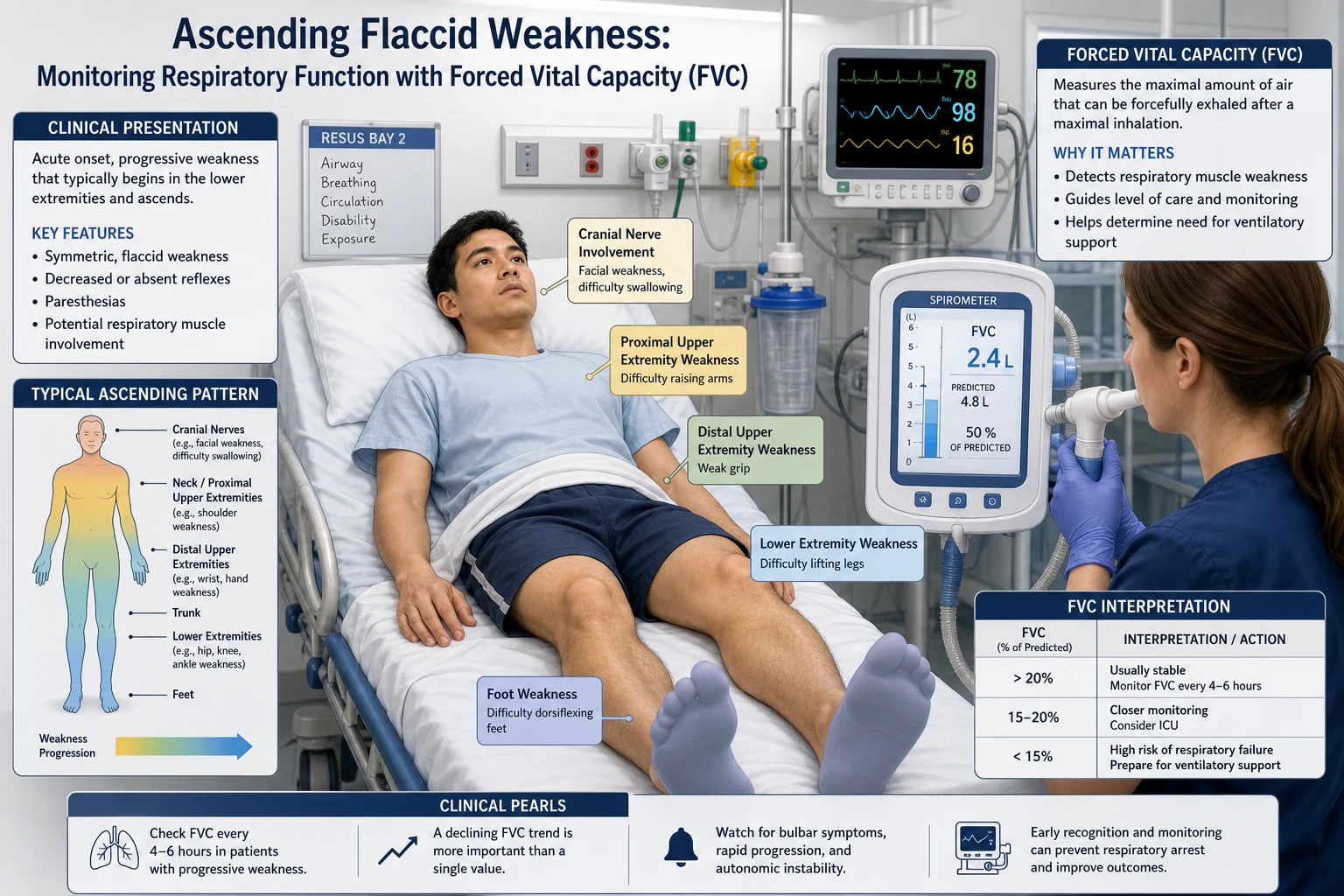
Guillain-Barré syndrome is the acute, monophasic, immune-mediated polyradiculoneuropathy of the peripheral nerves and the nerve roots, defined clinically by the progressive symmetric weakness of the limbs over hours to a maximum of 4 weeks, with the areflexia and the relative sparing of the sensation. The incidence is 1 to 2 per 100,000 per year. Two-thirds of the cases follow an antecedent infection in the preceding 1 to 3 weeks — Campylobacter jejuni (the commonest, the diarrhoeal illness), cytomegalovirus, Epstein-Barr virus, Mycoplasma pneumoniae, influenza, hepatitis E, and the Zika virus are the recognised triggers; the surgery and the vaccination are weaker associations.[1][2]
Myasthenia gravis is the antibody-mediated blockade of the post-synaptic acetylcholine receptor at the neuromuscular junction, producing a fatigable weakness that worsens with sustained activity and improves with rest. The prevalence is about 20 per 100,000, with a bimodal distribution — the young women (under 40) and the older men (over 60). A thymoma is present in 10 to 15 per cent of the patients, and the thymic hyperplasia in the majority of the early-onset disease. The myasthenic crisis — the respiratory failure severe enough to need the mechanical ventilation — occurs in 15 to 20 per cent of the MG patients at some point, most often within the first 2 years of the disease, and is the ED presentation that kills.[6][7]
Pathophysiology
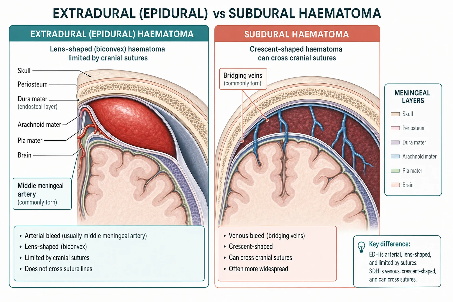
GBS is the molecular-mimicry disease: the antecedent infection shares an epitope with a component of the peripheral nerve, and the resulting antibody and the macrophage attack damages the myelin sheath (the acute inflammatory demyelinating polyradiculoneuropathy, AIDP, in 85 to 90 per cent of the Western cases) or the axon itself (the acute motor axonal neuropathy, AMAN, and the acute motor and sensory axonal neuropathy, AMSAN — commoner after Campylobacter and in Asia). The target gangliosides are the GM1 (in the AMAN and the AIDP with the motor predominance) and the GQ1b (in the Miller-Fisher syndrome). The demyelination slows and then blocks the nerve conduction; the axonal variant directly destroys the axon and carries a slower, less complete recovery. The cranial-nerve roots and the autonomic fibres are involved, explaining the facial diplegia and the autonomic instability.[1][2]
Myasthenia gravis is the post-synaptic NMJ disease. The antibodies bind the acetylcholine receptor (AChR) on the motor end-plate, causing the receptor degradation, the complement-mediated junctional damage, and the reduced safety factor for the neuromuscular transmission — the repetitive nerve stimulus arrives, but the post-synaptic response falls below the threshold for the muscle-fibre contraction, and the weakness fatigues with the sustained activity. In the AChR-antibody-negative patients, the antibodies against the muscle-specific kinase (MuSK) or the LRP4 are found; the MuSK-MG behaves differently (the prominent bulbar and the respiratory involvement, the relative sparing of the limb, the poorer response to the acetylcholinesterase inhibitors).[5][6]
The clinical presentation
The GBS-versus-MG bedside discriminators
Guillain-Barré syndrome begins with the symmetric weakness that typically starts in the legs and ascends to the arms, the trunk, the cranial nerves and the respiratory muscles over days. The weakness is flaccid, the reflexes are absent or markedly reduced (the clinical hallmark), and the sensation is relatively spared — a paraesthesia and the numbness are common early but the objective sensory loss is mild. The back pain and the myalgia are prominent in a third of the patients and may be the presenting complaint. The bilateral facial weakness (the facial diplegia) occurs in half the cases and is a strong clue when the limbs are also weak. The autonomic instability — the sinus tachycardia, the bradyarrhythmia, the blood-pressure swings (the hypertensive and the hypotensive episodes), the ileus, the urinary retention, the sweating and the pupillary changes — is the dangerous feature: sudden death from the cardiac arrhythmia or the asystole is a recognised event, and the patient needs the continuous cardiac monitoring from the moment the diagnosis is suspected. The respiratory failure develops in 25 per cent of the patients as the weakness ascends to the diaphragm and the intercostal muscles — the FVC falls, the cough weakens, the bulbar weakness invites the aspiration. The progression reaches its nadir by 4 weeks (by definition; over 4 weeks is the chronic inflammatory demyelinating polyradiculoneuropathy, CIDP).[1][2]
Myasthenia gravis presents with the fatigable weakness — the muscles that work hard weaken, and the muscles that rest recover. The ocular muscles are the first affected in half the patients: the ptosis (worse with the sustained upgaze) and the diplopia (the variable, the asymmetric, the fluctuating). The bulbar muscles produce the dysarthria (the nasal, the fatigable speech), the dysphagia, the weak cough and the nasal regurgitation — the bulbar weakness is the high-risk feature because it predicts the aspiration. The limb weakness is proximal and symmetric (the arms held up weaken over a minute; the head drop is a late sign of the neck-extensor fatigue). The respiratory failure of the myasthenic crisis is from the diaphragm and the intercostal fatigue, with the bulbar failure compounding the aspiration. The crisis is precipitated by the infection (the commonest), the surgery, the pregnancy and the puerperium, the emotional stress, and the drugs — the aminoglycosides, the macrolides, the fluoroquinolones, the beta-blockers, the magnesium, the neuromuscular blockers and the iodinated contrast.[5][6][7]
Differential diagnosis — the acute paralysis
The ED candidate must distinguish GBS, MG and Lambert-Eaton from each other and from the structural and the electrolyte causes of the acute paralysis, because the management diverges sharply. [1]
Guillain-Barré (AIDP)
- Subacute ascending flaccid paralysis over days to 4 weeks; areflexia the hallmark
- Antecedent Campylobacter / viral infection 1 to 3 weeks prior; CSF albuminocytologic dissociation
- Facial diplegia, autonomic instability, respiratory failure; NCS shows demyelination (prolonged distal latency, conduction block)
- Treatment: IVIG 0.4 g/kg/day x 5d OR plasma exchange; NOT steroids; ICU for FVC under 20 mL/kg or NIF under minus 30
Myasthenia gravis
- Fatigable weakness — worse with use, better with rest; ptosis and diplopia the opening features
- Reflexes preserved; sensation normal; no autonomic instability (unless from a precipitant)
- AChR antibody positive (85%); ice-pack and neostigmine tests positive; thymoma on the CT chest
- Treatment: pyridostigmine 60 mg PO QID; IVIG or plasma exchange plus steroids for crisis; avoid the precipitant drugs
Lambert-Eaton (LEMS)
- Proximal limb weakness that IMPROVES with repeated activity (the post-exercise facilitation); reflexes absent at rest, augment after brief exercise
- Autonomic features prominent (the dry mouth, the constipation, the impotence, the blurred vision)
- Paraneoplastic in 50 to 60 per cent — small-cell lung cancer is the association; anti-VGCC antibody positive
- Treatment: treat the tumour; 3,4-diaminopyridine; IVIG for the acute worsening; NOT pyridostigmine alone (weak response)
Botulism / tick paralysis
- Botulism — descending paralysis, cranial nerves first (the diplopia, the dysarthria, the dilated pupils), then the limbs; from the wound, the food, or the infant gut; antitoxin available
- Tick paralysis — ascending paralysis mimicking GBS; a tick is embedded (the scalp, the hidden fold); removal of the tick cures within hours
- Botulism — normal CSF, normal sensory exam; the pupillary paralysis and the postural hypotension distinguish it
- Treatment: botulism antitoxin + supportive; tick — find and remove the tick
Transverse myelitis / cord compression
- A sensory LEVEL (the band, the loss below) localises to the cord; the UMN signs (the spasticity, the hyperreflexia, the Babinski) once the spinal shock resolves
- The sphincter involvement (the urinary retention, the bowel incontinence) is early and prominent
- The MRI of the spine is the test — not the LP or the NCS
- Treatment: the urgent MRI, the steroids (the transverse myelitis) or the decompression (the compression)
Hypokalaemic periodic paralysis
- Episodic, sudden, painless weakness — minutes to hours, NOT days; the reflexes are absent during the attack
- Triggered by the heavy meal, the exercise, the cold, the emotion; the serum potassium is LOW (or HIGH in the hyperkalaemic variant)
- The thyroid (the thyrotoxic periodic paralysis in the Asian men), the familial and the renal-tubule causes
- Treatment: the potassium replacement (cautiously); the beta-agonist; the carbonic-anhydrase inhibitor for prevention
The three questions that separate them at the bedside: did it come on over days (GBS), over months with fluctuation (MG), or in minutes (the periodic paralysis, the stroke, the electrolyte)?, are the reflexes absent at rest and preserved after the brief exercise (LEMS)?, and is there a sensory level or the sphincter involvement (the cord lesion)? [1]
Bedside assessment
The ABCDE comes first, with two non-negotiable additions for the paralysed patient: the serial bedside spirometry and the continuous cardiac monitoring. The respiratory examination alone is dangerously late in the neuromuscular weakness — the patient retains the ability to talk and to maintain the saturation until the FVC is critically low, because the disease weakens the accessory muscles before the obvious respiratory distress appears. Measure the forced vital capacity (FVC), the negative inspiratory force (NIF), and the peak cough flow at the baseline and every 2 to 4 hours while the patient is deteriorating; document the trend. Examine the cranial nerves (the facial diplegia of the GBS, the fatigable ptosis and the ophthalmoplegia of the MG, the bulbar weakness of both), the limb power with the MRC grading (the proximal and the distal, the upper and the lower), the reflexes (the areflexia of the GBS versus the preserved reflexes of the MG), the sensation (the sparing in the MG, the paraesthesia in the GBS, the level in the cord lesion), and the autonomic signs (the heart rate, the blood pressure, the pupils). The fatigability tests for the MG — the sustained upgaze for 60 seconds (the ptosis appears), the sustained arm abduction for 60 seconds (the arm drops), the ice-pack test (the ptosis improves after 2 minutes of the cold applied to the eyelid, because the cold reduces the acetylcholinesterase activity).[5]
Investigations
The investigation of the suspected GBS confirms the diagnosis and excludes the mimics but must never delay the ICU admission or the disease-modifying therapy. The lumbar puncture shows the albuminocytologic dissociation — the CSF protein is elevated (often 0.55 to 2.0 g/L) with the normal cell count (under 5 white cells per microlitre) — but this change is absent in the first week and may take 2 to 3 weeks to appear; an early normal CSF does not exclude the GBS. A CSF pleocytosis over 50 cells per microlitre should prompt the reconsideration — the HIV-associated polyradiculopathy, the Lyme disease, the CMV polyradiculitis, the carcinomatous meningitis. The nerve conduction studies show the demyelination (the prolonged distal motor latencies, the conduction block, the slowed conduction velocity, the prolonged or the absent F-waves) in the AIDP, or the reduced CMAP amplitude with the normal conduction velocity in the axonal variants — but again, the NCS may be normal in the first 1 to 2 weeks and a normal early NCS does not exclude the diagnosis. The anti-ganglioside antibodies (the anti-GM1 in the AMAN and the motor-predominant AIDP, the anti-GQ1b in the Miller-Fisher syndrome) support the diagnosis when positive. The MRI of the spine (with the gadolinium) shows the enhancement of the nerve roots in the typical case and excludes the cord compression and the transverse myelitis when the clinical picture is uncertain. The bloods exclude the mimics — the electrolytes (the hypokalaemic periodic paralysis), the magnesium, the creatine kinase (the myositis), the lead level (the motor neuropathy), the toxicology, the thyroid function (the thyrotoxic periodic paralysis), the HIV.[1][2]
The investigation of the suspected MG confirms the antibody and excludes the thymoma. The AChR antibody is positive in 85 per cent of the generalised MG and about 50 per cent of the pure ocular MG — a highly specific test; the anti-MuSK antibody is tested in the AChR-negative patient (positive in 5 to 8 per cent, with the prominent bulbar and the respiratory phenotype). The anti-LRP4 is the third-line. The CT of the chest (or the MRI) excludes the thymoma in every new diagnosis of the MG. The bedside neostigmine test (the 1.5 mg IV with the atropine 0.6 mg, or the intramuscular neostigmine) and the older edrophonium (Tensilon) test are now rarely used in many centres because of the availability of the antibody testing and the risk of the bradycardia and the bronchospasm, but they remain a recognised bedside tool where the antibody result is awaited. The repetitive nerve stimulation (the decremental response at the 3 Hz stimulation) and the single-fibre EMG are the neurophysiological confirmations. The spirometry (the FVC, the NIF) and the peak cough flow quantify the respiratory reserve and the risk.[5][6]
The respiratory monitoring and the ICU thresholds
The single most important number in the ED management of the acute paralysis is the forced vital capacity. The thresholds for the ICU admission and the probable intubation are: [1]
The neuromuscular respiratory-failure thresholds
The other indications for the intubation in the GBS and the MG are the bulbar failure (the inability to protect the airway, the pooling of the secretions, the weak cough), the objective respiratory distress, the autonomic instability that is uncontrollable, and the rapid clinical deterioration. Elective, planned intubation in a controlled setting has a markedly better outcome than the emergency crash intubation of the exhausted patient — the trend, not the snapshot, decides. The NIF and the FVC are repeated every 2 to 4 hours while the patient is deteriorating.[2][7]
Immediate management — the resuscitation and the airway
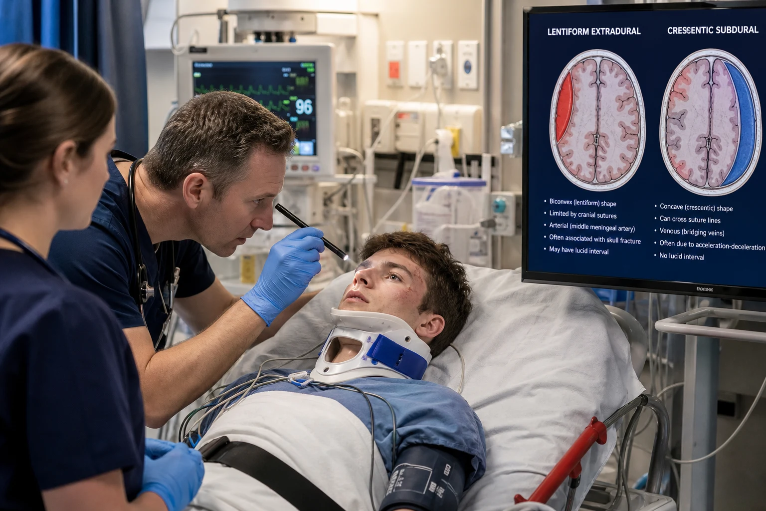
The intubation of the GBS or the MG patient carries a particular pharmacological hazard: the succinylcholine is contraindicated in the acute denervation and the paralytic states (the risk of the hyperkalaemic cardiac arrest), so the rapid-sequence intubation uses the rocuronium 1 mg/kg as the paralysing agent with the induction agent of choice (the propofol 2 mg/kg or the ketamine 1 to 2 mg/kg, with the cautious dosing for the haemodynamic instability).[2]
The disease-modifying therapy — GBS
The GBS disease-modifying therapy
The two equally effective first-line disease-modifying therapies for the GBS are the intravenous immunoglobulin (IVIG) 0.4 g/kg per day for 5 days (the total 2 g/kg) and the plasma exchange 200 to 250 mL/kg over 5 sessions in 1 to 2 weeks. The PSGBST trial established the equivalence of the IVIG and the plasma exchange, both superior to the supportive care alone, and the combined IVIG-plus-plasma-exchange confers no additional benefit over either alone — one or the other, not both. The therapy is started within the first 2 to 4 weeks of the onset, ideally within the first 2 weeks; the earlier the treatment the better the outcome. The corticosteroids are not effective as the monotherapy in the GBS — the Cochrane review confirms no benefit, and they should not be used.[2][3][4] The IVIG is preferred in most centres for the ease of the administration and the lower complication rate; the plasma exchange is reserved for the IVIG-resistant or the contraindicated case (the IgA deficiency, the severe renal impairment, the hyperviscosity). The supportive care is the mainstay: the airway, the ventilation, the autonomic monitoring, the DVT prophylaxis, the analgesia, the nutrition, the psychological support, and the early physiotherapy.
The disease-modifying therapy — myasthenia and the crisis
The symptomatic therapy for the MG is the pyridostigmine 30 to 60 mg PO four times daily (the acetylcholinesterase inhibitor that increases the acetylcholine at the NMJ); the onset of the action is 30 to 60 minutes, the duration 3 to 4 hours. The intravenous equivalent is the neostigmine 0.5 to 1 mg IV (one-thirtieth of the oral pyridostigmine dose). The pyridostigmine relieves the ocular and the limb symptoms but is rarely sufficient alone for the generalised MG, and it does not treat the underlying disease. The immunosuppression for the long-term control is the prednisolone (started at a low dose and titrated, because the early high dose causes the paradoxical worsening in the first 1 to 2 weeks), the azathioprine, the mycophenolate, the methotrexate, and the newer agents (the rituximab, the eculizumab, the efgartigimod). The thymectomy is indicated for the thymoma (every patient) and for the selected young generalised AChR-MG (the improved long-term outcome).[6]
The myasthenic crisis — the respiratory failure or the severe bulbar failure needing the ventilation — is treated with the IVIG 0.4 g/kg per day for 5 days or the plasma exchange, exactly as in the GBS, PLUS the glucocorticoid (the prednisolone 1 mg/kg per day or the methylprednisolone) added with the overlap. The crisis is treated first with the IVIG or the plasma exchange (the faster-acting, the stabilising therapy), and the steroid is added once the patient is stable, because the early steroid-induced worsening (in the first 1 to 2 weeks) can precipitate or deepen the crisis if it is started alone. The precipitant is identified and treated (the pneumonia, the urinary sepsis, the surgery); the precipitant drugs (the aminoglycosides, the macrolides, the magnesium, the beta-blockers, the neuromuscular blockers) are stopped and avoided. The pyridostigmine is continued at the pre-crisis dose during the IVIG or the plasma exchange, but it is sometimes held or reduced once the patient is intubated, because the excessive cholinergic secretions can complicate the airway management.[6][7]
The Miller-Fisher and the Bickerstaff variants
The Miller-Fisher syndrome is the GBS variant characterised by the triad of the ophthalmoplegia, the ataxia and the areflexia — often without the significant limb weakness, and almost always with the anti-GQ1b antibody positive. It may be the pure Miller-Fisher or it may overlap with the Bickerstaff brainstem encephalitis, which adds the altered consciousness and the hyperreflexia and the Babinski signs. The management is the same — the IVIG or the plasma exchange, the respiratory and the autonomic monitoring, the supportive care.[1]
Complications and the pitfalls
The complications of the GBS are the respiratory failure (25 per cent need the ventilation, often for weeks), the autonomic instability (the arrhythmia, the asystole, the blood-pressure swings — the cause of the in-hospital sudden death), the aspiration pneumonia (the bulbar weakness), the deep vein thrombosis and the pulmonary embolism (the immobility — the prophylactic enoxaparin), the critical illness neuropathy and myopathy (the prolonged ICU stay), the pressure sores, the contractures, the depression and the anxiety, and the prolonged rehabilitation. The pitfalls in the ED are: waiting for the CSF or the NCS to admit the ICU (the clinical trend and the spirometry decide); missing the autonomic storm (the patient on the ward arrests from the asystole); treating with the steroids (ineffective in the GBS); giving the succinylcholine for the intubation (the hyperkalaemic arrest); combining the IVIG and the plasma exchange (no benefit).[2]
The complications of the MG are the myasthenic crisis (the respiratory failure), the aspiration pneumonia (the bulbar weakness), the side-effects of the pyridostigmine (the cholinergic excess — the diarrhoea, the abdominal cramps, the salivation, the miosis, the bronchospasm, the muscle fasciculations), the side-effects of the long-term immunosuppression (the infection, the osteoporosis, the diabetes, the hypertension), and the thymoma (the paraneoplastic complication). The pitfalls are: giving the precipitant drugs (the aminoglycosides, the macrolides, the magnesium, the beta-blockers, the neuromuscular blockers); starting the high-dose steroid without the IVIG or the plasma-exchange cover (the early steroid-induced worsening); underestimating the bulbar failure (the silent aspiration); missing the thymoma (the CT chest on every new diagnosis).[6][7]
Prognosis and the disposition
The prognosis of the GBS: about 80 per cent recover fully or with the minor residual deficit, 20 per cent have the significant residual disability, and the mortality is 3 to 7 per cent (the autonomic instability, the pulmonary embolism, the sepsis, the respiratory failure). The predictors of the poor outcome are the older age, the severe weakness at the presentation, the need for the ventilation, the axonal variant on the NCS, and the preceding Campylobacter diarrhoea. The recovery takes months — the ICU and the rehabilitation are prolonged. The patient is admitted to the ICU or the HDU for the respiratory and the autonomic monitoring; the serial spirometry Q2 to 4H guides the escalation; the neurology and the ICU are involved early.[1][2]
The prognosis of the MG: with the modern therapy the mortality is under 5 per cent and the majority achieve the good functional outcome. The myasthenic crisis mortality is 4 to 8 per cent. The crisis patients are admitted to the ICU; the stable patients are admitted under the neurology for the workup and the titration of the immunosuppression. Every new diagnosis of the MG has the urgent CT chest for the thymoma; the thymectomy is the surgical referral when the thymoma is found.[6][7]
Special populations
The pregnant patient — the GBS and the MG are not more frequent in the pregnancy, but the pregnancy and the puerperium are recognised precipitants of the myasthenic crisis. The IVIG and the plasma exchange are safe in the pregnancy; the pyridostigmine is safe; the steroid and the azathioprine are used with the counselling. The elderly patient has the worse GBS outcome (the slower recovery, the higher mortality, the more frequent residual deficit) but the treatment is the same; the frailty assessment guides the shared decision-making. The paediatric GBS is less common and more often presents with the ataxia and the Miller-Fisher pattern; the diagnosis is the same and the IVIG is the therapy. The intubated GBS patient often needs the prolonged ventilation — the early tracheostomy (at 10 to 14 days) is discussed and often performed to facilitate the weaning and the airway care. The immunocompromised patient with the AIDP-like picture should raise the suspicion of the HIV-related polyradiculopathy, the CMV polyradiculitis and the lymphomatous infiltration — the CSF analysis and the specific testing are essential.[2][8]
Evidence and the regional guidelines
The contemporary evidence is the Brighton diagnostic criteria for the GBS (the international consensus), the PSGBST trial (the equivalence of the IVIG and the plasma exchange, the superiority to the supportive care), the Cochrane reviews (the plasma exchange, the IVIG, and the confirmation that the corticosteroids are not effective), the international consensus guidance for the myasthenia gravis 2020 update for the MG, and the systematic reviews on the Lambert-Eaton diagnosis. The drug doses, the ICU thresholds, and the disease-modifying pathways follow the neurology and the intensive-care guidelines; the regional variation is in the preferred first-line agent (the IVIG in most ANZ and the European centres, the plasma exchange in some).[2][3][4][6][8]
ANZ practice note. The GBS management in the ANZ practice follows the local neurology and the intensive-care protocol: the IVIG 0.4 g/kg per day for 5 days as the first-line, the plasma exchange as the alternative, the corticosteroids NOT used, the ICU admission for the FVC under 20 mL/kg or the NIF under minus 30 cmH2O, and the serial spirometry Q2 to 4H guiding the escalation. The MG management follows the international consensus guidance: the pyridostigmine 60 mg PO QID as the symptomatic therapy, the IVIG or the plasma exchange for the crisis, the steroid with the overlap, and the urgent CT chest for the thymoma on every new diagnosis. The succinylcholine is avoided for the RSI of the neuromuscular weakness — the rocuronium 1 mg/kg is the paralytic of choice. [1]
SAQs — exam practice
SAQ — Guillain-Barre syndrome with impending respiratory failure in the ED
10 minutes · 10 marks
A 54-year-old man presents to the emergency department with four days of progressive ascending weakness that began in his feet and has now reached his thighs and hands. He had a diarrhoeal illness two weeks ago. On arrival he is areflexic in all four limbs, has bilateral facial weakness, and is drooling secretions he cannot clear with a weak cough. Respiratory rate 24, SpO2 96 per cent on room air, heart rate 112 in sinus rhythm with frequent ectopics, blood pressure 184/96 then 92/58 within ten minutes. His forced vital capacity is 22 mL/kg and falling, the negative inspiratory force is minus 26 cmH2O, and the peak cough flow is 180 L/min.
SAQ — Disease-modifying therapy in Guillain-Barre syndrome: IVIG versus plasma exchange
10 minutes · 10 marks
A 47-year-old woman with the biopsy-proven Guillain-Barre syndrome (three days of the ascending paralysis, now unable to walk, FVC 25 mL/kg, areflexic, the CSF protein 1.1 g/L with 2 white cells, the nerve conduction studies showing the demyelination) is admitted to the high-dependency unit. She has the mild chronic kidney disease (creatinine 130 micromol per litre, eGFR 50), a known selective IgA deficiency from the childhood, and the indwelling dialysis-quality internal jugular catheter from a recent admission. The neurology team asks for the disease-modifying therapy plan.
Exam pearls
- The ascending flaccid paralysis with the areflexia over days to 4 weeks is the Guillain-Barré until proven otherwise — measure the FVC and the NIF every 2 to 4 hours and admit to the ICU for the FVC under 20 mL/kg or the NIF under minus 30 cmH2O.
- The albuminocytologic dissociation (the high CSF protein, the normal cell count) is the classic CSF finding but is absent in the first week — the early normal CSF does not exclude the GBS.
- The IVIG 0.4 g/kg per day for 5 days OR the plasma exchange — equally effective, NOT combined, NOT with the steroids.
- The fatigable ptosis and diplopia that worsen through the day are the myasthenia — the AChR antibody, the ice-pack test, the pyridostigmine 60 mg PO.
- The myasthenic crisis is the IVIG or the plasma exchange plus the steroid with the overlap — the early steroid-induced worsening is the trap if the steroid is started alone.
- The Lambert-Eaton improves with the repeated activity (the post-exercise facilitation), has the autonomic dry-mouth, and is the paraneoplastic from the small-cell lung cancer — anti-VGCC antibody positive.
- The succinylcholine is contraindicated in the acute paralysis — the rocuronium 1 mg/kg for the RSI. [1]
Red flags
[1]References
- [1]van den Berg B, Walgaard C, Drenthen J, et al. Guillain-Barré syndrome: pathogenesis, diagnosis, treatment and prognosis Nat Rev Neurol, 2014.PMID 25023340
- [2]Willison HJ, Jacobs BC, van Doorn PA. Guillain-Barré syndrome Lancet, 2016.PMID 26948435
- [3]Chevret S, Hughes RA, Annane D. Plasma exchange for Guillain-Barré syndrome Cochrane Database Syst Rev, 2017.PMID 28241090
- [4]Doets AY, Verboon C, van den Berg B, et al. Pharmacological treatment other than corticosteroids, intravenous immunoglobulin and plasma exchange for Guillain-Barré syndrome Cochrane Database Syst Rev, 2020.PMID 31981368
- [5]Lowie BJ, Hrachovinova I, Smith SR. Fluctuating, but Not Forgotten: Recognizing Neuromuscular Junction Disorders Emerg Med Clin North Am, 2026.PMID 41895886
- [6]Narayanaswami P, Sanders DB, Wolfe GI, et al. International Consensus Guidance for Management of Myasthenia Gravis: 2020 Update Neurology, 2021.PMID 33144515
- [7]Lacomis D. Myasthenic crisis Neurocrit Care, 2005.PMID 16377829
- [8]Motomura M, Nakamura Y, Yoshimura T. [How to Diagnose Lambert-Eaton Myasthenic Syndrome Patients Early: A Systematic Review of Japanese Case Reports] Brain Nerve, 2026.PMID 41700072