EM · Coma
Coma and GCS assessment
Also known as GCS · Altered conscious level · Decreased conscious state · Unconscious patient
The comatose patient and the Glasgow Coma Scale — the structured approach (the ABCDE, the rapid glucose, the reversible causes), the GCS (the eye, the verbal, the motor components; the best motor response is the most sensitive), the AVPU for the rapid triage, the differential (the toxic-metabolic, the structural, the infectious), the immediate investigations (the glucose, the blood gas, the CT, the drug screen), and the management (the airway, the naloxone, the thiamine, the specific antidote). ACEM-primary, globally tagged.
On this page & tools
Your progress
Saved locally on this device.
6 MCQs with explanations
Target exams
Red flags
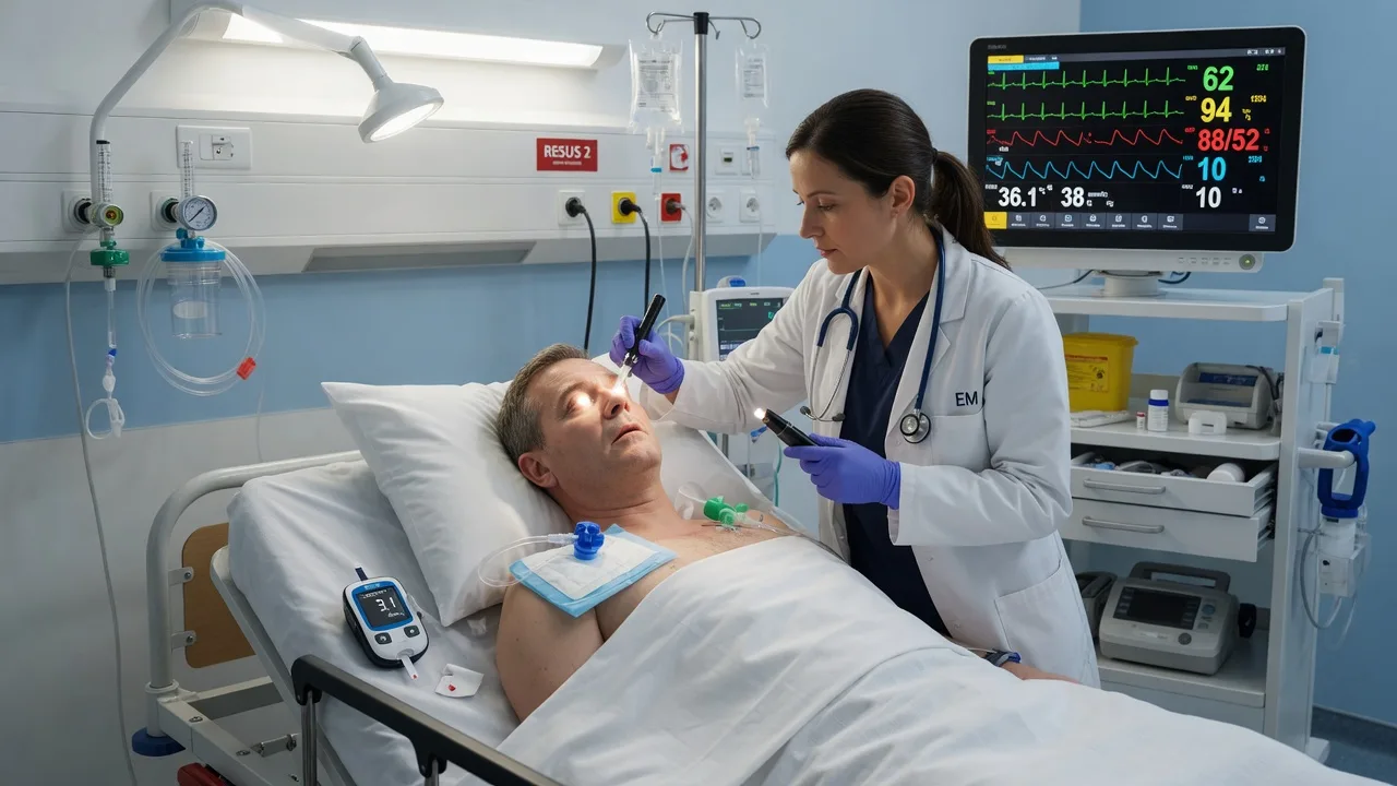
The comatose patient is one of the most challenging presentations in the emergency department, requiring the simultaneous resuscitation, the diagnosis, and the treatment. The Fellowship candidate must know the structured approach (the ABCDE, the rapid glucose, the reversible causes), the Glasgow Coma Scale and its limitations, the differential (the toxic-metabolic vs the structural), and the specific antidotes (the naloxone, the flumazenil, the thiamine), because the comatose patient is the patient in whom the seconds matter — the delayed reversal of the hypoglycaemia, the opioid, or the thiamine deficiency causes the irreversible brain injury.[1][2]
Definition
The coma is the state of unarousable unresponsiveness in which the patient does not open the eyes, does not follow commands, and does not utter comprehensible words. It is distinguished from the stupor (the patient can be briefly aroused with the vigorous stimulation), the confusion (the patient is awake but disoriented), the delirium (the acute fluctuating disturbance of the attention and the cognition), and the vegetative state (the wakefulness without the awareness). The coma is the medical emergency that demands the immediate structured approach. [1]
The structured approach — the ABCDE and the rapid glucose
The comatose patient is approached in parallel: the resuscitation and the diagnosis happen simultaneously. [1]
[1]The glucose is checked immediately — the hypoglycaemia is the commonest reversible cause of the coma, and the prolonged hypoglycaemia causes the irreversible brain injury. If the glucose is low (below 3 mmol/L), give glucose 25 g IV (50 mL of 50 per cent dextrose) immediately, repeated as needed. In the alcoholic patient, give thiamine 100 mg IV before or with the glucose to prevent the Wernicke encephalopathy (the glucose may precipitate the acute thiamine deficiency). [1]
The Glasgow Coma Scale
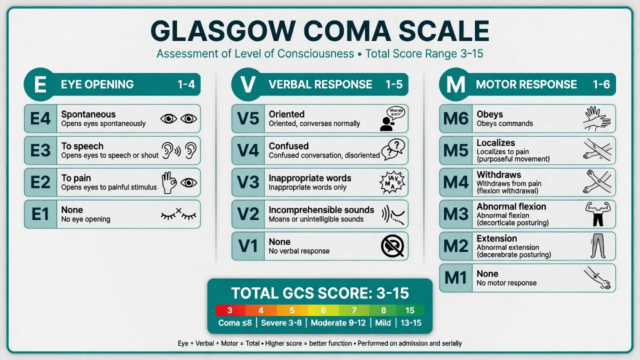
The GCS is the most widely used tool for the assessment of the conscious level, and it has three components: the eye opening (E, scored 1 to 4), the verbal response (V, scored 1 to 5), and the motor response (M, scored 1 to 6). The total ranges from 3 (the deepest coma) to 15 (the fully alert). The motor response is the most sensitive component — it is the last to deteriorate in the rising intracranial pressure and the first to improve in the recovery. The best motor response is recorded (the patient is tested in all four limbs and the best response in any limb is scored). [1]
GCS components
The GCS has limitations: it is poor at the moderate range (the score of 12 may mean the drunk patient or the patient with the expanding intracranial haematoma), the intubated patient scores V1 (tube), and the best motor response obscures the asymmetric deficit. The AVPU (Alert, responds to Voice, responds to Pain, Unresponsive) is the faster alternative for the triage and the serial assessment, and it correlates roughly with the GCS (A = 15, V = 13, P = 8, U = 3). [1]
GCS scoring in detail — the components, the maximum, the minimum
The GCS is recorded as the three components and the total — E4V5M6 = 15 (the maximum, the fully alert patient) and E1V1M1 = 3 (the minimum, the deepest coma). The minimum is 3, not zero — there is no score of 0 in any component; a patient with no eye opening, no verbalisation, and no motor response scores 3. The Fellowship candidate must record the components separately (the total alone is opaque — a score of 7 may be E2V1M4 or E1V1M5, with very different implications) and add the modifier "T" for the intubated patient (E1VTM5 = the verbal component is untestable because of the tube).[3][4]
Eye (E1–E4)
- E4 — Spontaneous eye opening
- E3 — Opens eyes to speech (or shout)
- E2 — Opens eyes to pain (the supraorbital pressure, the nail-bed pressure, the trapezius squeeze)
- E1 — No eye opening to any stimulus; the cerebral cortex or the ascending reticular activating system is profoundly depressed
Verbal (V1–V5)
- V5 — Oriented (knows the person, the place, the time)
- V4 — Confused conversation (attends and responds but disoriented)
- V3 — Inappropriate words (the random exclamatory words)
- V2 — Incomprehensible sounds (the moaning only)
- V1 — No verbal response; record "VT" if intubated, "V" with the tracheostomy
Motor (M1–M6)
- M6 — Obeys commands (the six-finger-peek, the close-the-eyes)
- M5 — Localises pain (reaches above the clavicle for the supraorbital stimulus, or the hand crosses the midline)
- M4 — Withdrawal to pain (the rapid normal flexion away from the stimulus)
- M3 — Abnormal flexion (the decorticate posturing — the slow stereotyped flexion of the arm)
- M2 — Abnormal extension (the decerebrate posturing — the adduction and the internal rotation of the arm, the extension of the leg)
- M1 — No motor response to pain
The AVPU — the rapid triage alternative
The AVPU (Alert, responds to Voice, responds to Pain, Unresponsive) is the faster, simpler alternative for the triage, the paramedic handover, and the serial ward assessment. It does not require the detailed testing and it correlates roughly with the GCS — A = GCS 15, V = GCS 13, P = GCS 8, U = GCS 3. The P (responds to Pain) is the threshold for the airway threat — the patient who responds only to pain has the GCS of around 8, and the airway should be reassessed and the intubation considered.[3]
Alert (A)
- Fully awake, the eyes open spontaneously, the conversation
- Equivalent to GCS 15 (E4V5M6)
- The patient can give the history; the airway is safe
Voice (V)
- Responds to the verbal stimulus but is not fully awake
- Equivalent to GCS 13 (E3V4M6)
- The patient is drowsy; the airway is at risk if the deterioration continues — the serial reassessment
Pain (P)
- Responds only to the painful stimulus; does not open the eyes to the voice
- Equivalent to GCS 8 (E2V2M4)
- The airway is threatened; the intubation threshold is reached — the preparation for the RSI
Unresponsive (U)
- No response to the voice or the pain
- Equivalent to GCS 3 (E1V1M1)
- The airway is unprotected; the immediate intubation and the mechanical ventilation
The FOUR score — the alternative that captures the brainstem
The FOUR (Full Outline of UnResponsiveness) score was developed by Wijdicks to address the limitations of the GCS — the loss of the verbal component in the intubated patient and the absence of the brainstem assessment. It has four components, each scored 0 to 4, with the maximum of 16 and the minimum of 0: the eye response, the motor response, the brainstem reflexes, and the respiration. The FOUR score can be performed in the intubated patient (it has no verbal component), it captures the brainstem function (the GCS does not), and it distinguishes the locked-in syndrome (the normal FOUR with the absent verbal) from the true coma.[5]
The FOUR score components (each 0 to 4, the max 16)
GCS
- Three components (the eye, the verbal, the motor)
- Range 3 to 15
- Cannot assess the intubated patient (the V1T)
- Does not assess the brainstem
- Universal, the trauma and the stroke guidelines use it
- The motor is the most sensitive
FOUR score
- Four components (the eye, the motor, the brainstem, the respiration)
- Range 0 to 16
- Can assess the intubated patient (no verbal component)
- Assesses the brainstem reflexes directly
- Used in the ICU and the neurology wards
- Captures the locked-in syndrome and the vegetative state
The causes of the coma — AEIOU TIPS
The differential of the coma is vast, but the Fellowship mnemonic AEIOU TIPS organises the causes into the eight rapid-reversibility categories. The mnemonic is the bedside checklist — every comatose patient is screened for each, because the missed reversible cause is the missed preventable death. [1]
AEIOU TIPS — the causes of the coma
A — Alcohol
- The ethanol intoxication (the smell, the history, the level)
- The Wernicke encephalopathy (the ataxia, the ophthalmoplegia, the confusion — the thiamine 100 mg IV)
- The hepatic encephalopathy (the asterixis, the jaundice, the elevated ammonia)
E — Epilepsy
- The post-ictal state (the history of the seizure, the gradual recovery over minutes to hours)
- The non-convulsive status epilepticus (the coma without the overt seizure — the EEG is diagnostic)
- The subtle status (the twitching of the face or the hand, the eye deviation)
I — Insulin / hypoglycaemia
- The commonest rapidly reversible cause — check the glucose immediately
- The glucose below 3 mmol/L: glucose 25 g IV (the 50 mL of the 50 per cent dextrose)
- The diabetic, the insulin overdose, the sulfonylurea, the sepsis, the alcohol, the liver failure
O — Overdose
- The opioid (the pin-point pupils, the reduced respiratory rate — the naloxone 400 mcg IV)
- The benzodiazepine (the flumazenil 200 mcg IV — caution the seizures)
- The tricyclic (the widened QRS, the anticholinergic signs — the sodium bicarbonate 50 mL of the 8.4 per cent)
U — Uraemia / metabolic
- The uraemia (the renal failure, the uremic frost, the elevated creatinine)
- The hyponatraemia (the sodium below 120 — the hypertonic saline if the seizure)
- The hypercalcaemia, the hepatic failure, the thyroid (the myxoedema, the thyrotoxic)
T — Trauma
- The traumatic brain injury (the C-spine immobilisation, the urgent CT)
- The intracranial haemorrhage (the subdural, the extradural, the intracerebral)
- The primary and the secondary survey, the trauma team activation
I — Infection
- The meningitis (the fever, the neck stiffness, the rash — the ceftriaxone 2 g IV before the LP)
- The encephalitis (the HSV — the aciclovir 10 mg/kg IV)
- The cerebral abscess, the sepsis, the cerebral malaria (the travel history)
P — Poisoning / environmental
- The carbon monoxide (the carboxyhaemoglobin — the high-flow oxygen)
- The organophosphate (the salivation, the lacrimation, the pinpoint — the atropine, the pralidoxime)
- The heat stroke, the hypothermia, the snake bite (the pressure-immobilisation)
S — Stroke
- The ischaemic stroke (the thrombolysis window, the basilar-artery occlusion)
- The intracerebral haemorrhage (the blood-pressure control, the neurosurgery)
- The subarachnoid (the thunderclap headache, the CT, the LP for the xanthochromia)
The differential by the speed of the onset
The Fellowship candidate must also frame the differential by the speed of the onset — the sudden onset (the vascular — the stroke, the subarachnoid, the intracerebral haemorrhage; the cardiac — the post-arrest, the arrhythmia), the rapid onset over minutes to hours (the metabolic — the hypoglycaemia, the hyponatraemia; the toxic — the overdose, the CO; the infectious — the meningitis), and the gradual onset over hours to days (the tumour, the abscess, the chronic subdural, the hepatic, the uraemic). The history from the family, the paramedic, and the nursing home is the key — the speed of the onset narrows the differential before any test. [1]
The first ten minutes of the comatose patient
- The airway — the jaw thrust (the C-spine until cleared), the nasopharyngeal airway; the suction; the high-flow oxygen 15 L/min via the non-rebreather.
- The breathing — the respiratory rate, the saturation, the chest rise; the bag-valve-mask if the rate is below 8 or the saturation is low; the capnography.
- The circulation — the pulse, the blood pressure, the capillary refill; the two large-bore cannulae; the IV fluid if the hypotensive; the ECG.
- The disability — the glucose first — the finger-prick glucose IMMEDIATELY; if below 3 mmol/L, the glucose 25 g IV and the thiamine 100 mg IV.
- The disability — the GCS, the pupils, the temperature — the GCS components (E_V_M_), the pupils (the size, the symmetry, the reactivity), the tympanic temperature.
- The exposure — the head-to-toe for the clues (the needle marks, the MedicAlert, the rash, the trauma, the bleeding, the tongue bite).
- The empiric antidotes — the naloxone 400 mcg IV (the opioid), the glucose if not already, the thiamine 100 mg IV (the alcoholic).
- The bloods and the drug screen — the VBG, the FBC, the U&E, the LFTs, the troponin, the paracetamol and the salicylate, the ethanol, the carboxyhaemoglobin.
- The CT brain — the non-contrast, after the stabilisation, if the structural cause is suspected or the metabolic screen is unrevealing.
- The disposition — the ICU if the GCS is 8 or below or the airway is threatened; the HDU for the moderate; the ward for the rapidly reversible who recovers fully.
Differential diagnosis — the toxic-metabolic vs the structural
The coma is divided into the toxic-metabolic (the bilateral cerebral dysfunction from the systemic disturbance) and the structural (the focal brainstem or the hemispheric lesion). [1]
Toxic-metabolic
- The bilateral, the symmetric signs; the pupils are typically equal and reactive
- The drug overdose, the hypoglycaemia, the uraemia, the hepatic, the sepsis, the post-ictal
- The CT is often normal; the treatment is the specific antidote or the correction
- The prognosis is often reversible
Structural
- The focal, the asymmetric signs; the pupil asymmetry, the unilateral weakness
- The intracerebral haemorrhage, the tumour, the abscess, the subdural, the brainstem stroke
- The CT shows the lesion; the neurosurgical referral
- The time-critical — the delay worsens the outcome
Infectious
- The fever, the neck stiffness, the rash (meningococcal)
- The meningitis, the encephalitis, the cerebral abscess
- The CT then the LP; the empirical antibiotics (ceftriaxone 2 g, vancomycin, aciclovir 10 mg/kg)
- The empirical treatment before the definitive diagnosis
Psychiatric
- The functional, the pseudocoma; the eye reflexes are intact, the EEG is normal
- The catatonia, the conversion, the dissociative state
- A diagnosis of exclusion; no structural or metabolic cause found
- The psychiatric assessment and the follow-up
The specific antidotes and the rapid reversibility
The comatose patient is given the specific antidotes empirically if the cause is suspected, because the diagnosis may be unclear at the presentation and the delay to the antidote may be the irreversible injury: [1]
- Naloxone 400 mcg IV (or 800 mcg IM) for the suspected opioid overdose (the pin-point pupils, the reduced respiratory rate, the needle marks). Titrate to the respiratory rate, not the full reversal — the rapid reversal causes the acute withdrawal and the agitation.
- Flumazenil 200 mcg IV for the suspected benzodiazepine overdose. Use cautiously — it may precipitate the seizures in the chronic user or the mixed overdose.
- Glucose 25 g IV for the hypoglycaemia (the finger-prick glucose below 3 mmol/L).
- Thiamine 100 mg IV for the suspected Wernicke encephalopathy (the alcoholic, the malnourished).
- Naloxone and the glucose are given to every comatose patient of unknown cause — the "coma cocktail." [1]
The investigations
The finger-prick glucose is the first test. The venous blood gas (the pH, the lactate, the electrolytes, the glucose, the carboxyhaemoglobin if the CO poisoning is suspected). The full blood count, the U&E, the LFTs, the troponin, the coagulation. The drug screen (the paracetamol level, the salicylate level, the ethanol level, the toxicology screen if the overdose is suspected). The urinalysis (the ketones for the DKA, the drug screen). The non-contrast CT of the brain if the structural cause is suspected or the toxic-metabolic screen is unrevealing. The lumbar puncture if the meningitis or the encephalitis is suspected (after the CT). The EEG if the non-convulsive status epilepticus is suspected (the patient who does not wake up after the seizure).[1]
The neurological examination
The comatose patient is examined for the clues to the cause: the pupils (the pin-point of the opioid or the pontine haemorrhage; the dilated of the atropine or the post-anoxic; the asymmetric of the third-nerve palsy or the uncal herniation), the eye movements (the doll's-eye reflex and the caloric testing — the intact brainstem moves the eyes conjugately), the corneal reflex (the intact facial nerve and the trigeminal nerve), the gag reflex (the intact glossopharyngeal and the vagus), the motor response (the purposeful movement vs the posturing — the decorticate flexion suggests the cortical lesion, the decerebrate extension suggests the brainstem lesion), the reflexes and the plantar responses (the Babinski sign in the upper-motor-neurone lesion), and the meningism (the neck stiffness of the meningitis or the subarachnoid haemorrhage). [1]
The pupils — the window to the brainstem
The pupils are the single most informative bedside sign in the comatose patient, because the pupillary light reflex is robust — it is the last reflex to be lost in the toxic-metabolic coma (the barbiturate, the ethanol) and it is the first reflex to be lost in the structural brainstem lesion. The Fellowship candidate must record the size, the symmetry, and the reactivity of both pupils, and the lesion is inferred from the pattern.[8]
Pin-point (<2 mm)
- The opioid overdose (the reactive, the reduced respiratory rate — the naloxone)
- The pontine haemorrhage (the fixed — the "pontine pupils")
- The organophosphate and the nerve agent (the cholinergic — the salivation, the lacrimation)
Small (2–4 mm) reactive
- The early uncal herniation (the slight asymmetry, the sluggish reactive)
- The metabolic and the toxic coma (the preserved reactivity — the distinguishing feature)
- The Horner syndrome (the unilateral small with the ptosis — the apical lung, the lateral medullary)
Mid-position fixed (4–6 mm, no reaction)
- The midbrain lesion (the tectal, the pineal)
- The brainstem herniation (the bilateral fixed)
- The brain death (the bilateral fixed mid-position — the confirmation requires the apnoea)
Dilated fixed (>6 mm, no reaction)
- The uncal herniation (the unilateral dilated fixed — the third-nerve palsy, the urgent neurosurgery)
- The post-anoxic (the bilateral dilated fixed — the poor prognosis)
- The anticholinergic (the atropine, the tricyclic — the dry skin, the delirium)
The brainstem reflexes — the systematic assessment
The brainstem reflexes are assessed to localise the level of the lesion (the diencephalon, the midbrain, the pons, the medulla) and to detect the progression of the herniation. There are four reflexes that the Fellowship candidate must elicit in every comatose patient: the pupillary, the corneal, the gag (and the cough), and the oculomotor (the doll's-eye and the cold-caloric). [1]
Pupillary reflex
- The afferent limb: the optic nerve (II). The efferent: the oculomotor nerve (III)
- The bright light in each eye; the size and the reactivity of both pupils
- Absent in the third-nerve palsy, the uncal herniation, the brain death; preserved in the metabolic
- Localises to the midbrain (the tectum, the Edinger-Westphal)
Corneal reflex
- The afferent: the trigeminal nerve (V1). The efferent: the facial nerve (VII)
- The cotton wisp on the cornea (not the sclera); the bilateral blink
- Absent in the pontine lesion, the facial-nerve palsy, the deep coma; preserved in the locked-in
- Localises to the pons
Gag and cough reflex
- The afferent: the glossopharyngeal (IX). The efferent: the vagus (X)
- The suction catheter against the posterior pharynx (the gag) and the carina (the cough)
- Absent in the medullary lesion, the deep coma; the last reflex to be lost
- Localises to the medulla — the confirmation of the brain death
Oculocephalic (Doll's eyes)
- The afferent: the vestibular and the proprioceptive. The efferent: III, IV, VI
- The rapid horizontal head turn — the intact brainstem moves the eyes conjugately opposite (the "doll's eyes")
- Contraindicated if the C-spine is not cleared; the test of the pontine gaze centres
- Absent in the brainstem lesion (the eyes move with the head)
Oculovestibular (cold caloric)
- The afferent: the vestibular nerve (VIII). The efferent: III, VI
- The 50 mL of the ice-cold water in the ear (after the tympanic check); the intact brainstem abducts and the slow tonic toward the irrigated ear, the fast nystagmus away
- The COWS (Cold Opposite, Warm Same) for the fast phase; the test of choice if the C-spine is uncleared
- Absent in the brainstem lesion, the brain death; preserved in the metabolic
COWS — the direction of the caloric nystagmus (the fast phase)
The motor response — the posturing and the level
The motor response to the pain localises the lesion: the purposeful movement (the localisation above the clavicle) indicates the intact cortex; the decorticate posturing (the M3 — the slow flexion, the adduction of the arm, the extension of the leg) indicates the hemispheric or the thalamic lesion with the intact brainstem; the decerebrate posturing (the M2 — the extension, the adduction, the internal rotation of the arm, the extension of the leg) indicates the midbrain or the upper-pons lesion. The flaccidity with no response (the M1) indicates the lower-pons or the medullary lesion — the deep brainstem involvement and the poor prognosis. [1]
Decorticate vs decerebrate — the arms
Intubation criteria — when to secure the airway
The comatose patient is intubated when the airway is threatened, the protective reflexes are lost, or the ventilation is inadequate. The Fellowship criteria are: [1]
- GCS 8 or below — the airway reflexes are unreliable below this threshold; the risk of the aspiration and the hypoventilation mandates the definitive airway.
- The loss of the gag or the cough reflex — the unprotected airway, regardless of the GCS.
- The hypoventilation — the respiratory rate below 8, the rising CO₂, the falling oxygen.
- The inability to maintain or protect the airway — the pooling of the secretions, the noisy breathing, the absent swallow.
- The predicted deterioration — the rising intracranial pressure, the expanding haematoma, the impending herniation. [1]
Herniation syndromes — the time-critical structural coma
The herniation is the displacement of the brain across the dural folds, and it is the final common pathway of the expanding intracranial mass — the minutes matter. There are four syndromes that the Fellowship candidate must recognise:[8]
Uncal (lateral)
- The commonest; the temporal lobe herniates through the tentorial notch
- The ipsilateral dilated fixed pupil (the third nerve), the contralateral hemiparesis
- Kernohan notch: the ipsilateral hemiparesis (the false-localising)
- The urgent CT, the mannitol/hypertonic saline, the neurosurgery
Central
- The diencephalon herniates downward through the tentorial notch
- The bilateral mid-position fixed pupils, the decorticate then decerebrate
- The progressive loss of the brainstem reflexes down the neuraxis
- The Cheyne-Stokes then the ataxic breathing
Subfalcine
- The cingulate gyrus herniates under the falx
- The anterior cerebral artery compression — the contralateral leg weakness
- Often silent; progresses to the central herniation
- The CT shows the midline shift
Tonsillar
- The cerebellar tonsils herniate through the foramen magnum
- The medullary compression — the respiratory arrest, the cardiac irregularity
- The posterior fossa mass — the CT before the LP (the LP herniates)
- The immediate decompression — the occipital and the high-cervical
The Cushing reflex — the late sign of the rising intracranial pressure
The Cushing reflex (the hypertension, the bradycardia, the irregular respiration) is the brainstem response to the rising intracranial pressure — the hypertension to maintain the cerebral perfusion, the bradycardia from the baroreceptor reflex, the irregular breathing from the brainstem distortion. It is a late sign — by the time it appears, the herniation is imminent. The hypertension with the bradycardia in the comatose patient is the raised intracranial pressure until proven otherwise — the head elevation 30 degrees, the hyperventilation, the mannitol or the hypertonic saline, the urgent CT and the neurosurgery. [1]
[1]Prognosis — the post-anoxic coma and the predictors
The prognosis of the coma depends on the cause, but the post-anoxic coma (after the cardiac arrest) is the one for which the evidence is strongest, because the systematic prognostication is the standard of care. The Fellowship candidate must know the ERC-ESICM stepwise algorithm and the predictors of the poor outcome.[6][7]
Prognostication after cardiac arrest — the ERC-ESICM algorithm
Intensive Care Medicine
PMID 32915254
Systematic review of the predictors of the poor neurological outcome in comatose survivors of cardiac arrest, at least 72 hours after the arrest, on or off targeted temperature management.
Key finding
The most reliable predictors of the poor outcome (the false-positive rate below 5 per cent): (1) the bilaterally absent N20 SSEP at ≥24 h; (2) the bilaterally absent pupillary reflex at ≥72 h; (3) the bilaterally absent corneal reflex at ≥72 h; (4) the highly malignant EEG at ≥24 h; (5) the serum NSE above 60 µg/L at 48 to 72 h. No single predictor is sufficient — the algorithm requires at least two concordant.
Practice change
The prognostication is the stepwise, the multimodal, and the deferred to 72 hours — the self-fulfilling prophecy is avoided by the no-single-test-decision rule.
The locked-in syndrome and the cognitive-motor dissociation
The locked-in syndrome (the de-efferented state — the quadriplegia and the anarthria with the preserved consciousness) is caused by the pontine lesion (the basilar-artery occlusion) that spares the cortex. The patient is awake and aware but can move only the vertical eyes and the eyelids — the command-following eye movement is the diagnostic sign. The Fellowship candidate must test the eye tracking in every comatose patient, because the misdiagnosis of the locked-in as the vegetative is the catastrophic error — the patient hears the conversation about the withdrawal of the care. The modern fMRI and the EEG reveal the cognitive-motor dissociation (the patient who is behaviourally unconscious but who can follow commands on the neuroimaging) in up to 25 per cent of the patients with the disorders of the consciousness — the implications for the prognostication and the end-of-life decisions are profound.[9]
[1]Management — the airway and the disposition
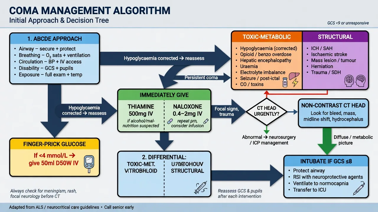
The airway is the priority — the comatose patient with the GCS of 8 or below, the loss of the protective reflexes, or the hypoventilation is intubated. The rapid sequence intubation with etomidate 0.3 mg/kg IV and rocuronium 1.2 mg/kg IV is used to protect the C-spine and to prevent the aspiration. The specific antidotes are given before the intubation if the rapidly reversible cause is suspected: naloxone 400 mcg IV for the opioid, glucose 25 g IV for the hypoglycaemia, flumazenil 200 mcg IV for the benzodiazepine. The comatose patient with the suspected meningitis is given ceftriaxone 2 g IV, vancomycin 1.5 g IV, dexamethasone 10 mg IV, and aciclovir 10 mg/kg IV before the LP. The comatose patient is admitted to the HDU or the ICU; the patient with the rapidly reversible cause (the hypoglycaemia, the opioid) who recovers fully may be discharged after the observation and the cause investigation. The raised intracranial pressure is treated with the head elevation 30 degrees, the mannitol 0.5 g/kg IV or the hypertonic saline 3 per cent 250 mL IV, and the urgent neurosurgical referral for the definitive decompression.[2]
Complications and prognosis
The complications are the aspiration pneumonia (the reduced consciousness and the loss of the protective reflexes — the commonest pulmonary complication, treated with the ceftriaxone 2 g IV), the pressure sores (the regular turning and the pressure-area care), the venous thromboembolism (the prophylactic enoxaparin 40 mg subcutaneously daily and the compression stockings), the rhabdomyolysis (from the prolonged immobility — check the creatine kinase, treat with the aggressive IV fluid to maintain the urine output), the corneal abrasion (from the incomplete eye closure — the eye taping and the lubricant), the seizures (the prophylactic levetiracetam), and the death. The prognosis depends on the cause and the depth of the coma: the toxic-metabolic coma is often fully reversible with the specific antidote; the structural coma (the intracerebral haemorrhage, the traumatic brain injury) has a variable prognosis depending on the lesion size and the GCS at the presentation; the post-anoxic coma (after the cardiac arrest) has the worst prognosis, particularly if the pupil reflexes are absent at 24 hours or the somatosensory evoked potentials are absent at 72 hours. [1]
Evidence and regional guidelines
The contemporary framework is the GCS for the serial assessment, the CT for the structural exclusion, the rapid glucose for the hypoglycaemia, and the coma cocktail (the naloxone, the glucose, the thiamine) for the empiric reversal.[1]
ANZ practice note. The comatose patient is managed with the ABCDE, the rapid glucose, the CT, and the empiric antidotes (naloxone 400 mcg IV, glucose 25 g IV, thiamine 100 mg IV); the intubation if the GCS is 8 or below; the neuroimaging and the LP if the structural or the infectious cause is suspected; and the ICU admission for the ongoing care. [1]
Exam practice
SAQ — The asymmetric motor response and the FOUR score
10 minutes · 10 marks
A 58-year-old man is brought to the emergency department after being found unconscious at home by his wife. On arrival his eyes remain closed, he makes no sound, and he does not open his eyes to the loud voice. On application of firm supraorbital pressure he withdraws his right arm with rapid normal flexion, while his left arm extends with adduction and internal rotation and his left leg extends. The finger-prick glucose is 6.4 mmol/L. Both pupils are 3 mm and briskly reactive. Blood pressure 172/94, heart rate 88, respiratory rate 6, SpO₂ 88 per cent on room air. There is no external sign of trauma.
SAQ — The toxidrome versus the structural coma: the pupil and the antidote
10 minutes · 10 marks
Two patients are brought to your resuscitation bay within minutes of each other. Patient A is a 26-year-old known heroin user, found slumped in a public toilet: GCS 6 (E1V1M4, withdraws symmetrically to pain), pupils 1.5 mm pin-point and reactive, respiratory rate 5, fresh needle-track marks in the left antecubital fossa, glucose 5.8 mmol/L. Patient B is a 64-year-old woman with the sudden thunderclap headache followed by the rapid loss of consciousness: GCS 6 (E1V1M4), RIGHT pupil 6 mm and fixed, LEFT pupil 3 mm and reactive, a left hemiparesis, glucose 6.2 mmol/L, blood pressure 190/100, heart rate 56, respiratory rate 10.
Exam pearls
- The glucose first — the hypoglycaemia is the commonest rapidly reversible cause.
- GCS 8 or below = intubate. The motor response is the most sensitive component.
- AVPU for the rapid triage: A = GCS 15, V = 13, P = 8, U = 3.
- The "coma cocktail": naloxone 400 mcg IV, glucose 25 g IV, thiamine 100 mg IV.
- Thiamine before the glucose in the alcoholic — to prevent the Wernicke encephalopathy.
- The asymmetric pupils or the unilateral weakness = the structural lesion until proven otherwise.
- The non-convulsive status epilepticus is the cause of the unexplained coma — the EEG is diagnostic.
- The doll's-eye reflex and the caloric testing assess the brainstem integrity.
- E4V5M6 = 15 (the max); E1V1M1 = 3 (the min) — there is no score of 0. Record the components separately, not just the total.
- The FOUR score (eye, motor, brainstem, respiration; 0–16) for the intubated patient and the suspected brainstem lesion — it has no verbal component.
- AEIOU TIPS — Alcohol, Epilepsy, Insulin, Overdose, Uraemia, Trauma, Infection, Poisoning, Stroke. The bedside checklist for every comatose patient.
- The pupils: pinpoint = opioids or pontine; small reactive = metabolic or early herniation; mid-position fixed = midbrain; dilated fixed = uncal herniation or post-anoxic.
- The pupillary reflex is the last to be lost in the metabolic coma and the first to be lost in the structural — equal reactive pupils point to the metabolic.
- The cold-caloric (COWS: Cold-Opposite, Warm-Same) for the brainstem if the C-spine is not cleared — never do the Doll's eyes if the C-spine is uncleared.
- Decorticate (M3, flexion) = above the red nucleus (the hemisphere); decerebrate (M2, extension) = at or below the red nucleus (the midbrain).
- The central pain stimulus (the supraorbital, the trapezius) — not the nail-bed, which gives the spinal-reflex withdrawal misread as M4.
- Cushing triad (hypertension, bradycardia, irregular breathing) — a late sign of the rising ICP; the herniation is imminent.
- Always CT before the LP in the comatose patient — the LP herniates the brain (the posterior fossa mass, the obstructive hydrocephalus).
- The locked-in syndrome (the pontine lesion) — the vertical eye movement is preserved; test the eye tracking in every comatose patient.
- The post-anoxic prognosis: the bilaterally absent pupil reflex at ≥72 h or the absent N20 SSEP at ≥24 h — the reliable poor-outcome predictors.
- The empirical antibiotics (the ceftriaxone 2 g, the vancomycin, the aciclovir 10 mg/kg) before the LP — the imaging delay is not the treatment delay.
- The flumazenil is the cautious antidote — it precipitates the seizures in the chronic user and the mixed overdose; reserve for the isolated iatrogenic or the pure benzodiazepine. [1]
Red flags
[1]References
- [1]Expert Panel on Neurological Imaging, Ledbetter LS, Shah LM, et al. ACR Appropriateness Criteria® Altered Mental Status, Coma, Delirium, and Psychosis: 2024 Update J Am Coll Radiol, 2024.PMID 39488349
- [2]Llorens P, Piqueras-Rodriguez F, Herrero P, et al. Prevalence, risk factors, and prognosis of the need for physical and chemical restraint in older patients attending emergency departments. EDEN-50 Am J Emerg Med, 2026.PMID 42000676
- [3]Teasdale G, Maas A, Lecky F, et al. The Glasgow Coma Scale at 40 years: standing the test of time Lancet Neurol, 2014.PMID 25030516
- [4]Aguilar-Fuentes V, Orozco-Puga P, Jiménez-Ruiz A The Glasgow Coma Scale: 50-year anniversary Neurol Sci, 2024.PMID 38436790
- [5]Wijdicks EFM, Bamlet WR, Maramattom BV, et al. Validation of a new coma scale: The FOUR score Ann Neurol, 2005.PMID 16178024
- [6]Sandroni C, D'Arrigo S, Cacciola S, et al. Prediction of poor neurological outcome in comatose survivors of cardiac arrest: a systematic review Intensive Care Med, 2020.PMID 32915254
- [7]Scarpino M, Lolli F, Lanzo G, et al. Do changes in SSEP amplitude over time predict the outcome of comatose survivors of cardiac arrest? Resuscitation, 2022.PMID 36375653
- [8]Hall WA, Munakomi S Uncal Herniation 2026.PMID 30725793
- [9]Bodien YG, Allanson J, Cardone P, et al. Cognitive Motor Dissociation in Disorders of Consciousness N Engl J Med, 2024.PMID 39141852