EM · Spinal cord compression
Spinal cord compression
Also known as Malignant spinal cord compression · MSCC · Epidural spinal cord compression
Spinal cord compression — the time-critical neurological emergency from the extrinsic compression of the spinal cord by the tumour, the disc, the abscess or the haematoma; the clinical (the progressive back pain, the motor weakness, the sensory level, the bladder and bowel dysfunction); the urgent MRI whole spine within 4 to 6 hours; the dexamethasone 10 mg IV loading then 4 mg every 6 hours; the neurosurgical and the oncological referral; and the time-to-treatment determining the ambulatory outcome. ACEM-primary, globally tagged.
On this page & tools
Your progress
Saved locally on this device.
Target exams
Red flags
Spinal cord compression is the time-critical neurological emergency where the spinal cord or the cauda equina is compressed by an extrinsic mass, most commonly the metastatic malignancy but also the disc herniation, the epidural abscess or the haematoma. The Fellowship candidate must recognise the presentation (the progressive back pain in the cancer patient, the motor weakness, the sensory level, the autonomic dysfunction), initiate the dexamethasone immediately, and arrange the urgent MRI whole spine, because the neurological outcome — ambulatory or not — is determined by the speed of the decompression.[1][2]
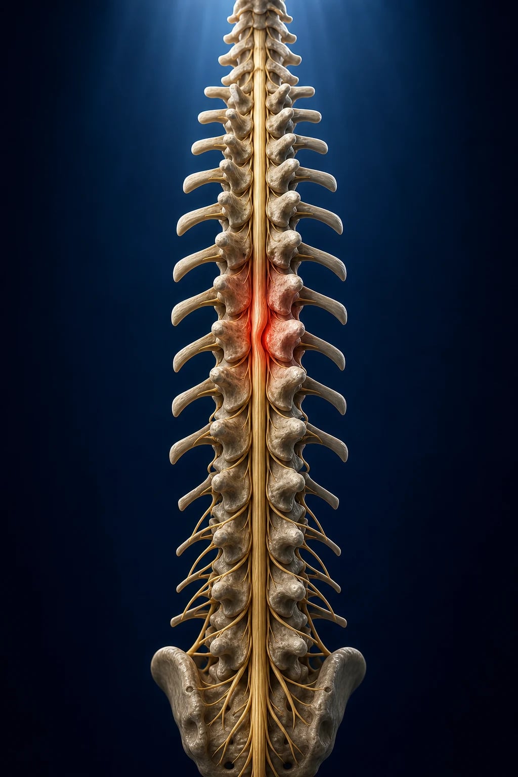
Definition and the epidemiology
The malignant spinal cord compression (MSCC) is the compression of the spinal cord or the cauda equina by the metastatic tumour, occurring in 5 to 10 per cent of the cancer patients. The commonest primary tumours are the breast, the lung, the prostate, the myeloma and the lymphoma.[2] The non-malignant causes include the intervertebral disc herniation (the central or the paracentral disc), the epidural abscess (from the Staphylococcus aureus bacteraemia, the IV drug use, the diabetes), the epidural haematoma (from the anticoagulation or the post-procedural), and the vertebral fracture (trauma, osteoporotic).
Differential diagnosis — the cause of the acute myelopathy
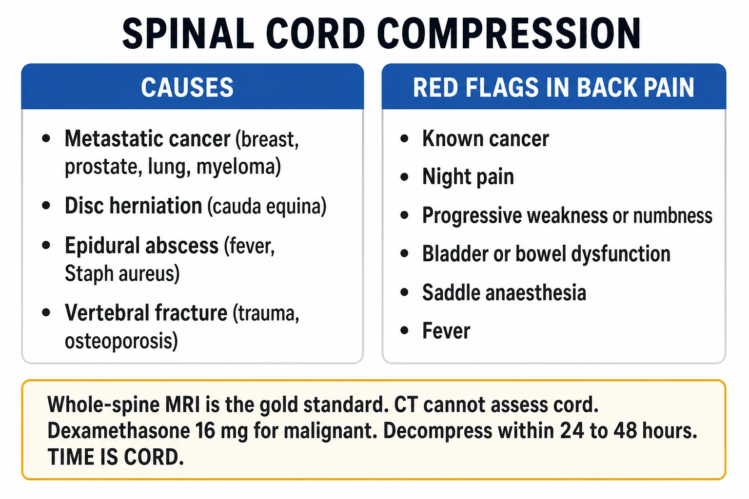
- Malignant spinal cord compression — the known cancer, the progressive pain, the vertebral collapse; the MRI shows the epidural mass.
- Epidural abscess — the fever, the IV drug use, the diabetes; the raised CRP; the MRI shows the collection.
- Acute disc herniation — the acute onset, the mechanical back pain; the MRI shows the disc protrusion.
- Transverse myelitis — the viral, the post-vaccination, the autoimmune; the bilateral symptoms; the MRI shows the cord signal change but no extrinsic mass.
- Guillain-Barre syndrome — the ascending flaccid paralysis, the areflexia; the normal MRI; the CSF shows the albuminocytologic dissociation. [1]
The clinical presentation
The progression is the pain (the earliest, the commonest, present in 95 per cent of the cases at the presentation — the localised back pain worse on lying flat, the nocturnal pain, the radicular pain in the dermatomal distribution, the worse on the Valsalva manoeuvre, the worse on the straight-leg raise) → the motor weakness (the progressive, the ascending, the bilateral, the UMN signs if above the conus — the spasticity, the hyperreflexia, the Babinski sign — the LMN if at the conus or the cauda — the flaccidity, the hyporeflexia, the fasciculations) → the sensory level (the pinprick and the light touch loss below the lesion, the finding that localises the level of the compression to within one or two segments) → the autonomic (the urinary retention with the overflow incontinence — the palpable bladder on the examination — the constipation, the erectile dysfunction, the loss of the perianal sensation). The cauda equina syndrome presents with the saddle anaesthesia, the bilateral sciatica, the bladder and bowel dysfunction, the lower-limb weakness, and the decreased or absent reflexes. The examination includes the digital rectal examination for the anal tone and the perianal sensation, the motor power grading (the MRC 0 to 5), the reflexes, the pinprick and the light touch, and the assessment of the gait. [1]
The investigations
The urgent MRI whole spine (not just the symptomatic level — the skip lesions are common in the malignancy, the multifocal disc disease, or the multilevel abscess) is the gold standard and should be performed within 4 to 6 hours of the presentation. The MRI demonstrates the site, the extent, the degree and the cause of the compression — the tumour (the extradural, the intradural extramedullary, the intramedullary), the disc (the central, the paracentral, the foraminal), the abscess (the rim-enhancing collection), or the haematoma (the heterogeneous signal). The CT is used for the vertebral bony detail — the fracture pattern, the canal compromise, the surgical planning. The CT myelogram is the alternative when the MRI is contraindicated (the pacemaker, the ferromagnetic implant) — it demonstrates the block to the contrast flow but does not show the cord parenchyma. The plain X-ray may show the vertebral collapse, the pedicle loss (the "winking owl" sign), or the sclerotic or lytic lesion, but it cannot assess the cord and is insufficient alone. The bloods — the FBC, the CRP, the ESR (for the abscess — a CRP over 50 with the fever and the back pain is the epidural abscess until proven otherwise), the coagulation (before the surgical decompression), the PSA (for the prostate), the myeloma screen (the serum protein electrophoresis, the serum free light chains, the urine Bence-Jones — if the primary is unknown), the calcium (the hypercalcaemia of the malignancy). The urine toxicology and the HIV test if the IV drug use is suspected. The ASIA impairment scale documents the neurological deficit at the baseline and at the serial assessments — the motor score (the 10 myotomes, the 0 to 5 scale, maximum 100), the sensory score (the 28 dermatomes, the pinprick and the light touch, maximum 112 each), and the impairment grade (A complete to E normal). [1]
Management — the approach and the drug doses
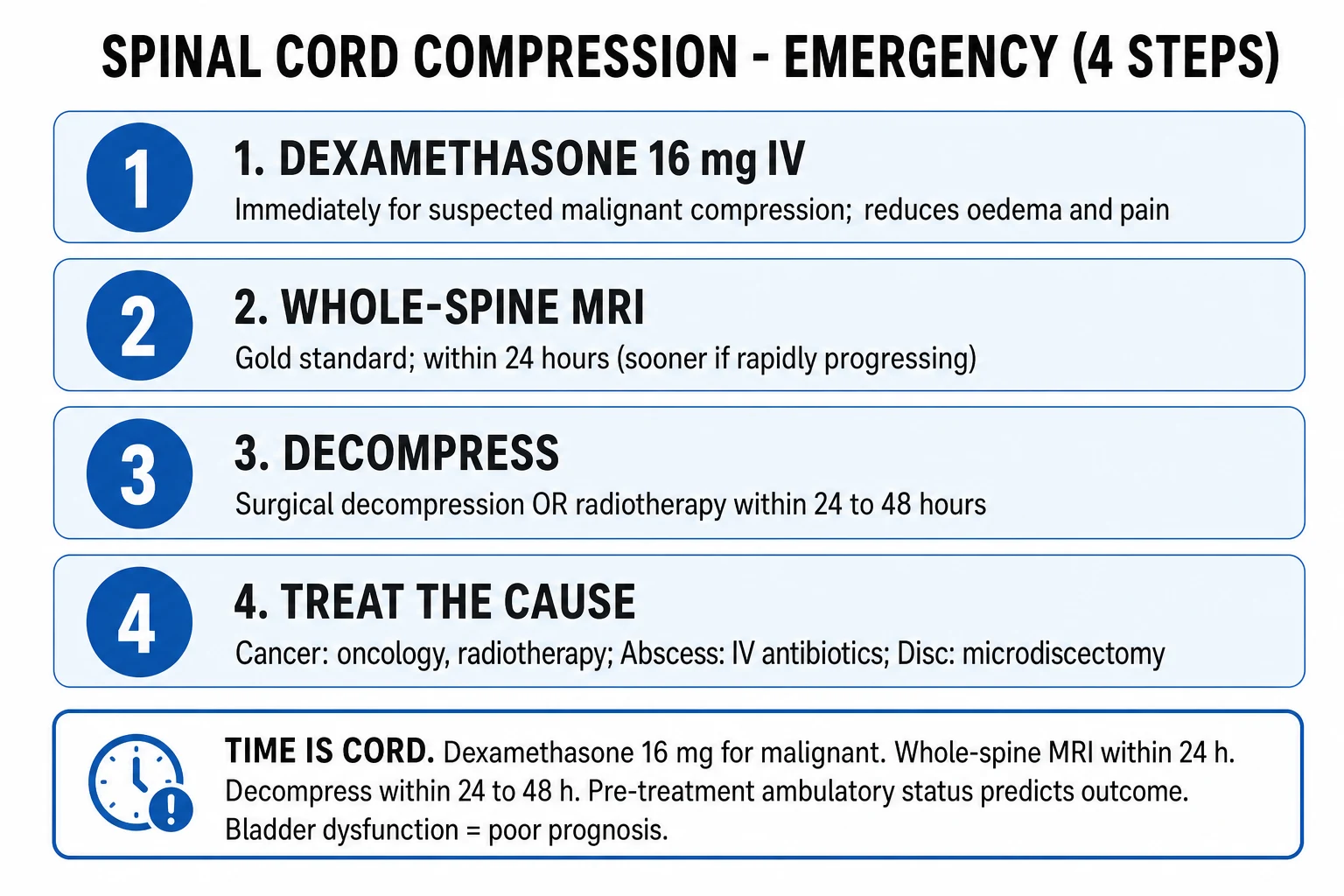
The SCC drug doses
Management — the time-critical sequence and the 24-hour window
The irreversible cord necrosis begins within 24 to 48 hours of the onset of the complete block, which places the spinal cord compression in the same category of time-critical neurological emergency as the ischaemic stroke and the extradural haematoma.[1] The two numbers that dominate the referral handover are the time of the onset of the deficit and the ambulatory status — the non-ambulatory patient decompressed within the 24-hour window may still regain the ambulation, but the patient decompressed after 48 hours of the complete paraplegia almost never does. The ED role is therefore fourfold and concurrent, not sequential: recognise the diagnosis at the bedside, give the dexamethasone immediately (before the MRI if the suspicion is high), arrange the urgent MRI whole spine, and make the parallel neurosurgical and oncological referrals so that the definitive treatment begins within the 24-hour window.
The ED workup for the suspected spinal cord compression
Recognise at the bedside
The known cancer with the new or the progressive back pain, the nocturnal pain that wakes the patient, any motor weakness, a sensory level on the examination, or the bladder and bowel dysfunction. The clock starts at the recognition — record the time of the onset of the deficit to the hour.
The focused neurological examination
The full motor grading of all the myotomes on the MRC 0-to-5 scale, the reflexes (the hyperreflexia and the Babinski above the conus; the hyporeflexia at the conus or the cauda), the pinprick and the light touch over all the dermatomes to define the sensory level, the digital rectal examination for the anal tone and the perianal sensation, and the gait. Document the ASIA impairment scale as the baseline.
Resuscitate and protect the cord
The ABCDE; the IV access; the cervical collar if the cervical lesion is suspected; the spinal precautions; the urinary catheter for the retention. Maintain the mean arterial pressure to perfuse the compressed cord — the hypotension worsens the ischaemia and must be corrected.
Dexamethasone 10 mg IV — before the MRI
Give the dexamethasone 10 mg IV immediately if the clinical suspicion is high, then 4 mg every 6 hours. Do not wait for the imaging. The steroid reduces the vasogenic oedema and buys the time for the decompression.
Analgesia
The morphine 5 mg IV titrated, the paracetamol 1 g PO. The pain of the cord compression is severe and the undertreated pain raises the blood pressure and the distress.
The blood panel
The FBC, the U&E, the CRP, the ESR, the coagulation, the calcium, the LFTs, the PSA for the prostate, the myeloma screen (the serum protein electrophoresis, the serum free light chains, the urine Bence-Jones) if the primary is unknown, the group and save. The blood cultures if the fever or the epidural abscess is suspected.
The urgent MRI whole spine with gadolinium — within 4 to 6 hours
The whole spine, not just the symptomatic level, because the skip lesions are common. The MRI defines the site, the extent, the degree and the cause. The CT myelogram is the alternative when the MRI is contraindicated.
The parallel referrals — neurosurgery, oncology, palliative care
Refer simultaneously, not in series. The target is the definitive treatment — the surgical decompression or the radiotherapy — within 24 hours. The Patchell criteria (the single-level compression, the fit patient, the expected survival greater than 3 months) favour the surgery.
The disposition
Admit to the neurosurgical or the oncological unit; the HDU or the ICU if the high cervical lesion compromises the respiration (the diaphragm is C3 to C5) or if the autonomic instability is present.
The direct decompressive surgical resection followed by the radiotherapy is superior to the radiotherapy alone for the single-level MSCC in the fit patient with the reasonable prognosis.[3]
Surgical decompression + radiotherapy
- Best for the single-level compression, the bony retropulsion, the spinal instability, the radioresistant tumour (the lung, the melanoma, the colon, the renal), the unknown primary, or the neurological deterioration during the radiotherapy
- The Patchell 2005 randomised trial was stopped early because the surgery plus the radiotherapy was superior to the radiotherapy alone in the ambulatory outcome
- Requires the fit patient with the reasonable prognosis — the expected survival of at least 3 to 6 months — and the single-level or the adjacent-level disease
- The rapid pain control and the preserved or the restored ambulation; the surgical stabilisation also permits the mobilisation and the earlier discharge
Radiotherapy alone
- Best for the radiosensitive tumour (the breast, the prostate, the myeloma, the lymphoma, the small-cell lung), the multi-level disease, the poor surgical candidate, or the very short prognosis
- The onset of the pain relief over the days to the weeks; the neurological recovery is slower and less complete than the surgery
- Ineffective for the radioresistant tumour; the deficit may progress during the treatment window
- The single 8 Gy fraction is now standard for the pain and is equivalent to the longer course for the ambulation in the short-prognosis patient
Conservative and palliative
- For the patient with the complete and the fixed paraplegia for over 48 hours, the very poor prognosis (the survival of days), or the patient who declines the intervention
- The focus shifts to the pain control, the pressure-area care, the bowel and bladder management, and the palliative-care input
- The dexamethasone is still given for the pain and the oedema even when the decompression is not pursued
- A goals-of-care conversation with the patient and the family is the centrepiece, not the omission of the care
Patchell 2005 — direct decompressive surgery for the single-level MSCC
Lancet
PMID 16112300
Key finding
In the randomised trial of 101 patients with the single-level MSCC and an expected survival of at least 3 months, the direct decompressive surgical resection followed by the radiotherapy was superior to the radiotherapy alone: more patients were ambulatory after the treatment (84 per cent vs 57 per cent), retained the ambulation for longer, and regained the ability to walk. The trial was stopped early by the data-monitoring committee for the clear benefit.
Practice change
The single-level MSCC in the fit patient with the reasonable prognosis receives the surgical decompression plus the radiotherapy, not the radiotherapy alone.
Complications and prognosis
The outcome is determined by the neurological status at the presentation — the patients who are ambulatory at the presentation have an 80 to 90 per cent chance of remaining ambulatory; the patients who are non-ambulatory have a 10 to 30 per cent chance of regaining the ambulation, and the patients who are paraplegic at the presentation have a less than 5 per cent chance of regaining the ambulation. This steep gradient — the ambulatory stay ambulatory and the non-ambulatory stay non-ambulatory — is the reason that the time to the decompression is the single most important determinant of the outcome, and the reason that every minute of delay in the ED is a minute of potential cord damage. [1]
The complications are the paraplegia (the irreversible if the treatment is delayed beyond 24 to 48 hours), the urinary and faecal incontinence (the autonomic disruption, the neurogenic bladder — managed with the intermittent catheterisation or the indwelling catheter), the pressure sores (from the immobility and the sensory loss — the regular turning and the pressure-area care), the deep vein thrombosis and the pulmonary embolism (from the immobility — the prophylactic enoxaparin 40 mg subcutaneously daily), the hypercalcaemia (from the bone metastases — treated with the IV fluid, the bisphosphonates, the denosumab), and the psychological impact (the grief and the anxiety about the prognosis, the loss of the independence, the palliative care referral). The median survival after the MSCC is 3 to 6 months (the breast and the prostate may survive years; the lung and the melanoma may survive weeks). The quality of life — the pain control, the mobility, the bowel and bladder function — is the priority alongside the survival. [1]
Cauda equina syndrome — the separate neurosurgical emergency
The cauda equina syndrome (CES) is anatomically distinct from the cord compression — it is the compression of the lumbosacral nerve roots below the conus medullaris (L1 to L2 in the adult), not the cord itself — but it shares the time-critical urgency and the same ED imperative: recognise, image, and decompress within the hours. Because the lesion is at the level of the nerve roots, the deficit is lower motor neurone: the flaccid weakness, the areflexia, the hypotonia, and the sensory loss in the saddle and the lumbosacral dermatomes. The cardinal red-flag features are the saddle anaesthesia (the loss of the sensation over the buttocks, the perineum and the posterior thighs), the bladder dysfunction (the urinary retention with the overflow incontinence — a palpable bladder and a post-void residual over 100 to 200 mL is the objective sign), the bowel dysfunction (the faecal incontinence from the loss of the anal tone), the bilateral sciatica or the bilateral neurological deficit, and the lower-limb weakness. The digital rectal examination for the anal tone and the perianal pinprick is mandatory in every patient with the suspected CES — its omission is a documented source of the missed diagnosis and the litigation. [1]
The MRI of the lumbosacral spine is the gold standard and should be performed within the hours of the suspected diagnosis. The management is the urgent surgical decompression (the laminectomy or the discectomy) — ideally within 24 to 48 hours, because the delay beyond 48 hours of the complete CES markedly reduces the chance of the recovery of the bladder and the bowel function. The dexamethasone is given as for the cord compression. The prognosis mirrors the cord compression: the patients who are ambulatory and continent at the presentation tend to remain so; the patients who present with the complete CES (the anaesthesia, the paralysis, the retention) have the poor recovery regardless of the timing, which is the reason to act before the syndrome becomes complete. [1]
[1]Cord compression (above the conus)
- The lesion at or above the conus medullaris (T12 to L1 in the adult); the upper motor neurone signs — the spasticity, the hyperreflexia, the Babinski, the clonus
- A clear sensory level on the trunk; the symmetrical and the progressive deficit; the autonomic features such as the urinary retention, but the anal tone is often preserved early
- The MRI whole spine; the metastatic malignancy is the commonest cause; the dexamethasone 10 mg IV then the decompression or the radiotherapy
- The ambulatory status at the presentation determines the ambulatory outcome; the 24 to 48 hour window
Cauda equina syndrome (below the conus)
- The lesion of the lumbosacral nerve roots below the conus; the lower motor neurone signs — the flaccidity, the areflexia, the hypotonia
- The saddle anaesthesia, the bilateral sciatica, the anal-tone loss on the digital rectal examination, the urinary retention with the overflow incontinence
- The MRI lumbosacral spine; the large central disc herniation is the commonest cause; the urgent surgical decompression within 24 to 48 hours
- The complete CES at the presentation has the poor recovery; act before it becomes complete
Conus medullaris syndrome (at the conus)
- The lesion at the conus (T12 to L1); the mixed upper and the lower motor neurone signs — the early and the prominent bladder and bowel involvement
- The saddle anaesthesia but the back pain is less prominent than in the CES; the asymmetrical weakness; the combination of the UMN and the LMN signs in the same patient is the clue
- The MRI thoracolumbar; the disc, the tumour or the trauma; the urgent decompression
- The early and the severe autonomic involvement distinguishes the conus from the pure cauda equina lesion
Pathophysiology
The spinal cord compression damages the cord by two mechanisms. First, the direct mechanical compression of the neural tissue by the tumour mass, the disc, the abscess or the haematoma distorts the white-matter tracts and the grey-matter neurons, disrupting the conduction. Second, the ischaemia from the compression of the epidural venous plexus and the radicular arteries produces the cord infarction — the venous congestion first, then the arterial compromise, then the irreversible necrosis. The oedema that follows the compression further increases the intramedullary pressure, creating a vicious cycle of the swelling, the ischaemia and the further damage. The dexamethasone breaks this cycle by reducing the vasogenic oedema, buying time for the definitive decompression. The time window for the reversibility is approximately 24 to 48 hours — after that, the necrosis is irreversible, and the neurological deficit becomes permanent. This is the biological basis for the urgency: the cord that is compressed but not yet infarcted can recover if the decompression is performed within hours; the cord that is infarcted cannot. [1]
The epidural abscess
The epidural abscess is the infectious cause of the spinal cord compression, and its incidence is rising with the IV drug use, the diabetes and the immunocompromise. The presentation is the triad of the back pain, the fever and the neurological deficit — but all three are present in only 15 per cent of the cases at the presentation. The raised inflammatory markers (the CRP over 50, the WCC raised) distinguish it from the malignancy. The blood cultures are positive in 60 per cent. The MRI with the gadolinium shows the rim-enhancing collection in the epidural space. The management is the urgent surgical decompression (the antibiotics alone do not penetrate the abscess cavity adequately) plus the empirical IV antibiotics (the vancomycin 1.5 g IV plus the ceftriaxone 2 g IV, or the piperacillin-tazobactam 4.5 g IV if the Pseudomonas is suspected) for 4 to 6 weeks. The mortality is 5 to 10 per cent, and the permanent neurological deficit occurs in 10 to 25 per cent. [1]
Special populations
The anticoagulated patient with the spinal cord compression — the anticoagulation is reversed (the PCC 25 to 50 IU/kg and the vitamin K 10 mg for the warfarin; the idarucizumab 5 g for the dabigatran) if the surgical decompression is planned or if the epidural haematoma is the cause. The pregnant patient — the MRI without the gadolinium is safe in the pregnancy; the radiotherapy is avoided; the surgical decompression is modified with the left lateral tilt. The elderly patient — the outcome is worse but the treatment should not be withheld; the frailty assessment guides the shared decision-making. The paediatric patient — the SCC is rare in children and is usually from the Ewing sarcoma, the neuroblastoma or the astrocytoma. [1]
Evidence and regional guidelines
The contemporary framework is the NICE guideline CG75 for the metastatic spinal cord compression and the ASCO guideline for the cancer patients. The dexamethasone 10 mg IV, the urgent MRI, the neurosurgical or radiotherapy referral within 24 hours, and the time-to-treatment determining the outcome are the global standards.[1][2]
ANZ practice note. The SCC management follows the local oncology and neurosurgical protocol; the dexamethasone 10 mg IV loading; the urgent MRI whole spine; the neurosurgical referral for the surgical decompression or the radiotherapy referral for the radiosensitive tumour; and the time-to-treatment within 24 hours determining the ambulatory outcome. [1]
Causes in detail — malignancy, disc, abscess, haematoma
The malignancy is the commonest cause of the cord compression in the adult, accounting for the majority of the cases that reach the ED. The metastatic disease reaches the spine by the haematogenous spread to the vertebral body — the rich, slow-flowing epidural venous plexus (the Batson plexus) is the conduit — and grows posteriorly into the epidural space, compressing the cord from the front in over 70 per cent of the cases. The breast, the lung, the prostate, the myeloma and the lymphoma are the five commonest primaries; the breast and the prostate are the most osteophilic (they seed the bone preferentially), the lung and the renal present late and progress rapidly, and the myeloma causes the multifocal lytic disease with the characteristic pattern on the serum screen. The thyroid, the melanoma and the colon are less common but characteristically radioresistant, which pushes the management towards the surgery. [1]
The intervertebral disc herniation is the commonest non-malignant cause — the large central or the paracentral disc, usually at the cervical or the thoracolumbar level, producing the acute myelopathy in the patient without the cancer history. The onset is more acute than the malignancy (the hours to the days rather than the weeks) and the MRI shows the disc material, not the tumour. The epidural abscess is the infectious cause, rising in the incidence with the IV drug use, the diabetes, the immunosuppression and the indwelling catheters — the Staphylococcus aureus (including the MRSA) is the organism in over 60 per cent; the triad of the back pain, the fever and the neurological deficit is complete in only 15 per cent at the presentation, so a raised CRP (over 50) with the back pain is the abscess until the MRI proves otherwise. The epidural haematoma is the spontaneous or the post-procedural bleed into the epidural space — the anticoagulation (the warfarin, the direct oral anticoagulants), the recent neuraxial procedure (the epidural, the spinal, the lumbar puncture), the coagulopathy and the thrombolysis are the risk factors, and the onset is the sudden severe back pain with the rapidly progressive deficit in the anticoagulated patient. [1]
[1] [1]Diagnosis — the MRI whole spine is the gold standard
The urgent MRI whole spine with gadolinium is the gold standard and the only imaging that directly visualises the cord and the lesion. It defines the site, the extent, the degree and the cause of the compression, and it is the mandatory investigation before any surgical or the radiotherapeutic decision. The whole-spine acquisition (not just the symptomatic level) is essential because the skip lesions are common in the malignancy (the multifocal metastatic disease), in the multilevel disc disease, and in the multilevel abscess; imaging only the symptomatic level misses the second lesion that may be the more critical one. The Bilsky epidural spinal cord compression scale grades the degree of the thecal-sac compromise (grade 0, the bone-only disease, through grade 3, the cord deformed) and standardises the communication between the radiologist, the surgeon and the oncologist.[4]
The CT is the adjunct for the bony detail — the fracture pattern, the canal compromise, the pedicle loss (the winking-owl sign of the pedicle metastasis), the surgical planning and the instrument fixation. The CT myelogram is the alternative when the MRI is contraindicated (the pacemaker, the cochlear implant, the ferromagnetic aneurysm clip): it demonstrates the block to the contrast flow and localises the level, but it cannot show the cord parenchyma, the oedema, or the intramedullary signal — it is a second-best investigation. The plain X-ray may show the vertebral collapse, the loss of the pedicle, or the lytic or the sclerotic lesion, but it cannot assess the cord and is insufficient alone; it is now rarely the primary investigation. The blood panel is driven by the suspected cause — the inflammatory markers for the abscess, the coagulation before the surgery and for the haematoma, the PSA and the myeloma screen for the occult primary, and the calcium for the hypercalcaemia of the malignancy. The ASIA impairment scale documents the deficit at the baseline and at the serial assessments and is the universal language of the outcome reporting. [1]
[1]SAQ — Malignant spinal cord compression and the 24-hour window
10 minutes · 10 marks
A 62-year-old woman with metastatic breast cancer presents to the emergency department with three weeks of progressive lower thoracic back pain that wakes her at night, and two days of difficulty walking. On examination she has a sensory level at T10, grade 3 of 5 weakness in both legs, and hyperreflexia with upgoing plantar responses. The blood pressure is 110 over 70 and the heart rate is 92.
SAQ — Cauda equina syndrome and the post-void residual
10 minutes · 10 marks
A 45-year-old man who is otherwise well presents with severe lower back pain radiating down both legs for one week, and for the last twelve hours he has noticed numbness around his genitals and perineum and an inability to pass urine. On examination he has grade 4 weakness of the ankle dorsiflexion, absent ankle reflexes, saddle anaesthesia and a reduced anal tone on the digital rectal examination.
Exam pearls
- The cancer patient with the new back pain has the SCC until the MRI proves otherwise.
- The MRI whole spine (not just the symptomatic level) — the skip lesions.
- The dexamethasone 10 mg IV loading then 4 mg every 6 hours.
- The ambulatory status at the presentation determines the ambulatory outcome.
- The neurosurgical referral for the unstable spine, the bony retropulsion, the single-level.
- The radiotherapy for the radiosensitive tumour (breast, prostate, myeloma, lymphoma).
- The cauda equina syndrome: the saddle anaesthesia, the bilateral sciatica, the urinary retention.
- The time of the onset of the deficit and the ambulatory status are the two numbers in the referral — record both to the hour.
- The whole-spine MRI, not the symptomatic level — the skip lesions in the metastatic disease.
- The Bilsky ESCC grade 3 (the cord deformed) is the surgical grade — learn the six-point scale.
- The Patchell trial: the single-level MSCC in the fit patient with the expected survival over 3 months receives the surgery plus the radiotherapy, not the radiotherapy alone.
- The anticoagulated patient with the sudden back pain — the epidural haematoma until the MRI proves otherwise; reverse and image in parallel.
- The epidural-abscess triad (the pain, the fever, the deficit) is complete in only 15 per cent — a CRP over 50 with the back pain is the abscess until proven otherwise.
- The dexamethasone is given before the MRI if the suspicion is high — never delay the steroid for the scan.
- The cauda equina syndrome is LMN; the cord compression above the conus is UMN — the digital rectal examination distinguishes them at the bedside.
- The post-void residual over 100 to 200 mL is the objective sign of the denervated bladder in the suspected CES — scan, do not rely on the history.
- The cervical cord compression at C3 to C5 compromises the diaphragm — anticipate the respiratory failure and the HDU or the ICU admission.
- The conus lesion (T12 to L1) gives the mixed UMN and the LMN signs in the same patient — the early and the severe bladder and bowel involvement is the clue.
- The hypercalcaemia of the malignancy accompanies the bone metastases — check the calcium and treat with the IV fluid, the bisphosphonate and the denosumab. [1]
Red flags
[1]References
- [1]Singer E, et al. Initial management and disposition of metastatic spinal cord compression in the emergency department: a review of the literature Ann Med, 2025.PMID 41033840
- [2]Patel DA, et al. Diagnostic and Therapeutic Strategies for Patients with Malignant Epidural Spinal Cord Compression Curr Treat Options Oncol, 2017.PMID 28795286
- [3]Patchell RA, Tibbs PA, Regine WF, et al. Direct decompressive surgical resection in the treatment of spinal cord compression caused by metastatic cancer: a randomised trial Lancet, 2005.PMID 16112300
- [4]Bilsky MH, Laufer I, Fourney DR, et al. Reliability analysis of the epidural spinal cord compression scale J Neurosurg Spine, 2010.PMID 20809724