EM · Haemoptysis
Haemoptysis (including massive haemoptysis)
Also known as Coughing blood · Massive haemoptysis · Pulmonary haemorrhage · Bronchial bleeding
Haemoptysis — the coughing of blood from the respiratory tract, the distinction between the non-massive (the bronchitis, the bronchiectasis, the cancer, the TB, the pneumonia) and the massive (over 100 mL in 24 hours — the life-threatening asphyxiation risk), the emergency management of the massive haemoptysis (the airway protection, the bleeding-side-down positioning, the large endotracheal tube or the double-lumen tube, the bronchoscopy, the bronchial artery embolisation), and the tranexamic acid. ACEM-primary, globally tagged.
On this page & tools
Your progress
Saved locally on this device.
Target exams
Red flags
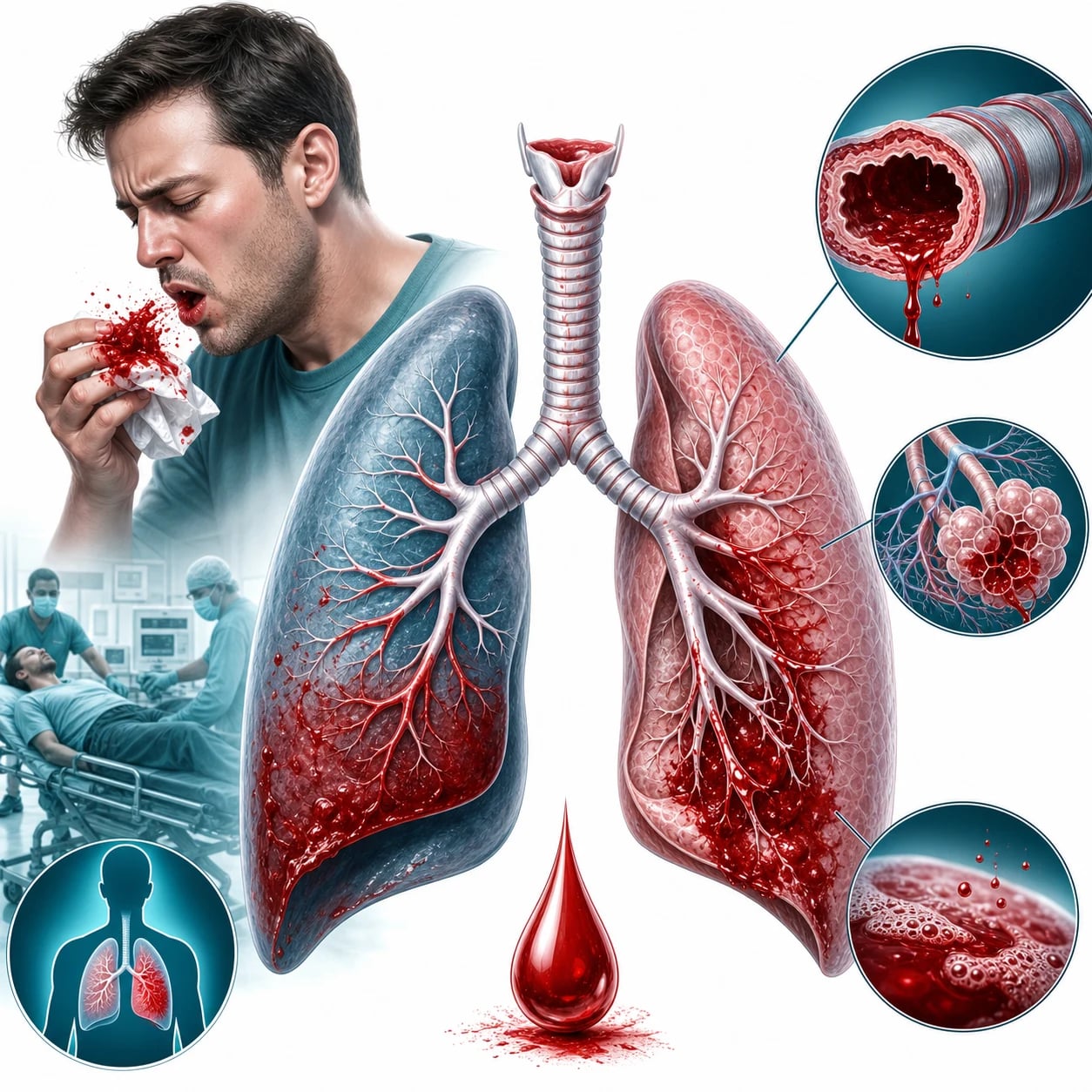
Haemoptysis — the coughing of blood from the respiratory tract below the larynx — is a presentation that ranges from the blood-streaked sputum of the bronchitis to the massive, life-threatening pulmonary haemorrhage that floods the alveoli and kills by asphyxiation within minutes. The Fellowship candidate must distinguish the non-massive (investigate the cause, manage as an outpatient or a ward) from the massive (the airway protection, the lung isolation, the bronchoscopy, the embolisation), because the massive haemoptysis is one of the few true respiratory emergencies where the minutes determine the survival.[1][2]
Definition and the classification by volume
Haemoptysis is the expectoration of blood from the respiratory tract below the vocal cords. It is classified by the volume: the scant or the non-massive (the blood-streaked sputum, up to about 30 mL in 24 hours) is the common presentation; the moderate (30 to 100 mL); and the massive (over 100 mL in 24 hours, or any volume that threatens the airway). The massive haemoptysis is the emergency — the blood floods the alveoli of the bleeding lung and, by the overflow, of the contralateral lung, producing a hypoxaemia and an asphyxiation that is the mechanism of the death. The exsanguination is rare — the blood clots within the airway and the loss is contained — the patient dies of the drowning, not the bleeding. [1]
Pathophysiology — the bronchial versus the pulmonary circulation
The lung is supplied by two circulations: the low-pressure pulmonary circulation (the pulmonary artery and its branches, which perfuse the alveoli for the gas exchange) and the high-pressure systemic bronchial circulation (the bronchial arteries, arising from the descending thoracic aorta, which perfuse the airways, the vessels, the supporting tissue, and — critically — the tumours). The dual circulation explains the haemoptysis: the bronchial arteries are the source of bleeding in approximately 90 per cent of the massive haemoptysis, because they are systemic, high-pressure vessels that rupture into the airway when eroded by a tumour, a cavity, or an inflamed bronchial wall. The pulmonary artery bleeds less often (the Rasmussen aneurysm in the TB cavity, the pulmonary-artery rupture from a catheter) but, when it does, the bleeding is brisk. This anatomical principle — the bronchial artery as the dominant source — is the rationale for the bronchial artery embolisation as the definitive treatment.[3][4]
The mechanism of the death clarifies the volume thresholds. The textbook massive haemoptysis is variously defined: over 100 mL in 24 hours, over 200 mL in 24 hours, or — the most clinically useful — any volume that threatens the airway. A single episode of 50 mL that floods the trachea is more dangerous than 300 mL spread over 24 hours that the patient expectorates between breaths. The Fellowship candidate should treat any bleeding that compromises the airway, the ventilation, or the haemodynamics as the massive haemoptysis regardless of the absolute volume, and reserve the volume-based thresholds for the audit and the classification.[1]
Differential diagnosis — the causes and the mimics
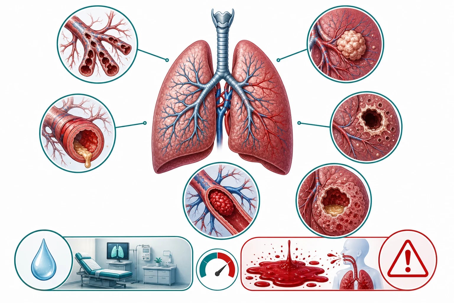
The first task is to distinguish the true haemoptysis from the mimics, and the second is to identify the cause. [1]
True haemoptysis (respiratory)
- Coughed up (not vomited); frothy, bright red, alkaline pH
- Causes: bronchiectasis, cancer, TB, pneumonia, PE, bronchitis
- Massive: bronchiectasis, cancer, TB, AV malformation, aspergilloma
- Bronchoscopy, CT angiography, embolisation
Haematemesis (GI)
- Vomited (not coughed); dark, coffee-ground, acidic
- A history of the upper-GI disease; the melena
- No frothy sputum; the NG aspirate confirms
- Treat the GI cause (the endoscopy)
Upper airway bleeding
- The epistaxis, the posterior nasal bleed, the gingival
- No cough; the blood drips down the posterior pharynx
- The nasendoscopy identifies the source
- Treat the ENT cause
Anticoagulation-related
- A patient on the warfarin/DOAC with a respiratory infection
- May be minor but persistent; the INR may be high
- Reverse the anticoagulation if massive
- Investigate the underlying cause — the anticoagulation is a cofactor, not the cause
The causes of the non-massive haemoptysis, in the order of the frequency: the bronchitis (the commonest in the smoker), the bronchiectasis (the chronic, the productive cough), the lung cancer (the smoker, the weight loss, the persistent haemoptysis), the tuberculosis (the endemic, the weight loss, the night sweats), the pneumonia (the fever, the consolidation), the pulmonary embolism (the pleuritic pain, the DVT), and the pulmonary oedema (the pink frothy sputum). The causes of the massive haemoptysis are the bronchiectasis, the cancer, the TB, the aspergilloma (the fungal ball in a pre-existing cavity), the pulmonary AV malformation (the hereditary haemorrhagic telangiectasia), and the iatrogenic (the bronchoscopic biopsy, the catheter). [1]
Non-massive causes (the common)
- Bronchitis (acute or chronic) — the commonest cause overall
- Bronchiectasis — the chronic productive cough, the recurrent infections
- Lung cancer — the smoker, the weight loss, the persistent streak
- Tuberculosis — the endemic, the night sweats, the cavity
- Pneumonia — the fever, the consolidation, the rusty sputum
- Pulmonary embolism — the pleuritic pain, the DVT, the infarct
Massive causes (the dangerous)
- Bronchiectasis — the hypertrophied bronchial arteries
- Lung cancer — the squamous-cell eroding a bronchial artery
- Tuberculosis — the Rasmussen aneurysm in a cavity wall
- Aspergilloma — the fungal ball in a pre-existing cavity
- Pulmonary AV malformation — the HHT, the high-flow shunt
- Iatrogenic — the bronchoscopic biopsy, the catheter trauma
The rare but examinable
- Pulmonary vasculitis (Goodpasture, ANCA — Wegener/GPA, MPA)
- Diffuse alveolar haemorrhage — the immunocompromised, the BMT
- Foreign body — the chronic child, the aspirated object
- Coagulopathy — the over-anticoagulated, the thrombocytopenic
- Catamenial — the endometriosis, the cyclical with the menses
- Trauma — the blunt or the penetrating chest injury
The major causes in detail
Bronchiectasis. The chronic, irreversible dilatation of the bronchi from the recurrent infection and inflammation (post-infective, the cystic fibrosis, the immunodeficiency, the allergic bronchopulmonary aspergillosis). The hypertrophied, friable bronchial arteries erode through the inflamed bronchial wall — the bronchiectasis is the commonest cause of the massive haemoptysis worldwide. The chest CT shows the signet-ring sign (the bronchus larger than its accompanying pulmonary artery). The bleeding is from the systemic bronchial circulation, which is why the BAE is so effective.[8]
Lung cancer. The squamous-cell carcinoma (the central, the cavitating) is the classic cause of the massive haemoptysis — the tumour erodes a bronchial artery. The adenocarcinoma (the peripheral) bleeds less dramatically. A haemoptysis in a smoker over 40 is a cancer until proven otherwise — the bronchoscopy and the CT are mandatory. The cancer-related bleed may be controlled by the BAE, but the definitive treatment is the resection, the radiotherapy, or the palliation depending on the stage. [1]
Tuberculosis. The active TB erodes the bronchial vessels; the post-TB cavity may harbour an aspergilloma or develop a Rasmussen aneurysm — a pseudoaneurysm of a pulmonary artery branch in the wall of a TB cavity that ruptures catastrophically. The TB is the leading cause of the massive haemoptysis in the endemic regions. The management is the acute control (the BAE) plus the anti-tubercular therapy; the Rasmussen aneurysm may need the surgical ligation. [1]
Aspergilloma. The fungal ball (the Aspergillus colonising a pre-existing cavity — the post-TB, the sarcoid, the bronchiectatic) is surrounded by the fragile, hypertrophied vessels that bleed briskly. The chest radiograph shows the air-crescent sign (the crescent of air around a mobile fungal ball). The aspergilloma-related bleed is a classic examinable cause of the massive haemoptysis — the BAE is the bridge, the surgical resection is the definitive treatment for the localised, the recurrent bleed.[9]
Pulmonary embolism. The PE causes the haemoptysis by the pulmonary infarction (the small, the peripheral embolus in a patient with the collateral bronchial circulation) — the classic presentation is the pleuritic pain, the haemoptysis, and the mild dyspnoea. The haemoptysis is usually small-volume; the massive haemoptysis from a PE is rare and paradoxical (the large PE causes the shock, not the bleed). The management is the anticoagulation — never embolise a PE-related bleed. [1]
The vasculitides — Goodpasture and the ANCA-associated diseases
The pulmonary-renal syndromes are the rare but examinable causes of the diffuse alveolar haemorrhage (DAH) — the bleeding from the pulmonary capillaries that presents as the haemoptysis, the diffuse infiltrates, and the rapidly progressive glomerulonephritis. [1]
Goodpasture (anti-GBM)
- Antibodies against the glomerular basement membrane
- DAH plus the rapidly progressive glomerulonephritis
- Anti-GBM antibody positive in the serum
- Treated with the plasmapheresis, the steroids, the cyclophosphamide
- Can relapse; the renal prognosis depends on the creatinine at the presentation
GPA (Wegener)
- ANCA-associated vasculitis (the PR3-ANCA, the c-ANCA)
- DAH plus the sinusitis, the saddle-nose, the renal disease
- The upper- and the lower-respiratory and the renal triad
- Treated with the steroids and the cyclophosphamide or the rituximab
- Relapsing; the long-term immunosuppression
MPA (microscopic polyangiitis)
- ANCA-associated vasculitis (the MPO-ANCA, the p-ANCA)
- DAH plus the renal and the pulmonary disease
- No the upper-respiratory involvement (distinguishes from the GPA)
- Treated with the steroids and the cyclophosphamide or the rituximab
- May have the pulmonary fibrosis as a late sequel
Pulmonary AVM (HHT)
- Hereditary haemorrhagic telangiectasia (Osler-Weber-Rendu)
- Direct the artery-to-vein shunt — the high-flow, the fragile
- Bleeds briskly; may cause the paradoxical embolism (the stroke, the abscess)
- Treated with the coil or the vascular-plug embolisation
- Screen the family — the autosomal dominant inheritance
The foreign body and the coagulopathy
A foreign-body aspiration (the chronic, the undiagnosed) causes the recurrent haemoptysis — the child or the adult with the chronic cough, the localised wheeze, and the unresolved pneumonia. The chest radiograph may show the foreign body or the post-obstructive changes; the bronchoscopy is both the diagnostic and the therapeutic (the removal). A coagulopathy (the over-anticoagulated patient on the warfarin or a DOAC, the thrombocytopenic, the leukaemic, the liver-failure patient) is a cofactor — it converts a minor respiratory bleed into a significant one. The coagulopathy is reversed (the prothrombin-complex concentrate, the specific reversal agents, the platelets), but the underlying lesion (the cancer, the bronchiectasis) is still sought — the coagulopathy alone rarely causes the haemoptysis without a lesion. [1]
Clinical assessment — the history and the examination
The history and the examination aim to (a) confirm the haemoptysis is true (not the haematemesis or the upper-airway bleed), (b) estimate the volume and the acuity, and (c) localise the bleeding side and the suspected cause. The history captures the volume (the teaspoons, the cupfuls, the continuous), the colour (the bright-red and frothy is the active arterial bleed; the dark and clotted is the older), the duration, the associated symptoms (the fever, the weight loss, the night sweats, the pleuritic pain, the chronic cough, the orthopnoea), and the risk factors (the smoking, the TB exposure, the anticoagulation, the recent bronchoscopy or biopsy, the cancer, the HIV, the HHT family history). The examination checks the vital signs (the hypoxia, the tachycardia, the hypotension), the chest (the localised wheeze or the crackles, the consolidation, the pleural rub), the lymphadenopathy, the clubbing (the cancer, the bronchiectasis, the fibrosis), the telangiectasia (the HHT — the lips, the tongue, the fingers), and the legs (the DVT). [1]
[1] [1]Investigations — the radiograph, the bronchoscopy and the CT
The chest radiograph is the first investigation — it may show the consolidation, the mass, the cavity, the bronchiectatic changes, or it may be normal (the small or the central lesion). The bronchoscopy is both the diagnostic and the therapeutic intervention — the flexible bronchoscopy identifies the bleeding site and delivers the local therapies (the cold saline lavage, the topical adrenaline 1:1000, the balloon tamponade); the rigid bronchoscopy (by the thoracic team) is used for the massive case to secure the airway and to allow the larger instruments. The CT angiography of the chest defines the bleeding site (the extravasation), the underlying lesion (the mass, the cavity, the bronchiectasis), and the bronchial artery anatomy for the embolisation planning. The bloods (the full blood count, the coagulation, the group and crossmatch, the troponin, the D-dimer) and the sputum for the AFB (if the TB is suspected) are sent. [1]
The diagnostic algorithm — the stable versus the unstable patient
The diagnostic strategy diverges by the haemodynamic stability. The unstable patient (the active massive bleed, the hypoxia, the shock) bypasses the diagnostic imaging and goes straight to the securing of the airway and the therapeutic flexible or rigid bronchoscopy — the CT scanner is an unsafe destination for the unstable patient. The stable patient (the non-massive or the controlled moderate bleed) undergoes the systematic workup — the chest radiograph, the CT angiography, and the timed bronchoscopy.[1][2]
The ED diagnostic algorithm for the haemoptysis
Step 1 — Is this a massive haemoptysis?
Assess the airway, the breathing, and the circulation. If the bleeding is brisk and the airway is threatened, this is the massive haemoptysis — secure the airway, isolate the lung, and proceed to the therapeutic bronchoscopy and the BAE. Do NOT send an unstable patient to the CT scanner.
Step 2 — Confirm the true haemoptysis
Distinguish from the haematemesis (the vomited, the coffee-ground, the acidic) and the upper-airway bleed (the epistaxis, the posterior nasal, the gingival). The frothy, the bright-red, the alkaline coughed-up blood is the true haemoptysis.
Step 3 — The chest radiograph
The first investigation. It localises the bleeding side (the infiltrate, the mass, the cavity, the bronchiectatic change) in about 50 to 80 per cent. A normal chest radiograph does not exclude a significant lesion — proceed to the CT.
Step 4 — The CT angiography of the chest
The CT angiography (the contrast-enhanced, the arterial phase) is the highest-yield investigation in the stable patient. It identifies the bleeding source (the contrast extravasation), the underlying lesion (the mass, the cavity, the aspergilloma, the AVM, the bronchiectasis), and it maps the bronchial artery anatomy for the embolisation planning.
Step 5 — The flexible bronchoscopy
Performed when the bleeding has slowed or for the non-massive case. It identifies the bleeding segment, allows the washings and the biopsy (the cancer, the TB), and delivers the local therapies (the iced saline, the adrenaline, the balloon). For the active moderate bleed, the bronchoscopy is both the diagnostic and the first-line therapeutic step.
Step 6 — The blood and the sputum
The full blood count (the anaemia, the thrombocytopenia), the coagulation (the INR, the APTT), the group and crossmatch (4 to 6 units for the massive case), the urea and electrolytes, the troponin, the D-dimer. The sputum for the AFB and the culture if the TB is suspected. The anti-GBM and the ANCA if the diffuse alveolar haemorrhage or the pulmonary-renal syndrome is suspected.
Step 7 — The disposition
The non-massive, the stable patient is investigated as an outpatient or a ward admission. The massive or the recurrent bleed is admitted to the ICU or the HDU after the bronchoscopy or the BAE, with the monitoring for the rebleed.
The investigations and the yields
Immediate management — the massive haemoptysis
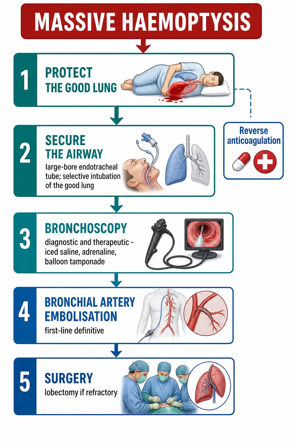
The massive haemoptysis is a true emergency — the airway is threatened by the blood, and the hypoxia is the mechanism of the death. [1]
The massive haemoptysis is a life-threatening emergency and the airway is the first priority — the blood floods the alveoli and the patient asphyxiates. The ABCDE is applied with three haemoptysis-specific modifications: position the bleeding side down (protect the good lung), intubate early with a large ETT or a double-lumen tube (allow the bronchoscopy and the lung isolation), and activate the interventional radiology and the thoracic team in parallel — do not wait for one step to finish before the next is begun.[1][3]
The first 30 minutes — the suspected massive haemoptysis
0 min — Recognise and call for help
Active brisk haemoptysis, a patient choking on blood, or any volume that threatens the airway is a massive haemoptysis. Call the senior ED doctor, the anaesthetist, the intensivist, the thoracic surgeon, and the interventional radiologist simultaneously — the parallel activation is essential, the haemoptysis pathway is run by the team, not the individual.
0 to 2 min — Position the bleeding side DOWN
Place the patient in the lateral decubitus position with the suspected bleeding side dependent (down). Gravity keeps the blood in the bleeding lung and prevents the spillage into the good lung — this is the single most important bedside manoeuvre and it is done before anything else. If the bleeding side is unknown, place the patient head-down or in the Trendelenburg tilt to keep the blood away from the upper airway.
0 to 5 min — A: Airway and oxygen
High-flow oxygen by a non-rebreather mask to hold the SpO₂ at 94 per cent or above. Suction the blood from the oropharynx. Prepare for the early intubation — do not wait for the patient to tire or arrest. Have the difficult-airway trolley and the large-bore suction ready.
2 to 10 min — A: Intubate with a large ETT or a double-lumen tube
A rapid-sequence intubation with a large endotracheal tube (8.0 mm internal diameter or above — the standard adult bronchoscope has an outer diameter of about 6 mm and will not pass a smaller tube). For the unilateral, localised bleed, a double-lumen tube or a single-lumen tube with a bronchial blocker isolates the bleeding lung from the good lung — the anaesthetist or the intensivist performs this.<Cite id="11" />
5 to 10 min — B and C: Ventilate, cannulate, sample
Ventilate with 100 per cent oxygen; titrate the PEEP cautiously (a high PEEP may displace the blood distally). Two large-bore cannulae; blood for the full blood count, the coagulation, the group and crossmatch (4 to 6 units), the urea and electrolytes, the troponin, the D-dimer, and the venous blood gas with the lactate. A balanced-crystalloid bolus for the hypovolaemia — but the hypovolaemia is rarely the threat; the asphyxiation is.
5 to 15 min — Stop the bleeding: TXA, antifibrinolytics, reversal
Give the tranexamic acid 1 g intravenously over 10 minutes. Reverse the anticoagulation if applicable (the prothrombin-complex concentrate for the warfarin; the specific reversal agents for the DOACs — andexanet alfa or the prothrombin-complex concentrate). Give the morphine 5 to 10 mg intravenously for the sedation and the cough suppression — the coughing worsens the bleeding.
10 to 30 min — Definitive intervention
In the intubated and stabilised patient, proceed to the flexible bronchoscopy (the therapeutic ladder) and the CT angiography if the bleeding site is unclear. Activate the bronchial artery embolisation — the definitive first-line intervention. Reserve the emergency thoracotomy for the catastrophic, uncontrolled bleed when the IR and the surgery are unavailable.
The haemoptysis targets and the doses
The bronchial artery embolisation is the definitive intervention for the massive haemoptysis — the interventional radiologist catheterises the bronchial artery (the usual source of the bleeding in the massive haemoptysis, as opposed to the pulmonary artery), identifies the extravasation, and embolises with the gelatin foam, the polyvinyl alcohol particles or the coils. The success rate is over 80 per cent; the recurrence is about 20 per cent (the rebleeding may need the repeat embolisation or the surgery). The surgical lobectomy is reserved for the uncontrolled bleeding from a localised, resectable lesion (the cancer, the aspergilloma) when the embolisation fails or is unavailable.[1][2]
Bronchoscopic interventions — the bedside therapeutic ladder
When the bleeding is active and the airway is secured, the bronchoscopy delivers a graded set of local therapies — each a bridge to the definitive embolisation or the surgery, and occasionally the definitive treatment in itself.[3]
The flexible-bronchoscopy therapeutic ladder for the active bleeding
Step 1 — Localise the bleeding lobe
Pass the scope through the ETT and systematically inspect each lobe. Blood tracking dependently means the patient must remain bleeding-side-down. Identify the segment of origin — this dictates the later embolisation target and the surgical field.
Step 2 — Iced saline lavage
Instil 10 to 20 mL aliquots of iced (4 °C) normal saline directly onto the bleeding segment — the cold induces vasoconstriction. Repeat to a total of 200 to 500 mL; the technique is cheap, universally available, and often temporarily effective.
Step 3 — Topical adrenaline 1:10,000
Instil 5 to 10 mL of adrenaline 1:10,000 (diluted from the 1:1000 cardiac-arrest formulation) onto the bleeding point for vasoconstriction. Some centres use topical tranexamic acid (500 mg in 5 mL). Monitor for systemic absorption — tachycardia, hypertension.
Step 4 — Balloon tamponade
Advance a bronchial-blocker or Fogarty catheter into the bleeding bronchus and inflate to occlude the segment — physically tamponades the vessel and prevents the spillage into the good lung. Leave inflated up to 24 hours while the BAE or surgery is arranged; deflate slowly and watch for rebleeding.
Step 5 — Argon plasma coagulation or laser
Thermal coagulation (argon plasma coagulation, Nd:YAG laser, or electrocautery) seals a visible discrete bleeding vessel — useful for the cancer, the AVM, and the post-biopsy ooze. Requires a dry field and a visible target; not suited to a torrential bleed.
Step 6 — Transition to rigid bronchoscopy
If the flexible scope cannot control the bleed, call the thoracic team for a rigid bronchoscopy — the larger working channel allows the larger suction, the rigid tamponade with the barrel of the scope, and the deployment of the blockers under direct vision.
Flexible bronchoscopy
- Performed under local anaesthesia or moderate sedation
- Diagnostic — identifies the bleeding site in 40 to 90 per cent
- Therapeutic — iced saline, adrenaline, balloon, APC
- Small working channel; overwhelmed by the massive bleed
- The first-line instrument for the moderate and the non-massive case
Rigid bronchoscopy
- Requires general anaesthesia and the thoracic or anaesthetic team
- Large-bore channel — clears the blood rapidly
- Tamponades the bleeding bronchus with the barrel
- Allows the larger blockers and forceps
- The instrument of choice for the massive and the torrential bleed
CT angiography
- Identifies the bleeding source (extravasation) in the stable patient
- Maps the bronchial artery anatomy for the embolisation
- Detects the underlying lesion — the mass, the cavity, the AVM
- The roadmap for the interventional radiologist
- Performed once the airway is secured and the patient is stable
Bronchial artery embolisation — the definitive IR treatment of choice
The bronchial artery embolisation (BAE) is the first-line definitive intervention for the massive and the refractory haemoptysis. The rationale is anatomical: the bronchial circulation (a high-pressure systemic circulation arising from the descending aorta) is the source of bleeding in approximately 90 per cent of massive haemoptysis cases, while the pulmonary circulation (low-pressure) accounts for most of the remainder.[3][4]
The bronchial artery embolisation procedure
1 — Femoral access and aortography
A femoral arterial puncture and a flush aortogram outline the bronchial artery origins — classically arising at the T5 to T6 level from the descending thoracic aorta, though variants are common and a missing artery must be actively sought.
2 — Selective catheterisation
A coaxial microcatheter is advanced selectively into the hypertrophied, tortuous bronchial artery that supplies the bleeding territory. The catheter tip is advanced beyond the origin of any spinal-artery branch (the artery of Adamkiewicz) to prevent the non-target embolisation and the catastrophic spinal-cord ischaemia.
3 — Identification of the culprit
The angiographic signs of the bleeding source are the hypertrophied and tortuous artery, the hypervascularity, the neovascularity, the arteriovenous shunting, the pseudoaneurysm, and — the direct sign — the contrast extravasation into the bronchus.
4 — Embolisation
The embolic agent — polyvinyl alcohol particles (150 to 500 micrometres), gelatin-foam slurry, microspheres, or the coil — is delivered until the stasis is achieved. The superselective coil embolisation achieves high immediate haemostasis with a low recurrence.<Cite id="7" />
5 — Check and re-catheterise
A completion angiogram confirms the stasis. Any additional culprit artery (a non-bronchial systemic artery such as an intercostal, a subclavian, or an internal mammary branch) is sought and embolised — the incomplete embolisation is a leading cause of the early rebleed.
6 —Post-procedure
The patient returns to the ICU or the HDU. A small chest radiograph is taken to exclude the pleural effusion or the infarction. The rebleed is monitored for 48 to 72 hours; a planned repeat BAE or the surgery is arranged for the uncontrolled case.
The immediate clinical success of the BAE (cessation of the active bleeding) is over 80 per cent across the published series; the long-term recurrence ranges from 10 to over 50 per cent depending on the underlying cause (the highest recurrence is in the active TB and the aspergilloma).[5][8] The complications are the post-embolisation syndrome (the fever, the pleuritic pain, the dysphagia — the self-limiting), the chest-wall or the pulmonary infarction, and the rare but feared spinal-cord ischaemia from the non-target embolisation of a spinal-artery branch (the artery of Adamkiewicz).[5]
Surgery — the lobectomy or the pneumonectomy for the refractory bleed
The surgical resection is the last-resort treatment for the massive haemoptysis — reserved for the patient in whom the BAE has failed, is unavailable, or the lesion is localised and resectable (the cancer, the aspergilloma, the destroyed lobe of the bronchiectasis). The operation is the lobectomy (the commonest) or, for the extensive and unilateral disease, the pneumonectomy. The mortality of the emergency lung resection for the massive haemoptysis is high (10 to 30 per cent), driven by the aspiration, the hypoxia, and the underlying disease; a preoperative BAE, even when it fails to stop the bleed definitively, may stabilise the patient and improve the surgical outcome by reducing the intraoperative spillage.[6]
Cahill and Ingbar — the landmark framework for the massive haemoptysis (Clin Chest Med 1994)
Clinics in Chest Medicine
PMID 8200191
Key finding
A definitive review that codified the modern framework for the massive haemoptysis: the priority of the airway protection over the haemostasis, the bleeding-side-down positioning, the large-bore or the double-lumen tube for the lung isolation, the graded bronchoscopic ladder, and the bronchial artery embolisation as the primary intervention. The review established that the death is by asphyxiation, not by exsanguination.
Practice change
Remains the conceptual backbone of the modern haemoptysis algorithm — the airway-first, lung-isolation, BAE-definitive framework that every Fellowship candidate must reproduce.<Cite id='3' />
Yoon — the embolic agents for the bronchial artery embolisation (Expert Opin Pharmacother 2004)
Expert Opinion on Pharmacotherapy
PMID 14996632
Key finding
A comprehensive review of the embolic materials — the gelatin sponge (resorbable, prone to recanalisation), the polyvinyl alcohol particles (permanent, the most-used), the microspheres, the coils, and the tissue adhesives — and their application in the BAE for the massive haemoptysis. The review established the polyvinyl alcohol particle and the microsphere as the durable agents of choice.
Practice change
Anchors the embolic-agent selection for the BAE and the principle that the permanent particles outperform the resorbable gelatin in the recurrence prevention.<Cite id='4' />
Frood et al. — the immediate and the long-term BAE outcomes (Pulm Ther 2020)
Pulmonary Therapy
PMID 32185642
Key finding
A retrospective single-centre series of the bronchial artery embolisation for the massive haemoptysis reporting the immediate clinical success (cessation of the active bleeding) in over 80 per cent, with the recurrence accumulating over time — higher in the active TB and the aspergilloma, lower in the cancer and the bronchiectasis. The repeat BAE was effective and safe in the rebleeding.
Practice change
Quantifies the success and the recurrence of the BAE and supports its role as the first-line definitive intervention, with the repeat embolisation as the standard management of the rebleed.<Cite id='8' />
Alexander — the emergency lung resection with and without a preoperative BAE (EJCTS 2014)
European Journal of Cardiothoracic Surgery
PMID 23918766
Key finding
A retrospective review comparing the emergency lung resection for the massive haemoptysis with and without a preoperative bronchial artery embolisation. The preoperative BAE, even when it did not definitively control the bleed, reduced the intraoperative blood spillage and improved the perioperative outcomes, supporting the BAE as a bridge even when the surgery is anticipated.
Practice change
Supports the BAE-first algorithm and the use of the preoperative BAE as a stabilising bridge to the definitive surgical resection in the localised refractory bleed.<Cite id='6' />
Ali et al. — the nebulised versus the intravenous tranexamic acid in the non-massive haemoptysis (Lung India 2026)
Lung India
PMID 42377174
Key finding
A randomised controlled trial comparing the nebulised against the intravenous tranexamic acid in the non-massive haemoptysis. Both routes reduced the bleeding; the nebulised route was at least as effective as the intravenous, with the practical advantage of the non-invasive administration in the alert and the cooperative patient.
Practice change
Provides the contemporary evidence for the tranexamic acid in the haemoptysis and the nebulised route as a reasonable option for the non-massive case.<Cite id='10' />
Tranexamic acid — the adjunct, not the definitive treatment
The tranexamic acid (TXA) is an antifibrinolytic that inhibits the plasminogen activation and stabilises the clot. The dose is 1 g intravenously (over 10 minutes) in the massive haemoptysis, repeatable at 8 hours; the nebulised route (500 mg in 5 mL saline, two to three times daily) is an option for the non-massive case. The evidence for the TXA in the haemoptysis is mixed — it reduces the bleeding duration and the volume in several trials, but a definitive mortality benefit in the massive haemoptysis is not established. The TXA is low-risk and is given as an adjunct alongside the definitive intervention (the bronchoscopy, the BAE), never in place of it.[10]
Complications and pitfalls
The complications are the asphyxiation (the death mechanism — the blood flooding the alveoli), the hypoxaemic respiratory failure, the aspiration of the blood into the uninvolved lung, the infection (the blood is a culture medium), and the complications of the bronchoscopy (the bleeding, the hypoxia, the arrhythmia) and the embolisation (the spinal-cord ischaemia from the non-target embolisation of a spinal-artery branch). The pitfalls are: not positioning the bleeding side down (the blood floods the good lung); using a small ETT that prevents the bronchoscopy; delaying the intubation until the patient arrests; not calling the interventional radiology early; and missing the underlying cause (the cancer, the TB, the aspergilloma). [1]
Prognosis and disposition
The mortality of the massive haemoptysis is 7 to 30 per cent, depending on the bleeding rate, the underlying cause, and the speed of the intervention. The non-massive haemoptysis is investigated as an outpatient or a ward admission (the bronchoscopy, the CT, the cause). The massive haemoptysis is admitted to the ICU or the HDU after the bronchoscopy or the embolisation; the patient is monitored for the rebleeding, and the definitive treatment of the underlying cause (the cancer resection, the TB therapy, the antifungal for the aspergilloma) is planned. [1]
Special populations
The smoker with the persistent haemoptysis is a cancer until proven otherwise — the bronchoscopy and the CT are the investigations. The TB-endemic patient (the migrant, the indigenous) with the haemoptysis is investigated for the active TB (the sputum AFB, the isolation). The anticoagulated patient with the haemoptysis has the coagulopathy as a cofactor — the underlying lesion (the cancer, the bronchiectasis) is the cause, not the anticoagulation alone; the anticoagulation is reversed for the massive case. The immunocompromised patient (the HIV) may have the aspergilloma or the TB. [1]
Evidence and regional guidelines
The contemporary framework is the bronchial artery embolisation as the first-line intervention for the massive haemoptysis,[1] and the bronchoscopy as the diagnostic-and-therapeutic bridge.[2] The tranexamic acid is given (the evidence is mixed — a mortality benefit is not confirmed, but it is low-risk and may help). The intervention pathway and the embolisation criteria follow the local respiratory, the interventional-radiology and the thoracic-surgical protocol.
ANZ practice note. The massive haemoptysis follows the local protocol: the bleeding-side-down positioning, the large ETT or the double-lumen tube, the bronchoscopy, and the bronchial artery embolisation by the interventional radiology; the tranexamic acid 1 g intravenously is given; the surgical lobectomy is the last resort for the uncontrolled, the localised bleeding. [1]
Exam practice
SAQ — Massive haemoptysis from bronchiectasis in the right lower lobe
10 minutes · 10 marks
A 54-year-old man with known post-tuberculous bronchiectasis and a chronic productive cough is brought to the emergency department after coughing up approximately 200 mL of bright-red frothy blood over the preceding hour. On arrival he is distressed, leaning forward and clutching a bowl of blood: BP 92/58, HR 124, RR 28, SpO2 88 per cent on room air, GCS 14. There are coarse crackles and a gurgling sound over the right mid and lower zones. The chest radiograph shows volume loss and bronchiectatic change in the right lower lobe. The interventional radiologist and the thoracic surgeon are 45 minutes away.
SAQ — Pulmonary-renal syndrome: the differential diagnosis of the diffuse alveolar haemorrhage
10 minutes · 10 marks
A 28-year-old man presents with a one-week history of worsening dyspnoea, a dry cough that produced two episodes of blood-streaked sputum this morning, and progressive malaise. He reports intermittent epistaxis over the preceding three weeks and notes that his urine has been dark and frothy. On arrival: RR 30, SpO2 90 per cent on room air, BP 148/92, HR 108, temperature 37.6 degrees C. The chest has bilateral crackles to the mid-zones. The urinalysis shows blood 3+, protein 2+, and the red-cell casts. The haemoglobin is 82 g/L (138 three months ago), the creatinine is 286 micromol per litre (baseline 80), and the chest radiograph shows bilateral alveolar infiltrates.
Exam pearls
- Massive haemoptysis kills by asphyxiation, not by exsanguination — the death is the drowning, not the bleeding.
- Position the bleeding side DOWN — protect the good lung. This is the single most important bedside manoeuvre; do it before the oxygen and the cannulation.
- Large ETT (8.0 mm internal diameter or above) or a double-lumen tube for the lung isolation and the bronchoscopy — a smaller tube blocks the scope.
- TXA 1 g IV; morphine 5 to 10 mg IV for the sedation and the cough suppression (the coughing worsens the bleed).
- Bronchial artery embolisation is the definitive first-line intervention (over 80 per cent immediate success); the surgery is the last resort.
- The bronchial artery (the systemic, high-pressure vessel), not the pulmonary artery, is the source in 90 per cent of the massive haemoptysis — this is why the BAE works.
- A haemoptysis in a smoker over 40 is a lung cancer until proven otherwise — the bronchoscopy and the CT are mandatory even with a normal chest X-ray.
- The rigid bronchoscopy is the airway-saver in the torrential bleed — escalate early when the flexible scope is overwhelmed.
- The Rasmussen aneurysm (a pulmonary-artery pseudoaneurysm in a TB cavity wall) is a classic examinable cause of the catastrophic bleed.
- The diffuse alveolar haemorrhage (the Goodpasture, the ANCA vasculitis) may present with no visible haemoptysis — the dyspnoea, the diffuse infiltrates, the anaemia, and the renal failure; send the anti-GBM and the ANCA.
- The aspergilloma (the fungal ball, the air-crescent sign) bleeds briskly — the BAE bridges, the surgery cures the localised case.
- A normal chest radiograph does not exclude the cause — up to a third of the significant lesions are missed on the plain film; the CT is the high-yield investigation.
- Activate the team in parallel — the anaesthetist, the intensivist, the thoracic surgeon, the interventional radiologist — the haemoptysis pathway is run by the team, not the individual.
- The spinal-cord ischaemia (the artery of Adamkiewicz embolisation) is the feared complication of the BAE — the superselective catheterisation beyond the spinal branch prevents it. [1]
Red flags
[1]References
- [1]Mishra PR, Choudhury K, Gopinath B. Advances and challenges in the management of haemoptysis in the emergency department: A narrative review Lung India, 2026.PMID 42377159
- [2]Samireddypalle Y, Arumulla M, Rahul A, et al. Emergency interventions for massive haemoptysis: a pictorial overview of life-saving endovascular procedures Emerg Radiol, 2025.PMID 40531434
- [3]Cahill BC, Ingbar DH. Massive hemoptysis. Assessment and management Clin Chest Med, 1994.PMID 8200191
- [4]Yoon W. Embolic agents used for bronchial artery embolisation in massive haemoptysis Expert Opin Pharmacother, 2004.PMID 14996632
- [5]Slattery MM, Keeling AN, Lee MJ. Outcome and complications of bronchial artery embolisation for life-threatening haemoptysis Ir J Med Sci, 2009.PMID 18953624
- [6]Alexander GR. A retrospective review comparing the treatment outcomes of emergency lung resection for massive haemoptysis with and without preoperative bronchial artery embolization Eur J Cardiothorac Surg, 2014.PMID 23918766
- [7]Ishikawa H, Hara M, Ryuge M, et al. Efficacy and safety of super selective bronchial artery coil embolisation for haemoptysis: a single-centre retrospective observational study BMJ Open, 2017.PMID 28213604
- [8]Frood R, Karthik S, Mirsadraee S, et al. Bronchial Artery Embolisation for Massive Haemoptysis: Immediate and Long-Term Outcomes-A Retrospective Study Pulm Ther, 2020.PMID 32185642
- [9]Ding WY, Chan T, Yadavilli RK, et al. Aspergilloma and massive haemoptysis BMJ Case Rep, 2014.PMID 24739651
- [10]Ali KNZ, Ravikumar KH, Swaika S. Efficacy of nebulised vs intravenous tranexamic acid in management of non-massive haemoptysis: A randomised controlled trial Lung India, 2026.PMID 42377174
- [11]Bahk JH, Lim YJ, Kim CS. Positioning of a double-lumen endobronchial tube without the aid of any instruments: an implication for emergency management J Trauma, 2000.PMID 11086783