EM · Red-flag headache
Red-flag headache (approach)
Also known as Dangerous headache · Secondary headache · Worst headache of my life
The red-flag headache approach — the structured identification of the dangerous secondary causes of the headache (the SNNOOPS/SNNOPT10 mnemonic); the thunderclap (the SAH), the fever and the neck stiffness (the meningitis), the age over 50 (the giant cell arteritis), the papilloedema (the idiopathic intracranial hypertension or the tumour), the positional (the low-CSF-pressure or the venous sinus thrombosis), the visual change (the angle-closure glaucoma); the diagnostic pathway (the CT, the LP, the ESR, the CRP); and the management of the specific cause. ACEM-primary, globally tagged.
On this page & tools
Your progress
Saved locally on this device.
5 MCQs with explanations
Target exams
Red flags
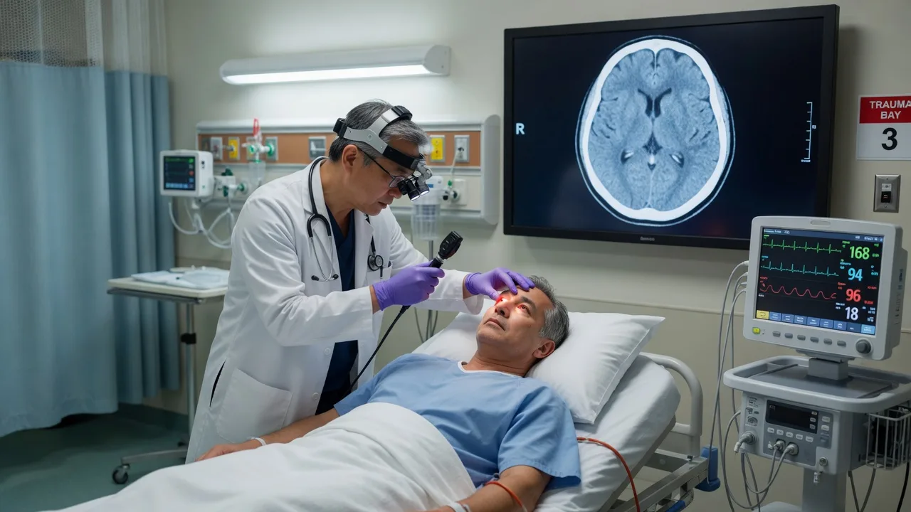
The headache is one of the commonest presentations to the emergency department, and the vast majority are benign primary headaches (the tension, the migraine, the cluster). The challenge is to identify the small but dangerous minority — the secondary headaches from the serious intracranial or systemic causes — and to investigate and treat them time-critically. The Fellowship candidate must know the red-flag mnemonic (SNNOOPS/SNNOPT10), the specific dangerous causes (the SAH, the meningitis, the giant cell arteritis, the venous sinus thrombosis, the CO poisoning), and the diagnostic pathway for each.[1][2]
The SNNOOPS mnemonic
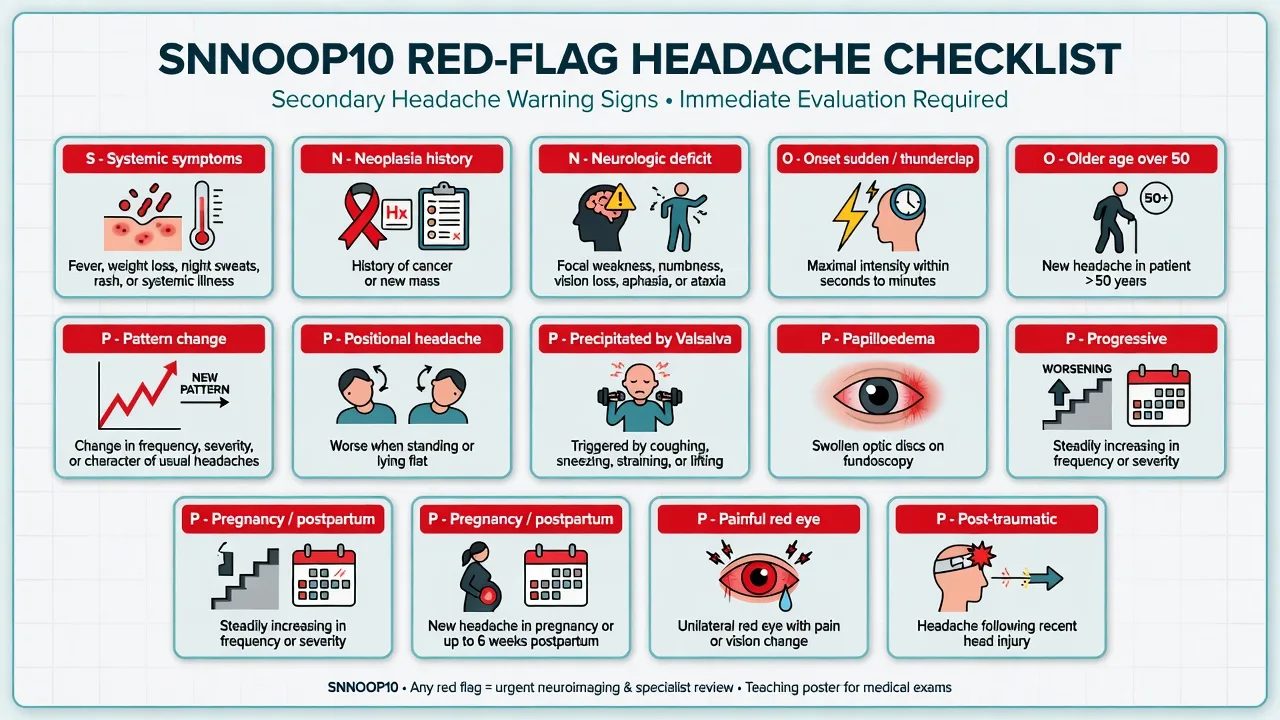
The SNNOOPS (or the newer SNNOPT10) is the checklist for the red flags that identify the secondary headache: [1]
SNNOOP10 — the red-flag headache checklist
Any positive red flag triggers the focused investigation for the specific dangerous cause. [1]
The dangerous secondary causes — the differential
SAH
- Thunderclap, occipital, maximal in seconds
- The neck stiffness, the photophobia, the brief loss of consciousness
- CT first, LP for xanthochromia if negative
- Nimodipine 60 mg q4h, BP control, urgent coiling or clipping
Meningitis
- Fever, headache, neck stiffness over hours
- The petechial rash of meningococcaemia
- CT then LP; empirical ceftriaxone 2 g IV
- Dexamethasone 10 mg IV for the pneumococcal
Giant cell arteritis
- Age over 50, the temporal headache, the jaw claudication
- The visual loss is the threat; the ESR over 50, the CRP raised
- Prednisolone 40 to 60 mg daily started immediately
- Temporal artery biopsy within 2 weeks
Venous sinus thrombosis
- The progressive headache, the papilloedema, the focal deficit
- The pregnant, the postpartum, the dehydrated, the prothrombotic
- MR or CT venography is the diagnostic test
- Anticoagulation even if the haemorrhagic infarct is present
CO poisoning
- The winter, the heater, the multiple family members
- The headache, the nausea, the confusion; the cherry-red skin is rare
- The carboxyhaemoglobin level confirms; high-flow oxygen
- Hyperbaric oxygen for the severe or the pregnant
Idiopathic intracranial hypertension
- Young, overweight woman; daily headache, the visual obscurations
- The papilloedema, the normal CT, the raised opening pressure on LP
- Acetazolamide 500 mg twice daily; the weight loss
- The neuro-ophthalmology referral for the visual-field monitoring
The diagnostic approach
The history identifies the red flags (the SNNOOPS). The examination includes the vital signs (the fever, the BP), the neurological (the GCS, the pupils, the focal deficit, the papilloedema), the temporal artery (the tenderness, the thickening, the reduced pulse in the suspected GCA), and the neck (the stiffness, the Kernig and the Brudzinski). The non-contrast CT is performed for any thunderclap headache, any focal deficit, any papilloedema, any altered consciousness, and any age over 50 with a new headache. The lumbar puncture (for the xanthochromia, the opening pressure, the cell count, the protein, the glucose) is performed after the CT for the suspected SAH (if the CT is negative or delayed), the suspected meningitis (after the CT), and the suspected IIH (to measure the opening pressure). The ESR and the CRP are checked for the suspected GCA. The carboxyhaemoglobin is checked for the suspected CO poisoning. The CT or MR venography is performed for the suspected venous sinus thrombosis.[1][2]
Management — the drug doses for the specific causes
The treatment is cause-specific: ceftriaxone 2 g IV plus vancomycin 1 g IV plus dexamethasone 10 mg IV for the bacterial meningitis; aciclovir 10 mg/kg IV three times daily for the HSV encephalitis; prednisolone 40 to 60 mg PO immediately for the giant cell arteritis (do NOT wait for the biopsy); nimodipine 60 mg PO every 4 hours for the SAH; acetazolamide 500 mg PO twice daily for the idiopathic intracranial hypertension; high-flow oxygen 100 per cent via the non-rebreather mask for the CO poisoning; and anticoagulation (the heparin then the warfarin) for the venous sinus thrombosis. The sumatriptan 50 to 100 mg PO (or the nasal spray 20 mg) for the acute migraine (not in the coronary disease, the pregnancy, or the uncontrolled hypertension). The oxygen 100 per cent via the non-rebreather mask at 12 to 15 L/min for 15 minutes for the cluster headache. [1]
The specific drug doses
The primary headaches — the benign differential
The primary headaches are the commonest cause of the ED presentation with the headache. The migraine is the unilateral, the throbbing, the moderate-to-severe headache with the photophobia, the phonophobia, the nausea, and the aura; treated with the triptan (sumatriptan 50 to 100 mg PO) and the antiemetic (metoclopramide 10 mg IV). The tension headache is the bilateral, the pressing, the mild-to-moderate band-like headache without the nausea or the photophobia; treated with the simple analgesia (paracetamol 1 g, ibuprofen 400 mg). The cluster headache is the unilateral, the severe, the orbital headache with the lacrimation and the nasal congestion, lasting 15 to 180 minutes, in the bouts (the cluster periods); treated with the 100 per cent oxygen at 12 to 15 L/min for 15 minutes and the sumatriptan 6 mg subcutaneously. [1]
The trigeminal autonomic cephalalgias (the cluster headache, the paroxysmal hemicrania, the SUNCT) share the unilateral pain and the autonomic features (the lacrimation, the conjunctival injection, the nasal congestion, the ptosis, the miosis). The paroxysmal hemicrania responds to the indomethacin 25 to 50 mg three times daily (the diagnostic response); the SUNCT and the SUNA are refractory and need the neurology referral for the lamotrigine or the IV lidocaine. The medication-overuse headache (the chronic daily headache from the overuse of the triptans, the opioids, or the combination analgesics on more than 10 days per month) is managed by the withdrawal of the causative drug, the bridge therapy, and the prophylactic agent (the propranolol, the amitriptyline, the topiramate). The cervicogenic headache (from the cervical spine, the occipital neuralgia) is managed with the nerve block (the greater occipital nerve block with the local anaesthetic and the steroid). [1]
Special populations
The pregnant patient with the headache is at a higher risk of the pre-eclampsia (the BP, the proteinuria, the hyperreflexia), the venous sinus thrombosis (the hypercoagulable state), and the pituitary apoplexy (the sudden severe headache with the visual change in the late pregnancy or the postpartum). The elderly patient with the new headache is at a higher risk of the giant cell arteritis, the subdural haematoma (from the minor trauma or the fall, often unrecalled), the intracerebral haemorrhage (from the hypertension or the amyloid angiopathy), and the metastatic disease. The immunocompromised patient (the HIV, the transplant, the chemotherapy) with the headache has the opportunistic infection (the cryptococcal meningitis, the toxoplasmosis, the CMV encephalitis) until proven otherwise. The anticoagulated patient with the new headache has the intracranial haemorrhage until the CT proves otherwise. [1]
Pitfalls and the missed diagnosis
The recurring errors are: dismissing the thunderclap headache as a migraine without the CT and the LP (this is the commonest missed diagnosis of the SAH); attributing the fever and the neck stiffness to a viral illness without the LP (the missed meningitis is the preventable death); not checking the ESR and CRP in the over-50-year-old with a new headache (the missed GCA causes the irreversible blindness); attributing the CO poisoning to a flu (the winter, the multiple family members, the pet that is also unwell — the CO detector in the home is the clue); and attributing the postpartum or the pregnancy headache to a tension headache without the venography (the missed venous sinus thrombosis). The positional headache (worse on standing, better on lying) suggests the low-CSF-pressure headache (from a dural tear after the LP, the epidural, or the trauma — treated with the caffeine, the bed rest, and the epidural blood patch); the positional headache (worse on lying, better on standing) suggests the raised intracranial pressure (the tumour, the IIH, the hydrocephalus). The precipitated-by-Valsalva headache (the cough, the exertion, the sneeze, the sexual activity) suggests the Chiari malformation or the posterior fossa lesion. The new daily persistent headache (the headache that is daily from the onset, in the patient with no headache history) warrants the neuroimaging and the LP to exclude the secondary cause. [1]
Evidence and regional guidelines
The contemporary framework is the IHS ICHD-3 classification for the headache types (the International Headache Society, updated in 2018), the SNNOOPS mnemonic for the red flags (the structured checklist derived from the American Headache Society and the International Headache Society guidelines), the CT then LP for the suspected SAH (the CT sensitivity over 98 per cent within 6 hours; the LP for the xanthochromia if the CT is negative or the presentation is delayed), and the ESR and CRP for the suspected giant cell arteritis (the sensitivity of the combined ESR and CRP is over 95 per cent; the temporal artery ultrasound is increasingly used as the first-line imaging). The neuroimaging is not needed for the headache without any red flags (the primary headache), but is mandatory for any positive SNNOOPS item.[1][2]
ANZ practice note. The red-flag headache is managed with the SNNOOPS checklist, the CT for the structural exclusion, the LP for the SAH and the meningitis, the ESR and CRP for the GCA, and the cause-specific treatment. The empirical ceftriaxone before the LP if the delay is over 1 hour is the standard. [1]
The ED diagnostic pathway — the structured approach
The emergency physician's task in the headache presentation is not to diagnose the migraine (the default) but to exclude the dangerous secondary cause through a structured, reproducible pathway. The history is the highest-yield tool — the temporal profile (the thunderclap, the progressive, the positional), the associated features (the fever, the neck stiffness, the focal deficit, the visual change), and the patient context (the age, the pregnancy, the immunocompromise, the anticoagulation) direct every subsequent decision. The examination confirms or refutes the suspected cause: the vital signs (the fever, the hypertensive crisis, the hypertensive encephalopathy), the neurological (the GCS, the pupils, the focal deficit, the meningeal signs, the papilloedema), and the targeted system (the temporal artery, the painful red eye, the skin rash). The investigation is then tailored — the non-contrast CT for the structural exclusion, the LP for the SAH and the meningitis, the ESR and CRP for the GCA, the venography for the venous sinus thrombosis, the carboxyhaemoglobin for the CO poisoning.[11]
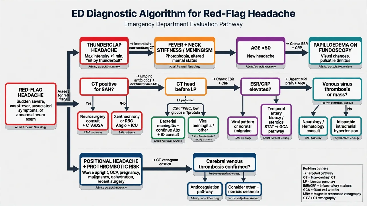
The ED diagnostic pathway for the red-flag headache
- Triage and the ABCDE. The airway, the breathing, the circulation, the disability (the GCS, the pupils), the exposure. The febrile and the altered, the thunderclap, and the focal deficit are the immediate high-acuity presentations.
- The history — the SNNOOP10. The onset (the thunderclap, the progressive, the positional), the character (the throbbing, the pressing, the stabbing), the severity (the worst-of-life), the pattern (the new, the changed, the progressive), the triggers (the Valsalva, the exertion, the sexual activity), and the associated features (the fever, the photophobia, the visual change, the neck stiffness).
- The examination. The vital signs, the full neurological (the GCS, the cranial nerves, the motor, the sensory, the coordination, the gait), the fundoscopy (the papilloedema), the temporal arteries (the tenderness, the thickening, the reduced pulse), the neck (the stiffness, the Kernig, the Brudzinski), the skin (the petechiae, the rash), and the painful eye (the red eye, the fixed pupil of the angle-closure glaucoma).
- The decision to image. Any positive SNNOOP10 item → the non-contrast CT. The thunderclap, the focal deficit, the papilloedema, the altered consciousness, the over-50 new headache, the HIV, the cancer, the anticoagulation, the post-traumatic, the pregnancy or the postpartum.
- The LP after the CT. For the suspected SAH (the xanthochromia, the opening pressure) if the CT is negative or the presentation is over 6 hours; for the suspected meningitis (the cell count, the protein, the glucose, the Gram stain, the culture) after the CT; for the suspected IIH (the raised opening pressure with the normal CT). The empirical antibiotics are given before the LP if the delay is over 1 hour.
- The targeted bloods. The ESR and the CRP for the suspected GCA; the carboxyhaemoglobin for the CO poisoning; the coagulation, the platelets, and the D-dimer for the suspected venous sinus thrombosis; the beta-hCG for the woman of reproductive age (the pre-eclampsia, the molar pregnancy).
- The disposition. The SAH → the neurosurgery and the ICU. The meningitis → the admission and the isolation. The GCA → the immediate steroids and the ophthalmology. The venous sinus thrombosis → the stroke unit and the anticoagulation. The CO poisoning → the high-flow oxygen and the disposition by the severity.
The specific dangerous causes — the detailed differential
Subarachnoid haemorrhage — the thunderclap
The subarachnoid haemorrhage is the ruptured berry aneurysm (the 85 per cent) or the non-aneurysmal perimesencephalic bleed (the 10 per cent), the arteriovenous malformation, or the traumatic bleed. The classic presentation is the thunderclap headache — the maximal intensity reached within seconds to one minute, often the occipital, the "worst headache of my life", with the neck stiffness, the photophobia, the nausea, the vomiting, and the brief loss of consciousness at the onset. The sentinel bleed (the warning leak) is the small SAH that resolves spontaneously but precedes the major rupture by days to weeks — the diagnosis is often missed as a migraine or a tension headache. The Ottawa SAH rule identifies the high-risk features for the CT: the age over 40, the neck pain or stiffness, the witnessed loss of consciousness, the onset during exertion, the thunderclap, and the limited neck flexion on the examination. The 6-hour CT rule — the non-contrast CT performed within 6 hours of the onset has the sensitivity of 100 per cent and the specificity of 100 per cent for the SAH (the third-generation scanner, the experienced radiologist); beyond 6 hours, the sensitivity declines to about 90 per cent by day 3, and the LP for the xanthochromia is required.[3][4][5]
The LP must be performed at least 12 hours after the onset (for the bilirubin — the haemoglobin breakdown product — to form) and within 2 weeks (after which the xanthochromia fades). The CSF is assessed for the xanthochromia (the yellow-tinged supernatant from the bilirubin, measured by the spectrophotometry — the visual inspection is unreliable), the red cell count (the stable count across the three bottles distinguishes the true SAH from the traumatic tap), and the opening pressure. The RCVS (the reversible cerebral vasoconstriction syndrome) is the important mimic — the thunderclap headache with the normal CT and the normal LP, but the segmental cerebral artery vasoconstriction on the angiography (the MRA or the CTA at 7 to 14 days); the RCVS is treated with the verapamil or the nimodipine and the avoidance of the vasoconstrictive drugs (the triptans, the SSRIs, the sympathomimetics).[12]
Perry et al, JAMA 2013 — the Ottawa SAH rule
PMID 24065011
Population: 2,131 alert neurologically intact adults with the non-traumatic headache peaking within 1 hour
Key finding
The Ottawa SAH rule (the age ≥40, the neck pain or stiffness, the witnessed loss of consciousness, the onset during exertion, the thunderclap, the limited neck flexion) — the sensitivity 100 per cent (95% CI 97.2–100), the specificity 27.7 per cent. The rule is designed to be the 100 per cent sensitive sieve for the CT decision.
Perry et al, BMJ 2011 — the 6-hour CT rule
PMID 21768192
Population: 593 patients with the acute headache peaking within 1 hour and the normal neurological examination
Key finding
The CT sensitivity 100 per cent (95% CI 99.0–100), the specificity 100 per cent (95% CI 98.9–100) for the SAH when performed within 6 hours on the third-generation scanner. The LP may be safely omitted in this subgroup.
Bacterial meningitis — the fever and the neck stiffness
The bacterial meningitis is the infection of the meninges by the Streptococcus pneumoniae (the commonest in the adult), the Neisseria meningitidis (the young adult, the petechial rash, the rapid sepsis), the Listeria monocytogenes (the elderly, the immunocompromised, the pregnant — cover with the amoxicillin or the ampicillin), and the Haemophilus influenzae (the post-splenectomy, the unvaccinated). The classic triad is the fever, the neck stiffness, and the altered mental status (the full triad in only about 44 per cent; the fever and the headache in 95 per cent; the neck stiffness in 70 per cent). The petechial or the purpuric rash of the meningococcaemia (the 30 to 50 per cent of the meningococcal meningitis) is the diagnostic clue — the rapidly progressive purpura with the septic shock is the medical emergency. The CT before the LP is performed for the immunocompromised, the new-onset seizure, the papilloedema, the altered consciousness, the focal deficit, and the known CNS disease; the empirical antibiotics (the ceftriaxone 2 g IV, the vancomycin 1 g IV, the amoxicillin 2 g IV for the Listeria cover in the over-50 and the immunocompromised) and the dexamethasone 10 mg IV (before or with the first antibiotic dose, for the suspected pneumococcal) are given before the CT if the delay is over 1 hour — the time to the antibiotic is the major determinant of the mortality.[6]
The HSV encephalitis is the important differential of the fever-and-the-altered-consciousness presentation — the temporal-lobe features (the aphasia, the hallucinations, the behavioural change), the CSF with the lymphocytic pleocytosis and the raised protein, and the aciclovir 10 mg/kg IV three times daily for 14 to 21 days (started empirically if the HSV PCR is pending). The empirical cover for the HSV is added to the meningitis regimen when the encephalitic features (the altered consciousness, the seizure, the focal deficit) are present. [1]
Giant cell arteritis — the over-50 new headache
The giant cell arteritis (the temporal arteritis, the GCA) is the granulomatous vasculitis of the medium and the large arteries (the temporal, the ophthalmic, the vertebral) in the over-50-year-old. The presentation is the new temporal headache (the unilateral, the tender scalp, the thickened pulseless temporal artery), the jaw claudication (the pain in the jaw on prolonged chewing that resolves with the rest — the most specific symptom), the polymyalgia rheumatica (the shoulder and the hip girdle stiffness), the visual symptoms (the amaurosis fugax, the diplopia, the visual loss from the anterior ischaemic optic neuropathy), and the systemic features (the fever, the weight loss, the malaise). The ESR is typically over 50 mm/h (but may be normal in up to 7 per cent — the " occult GCA"); the CRP is more sensitive and the combined ESR and CRP has the sensitivity over 95 per cent. The prednisolone 40 to 60 mg daily is started immediately on the clinical suspicion — do NOT wait for the temporal artery biopsy (the biopsy is performed within 2 weeks and may be positive for up to 2 to 4 weeks after the steroid start). The high-dose IV methylprednisolone 500 mg to 1 g daily for 3 days is given for the visual symptoms (the threat of the irreversible blindness). The temporal artery ultrasound (the halo sign) and the MRI are the non-invasive alternatives to the biopsy, increasingly recommended as the first-line imaging.[10]
Pituitary apoplexy — the sudden severe headache with the ophthalmoplegia
The pituitary apoplexy is the sudden haemorrhage or the infarction of the pituitary adenoma — the sudden severe headache (the thunderclap), the ophthalmoplegia (the third-nerve palsy — the ptosis, the diplopia, the dilated pupil — from the lateral expansion into the cavernous sinus), the visual field defect (the bitemporal hemianopia from the superior compression of the optic chiasm), and the acute adrenal insufficiency (the hypotension, the hyponatraemia from the acute cortisol deficiency — the stress dose hydrocortisone 100 mg IV is the immediate treatment). The MRI is the diagnostic test (the haemorrhage in the pituitary mass); the CT may miss it. The management is the urgent endocrine assessment (the cortisol, the TFTs, the prolactin, the growth hormone, the IGF-1), the stress-dose hydrocortisone 100 mg IV, and the surgical decompression for the severe or the progressive visual loss within 7 to 14 days (the conservative management for the mild or the improving visual deficit). The differential is the SAH (the thunderclap, the normal pituitary) and the cavernous sinus thrombosis (the ophthalmoplegia, the proptosis, the chemosis, the fever).[7]
Cerebral venous sinus thrombosis — the progressive headache in the prothrombotic
The cerebral venous sinus thrombosis (the CVST) is the thrombosis of the dural venous sinuses (the superior sagittal, the transverse, the sigmoid) or the cortical veins. The presentation is the progressive subacute headache (over days to weeks, the raised intracranial pressure), the papilloedema, the focal deficit (the motor, the sensory, the aphasia from the venous infarct), the seizure, and the altered consciousness. The risk factors are the pregnancy and the puerperium (the hypercoagulable state — the CVST is the commonest cerebrovascular complication of the pregnancy and the postpartum), the oral contraceptive pill, the dehydration, the prothrombotic state (the protein C, the protein S, the antithrombin deficiency, the antiphospholipid syndrome, the factor V Leiden, the prothrombin gene mutation), the malignancy, the infection (the otitis, the mastoiditis, the sinusitis), and the trauma. The MR venography or the CT venography is the diagnostic test — the empty delta sign (the non-opacification of the superior sagittal sinus on the contrast CT) is the classic finding. The anticoagulation (the heparin then the warfarin, or the DOAC) is given even if the haemorrhagic venous infarct is present (the haemorrhage is from the venous congestion, and the anticoagulation restores the venous drainage).[1][9]
Carbon monoxide poisoning — the winter headache
The carbon monoxide poisoning is the inhalation of the CO from the faulty heater, the blocked flue, the car exhaust, or the house fire. The CO binds the haemoglobin with the 200 to 250 times the affinity of the oxygen, causing the cellular hypoxia. The presentation is the headache (the commonest symptom), the nausea, the vomiting, the dizziness, the confusion, the flu-like symptoms (the misdiagnosis of the CO poisoning as the flu is the classic pitfall), and in the severe case the seizures, the coma, and the cardiac arrest. The cherry-red skin is the rare classic sign (the carboxyhaemoglobin gives the skin the pink colour, but the cyanosis is more common). The multiple family members affected (and the pet that is also unwell) is the critical clue — the CO poisoning is an environmental, not an individual, diagnosis. The carboxyhaemoglobin level confirms (the venous sample, the normal pulse oximetry is misleading as the COHb absorbs the same wavelength as the oxyhaemoglobin). The treatment is the high-flow 100 per cent oxygen via the non-rebreather mask (reduces the half-life of the COHb from 4 to 5 hours to 40 to 80 minutes) and the hyperbaric oxygen for the severe (the coma, the seizure, the syncope, the pregnancy, the cardiac ischaemia, the COHb over 25 per cent). [1]
The raised intracranial pressure — the mass and the hydrocephalus
The raised intracranial pressure from the mass lesion (the tumour, the abscess, the haematoma), the hydrocephalus, or the IIH presents with the progressive headache (worse in the morning, worse on lying, worse on coughing or the Valsalva), the papilloedema, the visual obscurations (the transient visual loss on the posture change), the sixth-nerve palsy (the false-localising sign — the diplopia from the stretched abducens nerve), and in the late stage the Cushing reflex (the hypertension, the bradycardia, the irregular respiration), the vomiting (the projectile, the effortless), and the altered consciousness. The non-contrast CT is the first-line test (the mass, the midline shift, the hydrocephalus, the haemorrhage); the MRI for the tumour characterisation. The idiopathic intracranial hypertension (the IIH) is the young, overweight woman with the daily headache, the pulsatile tinnitus, the visual obscurations, and the papilloedema — the normal CT (or the empty sella, the flattened posterior globe, the optic nerve sheath distension on the MRI), the raised opening pressure (over 25 cmH₂O on the LP), and the normal CSF composition. The acetazolamide 500 mg twice daily (up to 4 g daily) is the first-line treatment; the weight loss is the most effective long-term intervention. The visual-field monitoring is mandatory (the threat of the permanent visual loss from the chronic papilloedema).[8]
The hypertensive crisis and the posterior reversible encephalopathy syndrome
The hypertensive emergency with the headache is the malignant hypertension (the BP over 180/120 with the end-organ damage) — the hypertensive encephalopathy (the headache, the confusion, the visual change, the seizure), the posterior reversible encephalopathy syndrome (the PRES — the vasogenic oedema in the posterior parietal-occipital regions on the MRI, the reversible with the BP control), the intracerebral or the intraventricular haemorrhage, and the aortic dissection (the tearing chest-and-back pain, the pulse deficit, the BP differential). The BP control with the intravenous agent (the labetalol, the nicardipine, the clevidipine) is the treatment — the reduction by 10 to 20 per cent in the first hour, then to 160/100 over the next 2 to 6 hours (the over-rapid reduction causes the cerebral hypoperfusion and the stroke). The headache of the simple hypertension (without the end-organ damage) is not the hypertensive emergency — the BP and the headache may both be from the pain or the anxiety, and the over-aggressive treatment of the asymptomatic hypertension with the headache is the common pitfall. [1]
The cervicocephalic artery dissection — the neck pain and the Horner
The internal carotid artery dissection presents with the unilateral neck pain, the face pain, the Horner syndrome (the ptosis, the miosis, the anhidrosis — partial, from the disruption of the sympathetic chain), and the transient or the completed ischaemic stroke (the anterior circulation). The vertebral artery dissection presents with the posterior neck pain, the occipital headache, and the posterior circulation stroke (the wallenberg, the cerebellar, the vertebrobasilar). The trigger is the minor neck trauma (the chiropractic manipulation, the sports, the coughing, the roller coaster) in the young or the middle-aged patient (the dissection is the commonest cause of the stroke in the under-45-year-old). The CT angiography or the MR angiography is the diagnostic test. The management is the antithrombotic (the antiplatelet or the anticoagulation — the CADISS trial showed the equivalence), and the stroke-unit care. [1]
Angle-closure glaucoma — the painful red eye
The acute angle-closure glaucoma is the sudden rise in the intraocular pressure from the blocked aqueous outflow (the pupillary block in the patient with the anatomically narrow angle — the long-sighted, the elderly, the hypermetropic). The presentation is the severe unilateral eye pain (the headache), the red eye, the hazy cornea (the oedema), the fixed mid-dilated pupil (the ischaemic iris sphincter), the shallow anterior chamber, and the visual disturbance (the halos around the lights from the corneal oedema, the reduced acuity). The nausea and the vomiting are common and may misdirect the diagnosis to the abdominal cause. The intraocular pressure measurement (the tonometry — over 30 mmHg, often over 50 mmHg in the acute attack) confirms. The treatment is the acetazolamide 500 mg IV, the topical beta-blocker (the timolol), the topical alpha-2 agonist (the apraclonidine), the topical steroid, and the pilocarpine 2 per cent (after the pressure reduction — the ischaemic iris does not respond to the pilocarpine at the high pressure); the definitive treatment is the laser peripheral iridotomy. The differential is the cluster headache (the unilateral eye pain with the autonomic features, but the white eye and the normal pressure) and the optic neuritis (the painful eye movement, the reduced acuity, the relative afferent pupillary defect, the normal pressure). [1]
The diagnostic imaging — the modality for the question
The choice of the imaging modality depends on the suspected cause. The non-contrast CT is the first-line test for the thunderclap, the focal deficit, the papilloedema, the altered consciousness, and the over-50 new headache — it excludes the SAH, the intracerebral haemorrhage, the mass lesion, and the hydrocephalus. The CT angiography is added for the suspected aneurysm (the SAH) and the cervicocephical artery dissection. The CT venography or the MR venography is the test for the suspected venous sinus thrombosis. The MRI is the test for the tumour characterisation, the pituitary apoplexy, the posterior reversible encephalopathy syndrome, the cerebral abscess, the HSV encephalitis (the temporal-lobe signal change), and the chronic subdural. The lumbar puncture is the test for the SAH (the xanthochromia), the meningitis (the cells, the protein, the glucose, the Gram stain, the culture, the viral PCR), the IIH (the raised opening pressure), and the subarachnoid-pattern bleed that is over 6 hours old. The ESR and the CRP are the bloods for the GCA; the carboxyhaemoglobin for the CO poisoning. [1]
Non-contrast CT
- First-line for the thunderclap, the focal deficit, the papilloedema
- The SAH, the ICH, the mass, the hydrocephalus, the shift
- The 6-hour sensitivity 100 per cent for the SAH (Perry 2011)
- The limitation: misses the CVST, the small tumour, the dissection, the chronic subdural (MRI better)
CT venography / MR venography
- The test for the suspected CVST — the empty delta, the absent flow
- The progressive headache, the papilloedema, the pregnancy, the prothrombotic
- The MRV is the preferred (no radiation, the dural sinuses well seen)
- TheCTV is the faster alternative for the acutely unwell
MRI brain
- The tumour characterisation, the pituitary apoplexy, the PRES
- The HSV encephalitis (the temporal-lobe T2 hyperintensity)
- The chronic subdural (the isodense on CT, the bright on T1)
- The limitation: the longer scan time, the contraindication if the pacemaker
CT angiography
- The aneurysm in the confirmed SAH (the surgical planning)
- The cervicocephical artery dissection (the string sign, the flame)
- The moyamoya, the vasculitis (the beading)
- The RCVS (the segmental vasoconstriction — repeat at 7-14 days)
Lumbar puncture
- The xanthochromia for the SAH (≥12 hours, ≤2 weeks from the onset)
- The opening pressure for the IIH (over 25 cmH₂O) and the low-CSF-pressure
- The cells, protein, glucose, Gram stain, culture for the meningitis
- The HSV, the enterovirus, the VZV PCR for the viral encephalitis
ESR and CRP
- The combined sensitivity over 95 per cent for the GCA
- The ESR over 50 mm/h is the classic; the CRP is more sensitive
- The occult GCA (the normal ESR and CRP) is up to 7 per cent — treat on the clinical suspicion
- The temporal artery ultrasound (the halo sign) is the non-invasive adjunct
The lumbar puncture — the technique and the interpretation
The LP is performed at the L3–L4 or the L4–L5 interspace (the spinal cord ends at L1–L2 in the adult — Tuffier's line through the iliac crests marks the L4 spinous process). The opening pressure is measured in the lateral decubitus position with the legs extended (the prone position over-estimates) — the normal is 8 to 18 cmH₂O in the relaxed adult, up to 25 cmH₂O in the obese; the raised (over 25) is the IIH, the mass lesion, the venous sinus thrombosis, the hydrocephalus; the low (under 5) is the low-CSF-pressure headache (the dural tear, the post-LP leak). The CSF is collected in the four numbered bottles: bottle 1 for the cell count and the differential (the RBC, the WBC, the neutrophils, the lymphocytes); bottle 2 for the protein and the glucose (compare to the simultaneous serum glucose — the CSF glucose is normally 60 per cent of the serum); bottle 3 for the Gram stain, the culture, the viral PCR, the cryptococcal antigen; bottle 4 for the cell count (to compare with bottle 1 — the falling RBC across the bottles is the traumatic tap, the stable RBC is the true SAH) and the retain for the special tests (the cytology, the oligoclonal bands, the xanthochromia). The xanthochromia is the yellow-tinged supernatant from the bilirubin (the haemoglobin breakdown product, formed in vivo by the phagocytosis of the RBCs by the arachnoid cells, takes 12 hours to form, peaks at 48 hours, fades by 2 weeks) — measured by the spectrophotometry (the visual inspection in the white tube against the white background is the bedside method but is less reliable). [1]
Bacterial meningitis
- The neutrophilic pleocytosis (the polymorphs)
- The raised protein (over 1 g/L), the low glucose (under 40 mg/dL, under 60% of the serum)
- The Gram-positive diplococci (the pneumococcus), the Gram-negative diplococci (the meningococcus)
- The positive culture; the bacterial PCR (the pneumococcal, the meningococcal)
Viral meningitis / encephalitis
- The lymphocytic pleocytosis (the mononuclear)
- The mildly raised protein, the normal or mildly low glucose
- The HSV, the enterovirus, the VZV, the EBV PCR
- The negative Gram stain and the culture
Subarachnoid haemorrhage
- The stable RBC across the bottles (vs the falling of the traumatic tap)
- The xanthochromia (the bilirubin, ≥12 hours from the onset)
- The raised protein (from the RBC lysis), the normal glucose
- The xanthochromia fades by 2 weeks; the LP is unhelpful beyond 2 weeks
Tuberculous meningitis
- The lymphocytic pleocytosis with the neutrophils early
- The markedly low glucose (under 20 mg/dL), the raised protein
- The acid-fast bacilli on the Ziehl-Neelsen (rarely positive)
- The PCR for the M. tuberculosis; the culture takes 4-6 weeks
Fungal (cryptococcal)
- The lymphocytic pleocytosis (the immunocompromised, the HIV)
- The India ink (the classic), the cryptococcal antigen (the sensitive)
- The markedly raised pressure (the capsule blocks the arachnoid granulations)
- The culture on the Sabouraud agar; the antigen titre guides the treatment
Exam pearls
- SNNOOP10: Systemic, Neoplasia, Neurologic deficit, Onset sudden (thunderclap), Older (>50), Pattern change, Positional, Precipitated by Valsalva, Papilloedema, Progressive, Pregnancy, Painful eye, Post-traumatic.
- Thunderclap (maximal in seconds to 1 minute) = SAH until proven otherwise — the CT within 6 hours (100% sensitive), then the LP for the xanthochromia at 12 hours.
- Fever + neck stiffness = bacterial meningitis — the ceftriaxone 2 g IV (add amoxicillin for the Listeria in the over-50 and the immunocompromised) before the CT or LP if the delay is over 1 hour.
- Over-50 new headache = GCA — the ESR and CRP (combined sensitivity >95%), the prednisolone 40-60 mg before the biopsy; IV methylprednisolone for the visual symptoms.
- Jaw claudication (the pain on prolonged chewing) is the most specific symptom of the GCA — the headache, the polymyalgia, and the elevated ESR clinch the diagnosis.
- Pituitary apoplexy = thunderclap + ophthalmoplegia (third-nerve palsy) + bitemporal hemianopia — the cortisol is low; give the hydrocortisone 100 mg IV immediately.
- Venous sinus thrombosis = progressive headache + papilloedema in the pregnant, the postpartum, or the prothrombotic — the MR or CT venography is the diagnostic test; anticoagulate even if the haemorrhagic infarct.
- Cluster headache: 100 per cent oxygen at 12-15 L/min for 15 min + sumatriptan 6 mg SC — the trigeminal autonomic cephalalgia (the lacrimation, the conjunctival injection, the nasal congestion).
- CO poisoning: the winter, the heater, the multiple family members, the pet also unwell — check the venous carboxyhaemoglobin (the pulse oximetry is falsely normal); high-flow 100% oxygen.
- The positional headache: low-CSF-pressure (worse on standing, better on lying — the post-LP, the post-epidural, the dural tear) vs raised ICP (worse on lying, the morning, worse on the Valsalva).
- The Valsalva-precipitated headache (the cough, the sneeze, the exertion, the sexual activity) suggests the Chiari malformation, the posterior fossa lesion, or the RCVS.
- The carotid dissection = the unilateral neck/face pain + the Horner syndrome (the partial — ptosis, miosis, no anhidrosis) + the stroke; the vertebral dissection = the posterior neck pain + the posterior-circulation stroke.
- The angle-closure glaucoma = the painful red eye + the fixed mid-dilated pupil + the halos — check the intraocular pressure (the tonometry over 30 mmHg); give the acetazolamide 500 mg IV, the topical timolol, the laser iridotomy.
- The Ottawa SAH rule: age ≥40, the neck pain/stiffness, the witnessed LOC, the onset during exertion, the thunderclap, the limited neck flexion — the 100% sensitive sieve for the CT decision.
- The LP for the SAH: ≥12 hours from the onset (for the bilirubin), within 2 weeks; the xanthochromia by the spectrophotometry (not the visual inspection); the stable RBC across the bottles (vs the falling of the traumatic tap).
- The IIH: young, overweight woman, daily headache, pulsatile tinnitus, visual obscurations, papilloedema, normal CT, opening pressure over 25 cmH₂O — acetazolamide 500 mg BD, weight loss, the visual-field monitoring. [1]
Exam practice
SAQ — Thunderclap headache and the subarachnoid-haemorrhage workup
10 minutes · 10 marks
A 45-year-old woman presents with a sudden-onset, worst-of-life occipital headache that reached the maximal intensity within 30 seconds, with the neck stiffness and one episode of vomiting. GCS 15, afebrile, no focal deficit, normal fundoscopy. The CT is performed 3 hours after the onset.
SAQ — Giant cell arteritis with the threatened vision
10 minutes · 10 marks
A 68-year-old man presents with a two-week unilateral temporal headache, the jaw claudication on chewing, and a transient visual loss in the right eye this morning. He is afebrile, BP 142/86. ESR 88, CRP 64. The temporal artery is tender and pulseless on the right.
Red flags
[1]References
- [1]García-Azorín D, Monje MHG, González-García N, et al. Presence of red flags in patients with cerebral venous sinus thrombosis admitted to the emergency department because of headache: A STROBE compliant cohort-study Medicine (Baltimore), 2020.PMID 32702831
- [2]Jokubauskaite E, Bucaite A, Aukstakalnis V, et al. A Rare Cause of Headache in the Emergency Department: A Case Report J Emerg Med, 2025.PMID 40946370
- [3]Perry JJ, Stiell IG, Sivilotti ML, et al. Clinical decision rules to rule out subarachnoid hemorrhage for acute headache JAMA, 2013.PMID 24065011
- [4]Perry JJ, Stiell IG, Sivilotti ML, et al. Sensitivity of computed tomography performed within six hours of onset of headache for diagnosis of subarachnoid haemorrhage: prospective cohort study BMJ, 2011.PMID 21768192
- [5]van Gijn J, Rinkel GJ. Slip-ups in diagnosis of subarachnoid haemorrhage Lancet, 1997.PMID 9167456
- [6]Tunkel AR, Hartman BJ, Kaplan SL, et al. Practice guidelines for the management of bacterial meningitis Clin Infect Dis, 2004.PMID 15494903
- [7]Rajasekaran S, Vanderpump M, Baldeweg S, et al. UK guidelines for the management of pituitary apoplexy Clin Endocrinol (Oxf), 2011.PMID 21044119
- [8]Smith SV, Friedman DI. The Idiopathic Intracranial Hypertension Treatment Trial: A Review of the Outcomes Headache, 2017.PMID 28758206
- [9]Fam D, Saposnik G, Stroke Outcomes Research Canada Working Group. Critical care management of cerebral venous thrombosis Curr Opin Crit Care, 2016.PMID 26645556
- [10]Dejaco C, Ramiro S, Bond M, et al. EULAR recommendations for the use of imaging in large vessel vasculitis in clinical practice: 2023 update Ann Rheum Dis, 2024.PMID 37550004
- [11]Viera AJ, Antono B. Acute Headache in Adults: A Diagnostic Approach Am Fam Physician, 2022.PMID 36126007
- [12]Ducros A. Reversible cerebral vasoconstriction syndrome Lancet Neurol, 2012.PMID 22995694