EM · Acute ischaemic stroke
Acute ischaemic stroke
Also known as Acute ischemic stroke · Cerebral infarction · Acute stroke · Brain attack
Acute ischaemic stroke — sudden focal neurological deficit from arterial occlusion, the ischaemic penumbra and the rationale for permissive hypertension, recognition with FAST and the ROSIER score, non-contrast CT to exclude haemorrhage versus CT angiography to find the large-vessel occlusion, intravenous alteplase 0.9 mg/kg (maximum 90 mg, 10 per cent bolus) within 4.5 hours, mechanical thrombectomy within 6 hours or up to 24 hours with imaging mismatch, the BP thresholds and the exclusion criteria, and the mimics (Todd paresis, hypoglycaemia, hemiplegic migraine, functional). ACEM-primary, globally tagged.
On this page & tools
Your progress
Saved locally on this device.
8 MCQs with explanations
Target exams
Red flags
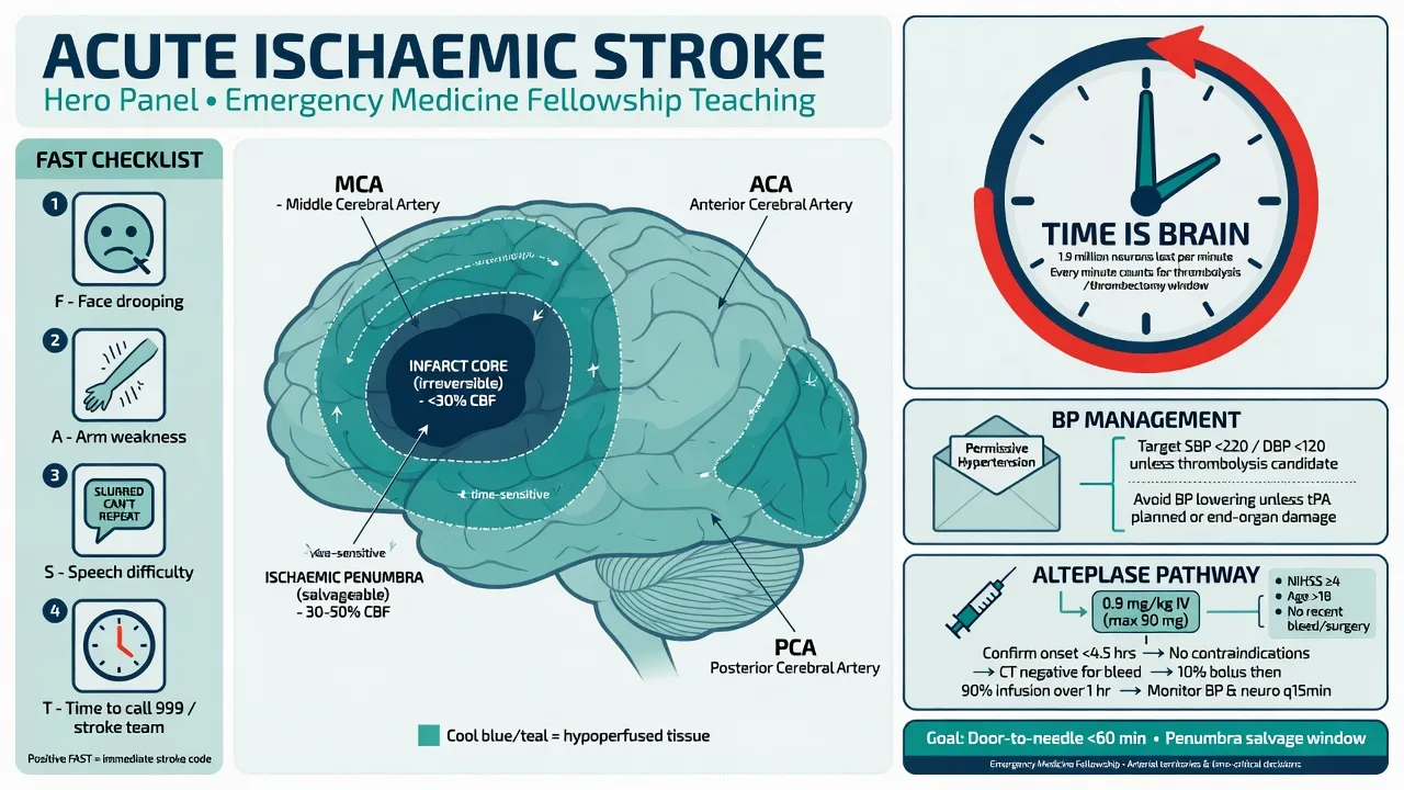
Acute ischaemic stroke is the sudden onset of a focal neurological deficit caused by interruption of arterial blood flow to a region of brain, producing an infarcted core of irreversibly dead tissue surrounded by a salvageable ischaemic penumbra — tissue that is dysfunctional but still alive, kept viable by collateral perfusion and the oxygen it carries. The entire emergency rests on one fact and one clock: the penumbra will die unless flow is restored, and it dies in minutes, not hours. The NINDS trial in 1995 showed that intravenous alteplase given within three hours restored that flow and improved outcome,[1] and ECASS III extended the window to four and a half hours.[2] Mechanical thrombectomy, validated by DAWN and DEFUSE 3, now salvages brain up to 24 hours after onset in selected patients with a perfusion mismatch.[3][4] For the Fellowship candidate the task is unambiguous: recognise the deficit at the door, exclude the mimics (above all hypoglycaemia and the postictal state), exclude haemorrhage on CT, hold the blood pressure within the safe envelope, and move the patient through thrombolysis and on to thrombectomy against a clock that does not pause.
Definition and classification
Stroke is defined by the World Health Organization as rapidly developing clinical signs of focal (at times global) disturbance of cerebral function lasting more than 24 hours (or leading to death) with no apparent cause other than a vascular origin. A transient ischaemic attack produces the same deficit resolving within 24 hours (and by modern tissue-based definition, without infarct on imaging); the distinction from stroke is one of duration and infarction, not of mechanism, and a TIA is a warning that demands urgent investigation rather than reassurance. Roughly 85 per cent of strokes are ischaemic and 15 per cent are haemorrhagic, a ratio that holds across ANZ, the United Kingdom and North America, and the first job of the emergency clinician is to separate them, because the treatments are opposite. [1]
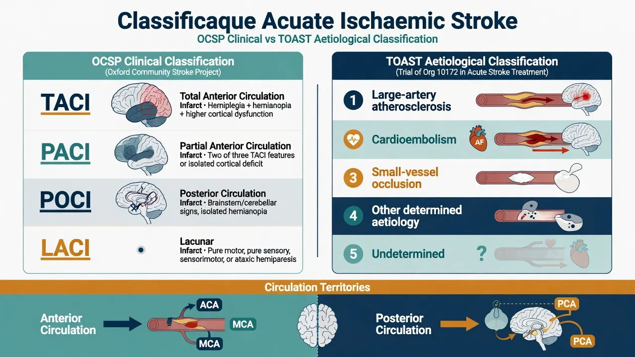
Ischaemic stroke is classified two ways. The Oxford Community Stroke Project classification divides by the clinical territory into total anterior circulation infarct (TACI — all of cortex, sensorimotor and higher function), partial anterior circulation infarct (PACI), posterior circulation infarct (POCI — brainstem, cerebellum, occipital) and lacunar infarct (LACI — pure motor, pure sensory or ataxic-hemiparesis from small deep vessel disease), and it predicts the underlying lesion and the prognosis at the bedside. The TOAST classification divides by aetiology into large-artery atherosclerosis, cardioembolism (atrial fibrillation being the dominant cause), small-vessel occlusion, other determined cause, and undetermined — and it directs the secondary-prevention workup. Both appear in vivas; the OCSP at triage, TOAST on the discharge summary. [1]
Epidemiology and risk factors
Stroke is the second commonest cause of death worldwide and a leading cause of adult disability, with incidence rising steeply with age. The non-modifiable risks are age, male sex, family history and prior stroke or TIA. The modifiable risks, in order of population-attributable importance, are hypertension, atrial fibrillation, smoking, diabetes, dyslipidaemia, obesity, physical inactivity and excess alcohol. Two are examined relentlessly because they are treatable and they change the acute plan: hypertension is the single biggest modifiable risk factor, and atrial fibrillation is the source of the cardioembolic clot that produces the most devastating large-vessel occlusions and dictates lifelong anticoagulation. Carotid stenosis, recent myocardial infarction (mural thrombus), patent foramen ovale in the young, sickle cell disease, and the hypercoagulable and vasculitic states complete the differential of cause. Identifying the mechanism is a secondary-prevention task, but it begins in the emergency department with the ECG and the history. [1]
Pathophysiology — the penumbra and why pressure matters
When a cerebral artery occludes, the territory it supplies falls into two zones. The infarct core is the region of blood flow below roughly 10 millilitres per 100 grams per minute, where energy failure is complete, neurons depolarise irreversibly, and cell death follows within minutes regardless of reperfusion. The penumbra surrounds it: blood flow between 10 and 20 millilitres per 100 grams per minute is enough to keep cells electrically silent but alive, and this tissue is the entire target of therapy — it will recover if flow is restored and it will die if it is not. The penumbra is metabolically precarious: it is maintained by collateral perfusion, and that collateral perfusion is pressure-dependent, which is the entire rationale for permissive hypertension. Lowering the blood pressure in acute ischaemic stroke (unless it is extreme, or unless thrombolysis demands it) drops the collateral pressure, shrinks the penumbra, and grows the infarct. [1]
[1]Clinical presentation
Stroke presents as the sudden onset of a focal deficit that maps to a vascular territory. An anterior-circulation middle-cerebral-artery occlusion — the commonest and the prototype — produces contralateral face and arm weakness (the arm worse than the leg, the cortical hand-over-foot gradient), contralateral sensory loss, a homonymous hemianopia, and, for dominant (left) hemisphere lesions, aphasia; for non-dominant lesions, neglect and anosognosia. An anterior-cerebral-artery occlusion produces leg-dominant weakness and abulia. A posterior-cerebral-artery occlusion produces a homonymous hemianopia with macular sparing. Posterior-circulation (brainstem, cerebellar) strokes are the dangerous atypical presentations: vertigo, diplopia, dysarthria, dysphagia, ataxia, crossed sensory or motor signs, a Horner syndrome, and the four Ds of vertebral dissection (sudden pain in the neck or head, then a posterior-circulation deficit). Lacunar strokes produce a pure motor, pure sensory, ataxic-hemiparesis or dysarthria-clumsy-hand syndrome with no cortical sign. A conscious-level drop is rare in a pure hemispheric stroke and should raise haemorrhage, brainstem stroke, seizure, or hypoglycaemia. [1]
Recognition — FAST and the ROSIER score
Pre-hospital recognition uses FAST — Face drooping, Arm weakness, Speech difficulty, Time — the public-facing tool that drives the ambulance bypass to a stroke centre. The extended BE-FAST adds Balance and Eyes (sudden vertigo or visual loss) and captures roughly 14 per cent more posterior-circulation strokes that FAST misses. [1]
In the emergency department the validated recognition tool is the ROSIER score (Recognition of Stroke in the Emergency Room).[5] It begins with a mandatory finger-prick glucose — hypoglycaemia mimics stroke exactly and must be excluded before any further assessment — and then scores seven bedside features. The two negative items weight against stroke; the five positive items weight for it.
ROSIER — Recognition of Stroke in the Emergency Room
ROSIER
Measure blood glucose and treat if low — hypoglycaemia mimics stroke and is reversed in minutes
If present, score -1 (weights against stroke)
If present, score -1 (weights against stroke — think postictal Todd paresis)
If present, score +1
If present, score +1
If present, score +1
If present, score +1 (aphasia or dysarthria)
If present, score +1 (homonymous hemianopia)
The score runs from minus 2 to plus 5. A score greater than zero is probable stroke (sensitivity around 92 per cent, specificity around 86 per cent in the derivation cohort), and these patients are fast-tracked to CT. A score of zero or below is a stroke mimic until proven otherwise, but the tool is a triage aid, not a rule-out — a convincing deficit with a low ROSIER score (for example a pure visual field loss with a posterior-circulation lesion) still warrants imaging. The NIH Stroke Scale (NIHSS, 0 to 42) is the structured severity measure that quantifies the deficit for the thrombolysis and thrombectomy decisions; a score of 6 or more with a large-vessel occlusion is the usual threshold to activate retrieval. [1]
Differential diagnosis
The mimics of stroke are the examiner's favourite trap, and the bedside job is to separate the true vascular deficit from its imitators by the history, the tempo, and the glucose. The single most dangerous mimic to miss is hypoglycaemia, which reproduces a focal deficit with complete fidelity and is reversed by glucose in minutes; the single most common mislabel is the postictal Todd paresis. [1]
Acute ischaemic stroke
- Sudden focal deficit maximal at onset, maps to a vascular territory
- Sustained; no rapid resolution; no seizure preceding
- Normal glucose; CT may be normal early, CTA may show occlusion
- Time-critical for thrombolysis/thrombectomy
Intracerebral haemorrhage
- Indistinguishable clinically from ischaemia — same focal deficit
- More often depressed conscious level, headache, vomiting, hypertensive
- Diagnosed ONLY on non-contrast CT (hyperdense mass, mass effect)
- Thrombolysis contraindicated — CT excludes it before any lytic
Hypoglycaemia
- Focal deficit reproduced exactly; may also cause confusion or coma
- Often in a diabetic on insulin or sulfonylurea
- Diagnosed by finger-prick glucose (typically below 2.5 mmol/L); reversed by IV glucose
- Must be excluded FIRST in every suspected stroke
Postictal Todd paresis
- Focal weakness after a seizure; resolves over hours
- History of seizure or epilepsy; may have been unwitnessed
- Glucose normal; CT often normal; EEG may show epileptiform activity
- A seizure at onset is a relative exclusion to thrombolysis unless residual deficit is clearly ischaemic
Hemiplegic migraine
- Aura with gradual spread over minutes; positive sensory or visual phenomena; family history
- Young patient; prior similar episodes; headache phase follows
- CT and CTA normal; diagnosis of exclusion
- Do not thrombolyse on history alone if any doubt — image first
Functional (non-organic) weakness
- Deficit does not fit a vascular territory; Hoover sign positive; give-way weakness
- Onset often after stress; fixed deficit; inconsistent on examination
- Imaging normal; diagnosis requires positive functional signs AND exclusion of organic disease
- Never assume functional before vascular is excluded
The discriminating moves at the bedside are: check the glucose; ask explicitly about a seizure in the preceding minutes to hours (including tongue-biting or incontinence); establish the tempo (stroke is maximal at onset, migraine marches, Todd paresis follows a seizure); and image every patient in whom stroke is a possibility, because the mimics overlap and a posterior-circulation lesion can be subtle. [1]
Investigations — CT, CT angiography, and glucose
The investigations run in parallel with resuscitation and against the clock. The first is the finger-prick glucose, done at the bedside before anything else. A 12-lead ECG screens for atrial fibrillation and an acute coronary syndrome (stroke and STEMI can coexist in the embolic patient). Bloods include a full blood count, coagulation screen (INR, aPTT), urea and electrolytes, and a group-and-save; troponin is sent but does not delay thrombolysis. Pregnancy testing is done in any woman of reproductive age. [1]
The pivotal imaging is non-contrast CT of the brain. Its purpose in the acute window is not to confirm ischaemia — early ischaemia is often invisible — but to exclude haemorrhage, which is the decision that permits or forbids thrombolysis. The early ischaemic signs to seek on plain CT within the first six hours are: the hyperdense MCA sign (the occluded middle cerebral artery seen as a bright dot or hyperdense band), loss of the insular ribbon, obscuration of the lentiform nucleus, loss of grey–white differentiation, and sulcal effacement. The ASPECTS (Alberta Stroke Program Early CT Score) grades these changes across ten territories (M1 to M6, caudate, lentiform, internal capsule, insula) starting at 10 and subtracting one per area of early ischaemia; a score of 7 or less indicates extensive early change, predicts poor outcome, and raises the bleeding risk with thrombolysis. [1]
Imaging thresholds and windows
CT angiography is the next layer and is now obtained in parallel in any patient with a suspected anterior-circulation deficit, to identify a large-vessel occlusion (intracranial internal carotid, M1 or basilar) that is amenable to mechanical thrombectomy. CT perfusion (or MRI diffusion–perfusion) maps the core against the penumbra and is the selection tool for the extended 6 to 24 hour thrombectomy window, defining a mismatch — a small irreversibly infarcted core with a large at-risk penumbra — that justifies retrieval beyond six hours.[3][4] MRI (diffusion-weighted imaging) is exquisitely sensitive to early ischaemia and is used for the wake-up stroke where the DWI–FLAIR mismatch (a lesion bright on diffusion but not yet on FLAIR) estimates onset within four and a half hours.
Immediate management — resuscitation and the time clock
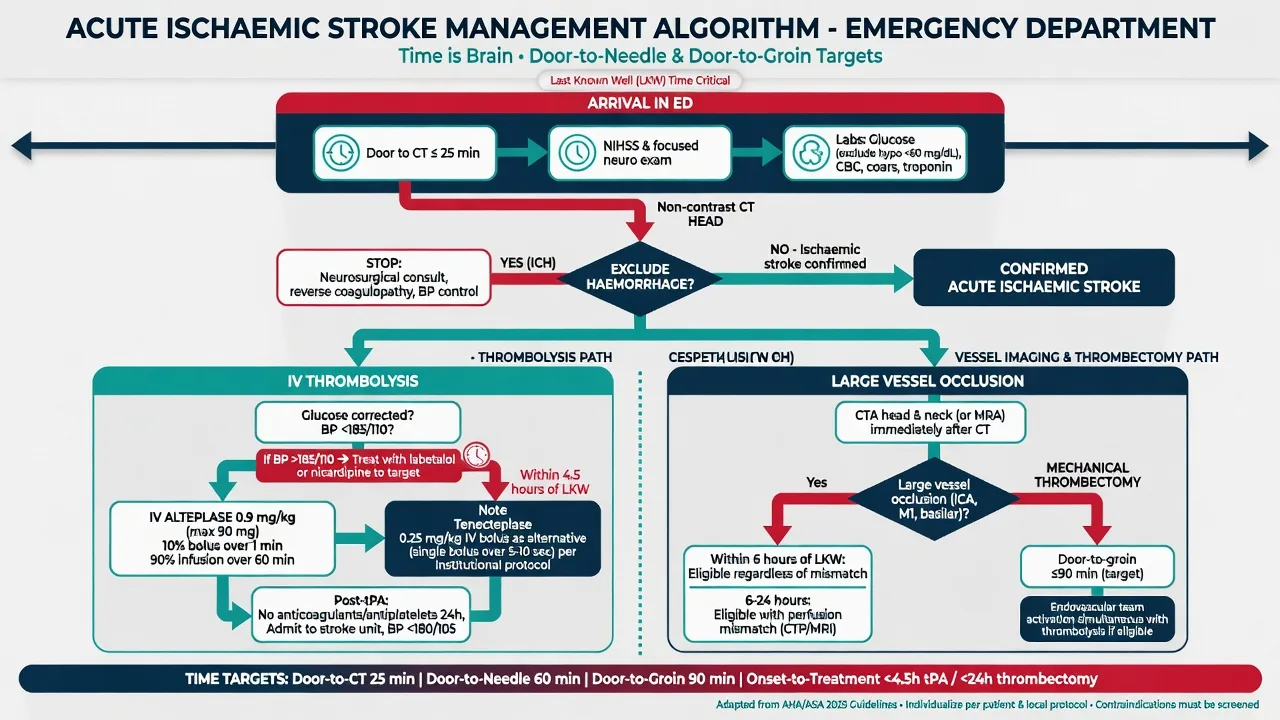
Resuscitation and the specific therapy are inseparable, and both obey the clock established at the door. Establish the last-known-well time with precision — not the time the patient was found collapsed, but the last moment they were seen normal — because this single fact decides every window. Notify the stroke team and retrieval service at the moment of suspicion, in parallel with assessment, not after it. Give oxygen only if oxygen saturation falls below 94 per cent (hyperoxia is harmful), secure intravenous access, keep the patient nil by mouth pending a swallow screen, and treat pain and agitation gently. A low finger-prick glucose is corrected with intravenous glucose (for example 25 to 50 mL of 50 per cent dextrose, or 100 mL of 10 per cent dextrose), never with hypotonic solutions. [1]
[1]Blood pressure control is the resuscitation manoeuvre most often examined. In the patient not receiving thrombolysis, permissive hypertension is the rule: the pressure is allowed to run high to support the penumbra, and is lowered only when it exceeds roughly 220 over 120, or in the setting of aortic dissection, acute myocardial infarction, acute pulmonary oedema, or a planned thrombolytic. In the patient receiving alteplase, the pressure must be brought under 185 over 110 before the bolus and held under 180 over 105 for 24 hours afterwards, because uncontrolled hypertension is the commonest cause of symptomatic intracranial haemorrhage after thrombolysis. Labetalol 10 to 20 mg intravenously every ten minutes to a maximum of 300 mg, or an infusion of 2 to 8 mg per minute, is first-line (it is alpha and beta balanced and does not raise intracranial pressure); nicardipine 5 to 15 mg per hour by infusion is the alternative and gives smooth titratable control. [1]
Definitive management — intravenous thrombolysis
Intravenous alteplase (recombinant tissue plasminogen activator) is the standard reperfusion therapy for ischaemic stroke within four and a half hours of onset. The NINDS trial established benefit within three hours[1] and ECASS III extended it to four and a half hours.[2] The dose is 0.9 mg per kilogram, to a maximum total dose of 90 mg, given as a 10 per cent bolus over one minute with the remainder infused over 60 minutes. No anticoagulant or antiplatelet is given for 24 hours afterwards, and the blood pressure is held under 180 over 105 throughout that day.
Tenecteplase at 0.25 mg per kilogram as a single five-second bolus is a non-inferior alternative validated in EXTEND-IA TNK and now preferred in several contemporary guidelines for anterior-circulation occlusion, because its single-bolus pharmacokinetics are simpler and it may produce better reperfusion before thrombectomy; the conventional agent examined at Fellowship is still alteplase, and the dose is the one to know cold. [1]
The extra exclusions in the 3 to 4.5 hour window (ECASS III)
The exclusion criteria for alteplase are extensive and are grouped into absolute and relative. The absolute exclusions are: intracranial haemorrhage on CT; subarachnoid haemorrhage or its clinical suspicion (thunderclap headache); serious head trauma or stroke in the previous three months; intracranial or intraspinal surgery in the previous three months; intracranial neoplasm, arteriovenous malformation or aneurysm; active internal bleeding; a known bleeding diathesis — platelets below 100,000, INR above 1.7, or heparin within 48 hours with a raised aPTT; current use of a direct oral anticoagulant within 48 hours (or with an unknown last-dose time); and blood pressure persistently above 185 over 110 despite treatment. The relative exclusions are recent major surgery or serious non-head trauma within 14 days, gastrointestinal or urinary bleeding within 21 days, myocardial infarction within three months, arterial puncture at a noncompressible site within seven days, seizure at onset (where the residual deficit may be postictal), and glucose below 2.7 or above 22 mmol per litre (correct and reassess). Pregnancy is a relative consideration: alteplase may still be given in pregnancy where the benefit outweighs the bleeding risk, in consultation with obstetrics. [1]
Definitive management — endovascular thrombectomy
Mechanical thrombectomy is now first-line for anterior-circulation large-vessel occlusion, and the evidence transformed practice after 2015. The principle is that an intracranial internal-carotid or M1 occlusion is often refractory to intravenous alteplase alone, and that mechanical retrieval restores flow in a majority within minutes of reperfusion. [1]
The windows are: within six hours of onset, a patient with a large-vessel occlusion, NIHSS of 6 or more and ASPECTS of 6 or more benefits from thrombectomy without the need for perfusion imaging, established by MR CLEAN, ESCAPE, EXTEND-IA, SWIFT PRIME and REVASCAT. Beyond six hours, two trials extended the window on the basis of imaging mismatch. DAWN showed benefit from thrombectomy at 6 to 24 hours in patients selected by a clinical–imaging mismatch (a small core on CT perfusion or MRI relative to the severity of the deficit),[3] and DEFUSE 3 confirmed benefit at 6 to 16 hours in patients selected by a perfusion–core mismatch (a small core with a penumbra at least 1.8 times the core).[4] The practical rule: a salvageable-mismatch patient is a retrieval patient up to 24 hours, and the emergency department's job is to obtain CT angiography and CT perfusion early enough to activate the neuroradiology team within that window. Alteplase is given first (the bridge) if the patient is within 4.5 hours, and thrombectomy follows; thrombectomy alone (without alteplase) is used for those outside the lytic window but within the retrieval window.
Blood-pressure management and the exclusion criteria revisited
The blood-pressure targets deserve their own emphasis because they recur in every exam form. Before alteplase: under 185 systolic and 110 diastolic, lowered if necessary with labetalol 10 to 20 mg intravenously every ten minutes (maximum 300 mg) or nicardipine 5 to 15 mg per hour. During and for 24 hours after alteplase: under 180 over 105. In the patient not receiving alteplase: permissive hypertension up to 220 over 120, lowered only for end-organ threats (dissection, infarction, pulmonary oedema) or to permit thrombolysis. After thrombectomy: the target is debated, but most pathways hold a systolic below 180 to protect the reperfused territory from haemorrhagic transformation. A common error is to lower the pressure reflexively on admission in an untreated stroke — this shrinks the penumbra and grows the infarct and must be resisted unless an indication forces it. [1]
[1]Complications
The feared complication of thrombolysis is symptomatic intracranial haemorrhage (symptomatic ICH), occurring in roughly 2 to 6 per cent of treated patients and carrying a mortality around 50 per cent. The risk rises with large infarct volume (low ASPECTS), high NIHSS, uncontrolled blood pressure, hyperglycaemia, and the use of anticoagulants; it presents as sudden neurological deterioration, headache, vomiting, or a rise in blood pressure during or after the infusion, and is confirmed on an immediate repeat CT. Management is to stop the alteplase infusion, send a coagulation screen, give cryoprecipitate (to raise fibrinogen) and tranexamic acid or prothrombin complex, lower the pressure to under 140 systolic, and involve neurosurgery. [1]
Haemorrhagic transformation also occurs spontaneously in infarcted tissue (petechial and usually clinically silent, versus frank parenchymal haematoma). Cerebral oedema peaks at 48 to 72 hours and, in a complete middle-cerebral-arterity infarct, produces the malignant MCA syndrome — a swollen hemisphere that herniates and kills within days. Young patients (under 60) with a dominant or non-dominant malignant MCA infarct benefit from decompressive hemicraniectomy within 48 hours (DECIMAL, DESTINY, HAMLET), a time-critical neurosurgical referral. Aspiration pneumonia (from dysphagia), seizures, deep-vein thrombosis, pressure injury, and recurrence of stroke complete the complication set, and each is anticipated rather than waited for — a swallow screen before any oral intake, subcutaneous heparin once the bleeding risk has settled, and early secondary-prevention therapy (antiplatelet, statin, anticoagulation where atrial fibrillation is the cause). [1]
Prognosis and disposition
Outcome tracks the severity at presentation (NIHSS), the territory (posterior-circulation and complete anterior-circulation strokes do worse), the patient's age and comorbidity, and above all whether reperfusion was achieved. A patient who reperfuses early can recover remarkably; a patient who does not may be left with dense hemiparesis, aphasia or visual loss. Every acute-stroke patient is admitted to a stroke unit with monitored care and early mobilisation by a multidisciplinary team. Secondary prevention — antiplatelet therapy (for example aspirin 300 mg then 75 mg daily, or clopidogrel) for non-cardioembolic stroke, anticoagulation for atrial fibrillation, a high-intensity statin, blood-pressure control, carotid endarterectomy for symptomatic severe stenosis, and smoking cessation — is begun in the acute admission. Early supported discharge and rehabilitation are planned from the first day. [1]
Special populations
The wake-up stroke — a deficit present on waking, with an unknown onset — is routed to CT perfusion or MRI rather than assumed outside the window; a DWI–FLAIR mismatch or a favourable perfusion profile identifies patients within the lytic or retrieval window. The posterior-circulation and basilar occlusion patient may present with reduced consciousness and quadriparesis and is a thrombectomy emergency even though the anterior-circulation trials did not include them; basilar thrombolysis and retrieval carry the same time imperative. Pregnancy is a relative indication rather than a contraindication to alteplase — the maternal benefit usually outweighs the fetal bleeding risk, in consultation with obstetrics and stroke teams. The anticoagulated patient is excluded from alteplase unless reversed, but remains a retrieval candidate. Young patients (under 50) demand a search for a cause — patent foramen ovale, carotid or vertebral dissection, thrombophilia, vasculitis, drug use — because the mechanism changes prevention. Paediatric stroke is rare, is managed by a paediatric stroke protocol, and is outside the adult alteplase pathway. [1]
Evidence and regional guidelines
The evidence base is one of the strongest in acute medicine. NINDS (1995) established intravenous alteplase within three hours[1] and ECASS III (2008) extended it to four and a half hours.[2] The thrombectomy revolution came in 2015 (MR CLEAN, ESCAPE, EXTEND-IA, SWIFT PRIME, REVASCAT) for the within-six-hour window, and DAWN[3] and DEFUSE 3[4] extended it to 16 to 24 hours with imaging selection in 2018. The ROSIER score for emergency recognition is from Nor and colleagues.[5] Regionally, the AHA/ASA 2019 acute ischaemic stroke guideline sets the global standard; ANZ practice follows the Stroke Foundation of Australia Clinical Guidelines for Stroke Management; the United Kingdom follows the NICE and Royal College of Physicians/SSNAP pathways (where ROSIER originated); Europe follows the ESO guidelines. The agents, the windows, and the blood-pressure targets are global; the local stroke pathway governs the exact retrieval logistics and the tenecteplase-versus-alteplase choice.
ANZ practice note. Australian and New Zealand practice follows the Stroke Foundation Clinical Guidelines for Stroke Management and the local state-wide stroke service (for example the Victorian Stroke Tele Medicine and retrieval network). Tenecteplase 0.25 mg per kilogram is increasingly the first-line lytic in eligible anterior-circulation occlusion, alteplase remains the Fellowship-examined standard, and the retrieval window extends to 24 hours for mismatch-selected patients via the regional endovascular clot retrieval service. Every Australian ED runs a stroke triage bypass protocol that fast-tracks a suspected stroke to CT within minutes of arrival. [1]
The hyperacute ED stroke pathway — the time targets
The whole treatment of acute ischaemic stroke is engineered around a small set of time targets that govern the system. The patient does not flow through the emergency department — they are propelled. The targets exist because of the decay of the penumbra, and because every minute between the door and the needle destroys brain that was salvageable at the door. The American Heart Association and the American Stroke Association Target: Stroke programme, the Stroke Foundation of Australia, and the United Kingdom Sentinel Stroke National Audit Programme all converge on the same milestones, and the Fellowship candidate is expected to recite them. [1]
The ED stroke time targets (the clock starts at the door)
The ED stroke first 30 minutes — door-to-CT and door-to-needle
0 to 5 minutes — recognise and activate
Triage straight to the resuscitation bay on any FAST-positive or sudden focal deficit; check the finger-prick glucose at the bedside before the cannula. Declare a code stroke aloud — the single call mobilises the stroke team, the CT scanner, the radiographer, the pharmacy (alteplase drawn up), and, for a suspected large-vessel occlusion, the endovascular service, all in parallel. Two large-bore cannulae, full monitoring, oxygen only if the saturation is under 94 per cent.
5 to 15 minutes — the focused history and exam
The one fact that decides every window: the last-known-well time, never the time the patient was found down. Establish it from a witness by phone if necessary. Determine the onset tempo (maximal at onset equals stroke), the territory (face, arm, leg, speech, vision, posterior-circulation signs), the NIHSS, and screen the alteplase exclusions and the anticoagulant history. Perform the swallow screen and keep the patient nil by mouth.
15 to 25 minutes — the bloods and the CT slot
Draw the full blood count, the coagulation (INR and aPTT), the urea and electrolytes, the glucose, the troponin, and the group-and-save; obtain a 12-lead ECG (atrial fibrillation changes the cause and the prevention, and an acute STEMI coexists in the embolic patient). Move the patient to CT with the team and the monitor. The non-contrast CT excludes haemorrhage — the single question that decides the lytic.
25 to 45 minutes — CT, CT angiography, and the decision
Read the CT for haemorrhage and for the early ischaemic signs (the hyperdense MCA, the lost insular ribbon, the ASPECTS). Obtain the CT angiography in parallel for any anterior-circulation deficit to find the large-vessel occlusion; add the CT perfusion for the wake-up or the 6 to 24 hour candidate. Apply the inclusion and exclusion checklist against the last-known-well time.
45 to 60 minutes — door-to-needle
If eligible and within 4.5 hours, give alteplase 0.9 mg/kg (10 per cent bolus over 1 minute, the remainder over 60 minutes, maximum 90 mg) the moment the CT excludes blood. Do not wait for the bloods unless the coagulation, the platelet or the anticoagulant status is in doubt. Notify the retrieval service before the infusion ends.
Thrombolysis in detail — dose, preparation, the bolus and the hour
Intravenous alteplase is the prototype and the Fellowship-examined standard. The dose and its partition are examined as automatically as an adrenaline dose, and they must be recited exactly. Every line of the dose is a line the examiner can quote. [1]
Alteplase — the numbers to know cold
Alteplase preparation and administration — the exact sequence
Weigh the patient on the actual body weight and calculate: the total is 0.9 mg/kg, capped at 90 mg. Split it — the bolus is 10 per cent over 1 minute, the infusion is 90 per cent over 60 minutes.
Confirm the CT excludes haemorrhage, the glucose is above 2.7 mmol/L, the INR is under 1.7, the platelets are above 100, and that no anticoagulant (warfarin, heparin within 48 h, or a direct oral anticoagulant within 48 h or with an unknown last-dose) is in play. If any are unknown or abnormal, do not bolus — wait for the result, because the bleeding risk is irreversible.
Confirm the blood pressure is under 185 over 110; lower it first with labetalol 10 to 20 mg intravenously every 10 minutes (maximum 300 mg) or nicardipine 5 to 15 mg per hour if it is not.
Draw up the alteplase and split it into the 10 per cent bolus syringe and the 90 per cent infusion; label both clearly and double-check the calculation with a second clinician.
Give the bolus over 1 minute, start the infusion over 60 minutes at time zero, set the blood-pressure and the neurocheck schedule, and send for retrieval. Give no antiplatelet, no anticoagulant, and place no urinary catheter or nasogastric tube for 24 hours.
Stay with the patient for the first 15 minutes and watch for the orolingual angio-oedema (stop the infusion, give antihistamine, steroid, and adrenaline if the airway is threatened) and for any acute neurological deterioration (stop, urgent repeat CT).
Alteplase
- Recombinant tissue plasminogen activator — the Fellowship-examined standard
- 0.9 mg/kg, 10% bolus over 1 min plus 90% over 60 min, maximum 90 mg
- Window 0 to 4.5 h from last-known-well
- A short half-life (about 5 min) demands the full 1-hour infusion
- Validated by NINDS (0 to 3 h) and ECASS III (3 to 4.5 h)
Tenecteplase
- A genetically modified tPA with a longer half-life and higher fibrin specificity
- 0.25 mg/kg as a single 5-second bolus, maximum 25 mg
- The same 0 to 4.5 h window; non-inferior to alteplase
- Single-bolus pharmacokinetics simplify delivery and speed the door-to-needle
- Increasingly first-line in ANZ and many contemporary pathways; alteplase remains the dose to know for exams
Tenecteplase at 0.25 mg/kg as a single five-second bolus is a non-inferior alternative validated in EXTEND-IA TNK[11] and NOR-TEST,[12] and it is now the first-line lytic for the eligible anterior-circulation occlusion in several contemporary pathways because the single-bolus pharmacokinetics are simpler and may produce better reperfusion before thrombectomy. The conventional agent examined at Fellowship is still alteplase, and the dose is the one to know cold.
EXTEND-IA TNK part 2 — tenecteplase versus alteplase before thrombectomy
New England Journal of Medicine, 2018
A multicentre randomised trial of tenecteplase 0.25 mg/kg versus alteplase 0.9 mg/kg in 300 patients with a large-vessel occlusion within 4.5 hours, before the mechanical thrombectomy.
Key finding
Tenecteplase was non-inferior for substantial reperfusion before thrombectomy, with better reperfusion (22 per cent versus 10 per cent) and no excess symptomatic intracranial haemorrhage. It is faster to give as a single 5-second bolus.
Practice change
Tenecteplase is increasingly the first-line lytic in the eligible anterior-circulation occlusion; alteplase remains the Fellowship-examined standard.
NOR-TEST — tenecteplase versus alteplase for acute ischaemic stroke
Lancet Neurology, 2017
A phase 3, randomised, open-label, blinded-endpoint trial of tenecteplase 0.4 mg/kg versus alteplase 0.9 mg/kg in 1,200 patients within 4.5 hours of onset, across the Norwegian centres.
Key finding
No significant difference in the favourable functional outcome at 90 days, and a similar safety profile; the higher tenecteplase dose (0.4 mg/kg) was not superior.
Practice change
Supported tenecteplase as a practical non-inferior alternative, with 0.25 mg/kg the dose adopted for the anterior-circulation stroke.
Absolute exclusions to alteplase
- Intracranial haemorrhage on CT, or a clinical suspicion of subarachnoid haemorrhage (the thunderclap headache)
- A serious head trauma or a stroke in the previous 3 months
- An intracranial or intraspinal surgery in the previous 3 months
- An intracranial neoplasm, arteriovenous malformation or aneurysm
- An active internal bleeding
- A known bleeding diathesis — platelets below 100,000, an INR above 1.7, or heparin within 48 h with a raised aPTT
- A direct oral anticoagulant within 48 h (or an unknown last-dose time)
- A blood pressure persistently above 185 over 110 despite the treatment
- A glucose below 2.7 or above 22 mmol/L (correct and reassess)
Relative exclusions
- A recent major surgery or a serious non-head trauma within 14 days
- A gastrointestinal or a genitourinary bleeding within 21 days
- A myocardial infarction within 3 months
- An arterial puncture at a non-compressible site within 7 days
- A seizure at onset (the residual deficit may be a postictal Todd paresis)
- Pregnancy — relative; give if the maternal benefit outweighs the bleeding risk, with obstetrics
- A large early infarct on CT (ASPECTS 7 or less) — a higher bleeding risk, weigh against the benefit
Post-thrombolysis monitoring — the first 24 hours
The hour after the bolus is the hour of greatest preventable harm. Two failures cause most of the bad outcomes: an unrecognised blood-pressure creep that bursts the reperfused territory, and a delayed recognition of the symptomatic haemorrhage. The 24-hour bundle is a defined protocol — the blood pressure, the neurochecks, the prohibition on the anticoagulants and the antiplatelets, and the repeat CT. [1]
Post-thrombolysis monitoring — the first 24 hours
Blood pressure — the 15-30-60 schedule
Measure the blood pressure every 15 minutes for 2 hours (during and immediately after the infusion), then every 30 minutes for 6 hours, then hourly for 16 hours. Hold the systolic under 180 and the diastolic under 105 throughout the 24 hours — a single reading above the target triggers a labetalol or a nicardipine bolus and a tighter monitoring, because an uncontrolled hypertension is the commonest cause of a symptomatic intracranial haemorrhage after the thrombolysis.
Neurochecks — every 15 minutes for the first hour
A neurocheck (the GCS, the pupils, the limb power, the speech, the visual fields) every 15 minutes during the infusion and the first hour, then hourly for 24 hours. Any acute deterioration — a fall in the GCS, a new headache, a vomiting, a rising blood pressure, a worsening deficit — is a symptomatic haemorrhage until proven otherwise: stop the infusion, send the coagulation, and obtain an immediate repeat CT.
No antiplatelet or anticoagulant for 24 hours
Aspirin, clopidogrel, heparin, the low-molecular-weight heparin and any anticoagulant are withheld for 24 hours after the alteplase. The first antiplatelet (aspirin 300 mg then 75 mg daily, or clopidogrel) is given only after a repeat CT at 24 hours has excluded a haemorrhagic transformation.
No invasive procedures for 24 hours
Avoid a urinary catheterisation, a nasogastric tube insertion, an arterial puncture at a non-compressible site, and a central line for 24 hours. Where a procedure is unavoidable, use a compressible site and document the reason.
A repeat CT at 24 hours (or sooner if indicated)
A routine repeat CT at 24 hours confirms the infarct and excludes a silent haemorrhagic transformation before the antiplatelet or the anticoagulant is started. An immediate CT is mandated for any deterioration, a blood pressure that will not come under control, or any new headache or vomiting.
Symptomatic intracranial haemorrhage after alteplase — the emergency sequence
Recognise the deterioration: an acute fall in the GCS, a new headache or a vomiting, a rising blood pressure, a worsening or a new deficit, or a seizure during or after the infusion.
Stop the alteplase infusion immediately — do not wait for the CT. Send an urgent coagulation screen, a full blood count, a fibrinogen, and a group-and-save.
Obtain an immediate repeat non-contrast CT to confirm the haemorrhage.
Reverse the lytic: give the cryoprecipitate (10 units, to raise the fibrinogen above 1.5 g/L) and the tranexamic acid 1 g intravenously (or the aminocaproic acid), and consider a prothrombin complex concentrate if available. There is no specific reversal agent for the alteplase.
Lower the blood pressure to a systolic under 140 mmHg with a labetalol or a nicardipine to limit the haematoma expansion.
Involve the neurosurgery early; transfer to a neurocritical care environment. The mortality of a symptomatic intracranial haemorrhage after the thrombolysis is around 50 per cent.
Thrombectomy in detail — the two windows
Mechanical thrombectomy is now first-line for an anterior-circulation large-vessel occlusion (an intracranial internal carotid, an M1, or a basilar occlusion), and the evidence base is one of the strongest in acute medicine. The 2015 trials — MR CLEAN[6], ESCAPE[7], EXTEND-IA[8], SWIFT PRIME[9] and REVASCAT[10] — established the within-6-hour window, and the two windows are examined separately because their entry criteria differ.
The thrombectomy windows — which patient, which imaging
The 0 to 6 hour window — occlusion plus severity
A large-vessel occlusion on the CT angiography with an NIHSS of 6 or more and an ASPECTS of 6 or more benefits from the thrombectomy without the need for a perfusion imaging, established by the 2015 trials. Give the alteplase first (the bridge) if within 4.5 hours; the thrombectomy follows.
The 6 to 24 hour window — the imaging mismatch
Beyond 6 hours the patient is selected by a mismatch between the infarcted core and the at-risk penumbra on the CT perfusion or the MRI. DAWN (6 to 24 h) used a clinical and imaging mismatch (a small core relative to the deficit); DEFUSE 3 (6 to 16 h) used a perfusion and core mismatch (a penumbra at least 1.8 times the core). The rule: a salvageable-mismatch patient is a retrieval patient up to 24 hours.
The late-window wake-up patient
A wake-up deficit or an unknown last-known-well time is routed to the CT perfusion or the MRI (a DWI to FLAIR mismatch) to estimate the onset and the salvageable tissue; WAKE-UP and EXTEND extended the thrombolysis and the thrombectomy into this group on the imaging grounds.
The basilar and the posterior circulation
A basilar occlusion with a reduced consciousness and a quadriparesis is a thrombectomy emergency despite lying outside the anterior-circulation trials; the time imperative is identical, and BASICS and ATTENTION support the retrieval. Do not defer the retrieval because the deficit is brainstem.
0 to 6 hour thrombectomy
- Entry: a large-vessel occlusion on the CTA plus an NIHSS of 6 or more plus an ASPECTS of 6 or more
- No perfusion imaging required — the occlusion and the deficit are enough
- The alteplase (the bridge) given first if within 4.5 h, then the thrombectomy
- Trials: MR CLEAN, ESCAPE, EXTEND-IA, SWIFT PRIME, REVASCAT (2015)
- Number needed to treat for a functional independence about 2.5 to 4
6 to 24 hour thrombectomy
- Entry: a large-vessel occlusion plus a salvageable mismatch on the CT perfusion or the MRI
- DAWN (6 to 24 h): a clinical and imaging mismatch; DEFUSE 3 (6 to 16 h): a core and penumbra mismatch (penumbra at least 1.8 times the core)
- The alteplase only if within 4.5 h; otherwise the thrombectomy alone
- Requires the perfusion imaging and the neuroradiology activation within the window
- The ED job is to obtain the CTA plus the CTP early enough to trigger the retrieval
MR CLEAN — the first positive thrombectomy trial
New England Journal of Medicine, 2015
A multicentre randomised trial of an intraarterial treatment (mostly a stent retriever) plus the usual care versus the usual care alone in 500 patients with a proximal anterior-circulation occlusion within 6 hours of onset.
Key finding
A functional independence (modified Rankin 0 to 2) at 90 days improved from 19 per cent to 33 per cent, with no excess symptomatic intracranial haemorrhage or death. The first trial to prove the thrombectomy works.
Practice change
Opened the thrombectomy era and triggered the other four 2015 trials.
ESCAPE — the rapid endovascular treatment
New England Journal of Medicine, 2015
A randomised trial of a rapid endovascular treatment plus the usual care versus the usual care in 315 patients with a proximal anterior-circulation occlusion and a favourable imaging (a small core, a good collateral) within 12 hours of onset.
Key finding
A functional independence at 90 days improved from 29 per cent to 53 per cent, with a halving of the mortality (19 per cent to 10 per cent). An imaging selection by the ASPECTS and the collateral score extended the effective window.
Practice change
Established the small-core and good-collateral imaging selection principle.
EXTEND-IA — the perfusion-imaging selection
New England Journal of Medicine, 2015
A randomised trial of an endovascular thrombectomy plus the alteplase versus the alteplase alone in 70 patients within 4.5 hours selected by a CT perfusion mismatch.
Key finding
An early reperfusion improved (89 per cent versus 34 per cent) and a functional independence at 90 days improved (71 per cent versus 40 per cent).
Practice change
Demonstrated the value of the perfusion selection and the bridging therapy.
SWIFT PRIME — the stent retriever after the t-PA
New England Journal of Medicine, 2015
A randomised trial of a stent-retriever thrombectomy plus the alteplase versus the alteplase alone in 196 patients with a proximal anterior-circulation occlusion within 6 hours.
Key finding
A functional independence at 90 days improved from 36 per cent to 60 per cent, with no significant difference in a symptomatic haemorrhage or a mortality.
Practice change
Confirmed the benefit of the stent retriever as the device of choice.
REVASCAT — the thrombectomy within 8 hours
New England Journal of Medicine, 2015
A randomised trial of a thrombectomy plus a medical therapy versus a medical therapy alone in 206 patients with a proximal anterior-circulation occlusion and an ASPECTS of 7 or more within 8 hours of onset.
Key finding
A functional independence at 90 days improved from 28 per cent to 44 per cent; the trial was stopped early for the efficacy.
Practice change
Added an ASPECTS-based selection and extended the window toward 8 hours.
DAWN — the thrombectomy 6 to 24 hours (the deficit and infarct mismatch)
New England Journal of Medicine, 2018
A randomised trial of a thrombectomy plus a standard care versus a standard care in 206 patients 6 to 24 hours after onset with a clinical and imaging mismatch (a small infarct core relative to the deficit and the age) on the CT perfusion or the MRI.
Key finding
A functional independence at 90 days improved from 14 per cent to 49 per cent, with no excess mortality.
Practice change
Established the extended 6 to 24 hour window selected by a clinical and imaging mismatch.
DEFUSE 3 — the thrombectomy 6 to 16 hours (the perfusion and core mismatch)
New England Journal of Medicine, 2018
A randomised trial of a thrombectomy plus a standard care versus a standard care in 182 patients 6 to 16 hours after onset selected by a perfusion and core mismatch (a penumbra at least 1.8 times the infarct core) on the CT perfusion or the MRI.
Key finding
A functional independence at 90 days improved from 17 per cent to 45 per cent, with a mortality reduction.
Practice change
Defined the core and penumbra mismatch ratio used to select the late-window retrieval.
WAKE-UP[13] opened the thrombolysis to the wake-up stroke on the DWI to FLAIR mismatch, and EXTEND[14] extended the alteplase window to 9 hours in the perfusion-selected patient. Both reframed the unknown-onset stroke from outside-the-window to imaging-defined-inside-the-window.
WAKE-UP — the MRI-guided thrombolysis for the unknown-onset stroke
New England Journal of Medicine, 2018
A randomised, double-blind trial of the alteplase versus a placebo in 503 patients with a wake-up stroke selected by a DWI to FLAIR mismatch (a lesion bright on the diffusion but not yet on the FLAIR, implying an onset within 4.5 hours).
Key finding
A functional independence at 90 days improved from 42 per cent to 53 per cent, with no significant excess symptomatic intracranial haemorrhage.
Practice change
Opened the thrombolysis to the wake-up patient selected by the DWI to FLAIR mismatch.
EXTEND — the thrombolysis 4.5 to 9 hours and the wake-up (the perfusion selection)
New England Journal of Medicine, 2019
A randomised trial of the alteplase versus a placebo in 225 patients 4.5 to 9 hours after onset (or a wake-up) selected by a favourable CT perfusion or MRI mismatch.
Key finding
A functional independence at 90 days improved from 35 per cent to 49 per cent; a symptomatic intracranial haemorrhage was higher with the alteplase but without a mortality difference.
Practice change
Extended the alteplase window to 9 hours in the perfusion-selected patient.
Malignant MCA syndrome — the time-critical neurosurgical referral
A complete middle-cerebral-artery infarct swells to its worst at 48 to 72 hours and can herniate and kill within days — the malignant MCA syndrome. The treatment is a decompressive hemicraniectomy within 48 hours, validated by the pooled analysis of DECIMAL, DESTINY and HAMLET.[15]
[1]The medical management of a malignant MCA
- A mortality up to 70 to 80 per cent without the surgery
- An osmotherapy (a mannitol 0.5 to 1 g/kg or a hypertonic saline) as a bridge, not a treatment
- A sedation, a head-of-bed at 30 degrees, a normocapnia, a normoglycaemia, a normothermia
- Reserved for the patient in whom the surgery is declined or futile (the very elderly, the devastating deficit)
The decompressive hemicraniectomy
- A benefit established in the patient under 60 within 48 hours (the DECIMAL/DESTINY/HAMLET pooled analysis)
- Roughly halves the mortality and improves the functional outcome versus the medical care
- The benefit extends to the dominant hemisphere — do not withhold for an aphasia
- A time-critical neurosurgical referral from the ED the moment the syndrome is recognised
Exam pearls
- Time is brain — the last-known-well time is the single fact that decides every window; hunt it down, never guess.
- Glucose first — in every suspected stroke, before anything else; hypoglycaemia mimics stroke and is reversed in minutes.
- Alteplase 0.9 mg/kg, max 90 mg, 10 per cent bolus over one minute, remainder over 60 minutes, within 4.5 hours — know this dose as automatically as an adrenaline dose.
- Thrombectomy: 0 to 6 hours by occlusion and severity, 6 to 24 hours by perfusion mismatch — DAWN and DEFUSE 3.
- BP under 185/110 before and under 180/105 for 24 h after alteplase; permissive up to 220/120 if not thrombolysed.
- Anticoagulant within 48 h (or INR above 1.7) excludes alteplase — reverse and consider retrieval.
- A normal early CT does not exclude ischaemic stroke — it excludes haemorrhage, which is the question that decides the lytic.
- Malignant MCA infarct in a patient under 60 — decompressive hemicraniectomy within 48 h (DECIMAL/DESTINY/HAMLET). [1]
SAQs
SAQ — Wake-up stroke with unknown onset
10 minutes · 10 marks
A 68-year-old man is brought to the ED by his wife at 07:30 after she woke to find him slumped in a chair with right-sided weakness and slurred speech. He was entirely normal when they went to bed at 23:00 the previous night and there is no interim witness. On arrival his blood pressure is 168/92 mmHg, GCS 14 (E4V4M6), NIHSS 11, with a dense right hemiparesis, right facial droop and expressive aphasia. Finger-prick glucose is 6.2 mmol/L. The non-contrast CT shows no intracranial haemorrhage and an ASPECTS of 9. His medications are ramipril 10 mg daily, atorvastatin 40 mg and aspirin 100 mg daily.
SAQ — Large-vessel occlusion in the extended thrombectomy window
10 minutes · 10 marks
A 72-year-old woman is transferred from a regional hospital 7 hours after last-known-well time with an acute left-sided hemiparesis and left neglect. On assessment her NIHSS is 16, blood pressure 174/96 mmHg, GCS 14. Non-contrast CT shows an ASPECTS of 8 with loss of the right insular ribbon; CT angiography demonstrates a proximal right M1 middle cerebral artery occlusion. CT perfusion shows an infarct core of 18 mL with a penumbra volume of 95 mL (mismatch ratio 5.3). Her past history includes atrial fibrillation, for which she takes apixaban 5 mg twice daily, with the last dose taken approximately 12 hours before onset.
Red flags
[1]References
- [1]National Institute of Neurological Disorders and Stroke rt-PA Stroke Study Group Tissue plasminogen activator for acute ischemic stroke N Engl J Med, 1995.PMID 7477192
- [2]Hacke W, Kaste M, Bluhmki E, et al. Thrombolysis with alteplase 3 to 4.5 hours after acute ischemic stroke N Engl J Med, 2008.PMID 18815396
- [3]Nogueira RG, Jadhav AP, Haussen DC, et al. Thrombectomy 6 to 24 Hours after Stroke with a Mismatch between Deficit and Infarct N Engl J Med, 2018.PMID 29129157
- [4]Albers GW, Marks MP, Kemp S, et al. Thrombectomy for Stroke at 6 to 16 Hours with Selection by Perfusion Imaging N Engl J Med, 2018.PMID 29364767
- [5]Nor AM, Davis J, Sen B, et al. The Recognition of Stroke in the Emergency Room (ROSIER) scale: development and validation of a stroke recognition instrument Lancet Neurol, 2005.PMID 16239179
- [6]Berkhemer OA, Fransen PSS, Beumer D, et al. A randomized trial of intraarterial treatment for acute ischemic stroke N Engl J Med, 2015.PMID 25517348
- [7]Goyal M, Demchuk AM, Menon BK, et al. Randomized assessment of rapid endovascular treatment of ischemic stroke N Engl J Med, 2015.PMID 25671798
- [8]Campbell BCV, Mitchell PJ, Kleinig TJ, et al. Endovascular therapy for ischemic stroke with perfusion-imaging selection N Engl J Med, 2015.PMID 25671797
- [9]Saver JL, Goyal M, Bonafe A, et al. Stent-retriever thrombectomy after intravenous t-PA vs. t-PA alone in stroke N Engl J Med, 2015.PMID 25882376
- [10]Jovin TG, Chamorro A, Cobo E, et al. Thrombectomy within 8 hours after symptom onset in ischemic stroke N Engl J Med, 2015.PMID 25882510
- [11]Campbell BCV, Ma H, Ringleb PA, et al. Tenecteplase versus Alteplase before Thrombectomy for Ischemic Stroke N Engl J Med, 2018.PMID 29694815
- [12]Logallo N, Novotny V, Alexandrov AV, et al. Tenecteplase versus alteplase for management of acute ischaemic stroke (NOR-TEST): a phase 3, randomised, open-label, blinded endpoint trial Lancet Neurol, 2017.PMID 28780236
- [13]Thomalla G, Simonsen CZ, Boutitie F, et al. MRI-Guided Thrombolysis for Stroke with Unknown Time of Onset N Engl J Med, 2018.PMID 29766770
- [14]Ma H, Campbell BCV, Parsons MW, et al. Thrombolysis Guided by Perfusion Imaging up to 9 Hours after Onset of Stroke N Engl J Med, 2019.PMID 31067369
- [15]Vahedi K, Hofmeijer J, Juettler E, et al. Early decompressive surgery in malignant infarction of the middle cerebral artery: a pooled analysis of three randomised controlled trials Lancet Neurol, 2007.PMID 17303527