EM · Subarachnoid haemorrhage
Subarachnoid haemorrhage
Also known as Aneurysmal SAH · Subarachnoid bleed · Thunderclap headache
Subarachnoid haemorrhage — the non-traumatic bleeding into the subarachnoid space, usually from a ruptured intracranial aneurysm; the thunderclap headache (the worst headache of my life, the maximal intensity within seconds to minutes); the CT sensitivity (98 per cent within 6 hours), the lumbar puncture for the xanthochromia if the CT is negative or the presentation is delayed; the nimodipine 60 mg every 4 hours for the vasospasm prophylaxis; the BP control; the aneurysm securing by the endovascular coiling or the surgical clipping; and the complications (the rebleeding, the vasospasm at day 4 to 14, the hydrocephalus, the hyponatraemia). ACEM-primary, globally tagged.
On this page & tools
Your progress
Saved locally on this device.
Target exams
Red flags
Subarachnoid haemorrhage is the non-traumatic bleeding into the subarachnoid space, most commonly from a ruptured saccular (berry) aneurysm, and it is one of the most devastating presentations in the emergency department: the mortality is 30 to 50 per cent, and the rebleeding — the commonest preventable cause of death — occurs in 4 per cent in the first 24 hours if the aneurysm is not secured. The Fellowship candidate must know the clinical presentation (the thunderclap headache), the diagnostic pathway (the CT, then the LP for the xanthochromia), the immediate management (the nimodipine, the BP control, the neurosurgical referral), and the complications (the rebleeding, the vasospasm, the hydrocephalus).[1][2]
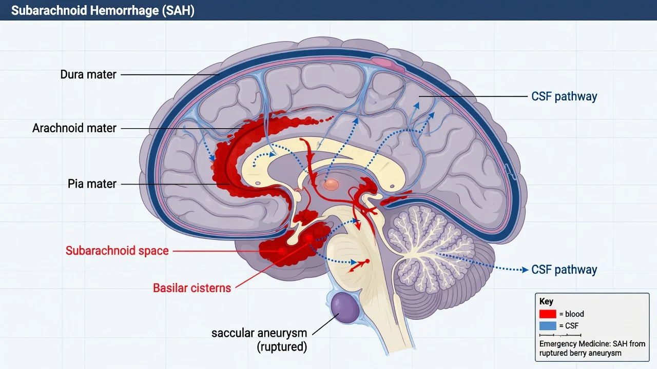
Definition and the epidemiology
The subarachnoid haemorrhage is the bleeding into the subarachnoid space — the space between the arachnoid and the pia mater, normally filled with the cerebrospinal fluid. The non-traumatic SAH accounts for approximately 5 per cent of all strokes, and 80 to 85 per cent are caused by the rupture of a saccular (berry) aneurysm at the bifurcation of the major cerebral arteries (the anterior communicating artery is the commonest site, followed by the posterior communicating and the middle cerebral artery). The other causes include the arteriovenous malformation, the vasculitis, the mycotic aneurysm (from the infective endocarditis), and the perimesencephalic non-aneurysmal SAH (a benign variant). The risk factors are the hypertension, the smoking, the excessive alcohol, the family history, the polycystic kidney disease, the connective tissue disease (the Ehlers-Danlos, the Marfan), and the coarctation of the aorta. [1]
The clinical presentation
The hallmark is the thunderclap headache — a headache that reaches its maximal intensity within seconds to minutes, described by the patient as "the worst headache of my life." It is typically occipital, it may radiate down the neck, and it may be accompanied by the vomiting, the neck stiffness, the photophobia, the brief loss of consciousness, and the seizure. The sentinel bleed — a minor leak days to weeks before the major rupture — presents as a sudden, atypical headache that resolves spontaneously and is often dismissed by the patient and the clinician; its recognition is the opportunity for the diagnosis before the catastrophic rupture. The examination may show the meningism (the neck stiffness, the Kernig and the Brudzinski signs), the depressed conscious level (from the raised intracranial pressure), the focal neurological deficit (the third-nerve palsy from the posterior communicating artery aneurysm), the subhyaloid haemorrhage (the preretinal bleed, pathognomonic), and the papilloedema (the raised ICP). [1]
Differential diagnosis — the thunderclap headache
The thunderclap headache has a broad differential, and the SAH must be distinguished from the other serious and the benign causes. [1]
SAH
- Thunderclap, occipital, maximal in seconds; the neck stiffness, the photophobia
- The CT shows blood in the basilar cisterns; the LP shows the xanthochromia
- The nimodipine, the BP control, the urgent neurosurgery
- The rebleeding risk is highest in the first 24 hours
Migraine
- May be sudden but usually builds over minutes; the aura, the photophobia
- The normal CT and LP; the recurrent pattern
- Treat with the analgesia, the antiemetic, the triptan
- No rebleeding risk; benign
Meningitis
- The fever, the headache over hours to days, the neck stiffness
- The CT may be normal; the LP shows the raised WCC, the low glucose
- Treat with the ceftriaxone, the vancomycin, the dexamethasone
- The bacterial meningitis is time-critical
Cervical artery dissection
- The sudden neck pain, the Horner syndrome, the focal deficit
- The CT or MRA shows the dissection (the flame sign, the double lumen)
- The anticoagulation or the antiplatelet; the stroke risk
- The traumatic or the spontaneous mechanism
The investigations
The non-contrast CT is the first-line investigation — the sensitivity is over 98 per cent within 6 hours of the onset. The blood appears as the hyperdense material in the basilar cisterns, the Sylvian fissure, and the interhemispheric fissure, and the pattern localises the aneurysm (the anterior interhemispheric blood suggests the ACom aneurysm; the unilateral Sylvian blood suggests the MCA aneurysm). If the CT is negative or the presentation is beyond 6 hours, the lumbar puncture is performed after at least 12 hours from the onset (to allow the bilirubin to form from the haemoglobin degradation). The xanthochromia (the yellow discolouration of the supernatant after the centrifugation) is the diagnostic finding — it persists for up to 2 weeks and distinguishes the true SAH from the traumatic tap.[2] The spectrophotometry is more sensitive than the visual inspection. The CT angiography identifies the aneurysm and guides the treatment (the coiling vs the clipping). The digital subtraction angiography is the gold standard for the aneurysm characterisation. The blood pressure, the ECG (the ST changes, the QT prolongation, the arrhythmia from the autonomic surge), the troponin, the coagulation, and the electrolytes are obtained.[1]
Management — the approach and the drug doses
The management has four pillars: the prevention of the rebleeding, the prevention of the vasospasm, the control of the intracranial pressure, and the securing of the aneurysm. [1]
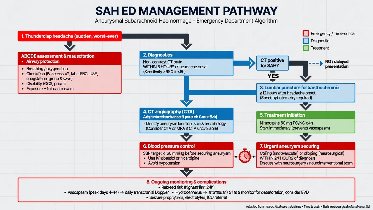
The SAH drug doses
The nimodipine and the vasospasm
The nimodipine is the calcium-channel blocker that reduces the incidence of the cerebral vasospasm and the delayed cerebral ischaemia, and it is the only drug shown to improve the outcome after the SAH. It is started immediately and continued for 21 days. The vasospasm occurs in 30 to 70 per cent of the patients, typically at day 4 to 14 after the bleed, and it manifests as the new neurological deficit, the confusion, or the fever. The treatment of the established vasospasm is the induced hypertension (the noradrenaline to raise the BP and the CPP), the volume expansion, and the angioplasty or the intra-arterial nimodipine for the refractory case. [1]
The aneurysm securing
The endovascular coiling (the platinum coils packed into the aneurysm sac via the catheter) is the preferred treatment for most aneurysms — the ISAT trial found that the coiling had a better outcome than the clipping at 1 year for the anterior circulation aneurysms amenable to either technique. The coiling is performed under the general anaesthetic via the femoral artery approach, and the patient is systemically heparinised during the procedure. The procedural complications include the aneurysm perforation (the catastrophic rebleed on the table, occurring in 1 to 2 per cent), the thromboembolic events (the stroke from the clot dislodged from the catheter), and the contrast nephropathy. The surgical clipping (the craniotomy and the placement of a clip across the aneurysm neck) is used for the MCA bifurcation aneurysms (where the surgical access is easy and the coiling is technically difficult), the wide-necked aneurysms, and the aneurysms not amenable to the coiling. The timing is urgent — within 24 hours of the diagnosis — to minimise the rebleeding risk. The flow-diverter stent is a newer option for the complex or the fusiform aneurysm, but it requires the dual antiplatelet therapy which may complicate any future surgical intervention. [1]
Complications and prognosis
The complications are the rebleeding (the highest risk is in the first 24 hours, 4 per cent; the rebleeding carries a 70 per cent mortality), the vasospasm and the delayed cerebral ischaemia (day 4 to 14, the commonest cause of the delayed morbidity), the hydrocephalus (the obstruction of the CSF drainage by the blood; treated with the EVD), the seizures (5 to 10 per cent), the hyponatraemia (the cerebral salt wasting or the SIADH), the cardiac (the neurogenic stunned myocardium, the arrhythmia, the troponin rise), and the pulmonary oedema (the neurogenic). The prognosis depends on the WFNS grade at presentation (the grade 1, the GCS 15 with no deficit, has a good prognosis; the grade 5, the GCS 3 to 6, has a poor prognosis), the amount of blood on the CT (the Fisher grade), and the complications. [1]
Special populations
The pregnant patient with the SAH is managed with the same protocol, with the obstetric and the neurosurgical teams involved jointly; the coiling is preferred to the clipping where feasible because it avoids the craniotomy and the general anaesthetic in pregnancy. The elderly patient has a worse prognosis but is still offered the treatment if the WFNS grade is favourable. The anticoagulated patient has the anticoagulation reversed urgently (the PCC 25 to 50 IU/kg and the vitamin K 10 mg for the warfarin; the idarucizumab 5 g for the dabigatran; the andexanet or the PCC for the apixaban or the rivaroxaban) before the aneurysm securing, because the anticoagulation increases the rebleeding risk and worsens the outcome. [1]
Evidence and regional guidelines
The contemporary framework is the AHA/ASA SAH guideline and the ESO guideline for the management of the aneurysmal SAH.[1] The nimodipine for 21 days, the BP control under 160 mmHg, the urgent coiling or clipping within 24 hours, and the monitoring for the vasospasm are the global standards.
ANZ practice note. The SAH management follows the local neurosurgical protocol via the state-wide neurosurgical referral; the CT first, the LP for the xanthochromia if the CT is negative or delayed; the nimodipine 60 mg every 4 hours for 21 days; the BP control under 160 mmHg; the coiling or clipping within 24 hours; and the neurosurgical referral for the definitive management. [1]
The aetiologies — the breakdown of the causes
The non-traumatic SAH is aetiologically heterogeneous, and the Fellowship candidate must state the proportions and the distinguishing features because the management and the prognosis differ sharply by the cause. [1]
The causes of the non-traumatic subarachnoid haemorrhage — the proportion, the source, the distinguishing features
| Cause | Proportion | The source / the mechanism | The distinguishing features on the imaging and the history |
|---|---|---|---|
| Saccular (berry) aneurysm | ~85 per cent | The congenital defect of the internal elastic lamina at the bifurcation of the major cerebral arteries, amplified by the haemodynamic stress, the hypertension, the smoking | The ACom (~30 per cent), the PCom (~25 per cent), the MCA bifurcation (~20 per cent); the CT shows the thick blood in the basal cisterns; the third-nerve palsy points to the PCom aneurysm; the CTA / the DSA confirms |
| Perimesencephalic non-aneurysmal SAH | ~10 per cent | The venous or the prepontine bleed of the uncertain origin, confined to the perimesencephalic cisterns | The blood is strictly in the perimesencephalic / the prepontine cisterns, the centre of the bleeding is immediately anterior to the midbrain, the angiogram is negative; the course is benign, the vasospasm is rare, the outcome is excellent |
| Arteriovenous malformation (AVM) | ~5 per cent (and a further fraction of the remainder) | The congenital tangle of the arteries and the veins without an intervening capillary bed; the rupture into the ventricle or the subarachnoid space | The younger patient, the seizure, the previous intracerebral haematoma; the CT may show the calcified nidus, the flow voids on the MRI; the DSA defines the nidus, the feeding arteries, the draining veins |
| Other (the mycotic, the traumatic, the vasculitic, the cocaine, the tumoural, the dural venous sinus thrombosis) | < 5 per cent combined | The infective endocarditis (the mycotic aneurysm — the distal MCA, the multiple, the changing on the serial angiography); the trauma; the vasculitis (the primary CNS angiitis, the polyarteritis nodosa); the cocaine (the acute hypertension, the vasospasm); the intracranial tumour (the metastatic melanoma, the glioblastoma) | The clinical context is the key — the fever and the murmur of the endocarditis, the trauma, the systemic vasculitis, the recreational drug use; the imaging is tailored (the infectious source, the vasculitic pattern) |
The perimesencephalic non-aneurysmal SAH is a high-yield discriminating point: the clinician who dismisses the perimesencephalic pattern as "a minor bleed" without excluding the aneurysm (the vertebrobasilar aneurysm can mimic it) misses a treatable lesion; the angiogram must be normal before the benign label is applied. The mycotic aneurysm of the infective endocarditis is the other trap — it is distal, it is multiple, and it may rupture or disappear on the serial angiography; the management is the antimicrobial therapy with the surgical or the endovascular intervention guided by the rupture status and the haemodynamic significance.[6]
Hunt and Hess and the WFNS grading — the clinical severity
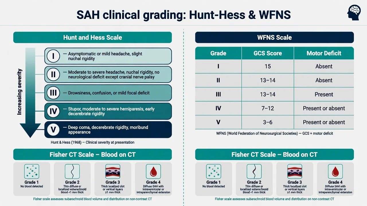
Two grading systems stratify the clinical severity at the presentation, and both are prognostic and they guide the management. The Hunt and Hess (1968) is the older, descriptive system; the WFNS (World Federation of Neurosurgical Societies, 1988) is the simpler, GCS-based system that is now more widely used because of the better inter-observer agreement. [1]
The Hunt and Hess grade — the clinical severity and the mortality
| Grade | The clinical description | The approximate mortality |
|---|---|---|
| I | The asymptomatic, or the minimal headache and the slight nuchal rigidity | ~5 to 10 per cent |
| II | The moderate to the severe headache, the nuchal rigidity, no neurological deficit other than the cranial-nerve palsy | ~10 to 15 per cent |
| III | The drowsy or the confused, the mild focal deficit | ~20 to 30 per cent |
| IV | The stupor, the moderate to the severe hemiparesis, the early decerebrate rigidity | ~40 to 50 per cent |
| V | The deep coma, the decerebrate rigidity, the moribund appearance | ~70 to 80 per cent |
The WFNS grade — the GCS and the motor deficit
| WFNS grade | The GCS | The major focal deficit | The prognostic implication |
|---|---|---|---|
| 1 | 15 | Absent | The best prognosis; the candidate for the early coiling or clipping |
| 2 | 13 to 14 | Absent | The good prognosis; the standard management |
| 3 | 13 to 14 | Present | The guarded prognosis; the higher vasospasm risk |
| 4 | 7 to 12 | Present or absent | The poor prognosis; the individualised decision |
| 5 | 3 to 6 | Present or absent | The very poor prognosis; the discussion of the futility and the goals of the care |
The exam point: the WFNS grade 1 (GCS 15, no deficit) is the good-prognosis patient in whom the urgent aneurysm securing is clearly indicated; the WFNS grade 5 (GCS 3 to 6) is the poor-prognosis patient in whom the discussion of the goals of the care, the family, and the futility is the priority. The higher the WFNS grade, the higher the mortality and the higher the risk of the vasospasm, the DCI, and the hydrocephalus. [1]
The Fisher and the modified Fisher CT grading — the prognostic pattern of the blood
The Fisher grade (1980) and the modified Fisher grade (Frontera 2006) quantify the amount and the distribution of the subarachnoid blood on the CT, and they predict the risk of the symptomatic vasospasm and the delayed cerebral ischaemia. The modified Fisher is preferred because it incorporates the intraventricular blood, which is the independent predictor of the DCI. [1]
The modified Fisher grade — the SAH thickness and the intraventricular haemorrhage, and the risk of the DCI
| mFisher grade | The SAH pattern | The IVH | The risk of the symptomatic vasospasm / the DCI |
|---|---|---|---|
| 0 | No blood | Absent | ~0 per cent |
| 1 | The focal or the diffuse thin SAH (≤ 1 mm thick) | Absent | ~5 to 10 per cent |
| 2 | The focal or the diffuse thin SAH (≤ 1 mm thick) | Present | ~15 to 25 per cent |
| 3 | The thick SAH (> 1 mm thick, the localised clot) | Absent | ~20 to 30 per cent |
| 4 | The thick SAH (> 1 mm thick) and / or the IVH | Present | ~30 to 40 per cent (the highest) |
The thick clot in the basal cisterns and the intraventricular blood are the highest-risk patterns; the clinician anticipates the vasospasm, the prophylactic nimodipine, the daily transcranial Doppler monitoring, and the low threshold for the induced hypertension if the new deficit appears. [1]
The Ottawa Subarachnoid Haemorrhage Rule — the clinical decision rule for the thunderclap headache
The Ottawa Subarachnoid Haemorrhage Rule (Perry 2013, validated 2017 and 2020) is the validated clinical decision rule that identifies the patients with the acute non-traumatic headache who need the investigation for the SAH. It is highly sensitive (approaching 100 per cent) and it is designed to rule OUT — a patient who meets NONE of the criteria can be safely discharged without the further testing. It is NOT specific (a positive rule does not confirm the SAH — most positives are not SAH).[9][8]
[1]The exam point: the Ottawa Rule is the rule-OUT tool — its strength is the sensitivity, not the specificity. The Fellowship candidate must state that a patient who meets NONE of the criteria is at the sufficiently low risk to forgo the CT and the LP, while any one criterion mandates the diagnostic ladder. The 6-hour CT rule (Perry 2020) showed that the modern multidetector CT within 6 hours of the onset has a sensitivity approaching 100 per cent and a negative predictive value approaching 100 per cent, permitting the safe discharge of the low-risk patient without the LP — but ONLY within the 6-hour window and ONLY in a centre with the modern CT and the radiologist reporting.[8]
The diagnostic ladder — the CT, the lumbar puncture, and the angiography
The diagnostic pathway is the ladder, and the Fellowship candidate must be able to walk up it step by step and to state the sensitivity and the timing of each rung. [1]
The SAH diagnostic ladder — the order of the investigations
- The non-contrast CT head — the FIRST test, immediately. (a) The sensitivity: over 98 per cent within 6 hours of the onset (the modern multidetector CT, reported by the experienced radiologist, approaches 100 per cent sensitivity and the negative predictive value within the 6-hour window). (b) The sensitivity falls with the time — ~85 per cent at 24 hours, ~50 per cent at 1 week. (c) The finding: the hyperdense material in the basal cisterns, the Sylvian fissure, the interhemispheric fissure, and the pattern localises the aneurysm. (d) The intraventricular blood and the intracerebral clot are the poor-prognosis signs. (e) If the CT is positive, the workup proceeds to the angiography; the LP is NOT required.[8][10]
- The lumbar puncture — the SECOND test, if the CT is negative OR the presentation is beyond 6 hours. (a) The timing: the LP is performed at least 12 hours after the onset (the British guideline; some centres use 6 hours) to allow the haemoglobin to degrade to the bilirubin (the oxyhaemoglobin is also released, but the bilirubin is the SAH-specific product, formed by the arachnoid macrophages and the CSF). (b) The three bottles are collected (to demonstrate the clearing cell count — the first to the third, the decreasing RBC in the traumatic tap; the SAH has the uniformly elevated RBC). (c) The CSF is centrifuged immediately and the supernatant is examined. (d) The xanthochromia — the yellow discolouration of the supernatant from the bilirubin and the oxyhaemoglobin — is the diagnostic finding. (e) The spectrophotometry (the absorbance at 410 to 415 nm for the bilirubin, 476 nm for the oxyhaemoglobin) is more sensitive than the visual inspection and is the preferred method. (f) The xanthochromia persists for up to 2 weeks. (g) The opening pressure is measured (the elevated opening pressure supports the SAH).[2]
- The CT angiography (CTA) — the THIRD step, to find and to characterise the aneurysm. (a) The CTA is performed once the SAH is confirmed (or the clinical suspicion is very high). (b) It identifies the aneurysm in ~85 per cent of the cases, and it guides the treatment (the coiling vs the clipping). (c) The CTA is fast, it is widely available, and it is the first-line angiographic test. (d) The limitation: the small aneurysm (< 3 mm), the vasospasm, and the vessel overlap may be missed.
- The digital subtraction angiography (DSA) — the GOLD standard, if the CTA is negative or the treatment is planned. (a) The DSA has the highest spatial and the temporal resolution, and it is the definitive test for the aneurysm characterisation, the vasospasm, and the AVM. (b) It is performed when the CTA is negative but the SAH is confirmed (the ~10 to 15 per cent of the confirmed SAH have a negative initial CTA — the second CTA or the DSA is needed, especially for the posterior circulation and the small aneurysm). (c) It is the test for the perimesencephalic vs the aneurysmal pattern. (d) The complication: the puncture-site, the contrast nephropathy, the stroke (1 to 2 per cent).[6]
The lumbar puncture and the xanthochromia — the detail
The lumbar puncture is the second-line test, and the Fellowship candidate must know the technique, the timing, the interpretation, and the pitfalls. The xanthochromia is the yellow discolouration of the CSF supernatant, and it is caused by the bilirubin (the SAH-specific, formed from the haemoglobin degradation by the arachnoid macrophages and the CSF, after ~12 hours) and the oxyhaemoglobin (the released from the RBC lysis, less specific, forms earlier).[2]
The true SAH vs the traumatic tap — the CSF findings
| Feature | The true SAH | The traumatic tap |
|---|---|---|
| The xanthochromia | Present (the bilirubin and the oxyhaemoglobin); persists for up to 2 weeks | Absent (the RBC have not yet lysed; no bilirubin) |
| The RBC count | The uniformly elevated in the bottle 1, 2, 3 (no clearing) | The decreasing from the bottle 1 to the bottle 3 (the clearing) |
| The opening pressure | Often elevated | Usually normal |
| The spectrophotometry | The elevated bilirubin absorbance (410 to 415 nm) | The normal bilirubin absorbance |
| The D-dimer / the ferritin in the CSF | May be elevated | Usually normal |
The spectrophotometry is more sensitive than the visual inspection, and it is the preferred method; the visual inspection misses up to 50 per cent of the cases, especially the mild xanthochromia. The exam point: a bloody tap (the traumatic LP) is a common pitfall — the decreasing RBC count across the three bottles and the absence of the xanthochromia distinguish it from the true SAH, but if there is any doubt, the patient is managed as the SAH until the imaging and the angiography are complete.[2]
The cardiac manifestations of the SAH — the neurogenic stunned myocardium
The SAH produces the profound sympathetic surge, and the catecholamine surge causes the neurogenic stunned myocardium (the Takotsubo-like, the transient myocardial dysfunction), the repolarisation abnormalities on the ECG, and the troponin elevation. The Fellowship candidate must recognise these because they mimic the acute coronary syndrome and they influence the anaesthetic and the procedural management. [1]
The cardiac manifestations of the SAH vs the acute coronary syndrome
| Feature | The neurogenic (the SAH-related) | The acute coronary syndrome |
|---|---|---|
| The ECG | The ST elevation / the depression, the deep T-wave inversions (the "cerebral T waves" — the wide, the deep, the symmetric), the QT prolongation, the U waves, the arrhythmia (the VT, the VF, the torsades) | The regional ST elevation, the reciprocal change, the Q waves, the T-wave inversion |
| The troponin | The mild to the moderate elevation (the peak at ~24 h, the resolves over the days) | The marked elevation, the rise and the fall |
| The echocardiogram | The apical ballooning (the Takotsubo pattern) or the global hypokinesis, the basal hyperkinesis, the reduced EF; the RECOVERS over the days to the weeks | The regional wall-motion abnormality in the coronary territory, the PERSISTS |
| The coronary angiography | The normal coronaries | The obstructive coronary disease |
| The management | The supportive; the avoid the vasoconstrictor; the treat the arrhythmia; the beta-blocker once the BP is controlled; the watchful waiting (the recovery is the rule) | The reperfusion (the PCI, the thrombolysis), the antiplatelet, the anticoagulant, the statin |
The exam point: the "cerebral T waves" (the deep, the symmetric, the wide T-wave inversions across the precordial leads) and the QT prolongation are the classic ECG findings of the SAH and the intracranial haemorrhage; they reflect the autonomic surge, not the ischaemia, and they are NOT treated with the reperfusion. The neurogenic pulmonary oedema (the sudden, the protein-rich, the bilateral, immediately after the ictus) is the pulmonary counterpart of the same sympathetic surge, and it is managed with the oxygen, the positive-pressure ventilation, and the diuresis once the BP is controlled. [1]
The hyponatraemia — the cerebral salt wasting vs the SIADH
The hyponatraemia develops in 30 to 50 per cent of the SAH patients, typically between the day 2 and the day 10, and it is the common cause of the deterioration. The two main mechanisms are the cerebral salt-wasting syndrome (CSWS) and the syndrome of the inappropriate antidiuretic hormone (SIADH), and they MUST be distinguished because the management is OPPOSITE (the CSWS is treated with the volume and the salt; the SIADH is treated with the fluid restriction).[6]
The cerebral salt wasting vs the SIADH in the SAH — the OPPOSITE management
| Feature | The cerebral salt wasting (CSWS) | The SIADH |
|---|---|---|
| The mechanism | The sympathetic surge and the BNP / the ANP release → the renal sodium loss and the volume depletion | The inappropriate ADH → the water retention and the dilutional hyponatraemia |
| The volume status | The HYPOVOLAEMIA (the volume depletion) | The EUVOLAEMIA or the mild hypervolaemia |
| The urine sodium | The high (> 40 mmol/L) | The high (> 40 mmol/L) |
| The urine osmolality | The high | The high |
| The serum osmolality | The low | The low |
| The CVP / the fluid balance | The LOW CVP, the negative fluid balance | The NORMAL or the HIGH CVP |
| The management | The VOLUME and the SALT — the normal saline, the hypertonic saline, the fludrocortisone; the DO NOT fluid-restrict (the restriction worsens the hypovolaemia and the vasospasm) | The FLUID RESTRICTION, the salt tablets, the vasopressin antagonist (the conivaptan, the tolvaptan); the hypertonic saline for the severe symptomatic hyponatraemia |
The exam point: the fluid restriction is DANGEROUS in the SAH because the CSWS is the commoner mechanism, and the fluid restriction precipitates the hypovolaemia, the worsening of the vasospasm, and the delayed cerebral ischaemia. The Fellowship candidate must state that the volume status (the CVP, the fluid balance, the clinical examination) is the discriminator, and that the SAH patient with the hyponatraemia is presumed to have the CSWS and is treated with the volume and the salt until the SIADH is confirmed. The hypertonic saline (the 1.5 to 3 per cent) and the fludrocortisone (0.1 to 0.2 mg orally twice daily) are the targeted therapy for the CSWS.[6]
The vasospasm and the delayed cerebral ischaemia — the monitoring and the management
The cerebral vasospasm occurs in 30 to 70 per cent of the SAH patients (the angiographic) and 20 to 30 per cent (the symptomatic), and it is the commonest cause of the delayed morbidity and mortality. The onset is the day 4 to 14 (the peak at the day 7 to 8), and it manifests as the new neurological deficit, the confusion, the drowsiness, or the fever. The delayed cerebral ischaemia (DCI) is the clinical syndrome (the new neurological deficit that is not attributable to the other cause), and it is the consequence of the vasospasm and the microvascular thrombosis and the cortical spreading ischaemia.[7][6]
The vasospasm monitoring and the management — the daily ICU workflow
- The nimodipine prophylaxis — started on the day 0, continued for 21 days. (a) The 60 mg orally or via the NG tube every 4 hours for 21 days — the only drug shown to improve the outcome (the British aneurysm nimodipine trial, 1989). (b) The mechanism: the calcium-channel blockade of the cerebral vasculature, and possibly the neuroprotective and the anti-inflammatory effect. (c) The intravenous nimodipine is an alternative if the oral route is unavailable, but it requires the careful BP monitoring (the hypotension). (d) The monitor for the nimodipine-induced hypotension — reduce the dose or the switch to the continuous infusion if the BP falls.[7]
- The daily transcranial Doppler (TCD) — the non-invasive vasospasm surveillance. (a) The mean flow velocity in the MCA: the < 120 cm/s is the normal, the 120 to 200 cm/s is the mild to the moderate spasm, the > 200 cm/s is the severe spasm. (b) The Lindegaard ratio (the MCA / the ICA flow velocity): the > 3 is the vasospasm, the > 6 is the severe. (c) The trend is more useful than the single value — the rising trend precedes the clinical deficit. (d) The limitation: the operator-dependent, the inadequate acoustic window in ~10 to 15 per cent.
- The clinical surveillance — the hourly neurology. (a) The GCS, the pupillary response, the motor power, the speech, the new deficit. (b) The new neurological deficit (not attributable to the rebleeding, the hydrocephalus, the electrolyte, the seizure, the sedation) raises the suspicion of the DCI. (c) The exclude the mimics — the CT to exclude the rebleed and the hydrocephalus, the electrolytes, the EEG.
- The induced hypertension and the volume expansion — for the established DCI. (a) The cardiac output is optimised (the euvolaemia to the mild hypervolaemia, the haemoglobin > 100 g/L). (b) The induced hypertension — the noradrenaline (or the phenylephrine, the vasopressin) to raise the systolic BP (the target is the 160 to 200 mmHg systolic, or the 20 per cent above the baseline, titrated to the neurological improvement). (c) The mechanism: the higher CPP overcomes the narrowed vessel, the improved collateral flow, the improved oxygen delivery. (d) The cardiac monitoring (the troponin, the ECG, the echocardiogram) — the neurogenic stunned myocardium may limit the induced hypertension. (e) The treatment is continued for the 24 to 48 hours of the neurological stability, then the slowly weaned.
- The endovascular rescue — for the refractory DCI. (a) The intra-arterial nimodipine (or the milrinone, the nicardipine, the verapamil) infused into the spasmodic vessel via the microcatheter — the chemical angioplasty. (b) The transluminal balloon angioplasty — the mechanical dilation of the proximal vessel spasm (the higher risk, the more durable, the reserved for the proximal MCA / ICA / basilar spasm). (c) The performed in the catheter laboratory, under the general anaesthetic. (d) The risk: the vessel rupture, the thromboembolic event, the reperfusion injury.[6]
The hydrocephalus and the external ventricular drain
The hydrocephalus complicates 20 to 30 per cent of the SAH (the acute, the obstructive, from the intraventricular blood and the clot obstructing the CSF outflow; and the delayed, the communicating, from the impaired CSF absorption over the weeks). The clinical signs are the deteriorating GCS, the headache, the vomiting, the pupillary changes, the bilateral Babinski, the upgaze palsy (the Parinaud syndrome from the tectal plate compression). The CT shows the dilated ventricles, the transependymal flow, the periventricular lucency.[6]
The external ventricular drain (EVD) is the treatment — the catheter inserted into the lateral ventricle (the Kocher point, the frontal) via the twist-drill craniostomy, connected to the closed drainage system set at the desired height (the ear tragus, the zero reference; the drain height determines the ICP). The EVD drains the CSF and the blood, it monitors the ICP, and it allows the CSF sampling. The complications are the infection (the ventriculitis, the EVD-related — the prophylactic antibiotic, the strict asepsis, the minimal manipulation, the early removal), the haemorrhage (the catheter tract), the malposition, and the over-drainage (the subdural haematoma from the rapid decompression, the upward herniation). The Fellowship candidate must know the EVD management: the level of the drain, the colour of the CSF, the ICP waveform, the weaning protocol, and the antibiotic-impregnated catheter to reduce the infection. [1]
The seizures — the prophylaxis and the management
The seizures complicate 5 to 10 per cent of the SAH (the generalised, the focal, the non-convulsive status in the comatose patient), and they worsen the outcome (the increased ICP, the hypoxia, the secondary brain injury, the rebleeding risk from the hypertensive surge). The prophylactic anticonvulsant is NOT routine (the AHA/ASA guideline) — it is reserved for the patient with the seizure at the presentation, the intracerebral haematoma, the middle cerebral artery aneurysm, or the previous seizure history. The levetiracetam (500 mg IV twice daily) is the preferred agent (the less interaction, the less sedation, the renal dosing). The phenytoin is avoided (the interaction with the nimodipine, the worse cognitive outcome, the fever masking the vasospasm). The non-convulsive status (the comatose patient with the unexplained deterioration) requires the urgent EEG and the treatment (the levetiracetam, the midazolam infusion for the refractory case).[6]
The rebleeding — the preventable catastrophe
The rebleeding is the commonest preventable cause of the death, and the risk is the highest in the first 24 hours (4 per cent) and continues through the first week (the day 1 to 7). The rebleeding carries the 70 per cent mortality. The risk factors are the unsecured aneurysm, the hypertension, the poorly controlled anxiety and the pain, the coughing and the retching, the seizure, the anticoagulation, and the early transfer. The prevention is the urgent aneurysm securing within 24 hours (the coiling or the clipping), the strict BP control (the systolic < 160 mmHg), the analgesia and the anxiolysis, the antiemetic, the seizure prophylaxis, and the reversal of the anticoagulation. The Fellowship candidate must state that the early securing is the priority — every hour the aneurysm is unsecured is the hour of the rebleeding risk.[3][4]
The evidence — the landmark trials
ISAT — International Subarachnoid Aneurysm Trial (Molyneux 2002, Lancet)
Design
Multicentre, randomised, controlled trial — 2143 patients with the ruptured intracranial aneurysm amenable to both the coiling and the clipping
Population
The good-grade SAH (mostly WFNS 1 to 2), the anterior circulation aneurysm
Intervention
The endovascular coiling vs the neurosurgical clipping
Primary outcome (1 year)
The dependency or the death (the modified Rankin 4 to 5): 23.7 per cent coiling vs 30.6 per cent clipping — the absolute risk reduction 6.9 per cent, the relative risk reduction 22.6 per cent, the number needed to treat ~14
Rebleeding
The slightly higher rebleeding risk in the coiling arm, but the net outcome still favoured the coiling
Bottom line
The endovascular coiling is the preferred treatment for the ruptured aneurysm amenable to both the techniques — the better 1-year outcome, the lower mortality, the shorter hospital stay
ISAT — the 1-year outcome and the subgroups (Molyneux 2005, Lancet)
Design
The 1-year follow-up of the ISAT cohort — 2143 patients
Key finding
The relative and the absolute risk reduction of the dependency or the death with the coiling persisted at the 1 year; the subgroups (the age, the WFNS grade, the aneurysm location) consistently favoured the coiling
The rebleeding after the treatment
The slightly higher rebleeding risk with the coiling (the incomplete occlusion, the aneurysm recurrence), but the overall outcome remained better
Bottom line
The coiling advantage is durable at the 1 year; the patient with the ruptured anterior circulation aneurysm amenable to both the techniques should be offered the coiling
ISAT — the 18-year follow-up (Molyneux 2015, Lancet)
Design
The long-term (18-year) follow-up of the UK ISAT cohort
Key finding
The rebleeding risk was higher in the coiling arm beyond the 1 year (the late rebleeding from the aneurysm recurrence), but the overall outcome (the survival, the dependency) remained comparable; the durability of the coiling depends on the complete occlusion and the follow-up angiography
Clinical implication
The coiling requires the lifelong surveillance angiography (the MRA / the CTA) and the re-treatment for the recurrent aneurysm; the clipping is more durable but the upfront risk is higher
Bottom line
The choice of the coiling vs the clipping is individualised — the aneurysm morphology, the age, the comorbidity, the centre expertise; the ISAT is the foundation, the long-term surveillance is the corollary
The British aneurysm nimodipine trial (Pickard 1989, BMJ)
Design
Multicentre, double-blind, randomised, placebo-controlled trial — 554 patients with the recent SAH
Intervention
The oral nimodipine 60 mg every 4 hours for 21 days vs the placebo
Primary outcome
The cerebral infarction (the CT) and the outcome (the Glasgow outcome scale)
Key finding
The nimodipine reduced the cerebral infarction (22 per cent to 17 per cent) and the poor outcome (33 per cent to 20 per cent) — the relative risk reduction of the poor outcome ~40 per cent
Bottom line
The oral nimodipine 60 mg every 4 hours for 21 days is the standard of care — the only drug shown to improve the outcome after the SAH
The Ottawa Subarachnoid Haemorrhage Rule — the validation (Perry 2017, CMAJ; Perry 2020, Stroke)
Design
The prospective, multicentre, cohort studies of the alert, neurologically intact ED patients with the acute non-traumatic headache
Population
The patients with the new, severe, non-traumatic headache reaching the maximal intensity within one hour
The rule
The six criteria (the age ≥ 40, the neck pain / stiffness, the witnessed loss of consciousness, the exertion, the thunderclap, the limited neck flexion) — any one criterion triggers the workup
Performance
The sensitivity approaching 100 per cent (the rule-out); the specificity ~15 to 30 per cent (most positives are not SAH)
The 6-hour CT rule
The modern multidetector CT within 6 hours of the onset has the sensitivity approaching 100 per cent and the negative predictive value approaching 100 per cent — the LP may be omitted in the low-risk patient within the 6-hour window
Bottom line
The Ottawa Rule is the validated rule-OUT tool for the thunderclap headache; the 6-hour CT rule permits the safe discharge without the LP in the selected low-risk patient
SHED — Subarachnoid Haemorrhage in the Emergency Department (TERN 2024, Emerg Med J)
Design
The prospective, observational, multicentre cohort study — the contemporary UK ED practice
Population
The ED patients with the suspected SAH across the multiple UK hospitals
Key findings
The contemporary rates of the SAH diagnosis, the use of the CT and the LP, the adherence to the diagnostic ladder, the outcomes; the study quantified the modern pre-test probability of the SAH in the thunderclap-headache population
Clinical implication
The ~1 in 10 of the patients presenting to the ED with the sudden severe headache has the SAH — the threshold to investigate is low; the CT-first, the LP-if-negative ladder remains the standard
Bottom line
The modern ED data reaffirm the SAH as the not-uncommon diagnosis in the thunderclap-headache population and the importance of the structured diagnostic pathway
Exam practice
SAQ — Thunderclap headache: the diagnostic pathway
12 minutes · 10 marks
A 48-year-old woman presents to the emergency department two hours after the sudden onset of the worst headache of her life, maximal within seconds, while lifting groceries. She has photophobia, neck stiffness and one episode of vomiting. She is alert and orientated with no focal deficit. GCS 15, T 37.0, HR 92, BP 158/94, no meningism other than neck stiffness. She has a history of migraine but says this headache is different.
SAQ — Day 7 after SAH: delayed cerebral ischaemia and hyponatraemia
12 minutes · 10 marks
A 56-year-old man is in the neurointensive care unit on day 7 after coiling of a ruptured anterior communicating artery aneurysm. He has become confused and drowsy over the past 12 hours with a new right-sided weakness. His sodium has fallen from 138 to 124 mmol/L over 48 hours. His ECG shows deep symmetric T-wave inversions and a QTc of 520 ms.
Exam pearls
- The thunderclap headache is a SAH until proven otherwise — the CT first, the LP for the xanthochromia if negative.
- CT sensitivity over 98 per cent within 6 hours — the LP is needed if the presentation is beyond 6 hours.
- The xanthochromia (the bilirubin in the CSF supernatant) is diagnostic — it persists for 2 weeks.
- Nimodipine 60 mg every 4 hours for 21 days — the only drug shown to improve the outcome.
- The BP under 160 mmHg systolic before the aneurysm is secured — the labetalol or the nicardipine.
- The rebleeding risk is 4 per cent in the first 24 hours — the urgent coiling or clipping.
- The vasospasm at day 4 to 14 — the induced hypertension for the treatment.
- The third-nerve palsy with the pupillary dilatation suggests the posterior communicating artery aneurysm.
- The ACom is the commonest aneurysm site (~30 per cent), then the PCom (~25 per cent), then the MCA bifurcation (~20 per cent) — the pattern of the blood on the CT localises the aneurysm.
- The perimesencephalic non-aneurysmal SAH (~10 per cent) is benign — the blood confined to the perimesencephalic cisterns, the negative angiogram, the rare vasospasm.
- The spectrophotometry is more sensitive than the visual inspection for the xanthochromia — the visual inspection misses up to 50 per cent.
- The hyponatraemia is usually the cerebral salt wasting, NOT the SIADH — the fluid restriction is DANGEROUS; the treat with the volume and the salt.
- The "cerebral T waves" (the deep, the symmetric, the wide T-wave inversions) and the QT prolongation are the classic ECG findings — the neurogenic stunned myocardium, NOT the ischaemia.
- The ISAT trial favours the coiling over the clipping for the anterior circulation aneurysm amenable to both — the absolute risk reduction of the poor outcome ~7 per cent, the NNT ~14.
- The Hunt and Hess and the WFNS grade at the presentation are the prognostic — the grade 1 is the good prognosis, the grade 5 is the poor prognosis and the goals-of-care discussion.
- The EVD for the hydrocephalus — the Kocher point, the level at the ear tragus, the ICP monitoring, the watch for the ventriculitis and the over-drainage.
The timing of the SAH complications — the day-by-day
| The complication | The typical timing | The hallmark | The intervention |
|---|---|---|---|
| The rebleeding | The day 1 to 7 (the highest in the first 24 h) | The sudden deterioration, the new headache, the fixed pupils | The urgent aneurysm securing (the coiling / the clipping) |
| The hydrocephalus (acute) | The day 1 to 3 | The deteriorating GCS, the headache, the upgaze palsy | The EVD |
| The vasospasm / the DCI | The day 4 to 14 (the peak day 7 to 8) | The new deficit, the confusion, the fever; the rising TCD velocities | The nimodipine, the induced hypertension, the endovascular rescue |
| The hyponatraemia | The day 2 to 10 | The confusion, the seizure; the low sodium, the high urine sodium | The CSWS → the volume and the salt; the SIADH → the restriction |
| The seizures | Any time, the commoner in the first week | The generalised / the focal; the non-convulsive in the comatose | The levetiracetam; the EEG for the non-convulsive |
| The hydrocephalus (delayed) | The weeks to the months | The gait apraxia, the dementia, the incontinence | The ventriculoperitoneal shunt |
Red flags
[1]References
- [1]Chen CY, Chen CY. Evaluating thunderclap headache Curr Opin Neurol, 2021.PMID 33661161
- [2]Sjulstad AS, Foss OA, Dons N, et al. Visual inspection versus spectrophotometry for xanthochromia detection in patients with sudden onset severe headache-A diagnostic accuracy study Headache, 2025.PMID 39087907
- [3]Molyneux A, Kerr R, Stratton I, Sandercock P, Clarke M, Shrimpton J, Holman R. International Subarachnoid Aneurysm Trial (ISAT) of neurosurgical clipping versus endovascular coiling in 2143 patients with ruptured intracranial aneurysms: a randomised trial Lancet, 2002.PMID 12414200
- [4]Molyneux AJ, Kerr RS, Yu LM, Clarke M, Sneade M, Yarnold JA, Sandercock P. International subarachnoid aneurysm trial (ISAT) of neurosurgical clipping versus endovascular coiling in 2143 patients with ruptured intracranial aneurysms: a randomised comparison of effects on survival, dependency, seizures, rebleeding, subgroups, and aneurysm occlusion Lancet, 2005.PMID 16139655
- [5]Molyneux AJ, Birks J, Clarke A, Sneade M, Kerr RS. The durability of endovascular coiling versus neurosurgical clipping of ruptured cerebral aneurysms: 18 year follow-up of the UK cohort of the International Subarachnoid Aneurysm Trial (ISAT) Lancet, 2015.PMID 25465111
- [6]Connolly ES Jr, Rabinstein AA, Carhuapoma JR, Derdeyn CP, Dion J, Higashida RT, Hoh BL, Kirkness CJ, Naidech AM, Ogilvy CS, Patel AB, Thompson BG, Vespa P. Guidelines for the management of aneurysmal subarachnoid hemorrhage: a guideline for healthcare professionals from the American Heart Association/american Stroke Association Stroke, 2012.PMID 22556195
- [7]Pickard JD, Murray GD, Illingworth R, Shaw MD, Teasdale GM, Foy PM, Humphrey PR, Lang DA, Nelson R, Richards P, et al. Effect of oral nimodipine on cerebral infarction and outcome after subarachnoid haemorrhage: British aneurysm nimodipine trial BMJ, 1989.PMID 2496789
- [8]Perry JJ, Sivilotti MLA, Émond M, Hohl CM, Caudarelli R, Sabbagh A, St-Hilaire C, Ormond M, Berthelot S, Eujayes C, Lemery J, Coyle D, Clement C, Worster A, Ducharme J, Borgundvaag B, Kitto S, Van M, McLeod SL, Thiruganasambandamoorthy V, Wells GA, Stiell IG. Prospective Implementation of the Ottawa Subarachnoid Hemorrhage Rule and 6-Hour Computed Tomography Rule Stroke, 2020.PMID 31805846
- [9]Perry JJ, Sivilotti MLA, Sutherland J, Hohl CM, Émond M, Caudarelli R, Sabbagh A, Lee JS, Worster A, Heslop C, Dryden D, Lesiuk H, Wells GA, Stiell IG. Validation of the Ottawa Subarachnoid Hemorrhage Rule in patients with acute headache CMAJ, 2017.PMID 29133539
- [10]Trainee Emergency Research Network (TERN). Subarachnoid haemorrhage in the emergency department (SHED): a prospective, observational, multicentre cohort study Emerg Med J, 2024.PMID 39266054