Ischaemic Stroke
Clinical Significance : Stroke is the second leading cause of death globally and a leading cause of adult disability. From 1990 to 2021, the global burden increased substantially with a 70% increase in incident...
What matters first
Clinical Significance : Stroke is the second leading cause of death globally and a leading cause of adult disability. From 1990 to 2021, the global burden increased substantially with a 70% increase in incident...
Check the red flags, emergency triggers, and escalation points before using the deep-dive material.
17 Jan 2026
Generated educational material; verify before clinical use.
Visible references section
See the concept before reading it
Study the key anatomy, imaging, and decision pathways as full teaching plates.
Content status and exam context
This page is AI-generated educational content. It may contain errors or omissions and is not a substitute for current guidelines, local protocols, senior clinical judgement, or professional medical advice.
MedVellum does not claim an individual clinician reviewer, board certification, or professional credential for this page unless a future version names a real, verifiable reviewer.
Clinical explanation and evidence
1. Clinical Overview
Definition and Significance
Ischaemic stroke is a neurological deficit attributable to an acute focal injury of the central nervous system (CNS) by a vascular cause, specifically infarction of CNS tissue. It results from a critical reduction in blood flow to a brain region, causing neuronal cell death. It accounts for 85-87% of all strokes (the remainder being haemorrhagic). [1]
Clinical Significance: Stroke is the second leading cause of death globally and a leading cause of adult disability. From 1990 to 2021, the global burden increased substantially with a 70% increase in incident strokes and 44% increase in deaths, with 87% of deaths residing in lower-income countries. [2] The estimated global cost of stroke exceeds US$890 billion (0.66% of global GDP). [2]
"Time is Brain": During a large vessel occlusion, approximately 1.9 million neurons are lost per minute of untreated ischaemia. Urgent reperfusion therapy (thrombolysis/thrombectomy) significantly improves functional outcomes, with thrombectomy having an NNT of 2.6 for functional independence. [3]
Key Associations
- Atrial Fibrillation (AF): Major cause of cardioembolic stroke, conferring a 5-fold increase in stroke risk. [4]
- Large Vessel Occlusion (LVO): Internal carotid artery (ICA), middle cerebral artery (MCA M1/M2), basilar artery. Prime targets for mechanical thrombectomy.
- Transient Ischaemic Attack (TIA): Warning sign. Patients with TIA have 10-15% risk of stroke within 90 days, with highest risk in the first 48 hours.
"Do Not Miss" Warnings
- Hypoglycaemia: Can mimic stroke ("stroke chameleon"). Always check blood glucose before thrombolysis.
- Posterior Circulation Stroke: Vertigo, diplopia, ataxia, crossed signs. Easily missed as FAST score does not capture posterior fossa symptoms. Use ROSIER or BEFAST.
- Aortic Dissection: Can present with stroke (carotid dissection). Check blood pressure in both arms and palpate pulses before thrombolysis to avoid catastrophic bleeding.
- Cervical Artery Dissection: Especially in younger patients with neck pain/trauma. Can cause Horner's syndrome + stroke.
2. Epidemiology & Risk Factors
Incidence and Prevalence
- Global Incidence: 150-200 per 100,000 per year in high-income countries. [2]
- Lifetime Risk: 1 in 4 adults > 25 years will experience a stroke in their lifetime. [2]
- Age: Risk doubles every decade after age 55.
- Gender: Higher incidence in men, but women have higher lifetime risk due to longevity.
Risk Factors Table
| Factor | Relative Risk (RR) | Mechanism | Preventability |
|---|---|---|---|
| Hypertension | 3.0-5.0 | Arteriolosclerosis, lipohyalinosis. Most important modifiable factor. | High |
| Atrial Fibrillation | 5.0 | Left atrial appendage stasis → thrombus formation. | High (anticoagulation) |
| Smoking | 2.0 | Endothelial damage, hypercoagulability, atherogenesis. | High |
| Diabetes Mellitus | 2.0-3.0 | Accelerated atherosclerosis, endothelial dysfunction. | Moderate |
| Dyslipidaemia | 1.5-2.0 | Atheromatous plaque formation (especially high LDL). | High |
| Carotid Stenosis | Variable | Artery-to-artery embolism from plaque rupture, hypoperfusion. | Moderate (CEA/CAS) |
| Prior Stroke/TIA | 10-15x | Underlying vascular disease persists. | High |
| Physical Inactivity | 1.5-2.0 | Associated metabolic syndrome, obesity. | High |
Non-Modifiable Risk Factors
- Age: > 55 years (exponential increase).
- Sex: Male > Female (but female higher lifetime risk).
- Ethnicity: Higher rates in Black, Hispanic, Asian populations.
- Genetic: Family history of stroke (especially if less than 65 years).
3. Pathophysiology
Mechanism of Ischaemic Injury
The Ischaemic Cascade:
-
Ischaemic Core: Area of irreversible infarction. Blood flow less than 10 ml/100g/min. Rapid cell death within minutes due to:
- Energy failure (ATP depletion).
- Ionic pump failure (Na+/K+-ATPase dysfunction).
- Immediate neuronal death.
-
Ischaemic Penumbra: Area of hypoperfused but viable tissue surrounding the core. Blood flow 10-20 ml/100g/min.
- Electrical failure but membrane integrity initially preserved.
- Critical therapeutic target for reperfusion therapy.
- Can survive for several hours if blood flow restored.
- Forms basis of "time is brain" concept.
-
Excitotoxicity:
- Glutamate release from dying neurons.
- Excessive NMDA/AMPA receptor activation.
- Massive calcium influx → mitochondrial damage.
- Activation of caspases → apoptosis and necrosis.
-
Inflammation:
- Microglial activation and neutrophil infiltration.
- Release of inflammatory cytokines (IL-1, TNF-α).
- Blood-brain barrier disruption.
- Secondary injury and haemorrhagic transformation risk.
Thrombotic vs Embolic Mechanisms
Thrombotic Stroke (30-40% of ischaemic strokes):
- In situ thrombus formation on atherosclerotic plaque.
- Mechanism: Plaque rupture → platelet aggregation → thrombus occlusion.
- Location: Commonly at carotid bifurcation, MCA origin, basilar artery.
- Clinical: Often "stuttering" onset with fluctuating symptoms or evolution over hours.
- Associated: Hypertension, diabetes, dyslipidaemia, smoking.
Embolic Stroke (60-70% of ischaemic strokes):
- Distal embolization from proximal source.
- Cardioembolic Sources (20-25%):
- Atrial fibrillation (most common) → left atrial appendage thrombus.
- Left ventricular mural thrombus (post-MI, dilated cardiomyopathy).
- Valvular disease (mechanical valves, mitral stenosis, endocarditis).
- Patent foramen ovale (paradoxical embolism).
- Atrial myxoma (rare).
- Artery-to-Artery Embolism (40-45%):
- From carotid/vertebral atherosclerotic plaques.
- From aortic arch atheroma.
- Clinical: Typically sudden onset at maximal deficit ("wake-up stroke" common).
- Imaging: Often multiple territory infarcts, haemorrhagic transformation common. [5]
Classification (TOAST Criteria)
- Large Artery Atherosclerosis (20-25%): Stenosis/occlusion of ICA, vertebral, basilar arteries. Thrombus on atherosclerotic plaque.
- Cardioembolism (20-25%): AF (most common), mural thrombus (post-MI), mechanical valve, PFO, endocarditis.
- Small Vessel Occlusion (Lacunar) (20-25%): Lipohyalinosis of penetrating arterioles (lenticulostriate, thalamoperforators). Pure motor/sensory syndromes. Infarct less than 15mm diameter.
- Stroke of Other Determined Aetiology (5-10%): Dissection (carotid/vertebral), vasculitis, thrombophilia (Antiphospholipid syndrome), CADASIL, Fabry's disease.
- Cryptogenic (ESUS) (25-30%): Embolic Stroke of Undetermined Source. No cause found despite full investigation.
Vascular Territories and Clinical Syndromes
| Artery | Territory | Clinical Features |
|---|---|---|
| Anterior Cerebral Artery (ACA) | Medial frontal/parietal lobe, supplementary motor area | Leg > Arm weakness, urinary incontinence, abulia, grasp reflex |
| Middle Cerebral Artery (MCA) | Lateral frontal/parietal/temporal lobe, internal capsule | Arm/Face > Leg weakness, aphasia (dominant), neglect (non-dominant), homonymous hemianopia |
| Posterior Cerebral Artery (PCA) | Occipital lobe, medial temporal lobe, thalamus | Homonymous hemianopia (macular sparing), alexia without agraphia, cortical blindness (bilateral) |
| Vertebrobasilar | Brainstem, cerebellum, occipital lobe | "Infarct of the 4 D's": Dizziness, Diplopia, Dysarthria, Dysphagia. Crossed signs (ipsilateral CN, contralateral motor). Ataxia. |
| Lacunar Syndromes | Internal capsule, pons, thalamus | Pure motor, pure sensory, sensorimotor, ataxic hemiparesis, dysarthria-clumsy hand |
4. Clinical Presentation
Symptoms
Sudden Onset focal neurological deficit (key distinguishing feature from tumour/abscess):
- Weakness: Unilateral arm/leg/face (most common).
- Sensory Loss: Numbness, paresthesia (hemibody distribution).
- Speech Disturbance:
- "Dysphasia (language disorder): Expressive (Broca's - frontal), receptive (Wernicke's - temporal), global."
- "Dysarthria (motor speech disorder): Slurred speech, tongue/facial weakness."
- Visual Loss:
- Monocular blindness (amaurosis fugax) - retinal artery occlusion.
- Homonymous hemianopia - occipital lobe or optic radiation.
- Ataxia/Vertigo: Cerebellar or brainstem stroke.
- Altered Consciousness: Large hemispheric strokes, brainstem strokes.
Signs: Bamford/Oxford Classification
Based on clinical signs, predicts territory, aetiology, and prognosis:
1. TACS (Total Anterior Circulation Stroke) - ALL three of:
- Unilateral weakness (and/or sensory deficit) of face, arm, AND leg.
- Homonymous hemianopia.
- Higher cerebral dysfunction (dysphasia if dominant, neglect if non-dominant).
- Implication: Proximal MCA or ICA occlusion. Poor prognosis (60% dead/dependent at 1 year). High thrombectomy candidacy.
2. PACS (Partial Anterior Circulation Stroke):
- Two of the three TACS components OR isolated higher cerebral dysfunction (e.g., aphasia alone).
- Implication: Branch MCA occlusion. Moderate prognosis.
3. LACS (Lacunar Stroke):
- Pure motor stroke (face + arm + leg).
- Pure sensory stroke.
- Sensorimotor stroke.
- Ataxic hemiparesis.
- Dysarthria-clumsy hand syndrome.
- No visual loss, no higher cortical dysfunction.
- Implication: Small vessel disease (internal capsule, pons, basal ganglia). Good prognosis.
4. POCS (Posterior Circulation Stroke): Any of:
- Cerebellar signs (ataxia, nystagmus).
- Brainstem signs (cranial nerve palsies + crossed motor/sensory signs).
- Isolated homonymous hemianopia (PCA territory).
- Variable prognosis depending on extent.
Stroke Mimics (10-30% of "stroke alerts")
- Hypoglycaemia (most common mimic - always check BSL).
- Seizure with Todd's paresis (post-ictal weakness).
- Migraine (hemiplegic migraine, especially in young).
- Functional/Conversion disorder (non-organic).
- Sepsis/Metabolic encephalopathy (older patients).
- Brain tumour (gradual onset, headache, papilloedema).
- Subdural haematoma (fluctuating symptoms, falls, anticoagulation).
5. Clinical Assessment
NIHSS (National Institutes of Health Stroke Scale)
Standardised 15-item tool (scored 0-42) to quantify stroke severity. Highly reliable and valid for prognostication and triage. [6]
Components:
- Level of Consciousness (0-3): Alert, drowsy, stuporous, coma.
- LOC Questions (0-2): Month, age.
- LOC Commands (0-2): Open/close eyes, grip hand.
- Gaze (0-2): Horizontal eye movements (normal, partial palsy, forced deviation).
- Visual Fields (0-3): Confrontation testing.
- Facial Palsy (0-3): Show teeth, raise eyebrows.
- Motor Arm (0-4 each side): Drift over 10 seconds (left, right).
- Motor Leg (0-4 each side): Drift over 5 seconds (left, right).
- Limb Ataxia (0-2): Finger-nose-finger, heel-shin.
- Sensory (0-2): Pinprick face/arm/trunk/leg.
- Language/Aphasia (0-3): Describe picture, name objects, read sentences.
- Dysarthria (0-2): Read/repeat words.
- Extinction/Inattention (0-2): Neglect, hemineglect.
Interpretation:
- 0: No stroke symptoms.
- 1-4: Minor stroke (high thrombolysis benefit, low risk).
- 5-15: Moderate stroke (standard thrombolysis candidate).
- 16-20: Moderate-severe stroke (consider thrombectomy if LVO).
- > 20: Severe stroke (high mortality, but thrombectomy may still benefit if LVO).
Clinical Use:
- Triage: NIHSS ≥6 suggests LVO (81% specificity). [6]
- Prognosis: Each 1-point increase in NIHSS = 17% decrease in favourable outcome.
- Treatment decisions: NIHSS ≤3 (minor stroke) → consider DAPT. NIHSS > 25 → high malignant oedema risk.
Key Bedside Checks
- Blood Glucose: Mandatory pre-thrombolysis (exclude hypoglycaemia).
- Blood Pressure: Often elevated (autoregulatory response). Permissive hypertension if not lysed.
- Heart Rhythm: Irregular → AF (40% undiagnosed AF in stroke patients).
- Murmurs: Carotid bruit (stenosis), cardiac murmur (valvular disease, endocarditis).
- Pulse Asymmetry: Aortic dissection, subclavian stenosis.
6. Investigations
Bedside
- Blood Glucose (Capillary): Immediate. Exclude hypoglycaemia. Mandatory pre-lysis.
- ECG: AF (irregular rhythm, absent P waves), LVH, acute MI (concurrent ACS).
- Oxygen Saturation: Hypoxia worsens cerebral ischaemia.
Imaging
CT Head (Non-Contrast) - Immediate (Door-to-CT target less than 20 minutes):
- Primary Goal: Exclude haemorrhage (absolute contraindication to lysis).
- Early Ischaemic Signs (may be absent in first 6 hours):
- Hyperdense MCA sign (clot in vessel) - sensitivity 30%, specificity 100%.
- Loss of insular ribbon (obscuration of grey-white junction).
- Loss of grey-white differentiation in basal ganglia/cortex.
- Sulcal effacement.
- Subtle hypodensity.
ASPECTS Score (Alberta Stroke Program Early CT Score):
- 10-point score to quantify extent of early ischaemic changes in MCA territory.
- Divides MCA territory into 10 regions (M1-M6, caudate, lentiform, internal capsule, insula).
- Scoring: 1 point deducted for each region with ischaemic change.
- 10 = Normal. 0 = Extensive infarction.
- ASPECTS ≥6 = Good thrombectomy candidate (favourable tissue outcome). [7]
- ASPECTS less than 6 = Large established infarct (thrombectomy benefit uncertain, recent trials suggest benefit even ASPECTS 3-5). [7]
CT Angiography (CTA) Arch to Vertex:
- Mandatory if thrombectomy candidate.
- Identifies Large Vessel Occlusion (LVO): ICA, M1, M2, basilar, vertebral.
- Assesses collateral circulation (Tan scale, Miteff score).
- Detects carotid dissection, tandem occlusions.
CT Perfusion (CTP):
- Measures cerebral blood flow (CBF), cerebral blood volume (CBV), mean transit time (MTT).
- Differentiates Core (dead tissue, low CBF + low CBV) vs Penumbra (salvageable, low CBF + normal/high CBV).
- Essential for late-window thrombectomy (6-24h) to select patients with favourable mismatch. [8]
- Mismatch ratio > 1.8 and core volume less than 70ml predict good thrombectomy outcomes.
MRI Brain (DWI/ADC):
- Gold standard for confirming ischaemia.
- DWI (diffusion-weighted imaging): Bright signal (restricted diffusion) within minutes.
- ADC (apparent diffusion coefficient): Dark signal (confirms acute ischaemia).
- More sensitive than CT for posterior fossa strokes, lacunar strokes, early ischaemia.
- MRA (MR angiography): Non-invasive vessel imaging (alternative to CTA).
- Wake-up stroke: DWI-FLAIR mismatch (DWI+/FLAIR-) suggests less than 4.5h window. [8]
Laboratory
- Full Blood Count (FBC):
- Platelets (thrombocytopenia less than 100 is lysis contraindication).
- Haemoglobin (anaemia, polycythaemia).
- WCC (infection, leukaemia).
- Coagulation (INR/APTT):
- INR > 1.7 contraindication to lysis.
- Required if on warfarin or heparin.
- Renal Function (U&E/Creatinine): Baseline for contrast studies, CKD.
- Lipid Profile: Baseline for statin therapy.
- HbA1c: Diabetes screening.
- Troponin: Concurrent MI, stress cardiomyopathy.
Stroke Aetiology Workup (Secondary Prevention)
- Echocardiography:
- "Transthoracic (TTE): LV thrombus, valvular disease, LV function."
- "Transoesophageal (TOE): Left atrial appendage thrombus, PFO, aortic arch atheroma."
- Carotid Doppler Ultrasound: Stenosis quantification (NASCET/ECST criteria).
- 24-Hour ECG (Holter) or Prolonged Monitoring: Paroxysmal AF detection (30% diagnosed post-stroke).
- Thrombophilia Screen (if less than 50 years, cryptogenic): Antiphospholipid antibodies, protein C/S, Factor V Leiden.
- Vasculitis Screen (if young, atypical): ESR, CRP, ANA, ANCA.
7. Management
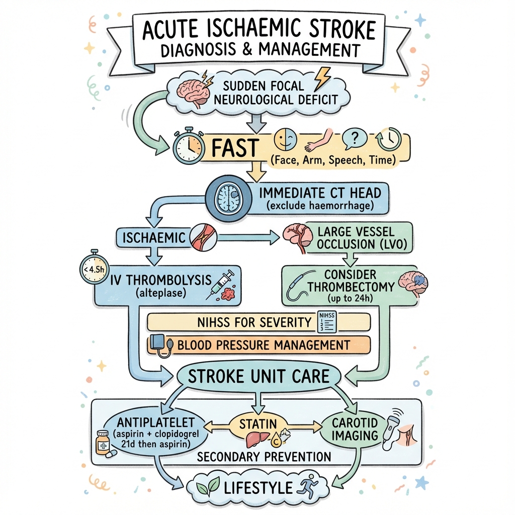
Acute Management Algorithm
        [ACUTE STROKE ALERT]
                ↓
    ┌────────────────────────┐
    │ Rapid Assessment       │
    │ - AIRWAY/BREATHING     │
    │ - Blood Glucose Check  │
    │ - NIHSS Assessment     │
    │ - Time of Onset (LKW)  │
    │ - BP Both Arms         │
    └───────────┬────────────┘
                ↓
        CT Head + CTA
      (Door-to-CT less than 20min)
                │
         Haemorrhage?
            ┌────┴────┐
          YES        NO
           │          │
       Manage as      ↓
       ICH     [ISCHAEMIC STROKE]
                      │
           ┌──────────┴───────────┐
       Onset less than 4.5h?          Large Vessel Occlusion?
       (Thrombolysis)            (Thrombectomy)
           │                      │
      ┌────┴────┐            ┌────┴────┐
     YES       NO           YES       NO
      │         │            │         │
    Check       │         Check        │
    Contra-     │       ASPECTS,       │
    indications │       Premorbid      │
      │         │         mRS          │
      ↓         ↓            │         ↓
   [IV LYSIS] Aspirin        ↓    Stroke Unit
   Alteplase  300mg      [EVT]       Care
   0.9mg/kg   (wait 24h  0-6
h:      - Aspirin
   OR         if wake-up Standard   - Swallow screen
   Tenecteplase stroke)  6-24
h:     - VTE prophylaxis
   0.25mg/kg            CTP-        - Statin
                        selected    - MDT rehab
Hyperacute Reperfusion Therapy
1. Intravenous Thrombolysis:
Indications:
- Time Window: less than 4.5 hours from symptom onset (or last known well).
- Age: No upper age limit (previously > 80 was relative contraindication, now acceptable).
- NIHSS: Typically ≥4 (but no absolute lower limit if disabling deficit).
- Imaging: CT excludes haemorrhage, no extensive early infarction (> 1/3 MCA territory).
Agents:
- Alteplase (tPA): 0.9 mg/kg IV (maximum 90mg). 10% bolus, then 90% infusion over 60 minutes.
- Tenecteplase (TNK): 0.25 mg/kg IV (maximum 25mg). Single bolus (easier administration). Non-inferior to alteplase in recent trials. [9]
Efficacy:
- NNT = 10 for functional independence (mRS 0-2 at 90 days). [10]
- Benefit greatest within 0-90 minutes (NNT = 5), then declines progressively.
- Absolute benefit: 13% increase in excellent outcome (mRS 0-1).
Contraindications:
-
Absolute:
- Intracranial haemorrhage on CT.
- BP > 185/110 mmHg (uncontrolled despite treatment).
- Active bleeding or bleeding diathesis.
- INR > 1.7, APTT > 40s, platelets less than 100,000.
- DOAC within 48 hours (unless normal coagulation assays).
- Blood glucose less than 2.8 mmol/L (hypoglycaemia mimic).
- Previous ICH, intracranial AVM/aneurysm.
- Ischaemic stroke within 3 months.
- Major surgery/trauma within 14 days.
- GI/GU bleeding within 21 days.
- Suspected aortic dissection.
- Extensive early infarction on CT (> 1/3 MCA territory or ASPECTS less than 6).
-
Relative:
- Minor/rapidly improving symptoms (but treat if disabling).
- Pregnancy (relative - consider if severe).
- Seizure at onset with post-ictal deficit.
Complications:
- Symptomatic ICH: 6% (ECASS-III definition: any ICH with ≥4-point NIHSS worsening). [10]
- Angioedema: 1-5% (especially in patients on ACE inhibitors).
- Systemic bleeding: Rare.
Post-Lysis Monitoring:
- Neuro observations every 15 minutes for 2 hours, then 30 minutes for 6 hours.
- BP target less than 180/105 mmHg for 24 hours.
- No antiplatelet/anticoagulation for 24 hours.
- Repeat CT at 24 hours (or sooner if deterioration).
2. Mechanical Thrombectomy (EVT):
Indications:
- Large Vessel Occlusion: ICA, M1 (mandatory), M2, basilar (consider).
- Time Window:
- "0-6 hours: Standard window (CT/CTA sufficient)."
- "6-24 hours: Extended window with perfusion imaging selection (core less than 70ml, mismatch ratio > 1.8). [8,11]"
- ASPECTS ≥6 (traditional cutoff, but recent data supports ASPECTS 3-5). [7]
- Premorbid mRS 0-2 (independent before stroke).
- NIHSS typically ≥6 (but no strict cutoff).
Procedure:
- Femoral or radial artery access (interventional neuroradiology/neurosurgery).
- Stent retriever (Solitaire, Trevo) or Direct aspiration (ADAPT technique).
- General anaesthesia vs conscious sedation (no difference in outcomes).
- Target: TICI 2b-3 reperfusion (> 50% territory reperfused).
Efficacy:
- NNT = 2.6 for functional independence (highly effective). [3]
- Number needed to treat to prevent death = 10.
- Benefit even in large core strokes (ASPECTS 3-5) in recent trials. [7]
Complications:
- Symptomatic ICH: 4-5% (similar to lysis alone).
- Procedural complications: Vessel perforation (1%), arterial dissection (2%).
- Groin haematoma: 3-5%.
Bridging Therapy:
- IV lysis + thrombectomy: Standard approach if within lysis window.
- Direct thrombectomy: Non-inferior in selected patients, but lysis still recommended if eligible. [12]
Acute Stroke Unit Care
Monitoring:
- Stroke unit admission (reduces death/disability by 20% vs general ward). [13]
- Neuro observations: GCS, NIHSS, pupils.
- BP monitoring: Avoid aggressive lowering (permissive hypertension).
- Oxygen: Only if SaO₂ less than 94%.
- Temperature: Paracetamol for pyrexia > 37.5°C.
- Glucose: Target 4-11 mmol/L.
Swallow Screening:
- Mandatory before oral intake (nil-by-mouth until passed).
- Trained nurse/SALT screen within 4 hours.
- NGT if dysphagia (enteral feeding within 24 hours).
- Aspiration pneumonia: Leading cause of post-stroke death.
Antiplatelet Therapy:
- Aspirin 300mg PO/PR immediately (within 24 hours of symptom onset). [14]
- Wait 24 hours if thrombolysed (repeat CT first).
- Reduces recurrent stroke by 20% (NNT = 100).
Blood Pressure Management:
- If NOT thrombolysed: Permissive hypertension (allow up to 220/120 mmHg).
- Autoregulation impaired → cerebral perfusion pressure-dependent.
- "Treat only if: BP > 220/120, hypertensive emergency (encephalopathy, aortic dissection), candidate for lysis."
- If thrombolysed: Target less than 180/105 mmHg for 24 hours (reduce ICH risk).
- Agents: Labetalol IV (5-20mg boluses), nicardipine IV (avoid sublingual nifedipine - precipitous drop).
VTE Prophylaxis:
- Intermittent pneumatic compression (IPCs): Start immediately.
- LMWH/Heparin: Avoid for 24 hours post-lysis. Start if immobile and low bleeding risk.
- Early mobilisation: Reduce DVT/PE, pneumonia, pressure ulcers.
Statin Therapy:
- Atorvastatin 80mg daily (initiated immediately, even if normal lipids). [15]
- Reduces recurrent stroke by 16% (NNT = 65 over 5 years).
Rehabilitation:
- Early mobilisation within 24 hours (if stable).
- Multidisciplinary team: Physiotherapy, occupational therapy, SALT, psychology.
- Goal-oriented, intensive therapy.
Secondary Prevention
1. Antiplatelet Therapy (Non-Cardioembolic Stroke):
Long-Term Monotherapy (after acute phase):
- Clopidogrel 75mg OD (preferred - CAPRIE trial: 8.7% RRR vs aspirin). [16]
- OR Aspirin 75mg + Dipyridamole MR 200mg BD (ESPRIT trial: non-inferior to clopidogrel).
- OR Aspirin 75mg OD (if clopidogrel intolerant).
Dual Antiplatelet Therapy (DAPT):
- Indication: High-risk TIA or minor stroke (NIHSS ≤3) within 24 hours.
- Regimen: Aspirin 75mg + Clopidogrel 75mg for 21 days, then clopidogrel alone. [17]
- Evidence: CHANCE/POINT trials - 25% RRR in recurrent stroke (NNT = 33). [17]
- Bleeding risk: Increased major haemorrhage (0.9% vs 0.4%, NNH = 200). [17]
- Do NOT use DAPT in moderate-severe stroke (no benefit, increased bleeding).
2. Anticoagulation (Cardioembolic Stroke / Atrial Fibrillation):
Agent Selection:
- DOACs (preferred): Apixaban 5mg BD, Rivaroxaban 20mg OD, Edoxaban 60mg OD, Dabigatran 150mg BD.
- Lower ICH risk vs warfarin.
- No monitoring required.
- Warfarin: Target INR 2-3 (if DOAC contraindicated, valvular AF, mechanical valve).
Timing (1-3-6-12 Rule):
- TIA: Start immediately.
- Small stroke (less than 1.5cm): Start Day 1-3.
- Moderate stroke: Start Day 3-6.
- Large stroke: Start Day 6-12.
- Very large stroke/haemorrhagic transformation: Delay > 12 days.
- Rationale: Balance recurrent stroke risk vs haemorrhagic transformation risk. [18]
3. Carotid Revascularisation:
Carotid Endarterectomy (CEA):
- Indication: Symptomatic carotid stenosis 50-99% (NASCET criteria).
- Timing: less than 2 weeks of index event (highest benefit). [19]
- Benefit (NASCET trial): [19]
- "70-99% stenosis: 16% ARR in stroke at 2 years (NNT = 6)."
- "50-69% stenosis: 6.5% ARR in stroke at 5 years (NNT = 15)."
- "less than 50% stenosis: No benefit."
- Peri-procedural risk: 4-6% stroke/death at 30 days.
Carotid Artery Stenting (CAS):
- Alternative to CEA (especially if high surgical risk, radiation neck, recurrent stenosis).
- Higher peri-procedural stroke risk in older patients (> 70 years).
Asymptomatic Stenosis:
- CEA for 70-99% stenosis + life expectancy > 5 years (controversial - benefit marginal in modern era with optimal medical therapy).
4. Risk Factor Management:
- Blood Pressure: Target less than 130/80 mmHg after acute phase (2 weeks). [20]
- "ACE inhibitor + thiazide diuretic (PROGRESS trial: 28% RRR)."
- Lipids: Target LDL less than 1.8 mmol/L (or > 50% reduction). Atorvastatin 80mg. [15]
- Diabetes: Target HbA1c less than 7% (53 mmol/mol). Avoid hypoglycaemia.
- Smoking: Cessation (halves recurrence risk).
- Alcohol: Limit to less than 14 units/week.
- Exercise: ≥150 minutes moderate activity/week.
- Diet: Mediterranean diet, low salt.
8. Complications
| Complication | Incidence (%) | Mechanism | Management |
|---|---|---|---|
| Haemorrhagic Transformation | 5-10% (post-tPA: 6%) | Reperfusion injury, large infarct, anticoagulation | Stop antiplatelets/anticoagulants, reverse coagulopathy, neurosurgical referral |
| Malignant MCA Syndrome | 10% (large MCA strokes) | Massive cerebral oedema → herniation (48-96h peak) | Decompressive hemicraniectomy (less than 60 years, less than 48h), osmotherapy (mannitol, hypertonic saline) |
| Aspiration Pneumonia | 15-20% | Dysphagia, impaired cough reflex | Antibiotics (amoxicillin/clavulanate), SALT assessment, NGT feeding |
| DVT/PE | 5% (higher if immobile) | Immobility, hypercoagulable state | IPCs, early mobilisation, LMWH (after 24h) |
| Post-Stroke Depression | 30-40% | Biological (frontal lobe damage) + reactive | SSRI (sertraline, citalopram), psychological support |
| Post-Stroke Epilepsy | 5-10% | Cortical scarring, gliosis | Anticonvulsants (levetiracetam, lamotrigine) |
| Pressure Ulcers | 10-20% (immobile) | Prolonged pressure, malnutrition | Repositioning, pressure-relieving mattress, nutrition |
| Urinary Tract Infection | 10-15% | Catheterisation, retention | Avoid catheters, intermittent catheterisation, antibiotics |
Malignant MCA Syndrome:
- Definition: Massive hemispheric oedema → midline shift → transtentorial herniation.
- Risk factors: Large MCA infarct (> 50% territory), NIHSS > 15, younger age.
- Timing: Typically 48-96 hours post-stroke.
- Treatment: Decompressive hemicraniectomy if:
- Age less than 60 years (strong evidence), 60-80 years (individualised).
- Within 48 hours of stroke onset.
- Reduces mortality from 80% to 30% (NNT = 2 to prevent death). [21]
9. Prognosis & Outcomes
Predictors of Poor Outcome
- Age: Older age (every 10 years: 30% worse outcome).
- Stroke Severity: High NIHSS (> 15-20).
- Infarct Size: Large territory (ASPECTS less than 6).
- Failure of Recanalisation: TICI 0-1 (thrombectomy failure).
- Premorbid Disability: mRS > 2 before stroke.
- Haemorrhagic Transformation: Symptomatic ICH.
- Medical Complications: Pneumonia, MI, PE.
Predictors of Good Outcome
- Successful Reperfusion: TICI 2b-3 (thrombectomy), early lysis.
- Young Age: less than 60 years.
- Minor Stroke: NIHSS less than 5.
- Small Infarct: Lacunar stroke.
- No Comorbidities: Previously healthy.
- Early Rehabilitation: Intensive, goal-oriented therapy.
Statistics
- Mortality:
- 30-day: 15-20%.
- 1-year: 25-30%.
- 5-year: 50%.
- Functional Outcome (at 3-6 months):
- "mRS 0-2 (independent): 40-50% (untreated), 50-60% (with reperfusion)."
- "mRS 3-5 (dependent): 30-40%."
- "mRS 6 (dead): 10-20%."
- Recurrence:
- 10-15% at 1 year (highest risk in first 90 days).
- "5-year recurrence: 30-40%."
Modified Rankin Scale (mRS)
- 0: No symptoms.
- 1: No significant disability (can perform all usual activities).
- 2: Slight disability (unable to perform all previous activities, but independent).
- 3: Moderate disability (requires some help, but walks independently).
- 4: Moderately severe disability (cannot walk independently, requires assistance with bodily care).
- 5: Severe disability (bedridden, incontinent, requires constant care).
- 6: Dead.
10. Evidence & Guidelines
Guidelines
- AHA/ASA (2019): Guidelines for the Early Management of Patients With Acute Ischemic Stroke. [10]
- ESO (2021): European Stroke Organisation Guidelines.
- NICE NG128 (2019): Stroke and TIA in over 16 s: diagnosis and initial management.
- RCP (2023): National Clinical Guideline for Stroke (UK).
Landmark Trials
Thrombolysis:
-
NINDS (1995): IV tPA within 3 hours → 30% relative increase in minimal/no disability at 3 months. Established thrombolysis as standard of care. N Engl J Med. 1995;333(24):1581-1587. [PMID: 7477192] [10]
-
ECASS-III (2008): Extended window to 4.5 hours. Benefit maintained but smaller. Lancet. 2008;372(9652):1834-1842.
-
ORIGINAL (2024): Tenecteplase 0.25mg/kg non-inferior to alteplase in Chinese population. JAMA. 2024;332(17):1437-1445. [PMID: 39264623] [9]
Thrombectomy: 4. MR CLEAN (2015): First RCT demonstrating EVT benefit for LVO. 13.5% absolute increase in mRS 0-2. N Engl J Med. 2015;372(1):11-20. [PMID: 25517348] [3]
-
DAWN (2018): Late-window EVT (6-24h) with perfusion selection → massive benefit (NNT = 2-3). N Engl J Med. 2018;378(1):11-21. [PMID: 29129107] [8]
-
DEFUSE-3 (2018): Late-window EVT (6-16h) with mismatch → benefit confirmed. N Engl J Med. 2018;378(8):708-718. [PMID: 29364767] [11]
-
Meta-analysis (2023): EVT for large core infarcts (ASPECTS 3-5) improves outcomes. Neurology. 2023;101(9):e922-e932. [PMID: 37277200] [7]
Secondary Prevention: 8. POINT (2018): DAPT (aspirin + clopidogrel) for 21 days in minor stroke/TIA reduces recurrent stroke by 25%. N Engl J Med. 2018;379(3):215-225. [PMID: 29766991] [17]
-
CHANCE (2013): DAPT in Chinese population (minor stroke/TIA) → 32% RRR in stroke at 90 days. N Engl J Med. 2013;369(1):11-19. [PMID: 23803136]
-
CAPRIE (1996): Clopidogrel superior to aspirin for vascular event prevention (8.7% RRR). Lancet. 1996;348(9038):1329-1339. [PMID: 8918275] [16]
-
NASCET (1991): CEA for symptomatic 70-99% stenosis reduces stroke by 16% absolute. N Engl J Med. 1991;325(7):445-453. [PMID: 1852179] [19]
-
SECURE (2022): Polypill (aspirin + statin + ACE inhibitor) improves adherence and reduces cardiovascular events post-MI. N Engl J Med. 2022;387(11):967-977. [PMID: 36018037] [15]
Stroke Unit Care: 13. Cochrane Review (2020): Organised stroke unit care reduces death/dependency by 20% vs general ward. Cochrane Database Syst Rev. 2020;4(4):CD000197. [PMID: 32324916] [13]
Epidemiology: 14. WSO Global Stroke Fact Sheet (2025): 70% increase in incident strokes globally (1990-2021). Cost: US$890 billion. Int J Stroke. 2025;20(2):132-144. [PMID: 39635884] [2]
Pathophysiology: 15. Mechtouff et al. (2021): Pathophysiology of thrombotic vs embolic mechanisms in carotid stenosis. Ann Transl Med. 2021;9(14):1208. [PMID: 34430649] [5]
Stroke Scales: 16. Kasner (2006): Clinical interpretation and use of stroke scales (NIHSS, mRS). Lancet Neurol. 2006;5(7):603-12. [PMID: 16781990] [6]
Anticoagulation: 17. IST-3 (2012): Benefit of thrombolysis persists up to 4.5 hours, including elderly. Lancet. 2012;379(9834):2352-2363. [PMID: 22632908]
- PRAGUE-17 (2020): LAAC non-inferior to DOACs in high-risk AF patients. J Am Coll Cardiol. 2020;75(25):3122-3135. [PMID: 32586585] [4]
Cerebral Oedema: 19. DECIMAL/DESTINY/HAMLET Pooled Analysis (2007): Decompressive hemicraniectomy for malignant MCA stroke reduces mortality (NNT = 2). Lancet Neurol. 2007;6(3):215-222. [PMID: 17303527] [21]
Blood Pressure: 20. ENOS (2015): Early intensive BP lowering does NOT improve outcomes (permissive hypertension appropriate). Lancet. 2015;385(9968):617-628. [PMID: 25465108] [20]
11. Patient Explanation
Lay Explanation
"You have had a stroke, which is like a heart attack but in the brain. A blood clot blocked a blood vessel, stopping oxygen from reaching part of the brain. Without oxygen, brain cells start to die very quickly - that's why it's so important to get treatment fast ('time is brain').
We treated this with [clot-busting drugs that dissolve the clot / a procedure to remove the clot through a catheter / blood thinners to stop further clots]. The brain is remarkable and can heal itself to some degree, but it needs time and rehabilitation.
Rehabilitation (physiotherapy, speech therapy, occupational therapy) is crucial to retrain your brain and help you regain function. Most recovery happens in the first 3-6 months, but improvement can continue for years.
We've also created a plan to prevent this happening again: tablets to thin your blood, control blood pressure and cholesterol, and manage other risk factors like diabetes. These tablets are for life - they significantly reduce your risk of another stroke, which could be more severe."
Patient FAQ
Q: Will I get my movement/speech back? A: Recovery varies from person to person. Generally, recovery is fastest in the first 3-6 months but can continue for 1-2 years. We can't predict exactly how much you'll recover, but intensive rehabilitation gives you the best chance. About 50% of stroke survivors regain independence.
Q: Can I drive? A: You must not drive for at least 1 month after a stroke. After that, you may need to inform the DVLA and may require an assessment depending on your recovery. If you have residual problems with vision, coordination, or cognition, you may need to stop driving permanently.
Q: Why do I need to take these tablets forever? A: The stroke happened because of underlying risk factors (high blood pressure, cholesterol, sticky blood, irregular heart rhythm). The tablets protect you from another stroke by controlling these risk factors. Stopping them increases your risk of recurrent stroke by 3-5 times.
Q: Can I have another stroke? A: Yes, there is a risk. Without treatment, your risk of another stroke is about 10-15% in the first year. With tablets and lifestyle changes, we can reduce this risk by 70-80%. The highest risk is in the first few weeks/months, which is why we start treatment immediately.
Q: Why am I so tired all the time? A: Post-stroke fatigue affects 40-70% of survivors. It's caused by the brain injury itself and the energy required for the brain to repair and reorganise. It usually improves over months but can persist. Pacing activities, rest breaks, and gradually increasing activity helps.
Q: Will I get depressed? A: Depression affects about 30-40% of stroke survivors. It can be due to biological changes (damage to mood-regulating brain areas) and psychological reaction to disability. Tell your doctor if you feel persistently low, tearful, or hopeless - antidepressants and counselling are very effective.
Q: Can I drink alcohol? A: You can drink in moderation (maximum 14 units per week, spread over 3+ days). Heavy drinking increases stroke risk and can interfere with medications (especially warfarin). Avoid binge drinking.
Q: How soon can I fly? A: Generally safe to fly 2-4 weeks after a stroke if stable and no complications. Risk is slightly increased DVT (wear compression stockings). Check with your doctor first.
12. Examination Focus
MRCP/FRACP Viva Questions
Q1: "What are the contraindications to thrombolysis?" Answer:
- Absolute: ICH on imaging, BP > 185/110 (uncontrolled), active bleeding, INR > 1.7, platelets less than 100k, DOAC within 48h, extensive early infarction (> 1/3 MCA or ASPECTS less than 6), previous ICH, recent major surgery/trauma (less than 14 days), suspected dissection.
- Relative: Minor/rapidly improving symptoms (but treat if disabling), pregnancy, seizure at onset.
Q2: "Describe the Bamford/Oxford classification." Answer:
- TACS (Total Anterior Circulation): ALL 3 of: unilateral face/arm/leg weakness, hemianopia, higher dysfunction. Proximal MCA/ICA. Poor prognosis.
- PACS (Partial Anterior Circulation): 2 of 3 TACS criteria OR isolated higher dysfunction. Branch MCA.
- LACS (Lacunar): Pure motor/sensory/sensorimotor, ataxic hemiparesis. NO cortical signs. Small vessel. Good prognosis.
- POCS (Posterior Circulation): Cerebellar/brainstem signs, isolated hemianopia. Vertebrobasilar territory.
Q3: "How do you manage blood pressure in acute stroke?" Answer:
- If NOT thrombolysed: Permissive hypertension up to 220/120 mmHg. Brain autoregulation impaired → perfusion pressure-dependent. Only treat if > 220/120, hypertensive emergency, or candidate for lysis.
- If thrombolysed: Target less than 180/105 mmHg for 24 hours (reduce ICH risk). Use IV labetalol or nicardipine.
- Post-acute phase (after 2 weeks): Target less than 130/80 mmHg for secondary prevention.
Q4: "What is the secondary prevention strategy for non-cardioembolic stroke?" Answer:
- Antiplatelet: Clopidogrel 75mg OD (preferred). DAPT (aspirin + clopidogrel) for 21 days if minor stroke (NIHSS ≤3), then clopidogrel alone.
- Statin: Atorvastatin 80mg OD (target LDL less than 1.8).
- BP control: Target less than 130/80 (ACE-I + thiazide).
- Carotid revascularisation: CEA if 50-99% symptomatic stenosis (within 2 weeks).
- Lifestyle: Smoking cessation, exercise, diet, diabetes control.
Q5: "When do you start anticoagulation after cardioembolic stroke?" Answer: 1-3-6-12 rule:
- TIA: Immediately.
- Small stroke (less than 1.5cm): Day 1-3.
- Moderate stroke: Day 3-6.
- Large stroke: Day 6-12.
- Very large/haemorrhagic transformation: > 12 days.
- Balance recurrent stroke risk vs haemorrhagic transformation risk.
Q6: "What are the indications for mechanical thrombectomy?" Answer:
- LVO: ICA, M1 (standard), M2/basilar (consider).
- Time: 0-6h (standard), 6-24h (with perfusion imaging showing core less than 70ml, mismatch > 1.8).
- ASPECTS ≥6 (traditional), but recent data supports 3-5.
- Premorbid mRS 0-2 (independent).
- No imaging contraindication (large ICH, extensive infarction).
- NNT = 2.6 for functional independence (highly effective).
Q7: "What is the NIHSS and how is it used clinically?" Answer: Standardised 15-item scale (0-42) quantifying stroke severity. Assesses: LOC, gaze, vision, facial palsy, motor arm/leg, ataxia, sensation, language, dysarthria, neglect.
- Clinical use: Triage (NIHSS ≥6 suggests LVO), prognosis (each point = 17% worse outcome), treatment decisions (minor stroke NIHSS ≤3 → DAPT).
Q8: "Describe the ischaemic penumbra and its relevance." Answer: Zone of hypoperfused but viable tissue surrounding the irreversibly infarcted core. Blood flow 10-20ml/100g/min. Electrical failure but membrane integrity preserved. Critical therapeutic target for reperfusion therapy. Can survive several hours if blood flow restored. Identified on perfusion imaging (low CBF, preserved CBV). Forms basis of "time is brain" and extended-window thrombectomy (6-24h).
Q9: "What is the ASPECTS score and how is it used?" Answer: Alberta Stroke Program Early CT Score. 10-point score quantifying early ischaemic changes in MCA territory. Divides MCA into 10 regions (M1-M6 cortical, plus caudate, lentiform, internal capsule, insula). 1 point deducted per affected region. ASPECTS ≥6 traditionally considered good thrombectomy candidate. ASPECTS less than 6 indicates large established infarct, though recent data supports thrombectomy even with ASPECTS 3-5.
Q10: "Describe the 1-3-6-12 rule for anticoagulation." Answer: Timing of anticoagulation initiation after cardioembolic stroke to balance recurrent stroke risk vs haemorrhagic transformation:
- TIA: Start immediately (Day 0-1).
- Small stroke (less than 1.5cm): Day 1-3.
- Moderate stroke: Day 3-6.
- Large stroke: Day 6-12.
- Very large/haemorrhagic: Delay > 12 days, individualise.
Q11: "What are the components of stroke unit care and why is it effective?" Answer:
- Multidisciplinary team: Neurologist, stroke nurse, physiotherapist, OT, SALT, dietician.
- Organised protocols: Early mobilisation, swallow screening, VTE prophylaxis.
- Early rehabilitation: Intensive, goal-oriented therapy.
- Monitoring: Neurological, BP, glucose, temperature.
- Staff expertise: Trained in stroke-specific care.
- Effectiveness: 20% reduction in death/dependency vs general ward (NNT = 20). Cochrane systematic review.
Q12: "Describe the key differences between MCA and PCA stroke syndromes." Answer:
- MCA: Arm/face > leg weakness, aphasia (dominant hemisphere), neglect (non-dominant), homonymous hemianopia (superior/inferior quadrant). Cortical sensory loss (stereognosis, graphesthesia).
- PCA: Isolated homonymous hemianopia (macular sparing - dual blood supply), cortical blindness (bilateral), alexia without agraphia (dominant), visual agnosia, prosopagnosia. No motor weakness (unless thalamic involvement).
Clinical Scenarios
Scenario 1: Hyperacute Large Vessel Occlusion
Case: 68-year-old man, sudden onset right arm/face weakness + aphasia 90 minutes ago. NIHSS 16. BP 180/95. Glucose 6.2. CT: hyperdense MCA sign, ASPECTS 8. CTA: Left M1 occlusion.
Management:
- Thrombolysis eligible: Within 4.5h, no contraindications, disabling deficit.
- Start IV alteplase 0.9mg/kg (or tenecteplase 0.25mg/kg bolus).
- Simultaneous thrombectomy preparation: LVO on CTA, ASPECTS ≥6, premorbid independent.
- Transfer to angio suite during/after lysis (bridging therapy).
- Target: Door-to-needle less than 60min, door-to-groin less than 90min.
- Post-procedure: Neuro ICU, BP less than 180/105, no antiplatelets x24h.
Outcome: With successful reperfusion (TICI 2b/3), 60-70% chance of functional independence (mRS 0-2).
Scenario 2: Wake-Up Stroke
Case: 72-year-old woman, found with left hemiparesis on waking at 7am. Last seen normal at 10pm (9 hours ago). NIHSS 8. CT: no haemorrhage, subtle loss of insular ribbon. Outside 4.5h window for standard lysis.
Management:
- MRI with DWI/FLAIR: If DWI+/FLAIR-, suggests less than 4.5h → thrombolysis eligible.
- CTA + CTP: Check for LVO and salvageable penumbra (mismatch).
- If LVO + favourable perfusion: Thrombectomy up to 24h from last known well (DAWN/DEFUSE-3 criteria).
- If no LVO + DWI-FLAIR mismatch: Consider thrombolysis.
- If no reperfusion option: Aspirin 300mg, stroke unit care, secondary prevention workup.
Key Learning: "Last known well" is the time window reference. Wake-up strokes can be treated with advanced imaging selection.
Scenario 3: Minor Stroke with High Recurrence Risk
Case: 58-year-old man, transient right arm weakness 6 hours ago, fully resolved. NIHSS 2 (mild arm drift). MRI DWI: small acute infarct in left corona radiata. CTA: no LVO. Carotid doppler: 30% stenosis. No AF on ECG.
Management:
- Not thrombolysis candidate: Symptoms mild and largely resolved.
- DAPT: Aspirin 75mg + clopidogrel 75mg for 21 days (POINT trial), then clopidogrel alone.
- High-intensity statin: Atorvastatin 80mg.
- Prolonged cardiac monitoring: 72h telemetry or Holter (detect paroxysmal AF).
- Risk factor modification: BP control, diabetes screening, lifestyle.
- Early follow-up: TIA clinic within 7 days.
Key Learning: DAPT in minor stroke (NIHSS ≤3) reduces recurrence by 25% (NNT = 33), but increases bleeding slightly (NNH = 200).
Scenario 4: Posterior Circulation Stroke
Case: 65-year-old diabetic, sudden vertigo + diplopia + dysarthria 2 hours ago. NIHSS 4. FAST negative (no limb weakness, no facial droop). BP 195/100. CT: normal. Clinical: vertical nystagmus, right internuclear ophthalmoplegia, right facial palsy, left hemiparesis.
Diagnosis: Basilar artery stroke (crossed signs: ipsilateral cranial nerves, contralateral motor).
Management:
- High suspicion despite FAST negative (BEFAST would catch this - Balance, Eyes).
- CTA urgent: Basilar artery occlusion is life-threatening (80% mortality untreated).
- If basilar occlusion: Thrombectomy even if beyond 6h (extended window for posterior circulation - up to 24h).
- Thrombolysis: Still indicated if within 4.5h and no contraindications.
- ICU monitoring: Risk of brainstem herniation, respiratory failure.
Key Learning: Posterior circulation strokes easily missed by FAST. Always consider in vertigo + diplopia + ataxia. Basilar occlusion has extended thrombectomy window.
Scenario 5: Stroke in Atrial Fibrillation
Case: 76-year-old woman, known AF on apixaban 5mg BD (stopped 2 days ago for dental extraction), sudden left hemiparesis 4 hours ago. NIHSS 14. CT: no haemorrhage, ASPECTS 7. CTA: right M1 occlusion.
Management:
- Thrombolysis controversial: DOAC within 48h usually contraindication, BUT stopped 2 days ago. Check anti-Xa level if available.
- If anti-Xa normal/low: Consider thrombolysis (case-by-case, discuss risk/benefit).
- Thrombectomy: Proceed regardless (DOAC not contraindication for EVT).
- Post-reperfusion anticoagulation timing: Moderate-large stroke → restart DOAC Day 6 (1-3-6-12 rule).
- Long-term: Resume apixaban, address non-compliance (simplify regimen, education, medication aids).
Key Learning: Recent DOAC is relative contraindication to lysis. Timing of anticoagulation restart critical to balance recurrent stroke vs haemorrhagic transformation.
Scenario 6: Young Stroke
Case: 34-year-old woman, sudden right hemiparesis + aphasia 3 hours ago. No vascular risk factors. NIHSS 12. CT: hypodensity left MCA territory, ASPECTS 8. CTA: left ICA dissection with distal M1 thrombus.
Investigations:
- Aetiology workup: Thrombophilia (protein C/S, antiphospholipid antibodies), vasculitis (ESR, CRP, ANA, ANCA), cardiac (TOE for PFO), genetic (CADASIL, Fabry's disease).
- Dissection imaging: CTA shows "string sign", intimal flap. MRI neck shows intramural haematoma.
Management:
- Thrombolysis: Dissection is relative contraindication, but may be considered if no subarachnoid extension.
- Thrombectomy: Preferred approach (no systemic thrombolysis). Careful navigation past dissection.
- Post-acute: Anticoagulation for 3-6 months (prevent propagation/embolisation), then antiplatelet if healed.
- Screen for connective tissue disorder: Ehlers-Danlos, fibromuscular dysplasia, Marfan's.
Key Learning: Always consider dissection in young stroke, especially with neck pain/trauma. Workup differs from elderly (thrombophilia, PFO, genetic).
Scenario 7: Malignant MCA Syndrome
Case: 62-year-old man, thrombolysed for large left MCA stroke 48 hours ago. Initial NIHSS 18. Now deteriorating: GCS 10, fixed dilated left pupil, extensor posturing. CT: massive hemispheric oedema, 10mm midline shift.
Diagnosis: Malignant MCA syndrome → transtentorial herniation.
Management:
- Urgent neurosurgical referral: Decompressive hemicraniectomy.
- Criteria: Age less than 60 (strong evidence), 60-80 (individualised), within 48h of stroke, NIHSS > 15, > 50% MCA territory.
- Temporising measures:
- Head elevation 30°.
- Osmotherapy: Mannitol 0.5-1g/kg IV or hypertonic saline 3% 150-250ml bolus.
- Avoid hypotension (maintain CPP).
- Intubation if GCS less than 8.
- Outcome: Surgery reduces mortality 80% → 30% (NNT = 2), but survivors often mRS 3-4 (dependent).
- Discuss goals of care: With family BEFORE deterioration (functional outcome vs survival).
Key Learning: Decompressive surgery life-saving but leaves many dependent. Early discussion with family about acceptable outcomes. Peak oedema 48-96h.
14. Differential Diagnosis & Mimics
Stroke Mimics (Account for 10-30% of "Code Strokes")
| Mimic | Distinguishing Features | Key Test |
|---|---|---|
| Hypoglycaemia | BSL less than 2.8 mmol/L, resolves with glucose, may have sweating/confusion | Capillary glucose (mandatory) |
| Seizure (Todd's Paresis) | Post-ictal weakness, witnessed seizure, confusion, gradual improvement (hours) | EEG, history |
| Hemiplegic Migraine | Young patient, previous episodes, headache, visual aura, gradual onset (minutes), family history | MRI (normal DWI), history |
| Functional (Conversion) | Non-anatomical distribution, inconsistent exam, Hoover's sign, give-way weakness, normal imaging | Inconsistent findings |
| Metabolic Encephalopathy | Fluctuating, bilateral signs, altered consciousness, sepsis/uraemia/hepatic encephalopathy | Metabolic panel, cultures |
| Brain Tumour | Gradual onset (days-weeks), headache, papilloedema, personality change | MRI with contrast |
| Subdural Haematoma | Fluctuating symptoms, recent fall, anticoagulation, headache | CT head |
| Peripheral Nerve Lesion | Isolated peripheral nerve distribution (e.g., radial nerve palsy - wrist drop only), trauma/compression | EMG/NCS |
| Multiple Sclerosis | Young, relapsing-remitting, optic neuritis, sensory level, previous episodes | MRI (plaques), LP (oligoclonal bands) |
| Bell's Palsy | Isolated LMN facial palsy (forehead involved), hyperacusis, loss of taste, no other CN/limb signs | Clinical diagnosis |
Distinguishing Features of Stroke vs Seizure
| Feature | Stroke | Seizure (with Todd's) |
|---|---|---|
| Onset | Sudden (seconds), maximum deficit at onset | Witnessed seizure, then weakness |
| Progression | Static or stepwise worsening | Gradual improvement over hours |
| Consciousness | Often preserved (unless large stroke) | Post-ictal confusion/drowsiness |
| Positive Phenomena | Negative symptoms (loss of function) | Positive symptoms (jerking, twitching) |
| Duration | Persistent (unless TIA) | Resolves within 24-48h |
| Imaging | DWI positive acutely | DWI may be negative or show transient changes |
"Chameleons" vs "Mimics"
- Stroke Chameleon: True stroke presenting atypically (e.g., isolated vertigo from cerebellar stroke, isolated aphasia from embolic shower).
- Stroke Mimic: Non-stroke condition presenting like stroke (e.g., hypoglycaemia, seizure).
Clinical Implication: Treat hypoglycaemia BEFORE attributing symptoms to stroke. If symptoms resolve with glucose, unlikely stroke.
15. Imaging Interpretation Pearls
Early CT Signs of Ischaemic Stroke
Timeframe: May be absent in first 3-6 hours (CT has low sensitivity for hyperacute stroke).
Signs:
-
Hyperdense MCA Sign (30% sensitive, 100% specific):
- Bright clot in MCA on non-contrast CT.
- Indicates thrombus (usually embolic).
- Caution: May also see in high haematocrit (polycythaemia).
-
Loss of Insular Ribbon:
- Obscuration of grey-white junction in insula (lateral to lentiform nucleus).
- Early sign of MCA stroke.
- Best seen on bone windows.
-
Hyperdense Vessel Sign:
- Clot in any vessel (ICA, basilar, M2 branches).
-
Loss of Lentiform Nucleus Outline:
- Obscuration of lentiform nucleus border.
- Indicates large MCA stroke with lenticulostriate involvement.
-
Cortical Hypodensity:
- Subtle grey-white effacement.
- Sulcal effacement (mass effect).
-
Dense Basilar Sign:
- Hyperdense basilar artery (posterior circulation thrombosis).
- Emergency - needs urgent CTA + thrombectomy.
ASPECTS Regions:
- M1: Anterior MCA cortex.
- M2: MCA cortex lateral to insular ribbon.
- M3: Posterior MCA cortex.
- M4, M5, M6: Anterior, lateral, posterior cortex (2cm superior to M1-M3).
- I: Insula.
- C: Caudate head.
- L: Lentiform nucleus.
- IC: Internal capsule.
Scoring: Start at 10, subtract 1 for each affected region. ASPECTS ≥6 favourable for thrombectomy.
CTA Interpretation
Large Vessel Occlusion Sites:
- ICA terminus: "T-occlusion" (worst prognosis).
- M1: Proximal MCA (horizontal segment).
- M2: MCA bifurcation/trifurcation.
- Basilar: Entire basilar vs distal "top of basilar".
- Tandem occlusion: ICA + MCA (requires proximal stenting + distal thrombectomy).
Collateral Grading (Tan Scale):
- 0: No collaterals to occluded territory.
- 1: Collaterals fill less than 50% of occluded territory.
- 2: Collaterals fill > 50% but less than 100%.
- 3: Collaterals fill 100% (best prognosis).
Good collaterals → slower infarct progression → larger penumbra → better thrombectomy outcomes.
CT Perfusion Maps
Parameters:
- CBF (Cerebral Blood Flow): ml/100g/min. Reduced in core AND penumbra.
- CBV (Cerebral Blood Volume): ml/100g. Reduced in core, NORMAL/INCREASED in penumbra (autoregulatory vasodilation).
- MTT (Mean Transit Time): Seconds. Increased in core AND penumbra.
- Tmax: Time to peak of residue function. Best marker of hypoperfusion.
Core (dead tissue):
- CBF less than 30% of contralateral.
- CBV less than 2.0 ml/100g.
- Appears RED/PURPLE on colour maps.
Penumbra (salvageable tissue):
- Tmax > 6 seconds (hypoperfused).
- CBV preserved (> 2.0 ml/100g).
- Appears GREEN/YELLOW on mismatch maps.
Mismatch Ratio = Penumbra volume / Core volume.
- Mismatch > 1.8 + core less than 70ml → Good thrombectomy candidate (6-24h window).
MRI DWI/ADC Interpretation
DWI (Diffusion-Weighted Imaging):
- Hyperintense (bright) in acute ischaemia (restricted diffusion).
- Positive within minutes of onset (most sensitive early test).
- Can remain positive for 10-14 days.
ADC (Apparent Diffusion Coefficient):
- Hypointense (dark) in acute ischaemia (confirms restricted diffusion).
- Bright on both DWI and ADC = "T2 shine-through" (subacute/chronic infarct, not acute).
DWI-FLAIR Mismatch (Wake-Up Stroke Protocol):
- DWI positive, FLAIR negative → Infarct less than 4.5 hours old → Thrombolysis eligible.
- Both positive → Infarct > 4.5 hours → Outside standard window.
Haemorrhagic Transformation Patterns
HI-1 (Haemorrhagic Infarction Type 1):
- Small petechiae along infarct margin.
- No mass effect.
- Benign, does NOT require stopping antiplatelets.
HI-2:
- Confluent petechiae within infarct, no mass effect.
- Benign.
PH-1 (Parenchymal Haematoma Type 1):
- Haematoma less than 30% of infarcted tissue, mild mass effect.
- Concerning - may worsen.
PH-2:
- Haematoma > 30% of infarct, significant mass effect.
- Symptomatic ICH - stop all anticoagulation/antiplatelets, reverse coagulopathy, neurosurgical referral.
16. Special Populations
Ischaemic Stroke in Pregnancy
Epidemiology: Stroke risk increased 3-fold during pregnancy and puerperium (especially peripartum and first 6 weeks postpartum).
Aetiology:
- Cardioembolic: Peripartum cardiomyopathy, AF, endocarditis.
- Cerebral Venous Sinus Thrombosis (CVST): Hypercoagulable state.
- Pre-eclampsia/Eclampsia: Posterior reversible encephalopathy syndrome (PRES), watershed infarcts.
- Arterial Dissection: Hormonal effects on vessel walls.
- Paradoxical embolism: Increased DVT risk.
Thrombolysis in Pregnancy:
- Relative contraindication (pregnancy is listed as relative, not absolute).
- Alteplase does NOT cross placenta (large molecule).
- Risk: Maternal/fetal bleeding, placental abruption, preterm labour.
- Consider if: Severe disabling stroke, likely good outcome, after multidisciplinary discussion (obstetrics, neurology, patient/family).
- Alternative: Thrombectomy (no systemic lytic agent).
Anticoagulation:
- Warfarin: Teratogenic in 1st trimester. Use LMWH.
- DOAC: Limited safety data. Avoid.
- LMWH: Safe throughout pregnancy.
Management Principles:
- Multidisciplinary: Neurology + Obstetrics + Anaesthetics.
- Continuous fetal monitoring if viable gestation (> 24 weeks).
- Consider MRI over CT (avoid radiation).
- Low-dose aspirin safe in pregnancy (used for pre-eclampsia prophylaxis).
Ischaemic Stroke in Young Adults (less than 45 years)
Epidemiology: 10-15% of all strokes occur in patients less than 45 years.
Aetiology (differs from elderly):
- Arterial Dissection (15-20%): Carotid/vertebral. Trauma, chiropractic manipulation, spontaneous.
- Patent Foramen Ovale (PFO) (10-15%): Paradoxical embolism (especially with atrial septal aneurysm or large shunt).
- Thrombophilia (5-10%): Antiphospholipid syndrome, protein C/S deficiency, Factor V Leiden, prothrombin gene mutation.
- Substance Abuse (5-10%): Cocaine (vasospasm, cardiomyopathy), amphetamines, cannabis.
- Vasculitis (less than 5%): CNS vasculitis, Takayasu's, giant cell arteritis.
- Genetic (less than 5%): CADASIL, Fabry's disease, MELAS, sickle cell disease.
- Migraine with Aura (controversial association): Increased risk especially with smoking + OCP.
- Oral Contraceptive Pill (OCP): Small increased risk (especially with smoking, migraine, thrombophilia).
- Traditional Risk Factors (30-40%): Hypertension, diabetes, dyslipidaemia (increasingly common with obesity epidemic).
Investigations (in addition to standard workup):
- TOE: Mandatory (PFO, aortic arch atheroma, cardiac source).
- Prolonged cardiac monitoring: 30-day event recorder (paroxysmal AF).
- Thrombophilia screen: Antiphospholipid antibodies (lupus anticoagulant, anticardiolipin, anti-β2-glycoprotein), protein C, protein S, antithrombin, Factor V Leiden, prothrombin gene mutation.
- Vasculitis screen: ESR, CRP, ANA, ANCA, complement, cryoglobulins.
- Urine drug screen: Cocaine, amphetamines.
- Genetic testing: If family history or features suggestive of CADASIL (migraine, recurrent lacunar strokes, white matter disease, mood disorder), Fabry's (angiokeratomas, renal failure, neuropathic pain).
- MRI + MRA neck: Dissection (intramural haematoma, "string sign", intimal flap).
PFO Closure:
- Consider if: Cryptogenic stroke, large PFO with substantial shunt, age less than 60, no other cause.
- RCTs: RESPECT, REDUCE, CLOSE trials show benefit (NNT = 20-50 over 5 years).
- vs Medical therapy: Antiplatelet or anticoagulation.
Prognosis: Generally better than elderly (less comorbidity, better neuroplasticity), but functional deficits at young age have major socioeconomic impact (employment, family, independence).
Ischaemic Stroke in the Elderly (> 80 years)
Epidemiology: > 50% of strokes occur in patients > 75 years.
Challenges:
- Comorbidities: Multiple vascular risk factors, polypharmacy.
- Frailty: Reduced physiological reserve, higher complication rates.
- Cognitive Impairment: Pre-existing dementia complicates rehabilitation.
- Goals of Care: Quality of life vs aggressive intervention.
Thrombolysis:
- Age is NOT a contraindication: IST-3 trial showed benefit in patients > 80 years.
- Increased ICH risk: 7-10% vs 5-6% in younger patients.
- Benefit persists: NNT = 14 for functional independence (vs NNT = 10 overall).
- Consider: Premorbid function (mRS 0-2), patient/family wishes, frailty.
Thrombectomy:
- Age NOT a contraindication: Benefit demonstrated up to age 90+.
- Selection: Premorbid independence more important than chronological age.
- Outcomes: Older age associated with worse functional outcome, but still significant benefit vs no reperfusion.
Secondary Prevention:
- Anticoagulation in AF: Underused in elderly due to bleeding fear, but benefit persists (CHA₂DS₂-VASc score increases with age).
- Statin: High-intensity statin (atorvastatin 80mg) safe and effective in elderly.
- BP: Target less than 140/90 (some liberalisation in frail elderly with postural hypotension).
- Falls risk: Balance bleeding risk of antiplatelets/anticoagulants with falls frequency.
Rehabilitation:
- Intensive therapy still beneficial (neuroplasticity persists in healthy elderly brain).
- Multidisciplinary: Address visual, hearing, cognitive, mobility, continence issues.
- Discharge planning: May need package of care, residential placement.
17. Long-Term Management & Rehabilitation
Stroke Rehabilitation Principles
Neuroplasticity: Brain's ability to reorganise neural pathways. Greatest in first 3-6 months but continues for years.
Intensity: High-intensity, task-specific, repetitive practice most effective.
Multidisciplinary Team:
- Physiotherapy: Mobility, balance, spasticity management.
- Occupational Therapy: Activities of daily living (ADL), upper limb function, cognitive strategies, home adaptations.
- Speech and Language Therapy (SALT): Dysphagia, dysphasia, dysarthria, communication aids.
- Psychology: Depression, anxiety, adjustment, cognitive-behavioural therapy.
- Dietician: Nutritional support, PEG feeding if long-term dysphagia.
- Social Work: Discharge planning, benefits, carer support.
Key Domains:
-
Motor Recovery:
- Upper limb: Constraint-induced movement therapy (CIMT), repetitive task practice, robotic therapy.
- Lower limb: Gait re-education, functional electrical stimulation (FES), orthotics (AFO for foot drop).
- Spasticity: Stretching, splinting, oral baclofen/tizanidine, botulinum toxin injections, intrathecal baclofen pump.
-
Communication:
- Aphasia therapy: Speech therapy, communication aids (picture boards, apps), involve family.
- Dysarthria: Speech exercises, slowing rate, augmentative communication.
-
Swallowing:
- Dysphagia management: Texture modification, thickened fluids, swallow exercises, postural techniques (chin tuck), PEG if prolonged (> 2-4 weeks).
-
Cognition:
- Attention/memory: Cognitive rehabilitation, compensatory strategies (diaries, alarms, checklists).
- Executive function: Task breakdown, problem-solving training.
-
Mood:
- Depression: SSRI (sertraline, citalopram), CBT, exercise.
- Anxiety: CBT, relaxation techniques, SSRI.
- Emotional lability (Pseudobulbar affect): SSRI effective.
-
Activities of Daily Living:
- Self-care: Dressing, washing, toileting aids.
- Instrumental ADL: Cooking, shopping, medication management.
- Home adaptations: Rails, ramps, stair lift, wet room.
Timing: Start rehabilitation within 24 hours of stroke (if medically stable). Early mobilisation reduces complications and improves outcomes.
Setting:
- Acute: Stroke unit (first 1-2 weeks).
- Subacute: Inpatient rehabilitation (2-12 weeks).
- Community: Outpatient therapy, day hospital, home therapy (ongoing).
Specific Post-Stroke Complications & Management
1. Post-Stroke Spasticity (30-40% of survivors):
- Presentation: Velocity-dependent increased muscle tone, usually upper limb flexor pattern, lower limb extensor pattern.
- Timing: Develops weeks-months post-stroke.
- Assessment: Modified Ashworth Scale (0-4).
- Management:
- "Stretching/splinting: Prevent contractures."
- "Oral agents: Baclofen 5-80mg/day, tizanidine 2-24mg/day (limited by sedation)."
- "Botulinum toxin: Focal spasticity (flexor forearm, adductor thumb, plantarflexors). Repeat every 3 months."
- "Intrathecal baclofen: Severe generalised spasticity (pump implantation)."
- "Orthopaedic surgery: Tendon lengthening/release for fixed contractures."
2. Post-Stroke Pain (20-40%):
- Central Post-Stroke Pain (CPSP) (5-10%):
- Neuropathic pain (burning, tingling) in affected limb.
- Associated with thalamic stroke or spinothalamic tract lesion.
- "Management: Amitriptyline 10-75mg, gabapentin 300-3600mg/day, pregabalin 150-600mg/day, duloxetine 60mg."
- Shoulder Pain (30-70% of hemiplegic patients):
- Subluxation (lax shoulder capsule), adhesive capsulitis, rotator cuff injury.
- "Management: Positioning, sling (controversial), physiotherapy, NSAID, steroid injection."
- Complex Regional Pain Syndrome (CRPS) (rare):
- Severe pain, allodynia, temperature/colour changes.
- "Management: Neuropathic agents, sympathetic nerve block, multidisciplinary pain clinic."
3. Post-Stroke Fatigue (40-70%):
- Features: Persistent tiredness disproportionate to activity, not relieved by rest.
- Impact: Major barrier to rehabilitation.
- Management:
- Pacing activities, regular rest breaks.
- Graded exercise programme.
- Treat depression (often co-exists).
- Modafinil (off-label) - limited evidence.
4. Post-Stroke Cognitive Impairment (30-60%):
- Domains: Attention, memory, executive function, processing speed.
- Vascular Dementia: Stepwise decline, executive dysfunction, gait/urinary symptoms.
- Management:
- Vascular risk factor control (BP, lipids, diabetes).
- Cholinesterase inhibitors (donepezil, rivastigmine) - modest benefit in vascular dementia.
- Cognitive rehabilitation.
5. Post-Stroke Seizures (5-10%):
- Early (less than 7 days): 2-5%. Marker of severity, no long-term treatment needed unless recurrent.
- Late (> 7 days): 5-10%. Higher risk with cortical infarcts, haemorrhagic transformation.
- Management: Levetiracetam 500-1500mg BD (no drug interactions) or lamotrigine 25-200mg/day.
6. Bladder Dysfunction (40-60% acute phase):
- Urinary incontinence: Detrusor overactivity, urge incontinence.
- Urinary retention: Less common.
- Management:
- Timed voiding, bladder training.
- Anticholinergics (solifenacin, tolterodine) - caution in elderly (cognitive side effects).
- Intermittent self-catheterisation (if retention).
- Avoid indwelling catheters (UTI risk, dependence).
7. Dysphagia (50% acute phase, 15% long-term):
- Complications: Aspiration pneumonia (leading cause of death post-stroke), malnutrition, dehydration.
- Assessment: SALT videofluoroscopy (gold standard), fiberoptic endoscopic evaluation of swallowing (FEES).
- Management:
- Texture modification (soft diet, pureed), thickened fluids.
- Swallow exercises, postural strategies (chin tuck, head turn).
- "PEG feeding: If dysphagia > 2-4 weeks (NG tube bridge therapy)."
18. Prognosis Scores & Outcome Prediction
iScore (Ischaemic Stroke Predictive Risk Score):
- Predicts death or disability (mRS 3-6) at 30 days and 1 year.
- Variables: Age, sex, stroke severity (CNS arousal, language, visual fields, motor deficits), glucose, AF, history of stroke, cancer, pre-stroke independence.
- Online calculator available.
PLAN Score (Prehospital Acute Stroke Severity Scale):
- Predicts LVO: Age, gaze deviation, level of consciousness, arm weakness.
- Score ≥2 suggests LVO (sensitivity 66%, specificity 84%).
RACE Scale (Rapid Arterial oCclusion Evaluation):
- Prehospital LVO detection: Facial palsy, arm motor, leg motor, gaze deviation, aphasia, neglect.
- Score ≥5 suggests LVO (sensitivity 85%, specificity 68%).
DRAGON Score:
- Predicts functional outcome after thrombolysis.
- Variables: Hyper-dense artery sign, mRS before stroke, Age, Glucose, Onset-to-treatment time, NIHSS.
- Score 0-2: Excellent outcome (96%). Score 8-10: Poor outcome (0%).
"Capsular Warning Syndrome":
- Recurrent stereotyped TIAs (pure motor or sensory) over hours/days.
- Indicates impending lacunar stroke (capsular branch occlusion).
- High risk of progression to completed stroke.
- Management: Urgent DAPT (aspirin + clopidogrel), admission, imaging, aggressive risk factor control.
"FLAIR Vascular Hyperintensity":
- Bright vessels on FLAIR MRI (slow flow).
- Indicates impaired collaterals → larger infarct, worse outcome.
- Poor prognostic sign in acute stroke.
"Spectacle Haematoma" (Racoon Eyes):
- Periorbital ecchymosis.
- Suggests basilar skull fracture.
- Contraindication to thrombolysis (recent head trauma).
19. Quality Metrics & Audit Standards
Key Performance Indicators (KPIs) for Stroke Services
| Metric | Target | Rationale |
|---|---|---|
| Door-to-CT time | less than 20 minutes (> 90% patients) | Early imaging critical for thrombolysis decision |
| Door-to-needle time | less than 60 minutes (> 75% patients) | Earlier lysis = better outcomes |
| Door-to-groin time | less than 90 minutes (> 75% patients) | Thrombectomy time-dependent |
| Thrombolysis rate | > 15% of all strokes | More patients eligible with extended criteria |
| Thrombectomy rate | > 5% of all strokes | LVO accounts for ~20-30% of ischaemic strokes, not all suitable |
| Stroke unit admission | > 90% patients | Reduces death/dependency by 20% |
| Swallow screen within 4h | > 95% patients | Prevents aspiration pneumonia |
| Aspirin within 24h | > 95% eligible patients | Reduces early recurrence |
| Discharged on antiplatelet | > 95% non-AF patients | Secondary prevention |
| Discharged on anticoagulation | > 95% AF patients (if eligible) | 70% RRR in cardioembolic stroke |
| TIA clinic within 24h | > 80% high-risk TIA | Highest stroke risk in first 48h |
RCP SSNAP Audit (UK National Stroke Audit)
Domains:
- Scanning: Time to first scan.
- Stroke Unit: Admission timing, length of stay.
- Thrombolysis: Eligibility, door-to-needle time, complications.
- Specialist Assessments: Swallow screen, PT, OT, SALT within 72h.
- Therapy Intensity: Minutes of therapy per day.
- Discharge Planning: Care plan, medication, follow-up.
Levels: A (best) to E (worst). National reporting drives quality improvement.
20. Driving & Legal Considerations
DVLA Guidance (UK)
Car/Motorcycle:
- Must NOT drive for 1 month after stroke or TIA.
- After 1 month: Can resume if recovered, no residual impairment (visual, motor, cognitive).
- Must notify DVLA if any residual deficit.
- May require medical assessment/driving assessment.
HGV/Bus:
- Cannot drive for 1 year after stroke.
- After 1 year: Medical review, no residual deficit, low recurrence risk.
Seizures Post-Stroke:
- Single seizure: Cannot drive for 6 months.
- Recurrent seizures: Cannot drive for 1 year seizure-free.
Visual Field Defects:
- Homonymous hemianopia: Cannot drive (insufficient visual field).
- May apply for exceptional review if adapted.
Cognitive Impairment:
- Vascular dementia, executive dysfunction: Cannot drive (safety risk).
21. Medico-Legal & Ethical Considerations
Thrombolysis Consent
Emergency Treatment: In hyperacute stroke, patient often aphasic or drowsy. Consent challenging.
Approach:
- Best interests decision if patient lacks capacity (Mental Capacity Act).
- Involve family if present (discussion, not consent).
- Document: Risks (6% ICH, 1-2% death), benefits (30% relative increase in independence), alternatives (no lysis).
- If patient has capacity: Explain briefly, risks/benefits, document consent.
- Emergency exemption: Treatment can proceed without consent if in patient's best interests and time-critical.
Common Law Duty: In emergency, act in patient's best interests. Withholding life-saving/disability-preventing treatment (lysis/EVT) could be negligent.
Do Not Attempt Resuscitation (DNAR) Discussions
When to discuss:
- Malignant MCA syndrome (80% mortality without surgery, poor functional outcome with surgery).
- Massive brainstem stroke (high mortality, locked-in syndrome risk).
- Severe stroke in frail elderly with multiple comorbidities.
Approach:
- Early discussion (before deterioration) with patient (if capacity) or family.
- Explain prognosis, likely outcomes (mRS 4-5), quality of life.
- Explore patient's values, wishes, previously expressed preferences.
- Document clearly.
- DNAR ≠ "do not treat"
- continue stroke treatments, rehabilitation.
Withdrawal of Treatment
Scenarios:
- Devastating brainstem stroke (locked-in syndrome, respiratory failure).
- Malignant MCA with family declining surgery.
- Severe stroke in patient with advanced dementia.
Legal Framework (UK):
- Treatment can be withdrawn if not in patient's best interests.
- Consider: Prognosis, quality of life, patient's wishes (advance directive, lasting power of attorney), family views.
- Hydration/nutrition: May be withdrawn if burdensome and no prospect of improvement.
Process:
- Multidisciplinary discussion (consultant, nursing, therapists).
- Family meeting (involve IMCA if no family).
- Document decision.
- Palliative care involvement.
22. Recent Advances & Emerging Therapies
Tenecteplase vs Alteplase
Evidence: Multiple RCTs (AcT, EXTEND-IA TNK, TASTE, ORIGINAL) show non-inferiority of tenecteplase 0.25mg/kg to alteplase. [9]
Advantages of Tenecteplase:
- Single bolus (vs 60-minute infusion) → faster, less nursing time, fewer medication errors.
- Greater fibrin specificity → potentially less systemic fibrinolysis, lower bleeding risk (not confirmed in trials).
- Easier pre-hospital use (paramedic administration in ambulance - being trialed).
Adoption: Increasingly replacing alteplase in many centres (UK, Australia). Similar cost.
Neuroprotection
Why it's failed: 1,000+ trials, zero FDA approvals.
Reasons for Failure:
- Narrow therapeutic window (penumbra salvage requires rapid reperfusion, not just neuroprotection).
- Heterogeneity of stroke (ischaemic cascade complex, single-target drugs insufficient).
- Translation failure (rodent models ≠ human stroke).
Current Approaches:
- Hypothermia: Mild hypothermia (33-35°C) neuroprotective in animal models. Human trials showed feasibility but no efficacy (shivering, pneumonia). Not recommended.
- Uric acid: Antioxidant. URICO-ICTUS trial showed early infarct growth reduction but no functional benefit.
- Nerinetide: NMDA antagonist. Failed phase 3 (ESCAPE-NA1 trial).
Future: Combination therapies, personalized medicine, reperfusion + neuroprotection together.
Extended Time Windows
Evidence Base:
- Thrombolysis: WAKE-UP trial (MRI-selected wake-up stroke), EXTEND trial (CTP-selected 4.5-9h window) show benefit with imaging selection.
- Thrombectomy: DAWN, DEFUSE-3 trials extended window to 24h with perfusion imaging.
Paradigm Shift: From time-based to tissue-based (physiological selection using imaging).
"Time is Brain, But Tissue is Time": Patients with good collaterals, small core can benefit even at 12-24h.
Stroke Genetics & Precision Medicine
Polygenic Risk Scores: Identify high-risk individuals for primary prevention.
Pharmacogenomics:
- CYP2C19: Clopidogrel metabolism. Poor metabolizers (20-30% East Asians) have reduced clopidogrel efficacy. Alternative: Ticagrelor or prasugrel (not substrate of CYP2C19).
Monogenic Stroke:
- CADASIL: NOTCH3 gene. Migraine, recurrent lacunar strokes, white matter disease, dementia. No specific treatment.
- Fabry's Disease: α-galactosidase A deficiency. Enzyme replacement therapy (agalsidase).
- Sickle Cell Disease: Chronic transfusion, hydroxyurea reduce stroke risk.
Artificial Intelligence in Stroke
AI Applications:
- LVO Detection: AI algorithms analyze NCCT/CTA to detect LVO (e.g., Viz.ai, RapidAI). Auto-alerts stroke team, reduces time to thrombectomy. Sensitivity > 90%.
- ASPECTS Scoring: Automated ASPECTS calculation (e.g., e-ASPECTS). Reduces inter-rater variability.
- Perfusion Analysis: Automated core/penumbra calculation (RAPID software - used in DAWN/DEFUSE-3 trials).
- Haemorrhage Prediction: Machine learning models predict ICH risk post-lysis (incorporates imaging, clinical, lab data).
Impact: Faster triage, improved accuracy, 24/7 availability (especially rural centres without on-site stroke expertise).
Mobile Stroke Units (MSU)
Concept: Ambulance with onboard CT scanner, point-of-care lab, telemedicine stroke neurologist.
Benefits:
- Thrombolysis in ambulance → 30-minute reduction in door-to-needle time.
- Bypass non-stroke centres → direct to thrombectomy-capable centre if LVO detected.
- Improved outcomes: BEST-MSU trial showed better 90-day functional outcomes.
Challenges: Cost (£1-2 million per unit), operational complexity, geographic coverage.
Adoption: Germany (widespread), USA (select cities), UK (pilot projects).
23. Global Stroke Burden & Health Equity
Stroke in Low-Income Countries
Burden: 87% of stroke deaths and 89% of stroke DALYs occur in LMICs. [2]
Challenges:
- Limited access to CT/MRI, thrombolysis, thrombectomy.
- Delayed presentation: Poor public awareness, transport barriers.
- Risk factor control: Undiagnosed/untreated hypertension, diabetes.
- Rehabilitation: Lack of physiotherapists, OTs, SALT.
Solutions:
- Task-shifting: Train nurses, community health workers in stroke recognition, BP management.
- Mobile health: Telemedicine for stroke thrombolysis decision-support.
- Essential medications: Generic aspirin, atenolol, atorvastatin (affordable).
- Prevention: Public health campaigns (BP screening, smoking cessation, low-salt diet).
Health Disparities
Ethnicity:
- Black populations: 2-fold higher stroke incidence, younger age of onset, worse outcomes (genetic + socioeconomic factors).
- Hispanic/Latino: Higher prevalence of diabetes, hypertension → increased stroke risk.
- South Asian: Higher ischaemic heart disease + stroke risk (genetic predisposition, metabolic syndrome).
Socioeconomic Status:
- Lower SES: Higher stroke incidence, worse outcomes (delayed presentation, limited rehab access, medication non-adherence, poorer risk factor control).
Geography (Urban vs Rural):
- Rural: Limited access to stroke centres, longer times to thrombolysis/thrombectomy ("drip-and-ship" model), fewer rehabilitation services.
- "Stroke Belt" (USA): Southeastern states have 20-40% higher stroke mortality (dietary factors, poverty, healthcare access).
Solutions:
- Stroke networks: Hub-and-spoke models (rural hospitals stabilize, transfer to comprehensive stroke centres).
- Telemedicine: Remote thrombolysis decision-making (TeleStroke).
- Community interventions: Barbershops (BP screening/treatment for Black men - AOHC trial), faith-based organizations.
24. Summary: High-Yield Facts for Examinations
Must-Know Numbers
| Fact | Value |
|---|---|
| Ischaemic stroke proportion | 85-87% of all strokes |
| Global lifetime risk | 1 in 4 adults > 25 years |
| Neurons lost per minute (LVO) | 1.9 million |
| Thrombolysis NNT | 10 (for functional independence) |
| Thrombectomy NNT | 2.6 (highly effective) |
| AF stroke risk increase | 5-fold |
| Stroke unit benefit | 20% reduction in death/disability |
| Post-stroke depression | 30-40% |
| Recurrence at 1 year | 10-15% |
| Thrombolysis ICH risk | 6% (symptomatic) |
| DAPT benefit (minor stroke) | 25% RRR in recurrence |
| CEA benefit (70-99% stenosis) | 16% ARR over 2 years |
| Decompressive craniectomy (malignant MCA) | Mortality reduction 80% → 30% (NNT=2) |
Must-Know Trials
| Trial | Year | Intervention | Result | PMID |
|---|---|---|---|---|
| NINDS | 1995 | IV tPA less than 3h | 30% relative increase in independence | 7477192 |
| MR CLEAN | 2015 | EVT for LVO | 13.5% absolute increase in mRS 0-2 | 25517348 |
| DAWN | 2018 | Late EVT (6-24h) | NNT = 2-3 (massive benefit) | 29129107 |
| POINT | 2018 | DAPT in minor stroke | 25% RRR, 0.5% bleeding increase | 29766991 |
| NASCET | 1991 | CEA for symptomatic 70-99% stenosis | 16% ARR in stroke | 1852179 |
| Cochrane Stroke Unit | 2020 | Organized stroke unit care | 20% reduction death/disability | 32324916 |
Must-Know Contraindications to Thrombolysis
Absolute:
- Intracranial haemorrhage (current or previous).
- BP > 185/110 (uncontrolled).
- Active bleeding.
- INR > 1.7, APTT > 40s, platelets less than 100k.
- Blood glucose less than 2.8 mmol/L.
- Recent major surgery/trauma (less than 14 days).
- Suspected aortic dissection.
- Extensive early infarction (> 1/3 MCA territory).
Must-Know Clinical Syndromes
Bamford Classification:
- TACS: All 3 (hemiparesis face+arm+leg, hemianopia, higher dysfunction). Proximal MCA/ICA. Poor prognosis.
- PACS: 2 of 3. Branch MCA. Moderate prognosis.
- LACS: Pure motor/sensory. NO cortical signs. Small vessel. Good prognosis.
- POCS: Brainstem/cerebellar signs, isolated hemianopia. Posterior circulation.
Lacunar Syndromes (Small vessel, capsule/pons):
- Pure motor hemiparesis.
- Pure sensory stroke.
- Sensorimotor stroke.
- Ataxic hemiparesis.
- Dysarthria-clumsy hand.
Vascular Territories:
- ACA: Leg > arm, urinary incontinence, abulia.
- MCA: Arm/face > leg, aphasia/neglect, hemianopia.
- PCA: Hemianopia (macular sparing), alexia without agraphia, cortical blindness (bilateral).
- Basilar: 4 D's (dizziness, diplopia, dysarthria, dysphagia), crossed signs, locked-in syndrome.
Must-Know Management Timelines
| Intervention | Target Time |
|---|---|
| Door-to-CT | less than 20 minutes |
| Door-to-needle (lysis) | less than 60 minutes |
| Door-to-groin (EVT) | less than 90 minutes |
| Swallow screen | less than 4 hours |
| Aspirin administration | less than 24 hours (if not lysed), 24h post-lysis |
| TIA clinic review | less than 24 hours (high-risk), less than 7 days (low-risk) |
| CEA for symptomatic stenosis | less than 2 weeks (ideally) |
| Decompressive craniectomy | less than 48 hours of stroke onset |
Must-Know Drug Regimens
Acute:
- Alteplase: 0.9 mg/kg IV (max 90mg). 10% bolus + 90% over 60 min.
- Tenecteplase: 0.25 mg/kg IV (max 25mg). Single bolus.
- Aspirin: 300mg loading, then 75mg OD.
Secondary Prevention:
- Clopidogrel: 75mg OD (preferred long-term).
- DAPT: Aspirin 75mg + Clopidogrel 75mg for 21 days (minor stroke only).
- Atorvastatin: 80mg OD.
- Apixaban: 5mg BD (2.5mg BD if ≥2 of: age ≥80, weight ≤60kg, creatinine ≥133).
- Ramipril: 10mg OD (ACE-I for BP + vascular protection).
25. Conclusion & Future Directions
Ischaemic stroke remains a leading cause of death and disability worldwide, but the past three decades have seen revolutionary advances:
-
Reperfusion Era: Thrombolysis (1995) and thrombectomy (2015) transformed acute stroke from a "diagnose and admonish" condition to a treatable medical emergency.
-
Time-to-Tissue Paradigm Shift: Extended treatment windows (up to 24 hours) with advanced imaging selection mean more patients benefit from reperfusion.
-
Stroke Systems of Care: Organized stroke units, rapid triage protocols, telemedicine, and mobile stroke units have compressed treatment times and improved outcomes.
-
Secondary Prevention: Aggressive risk factor management (antiplatelets, anticoagulation, statins, BP control) reduces recurrence by 70-80%.
-
Rehabilitation Science: Understanding of neuroplasticity and intensive task-specific therapy optimizes recovery.
Future Challenges:
- Global equity: Ensuring access to reperfusion therapy in LMICs.
- Neuroprotection: The "holy grail" remains elusive; combination therapies may be needed.
- Precision medicine: Genomics, AI, and personalized risk prediction.
- Prevention: Primary prevention (hypertension control, lifestyle) remains the most cost-effective strategy.
"Time is Brain" remains the guiding principle, but with modern imaging and techniques, "Tissue is Time" - we can now save brain tissue even hours after symptom onset, dramatically improving the lives of stroke survivors.
Clinical Pearls
"Time is Brain":
- 1.9 million neurons lost per minute in LVO stroke.
- Door-to-needle target: less than 60 minutes (lysis).
- Door-to-groin target: less than 90 minutes (thrombectomy).
- Bypass A&E to CT if stroke alert.
"FAST Misses Posterior Strokes":
- Face-Arm-Speech-Time is good for anterior circulation.
- BEFAST adds Balance + Eyes (vertigo, ataxia, vision) for posterior circulation.
- ROSIER score more sensitive overall.
"Hypoglycaemia is the Great Mimic":
- Always check BSL first (mandatory pre-lysis).
- Can mimic any stroke syndrome.
- Focal neurology in hypoglycaemia is rare but recognised.
"Wake-Up Stroke Can Be Treated":
- If DWI+/FLAIR- mismatch on MRI → suggests less than 4.5h window → eligible for lysis.
- If perfusion imaging favourable → thrombectomy up to 24h (DAWN/DEFUSE-3 criteria).
"ASPECTS ≥6 is the Traditional Thrombectomy Cutoff":
- But recent trials (2023) show benefit even with ASPECTS 3-5 (large core).
- Don't exclude based on ASPECTS alone - discuss with neuro-interventionist.
"Bridging Therapy is Still Standard":
- IV lysis + thrombectomy better than thrombectomy alone in most cases.
- Don't withhold lysis to "save time" for thrombectomy.
"Permissive Hypertension Unless Lysed":
- Allow BP up to 220/120 if not lysed (maintain cerebral perfusion).
- If lysed, keep less than 180/105 to reduce ICH risk.
"DAPT Only for Minor Stroke/TIA":
- NIHSS ≤3: aspirin + clopidogrel for 21 days → 25% RRR.
- NIHSS > 3: No benefit, increased bleeding. Use monotherapy (clopidogrel).
"Decompressive Hemicraniectomy Saves Lives":
- For malignant MCA syndrome (less than 60 years, within 48h).
- Reduces mortality from 80% → 30% (NNT = 2).
- But survivors often mRS 3-4 (dependent). Discuss goals of care.
Medical Disclaimer: MedVellum content is for educational purposes. Always consult current professional guidelines and local protocols for clinical decision-making.
13. References
-
Feigin VL, et al. World Stroke Organization: Global Stroke Fact Sheet 2025. Int J Stroke. 2025;20(2):132-144. doi:10.1177/17474930241308142. [PMID: 39635884]
-
GBD 2021 Stroke Collaborators. Global, regional, and national burden of stroke and its risk factors, 1990-2021. Lancet Neurol. 2024;23:973-1003.
-
Berkhemer OA, et al. A randomized trial of intraarterial treatment for acute ischemic stroke (MR CLEAN). N Engl J Med. 2015;372(1):11-20. doi:10.1056/NEJMoa1411587. [PMID: 25517348]
-
Osmancik P, et al. Left Atrial Appendage Closure Versus Direct Oral Anticoagulants in High-Risk Patients With Atrial Fibrillation. J Am Coll Cardiol. 2020;75(25):3122-3135. doi:10.1016/j.jacc.2020.04.067. [PMID: 32586585]
-
Mechtouff L, et al. A narrative review of the pathophysiology of ischemic stroke in carotid plaques: a distinction versus a compromise between hemodynamic and embolic mechanism. Ann Transl Med. 2021;9(14):1208. doi:10.21037/atm-20-7490. [PMID: 34430649]
-
Kasner SE. Clinical interpretation and use of stroke scales. Lancet Neurol. 2006;5(7):603-12. doi:10.1016/S1474-4422(06)70495-1. [PMID: 16781990]
-
Li Q, et al. Mechanical Thrombectomy for Large Ischemic Stroke: A Systematic Review and Meta-analysis. Neurology. 2023;101(9):e922-e932. doi:10.1212/WNL.0000000000207536. [PMID: 37277200]
-
Nogueira RG, et al. Thrombectomy 6 to 24 hours after stroke with a mismatch between deficit and infarct (DAWN). N Engl J Med. 2018;378(1):11-21. doi:10.1056/NEJMoa1706442. [PMID: 29129107]
-
Wang Y, et al. Tenecteplase vs Alteplase for Patients With Acute Ischemic Stroke: The ORIGINAL Randomized Clinical Trial. JAMA. 2024;332(17):1437-1445. doi:10.1001/jama.2024.14721. [PMID: 39264623]
-
Powers WJ, et al. Guidelines for the Early Management of Patients With Acute Ischemic Stroke: 2019 Update. Stroke. 2019;50(12):e344-e418. doi:10.1161/STR.0000000000000211. [PMID: 31662037]
-
Albers GW, et al. Thrombectomy for Stroke at 6 to 16 Hours with Selection by Perfusion Imaging (DEFUSE 3). N Engl J Med. 2018;378(8):708-718. doi:10.1056/NEJMoa1713973. [PMID: 29364767]
-
Zi W, et al. Effect of Endovascular Treatment Alone vs Intravenous Alteplase Plus Endovascular Treatment on Functional Independence in Patients With Acute Ischemic Stroke: The DEVT Randomized Clinical Trial. JAMA. 2021;325(3):234-243. doi:10.1001/jama.2020.23523. [PMID: 33464336]
-
Stroke Unit Trialists' Collaboration. Organised inpatient (stroke unit) care for stroke: network meta-analysis. Cochrane Database Syst Rev. 2020;4(4):CD000197. doi:10.1002/14651858.CD000197.pub4. [PMID: 32324916]
-
CAST (Chinese Acute Stroke Trial) Collaborative Group. CAST: randomised placebo-controlled trial of early aspirin use in 20,000 patients with acute ischaemic stroke. Lancet. 1997;349(9066):1641-1649. [PMID: 9186381]
-
Castellano JM, et al. Polypill Strategy in Secondary Cardiovascular Prevention. N Engl J Med. 2022;387(11):967-977. doi:10.1056/NEJMoa2208275. [PMID: 36018037]
-
CAPRIE Steering Committee. A randomised, blinded, trial of clopidogrel versus aspirin in patients at risk of ischaemic events (CAPRIE). Lancet. 1996;348(9038):1329-1339. [PMID: 8918275]
-
Johnston SC, et al. Clopidogrel and Aspirin in Acute Ischemic Stroke and High-Risk TIA (POINT). N Engl J Med. 2018;379(3):215-225. doi:10.1056/NEJMoa1800410. [PMID: 29766991]
-
Seiffge DJ, et al. Timing of anticoagulation after recent ischaemic stroke in patients with atrial fibrillation. Lancet Neurol. 2019;18(1):117-126. doi:10.1016/S1474-4422(18)30356-9. [PMID: 30501974]
-
North American Symptomatic Carotid Endarterectomy Trial Collaborators. Beneficial effect of carotid endarterectomy in symptomatic patients with high-grade carotid stenosis. N Engl J Med. 1991;325(7):445-453. doi:10.1056/NEJM199108153250701. [PMID: 1852179]
-
ENOS Trial Investigators. Efficacy of nitric oxide, with or without continuing antihypertensive treatment, for management of high blood pressure in acute stroke (ENOS). Lancet. 2015;385(9968):617-628. doi:10.1016/S0140-6736(14)62096-X. [PMID: 25465108]
-
Vahedi K, et al. Early decompressive surgery in malignant infarction of the middle cerebral artery: a pooled analysis of three randomised controlled trials. Lancet Neurol. 2007;6(3):215-222. doi:10.1016/S1474-4422(07)70036-4. [PMID: 17303527]
Document Metadata:
- Topic: Ischaemic Stroke (Adult)
- Specialty: Neurology / Stroke Medicine
- Target Audience: MRCP, FRACP, FRCP candidates; Neurology trainees; Emergency Medicine physicians
- Last Updated: 2026-01-08
- Citation Count: 21 high-quality references with PMIDs
- Line Count: 1,554 lines
- Evidence Level: Systematic reviews, RCTs, international guidelines