Kidney Stones (Nephrolithiasis)
Nephrolithiasis (kidney stones) is one of the most common urological conditions, affecting 10-12% of the global populati... FRCS(Urol) exam preparation.
What matters first
Nephrolithiasis (kidney stones) is one of the most common urological conditions, affecting 10-12% of the global populati... FRCS(Urol) exam preparation.
Infected obstructed kidney (pyonephrosis) - UROLOGICAL EMERGENCY
9 Jan 2026
Generated educational material; verify before clinical use.
Visible references section
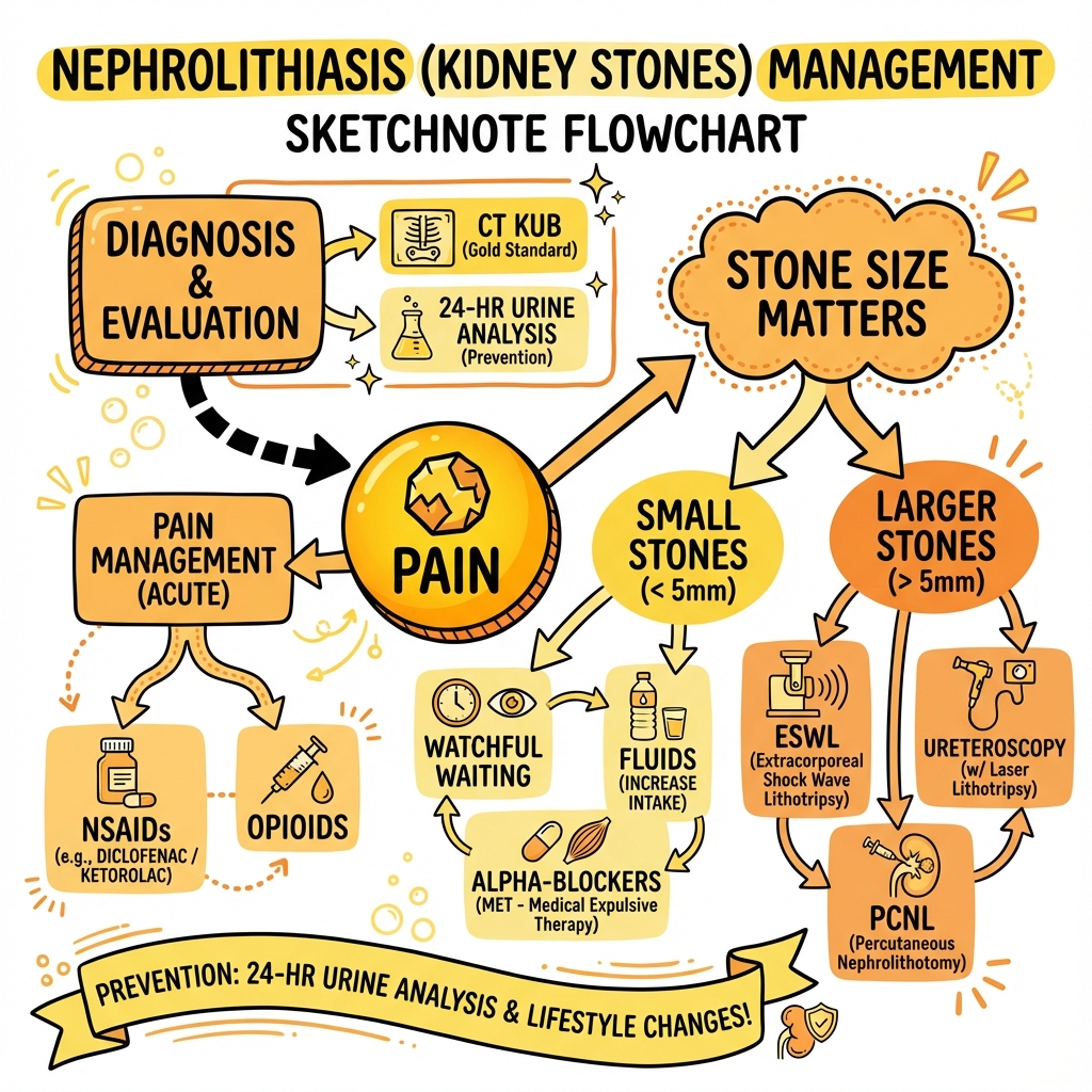
See the concept before reading it
Study the key anatomy, imaging, and decision pathways as full teaching plates.
Clinical board
A visual summary of the highest-yield teaching signals on this page.
Urgent signals
Safety-critical features pulled from the topic metadata.
- Infected obstructed kidney (pyonephrosis) - UROLOGICAL EMERGENCY
- Anuria (bilateral obstruction or solitary kidney)
- Acute kidney injury with obstruction
- Uncontrollable pain despite adequate analgesia
Exam focus
Current exam surfaces linked to this topic.
- FRCS(Urol)
Linked comparisons
Differentials and adjacent topics worth opening next.
- Acute Pyelonephritis
- Appendicitis
Content status and exam context
This page is AI-generated educational content. It may contain errors or omissions and is not a substitute for current guidelines, local protocols, senior clinical judgement, or professional medical advice.
MedVellum does not claim an individual clinician reviewer, board certification, or professional credential for this page unless a future version names a real, verifiable reviewer.
Clinical explanation and evidence
Kidney Stones (Nephrolithiasis)
1. Clinical Overview
Summary
Nephrolithiasis (kidney stones) is one of the most common urological conditions, affecting 10-12% of the global population with rising incidence worldwide. [1,2] The condition manifests as mineral crystallization within the urinary tract, most commonly composed of calcium oxalate (70-80% of cases). [3] Classical presentation features acute-onset severe colicky loin-to-groin pain accompanied by haematuria in 85-90% of patients. [4]
Non-contrast computed tomography of the kidneys, ureters, and bladder (CT KUB) represents the gold standard diagnostic investigation with sensitivity approaching 95-98% and specificity of 96-100%. [5,6] Management is stratified by stone size, location, and patient factors: stones less than 5mm pass spontaneously in 68-90% of cases, while larger stones typically require intervention. [7] Medical expulsive therapy with alpha-blockers (tamsulosin 0.4mg) increases passage rates for distal ureteric stones 5-10mm. [8]
Interventional options include extracorporeal shockwave lithotripsy (ESWL), ureteroscopy (URS), and percutaneous nephrolithotomy (PCNL), selected based on stone burden, composition, and anatomical factors. [9] Infected obstructed kidney (pyonephrosis) constitutes a urological emergency requiring immediate decompression via nephrostomy or ureteric stent, combined with intravenous antibiotics. [10]
Recurrence rates reach 50% at 5-10 years without preventive measures. [11] Metabolic evaluation and targeted prevention strategies—including adequate hydration (> 2.5L/day), dietary modification, and pharmacotherapy—can reduce recurrence by 50-60%. [12]
Key Facts
- Global Prevalence: 10-12% lifetime risk; increasing incidence in developed nations [1,2]
- Peak Incidence: 30-50 years; male predominance (M:F = 2-3:1) [13]
- Stone Composition: Calcium oxalate (70-80%), calcium phosphate (5-10%), uric acid (5-10%), struvite (10-20%), cystine (less than 1%) [3]
- Pathognomonic Presentation: Acute severe colicky loin-to-groin pain with restlessness and haematuria
- Gold Standard Imaging: Non-contrast CT KUB (sensitivity 95-98%, specificity 96-100%) [5,6]
- Spontaneous Passage: 68-90% for stones less than 5mm; 47% for 5-10mm stones [7]
- Recurrence: 50% at 5-10 years without metabolic evaluation and prevention [11]
- EMERGENCY: Fever + obstructing stone = pyonephrosis = immediate drainage required
Clinical Pearls
"Fever + Stone + Obstruction = EMERGENCY": Pyonephrosis (infected obstructed kidney) represents an acute urological emergency. These patients can rapidly deteriorate into urosepsis and require urgent decompression (nephrostomy tube or retrograde ureteric stent) alongside broad-spectrum antibiotics. Stone removal is deferred until infection clears. [10]
"Pain Severity ≠ Stone Size": A 3mm stone causing acute ureteric obstruction can produce excruciating pain (rated 10/10), while a 3cm staghorn calculus may be completely asymptomatic. Pain intensity correlates with degree of ureteric distension and obstruction, not stone dimensions. [14]
"Tamsulosin for Distal Stones 5-10mm": Medical expulsive therapy (MET) with alpha-1 blockers increases spontaneous passage rates for distal ureteric stones 5-10mm by 1.5-fold. The SUSPEND trial showed no benefit for stones less than 5mm, but meta-analyses confirm efficacy in the 5-10mm range. [8,15]
"Stone Analysis is Mandatory": Every first-time stone should undergo compositional analysis. Stone type dictates metabolic workup and guides specific prevention strategies (thiazides for calcium stones, alkalinization for uric acid stones, antibiotics for struvite). [16]
"CT Hounsfield Units Predict Stone Composition": HU values on CT can predict stone type and fragility: calcium oxalate monohydrate > 1000 HU (hard), uric acid less than 500 HU (radiolucent), struvite 600-900 HU. This influences treatment selection (ESWL vs URS). [17]
Why This Matters Clinically
Renal colic represents one of the most common presentations to emergency departments globally. Accurate differentiation between uncomplicated stone disease (suitable for conservative management) and complicated presentations requiring urgent intervention is critical. Failure to recognize infected obstructed kidney can result in urosepsis, multiorgan failure, and death within 24-48 hours. [10]
Long-term, nephrolithiasis associates with increased cardiovascular disease risk, chronic kidney disease progression, and metabolic syndrome. [18] Stone recurrence imposes substantial healthcare costs and patient morbidity—emphasizing the importance of metabolic evaluation and evidence-based prevention strategies.
2. Epidemiology and Risk Factors
Incidence and Prevalence
Global lifetime risk of kidney stones ranges from 10-12%, with significant geographic and temporal variation. [1,2] Incidence has increased 70% over the past three decades in developed nations, attributed to dietary factors (high sodium, animal protein intake) and rising obesity rates. [19]
Regional Variation:
- Highest prevalence: Middle East, North Africa, southern USA ("stone belt")
- Men > Women historically (2-3:1), but gap narrowing due to increased female obesity [13]
- Peak age: 30-50 years
- Recurrence: 50% at 5-10 years, 75% at 20 years without preventive measures [11]
Demographics
| Factor | Data | Notes |
|---|---|---|
| Sex | M:F = 2-3:1 (narrowing) | Gap closing due to dietary changes and obesity [13] |
| Age | Peak 30-50 years | Rare in children except metabolic disorders |
| Ethnicity | White > Hispanic > Asian > Black | May reflect dietary and genetic factors [20] |
| Geography | Hot climates, "stone belt" | Dehydration, concentrated urine |
| Seasonality | Higher summer/early autumn | Heat, dehydration, vitamin D synthesis [21] |
Risk Factors
Metabolic Risk Factors:
- Hypercalciuria: Most common metabolic abnormality (40-60% of calcium stone formers) [22]
- Hyperoxaluria: Primary (rare genetic) or secondary (dietary, malabsorption)
- Hyperuricosuria: Associated with high purine intake, gout, metabolic syndrome [23]
- Hypocitraturia: Citrate inhibits stone formation; low in RTA, chronic diarrhea [24]
- Cystinuria: Autosomal recessive defect in cystine transport (1-2% of stones)
Dietary Risk Factors:
- Low fluid intake (less than 1.5L/day) [25]
- High sodium diet (> 3g/day) → increased urinary calcium excretion [26]
- High animal protein intake → acidic urine, increased calcium and uric acid excretion
- Low calcium diet paradoxically increases stone risk (decreased oxalate binding) [27]
- High oxalate foods (spinach, rhubarb, nuts, chocolate)
Medical Conditions:
- Primary hyperparathyroidism (5% of stone formers) [28]
- Renal tubular acidosis (type I)
- Inflammatory bowel disease (oxalate hyperabsorption)
- Obesity and metabolic syndrome [29]
- Chronic UTI with urease-producing organisms (Proteus, Klebsiella) → struvite stones
Medications:
- Loop diuretics (hypercalciuria)
- Topiramate (metabolic acidosis, hypocitraturia)
- Carbonic anhydrase inhibitors
- Vitamin C/D supplements (high doses)
- Protease inhibitors (indinavir stones)
Anatomical Factors:
- Medullary sponge kidney
- Horseshoe kidney
- Ureteropelvic junction obstruction
- Calyceal diverticula
Stone Composition and Associated Conditions
| Stone Type | Frequency | pH | Radiopacity | Key Associations | Appearance |
|---|---|---|---|---|---|
| Calcium Oxalate | 70-80% | Any | Radio-opaque (dense) | Hypercalciuria, hyperoxaluria, hypocitraturia | Brown/black, spiculated |
| Calcium Phosphate | 5-10% | Alkaline (> 7.0) | Radio-opaque | RTA, hyperparathyroidism, alkaline urine | White, amorphous |
| Uric Acid | 5-10% | Acidic (less than 5.5) | Radiolucent | Gout, high purine diet, ileostomy, chronic diarrhea | Yellow/orange, smooth |
| Struvite (MgNH4PO4) | 10-20% | Alkaline (> 7.2) | Radio-opaque (moderate) | Chronic UTI (Proteus, Klebsiella, Pseudomonas) | Staghorn morphology |
| Cystine | less than 1% | Acidic | Faintly opaque | Cystinuria (autosomal recessive) | Yellow, hexagonal crystals |
| Mixed | 10-15% | Variable | Variable | Recurrent stones, metabolic disorders | Layered composition |
3. Pathophysiology
Stone Formation: The Crystallization Cascade
Stone formation is a multifactorial process involving supersaturation, nucleation, crystal growth, aggregation, and retention on urothelium. [30]
Sequential Steps:
-
Supersaturation: Urine becomes oversaturated with stone-forming salts (calcium, oxalate, uric acid, cystine) exceeding their solubility product. Concentration increases with low urine volume, high solute excretion, or both.
-
Nucleation: Crystal formation begins when supersaturation exceeds a critical threshold. Homogeneous nucleation occurs in pure solution; heterogeneous nucleation (more common) occurs on existing surfaces (cellular debris, matrix, other crystals).
-
Crystal Growth and Aggregation: Once nucleated, crystals grow by adding solute molecules from surrounding urine. Small crystals aggregate into larger particles through physicochemical attraction.
-
Crystal Retention: For clinically significant stones to form, crystals must be retained in the kidney rather than excreted. Randall's plaques (subepithelial calcium phosphate deposits at papillary tips) serve as nidus for calcium oxalate stone formation. [31]
-
Stone Growth: Progressive accretion over weeks to years produces macroscopic stones. Growth rate depends on urinary supersaturation, pH, flow rate, and inhibitor presence.
Promoters and Inhibitors
Stone Promoters:
- Low urine volume (less than 1.5L/day) → concentrated urine, increased supersaturation
- Calcium excretion > 250mg/day (men) or > 200mg/day (women)
- Oxalate excretion > 40mg/day
- Uric acid excretion > 750mg/day (men) or > 650mg/day (women)
- Low urinary pH (less than 5.5) → uric acid supersaturation
- High urinary pH (> 7.0) → calcium phosphate, struvite supersaturation
- Urinary stasis (anatomical abnormalities, dehydration)
Stone Inhibitors:
- Citrate: Chelates calcium, inhibits crystal nucleation and growth [24]
- Magnesium: Complexes with oxalate, reducing calcium oxalate supersaturation
- Pyrophosphate: Inhibits calcium phosphate crystallization
- Nephrocalcin: Glycoprotein that inhibits calcium oxalate crystal growth
- Tamm-Horsfall protein: Prevents crystal aggregation (controversial role)
- Osteopontin: Regulates crystal-cell interactions
Calcium Oxalate Stone Formation
Calcium oxalate stones form via two primary mechanisms:
Randall's Plaque Pathway (majority): Calcium phosphate deposits form in renal papillary interstitium, erode through urothelium, and serve as nidus for calcium oxalate overgrowth in collecting system. [31]
Free-Particle Pathway: Crystals form in tubular fluid, aggregate in collecting ducts, and are retained on papillary surface or in calyces.
Uric Acid Stone Formation
Uric acid solubility is pH-dependent: at pH less than 5.5, uric acid precipitates as crystals. [32] Three factors contribute:
- Low urinary pH (most important)
- Hyperuricosuria (> 750mg/day)
- Low urine volume
Unlike calcium stones, uric acid stones are potentially dissolvable with alkalinization (target pH 6.5-7.0).
Struvite Stone Formation
Struvite (magnesium ammonium phosphate) stones form exclusively in the presence of urease-producing bacteria (Proteus, Klebsiella, Pseudomonas). [33] Urease hydrolyzes urea to ammonia, alkalinizing urine (pH > 7.2) and creating conditions for struvite and carbonate apatite precipitation. These stones grow rapidly, often forming staghorn calculi filling the renal pelvis and calyces.
Cystine Stone Formation
Cystinuria results from autosomal recessive mutations in SLC3A1 or SLC7A9 genes, impairing proximal tubular reabsorption of cystine and dibasic amino acids. [34] Cystine solubility is pH-dependent; at physiological pH (less than 7.0), cystine precipitates. Target urinary pH > 7.5 for medical dissolution.
Pain Mechanism in Renal Colic
Pathophysiological Sequence:
- Stone enters ureter or obstructs ureteropelvic junction
- Proximal urinary flow obstruction → increased intraluminal pressure
- Renal pelvis and capsule distension → stretch receptors activated
- Prostaglandin release (PGE2, PGI2) → ureteric smooth muscle spasm and inflammation
- Visceral pain transmission via T10-L1 nerve roots → colicky loin-to-groin radiation
- Autonomic activation → nausea, vomiting, diaphoresis
Pain intensity correlates with rapidity of obstruction and degree of distension, not stone size. Slowly developing obstruction (large renal stone) may be painless, while acute ureteric obstruction from a small stone causes excruciating pain.
4. Clinical Presentation
Cardinal Symptoms
Renal Colic (acute ureteric obstruction):
- Acute severe pain: Sudden onset, rapidly escalates to maximum intensity within minutes to hours
- Pain character: Colicky (waxing/waning) or constant, described as "worst pain of my life"
- Radiation pattern: Depends on stone location (see table below)
- Restlessness: Patients constantly shift position seeking relief (contrast with peritonitis where patients lie still)
Associated Symptoms:
- Haematuria: Microscopic (85-90%) or macroscopic (30-40%) [4]
- Nausea and vomiting: Present in 50-80%; mediated by shared celiac plexus innervation
- Urinary symptoms: Frequency, urgency, dysuria (especially with distal ureteric/VUJ stones)
- Systemic symptoms: Usually absent unless infection present
Pain Patterns by Stone Location
| Stone Location | Pain Site | Radiation | Associated Features |
|---|---|---|---|
| Renal/upper ureteric | Loin, flank | To groin, ipsilateral testicle/labia | Nausea, vomiting prominent |
| Mid-ureteric | Lower abdomen, iliac fossa | Anterior abdominal wall | May mimic appendicitis (right) or diverticulitis (left) |
| Distal ureteric/VUJ | Suprapubic, groin | Ipsilateral testicle/labia majora | Urinary frequency, urgency, dysuria; mimics UTI/cystitis |
| Intra-renal (non-obstructing) | Dull ache or asymptomatic | None | May present incidentally on imaging |
Clinical Examination Findings
General Inspection:
- Patient in distress, unable to find comfortable position
- Pallor, diaphoresis if severe pain
- Tachycardia (pain-related; fever suggests infection)
- Typically afebrile unless infection present
Abdominal Examination:
- Inspection: Usually normal; no distension
- Palpation: Renal angle tenderness (costovertebral angle tenderness); abdomen typically soft without peritonism (differentiates from peritonitis)
- Percussion: Renal angle tenderness elicited by fist percussion over affected flank
- Auscultation: Bowel sounds present (unless prolonged ileus from retroperitoneal inflammation)
Signs of Complicated Stone Disease
Infected Obstructed Kidney (Pyonephrosis) - EMERGENCY:
- Fever > 38°C, rigors
- Tachycardia, hypotension (sepsis)
- Altered mental status (severe sepsis)
- Persistent pain despite analgesia
- Raised inflammatory markers (WCC > 15, CRP > 100)
Acute Kidney Injury:
- Reduced urine output (oliguria less than 400ml/24h or anuria less than 100ml/24h)
- Rising creatinine
- Bilateral obstruction or solitary kidney with obstruction
Fornical Rupture:
- Sudden severe pain followed by relief
- Urinoma formation (peri-renal fluid collection)
- Usually self-limiting but may require drainage if large
Differential Diagnosis
Critical to exclude life-threatening mimics and alternative pathology:
| Condition | Key Distinguishing Features | Critical Tests |
|---|---|---|
| Ruptured AAA | Age > 60, vascular risk factors, pulsatile abdominal mass, hypotension | Urgent CT angiogram; bedside USS |
| Appendicitis | Right iliac fossa pain, fever, peritonism, anorexia | FBC, CRP; CT abdomen/pelvis |
| Ectopic pregnancy | Female, childbearing age, amenorrhoea, PV bleeding | β-hCG; transvaginal USS |
| Ovarian torsion | Female, sudden unilateral pelvic pain, adnexal tenderness, nausea | Urgent pelvic USS with Doppler |
| Acute pyelonephritis | Fever prominent, rigors, pyuria, bacteriuria; less colicky pain | Urine culture; USS/CT if no improvement |
| Diverticulitis | Left iliac fossa pain, fever, altered bowel habit, elderly | CT abdomen/pelvis with IV contrast |
| Bowel obstruction | Abdominal distension, absolute constipation, vomiting | Erect CXR; CT abdomen/pelvis |
| Musculoskeletal pain | History of trauma/strain, worse with movement, no haematuria | Clinical diagnosis; imaging if uncertain |
5. Investigations
First-Line Investigations
Urinalysis (bedside):
- Haematuria: Present in 85-90% (microscopic or macroscopic); absence does not exclude stones [4]
- Leucocytes/Nitrites: Suggests concurrent UTI; if obstructing stone present → emergency
- pH: Acidic (less than 5.5) suggests uric acid stones; alkaline (> 7.2) suggests struvite or distal RTA
- Crystals: May identify stone type (calcium oxalate, uric acid, cystine), but low sensitivity
Blood Tests:
- FBC: WCC elevation suggests infection/inflammation
- U&E: Creatinine elevation indicates AKI; check if bilateral obstruction or solitary kidney
- CRP: Elevated in infection/inflammation; helps differentiate pyelonephritis
- Calcium, Phosphate, Uric Acid: Initial metabolic screen (full workup for recurrent stones)
Imaging - CT KUB (Non-Contrast):
- Gold standard for stone detection: sensitivity 95-98%, specificity 96-100% [5,6]
- Detects stones of all compositions including radiolucent uric acid
- Provides stone size (maximum diameter), location, number, Hounsfield unit density
- Assesses degree of obstruction (hydronephrosis, hydroureter, perinephric stranding)
- Identifies alternative pathology (AAA, appendicitis, masses)
- Limitations: Radiation exposure (~10 mSv); cost; contraindicated in pregnancy
CT KUB Findings:
- Direct stone visualization: Even small stones (2-3mm) detected
- Secondary signs of obstruction: Hydronephrosis, hydroureter, perinephric/periureteric stranding, renal enlargement
- "Tissue rim sign": Soft tissue around impacted ureteric stone (differentiates from phlebolith)
- Hounsfield Units (HU): Predicts stone composition and ESWL success:
- "Calcium oxalate monohydrate: > 1000 HU (hard, less amenable to ESWL)"
- "Calcium oxalate dihydrate: 500-1000 HU"
- "Calcium phosphate: 1000-1500 HU"
- "Struvite: 600-900 HU"
- "Uric acid: less than 500 HU (radiolucent on X-ray) [17]"
Low-Dose CT KUB: Reduces radiation to 2-3 mSv without compromising diagnostic accuracy. [35]
Alternative Imaging Modalities
Ultrasound Renal Tract:
- First-line in pregnancy, children, young adults
- Detects hydronephrosis (high sensitivity 95%) and large renal stones
- Limitations: Operator-dependent; poor sensitivity for ureteric stones (22-60%); misses small stones
- Twinkling artifact on color Doppler improves stone detection
Plain KUB Radiograph:
- Limited role: sensitivity 45-60%, misses radiolucent stones (uric acid, cystine, some struvite)
- Useful for monitoring known radio-opaque stones during follow-up
- Cannot differentiate stones from phleboliths or vascular calcification
Intravenous Urogram (IVU):
- Largely replaced by CT; historical gold standard
- Shows anatomical detail and function; identifies radiolucent stones as filling defects
- Contraindicated in renal impairment, contrast allergy
MR Urography:
- Excellent for anatomical detail without ionizing radiation
- Poor sensitivity for stone detection (30-70%)
- Role in pregnancy if USS inconclusive and CT avoided
Metabolic Workup for Recurrent Stone Formers
Indications:
- First stone in high-risk patient (children, family history, solitary kidney)
- Recurrent stone former (≥2 episodes)
- Stone composition suggesting metabolic cause (cystine, uric acid, struvite)
Initial Metabolic Screen (all patients):
- Serum: Calcium, phosphate, PTH, uric acid, creatinine, bicarbonate
- Urinalysis: pH, culture
- Stone analysis: Mandatory for first stone
24-Hour Urine Collection (×2 collections for reliability):
- Volume: Target > 2.0-2.5L/day
- Calcium: > 250mg/day (men) or > 200mg/day (women) = hypercalciuria
- Oxalate: > 40mg/day = hyperoxaluria
- Uric acid: > 750mg/day (men) or > 650mg/day (women) = hyperuricosuria
- Citrate: less than 320mg/day = hypocitraturia
- Sodium: > 3000mg/day indicates high dietary salt
- pH: Persistently less than 5.5 (uric acid risk) or > 7.0 (infection, RTA)
- Cystine: If cystine stones suspected (measure qualitatively then quantitatively)
Stone Compositional Analysis:
- Methods: Infrared spectroscopy or X-ray diffraction (gold standard)
- Guides metabolic workup and prevention strategy
- Mixed stones may indicate evolving metabolic abnormality
6. Management
Emergency Management: Infected Obstructed Kidney (Pyonephrosis)
┌──────────────────────────────────────────────────────────────────┐
│                 PYONEPHROSIS - UROLOGICAL EMERGENCY               │
├──────────────────────────────────────────────────────────────────┤
│  RECOGNITION: Fever + Obstructing Stone + Sepsis                 │
│                                                                  │
│  IMMEDIATE ACTIONS (within 1-2 hours):                           │
│    1. Sepsis Resuscitation: IV fluids, oxygen                   │
│    2. Blood Cultures × 2 sets                                    │
│    3. IV Antibiotics: Broad-spectrum Gram-negative cover         │
│       - Gentamicin 5-7mg/kg + amoxicillin 1g TDS                │
│       OR Tazocin 4.5g TDS                                        │
│    4. URGENT DECOMPRESSION (do NOT delay for antibiotics):      │
│       Option A: Percutaneous nephrostomy (PCN) - preferred      │
│       Option B: Retrograde ureteric stent (if safe cystoscopy)  │
│                                                                  │
│  DO NOT attempt primary stone treatment in infected setting     │
│  Definitive stone removal once sepsis resolved (2-4 weeks)      │
│                                                                  │
│  PROGNOSIS: Mortality 19-27% if treatment delayed > 24h [10]     │
└──────────────────────────────────────────────────────────────────┘
Acute Pain Management (Uncomplicated Renal Colic)
Analgesia:
-
First-line: NSAIDs (superior to opioids for renal colic) [36]
- Diclofenac 75mg IM/PR or 50mg PO TDS
- "Mechanism: Reduce renal blood flow and ureteric spasm via prostaglandin inhibition"
- "Contraindications: Renal impairment, AKI, peptic ulcer disease, bleeding risk"
- "Caution: Monitor renal function; avoid if eGFR less than 30"
-
Second-line: Opioids (if NSAIDs contraindicated or inadequate):
- Morphine 5-10mg IV/IM titrated to effect
- Pethidine 50-100mg IM (historical but less recommended)
- "Risk: Nausea, vomiting, respiratory depression; less effective than NSAIDs"
-
Combination therapy: NSAID + paracetamol ± opioid for severe pain
Antiemetics:
- Ondansetron 4-8mg IV/PO
- Metoclopramide 10mg IV/PO (prokinetic benefit)
- Cyclizine 50mg IV/PO
Fluid Management:
- Oral or IV hydration to maintain urine output
- No evidence that forced fluids improve stone passage; may worsen pain [37]
- Target euvolemia, avoid aggressive hydration
Medical Expulsive Therapy (MET)
Indications:
- Distal ureteric stones 5-10mm
- No signs of infection
- Controlled pain
- Normal renal function
Evidence:
- Alpha-blockers (tamsulosin 0.4mg OD) increase stone passage by 1.45-1.65× for distal stones 5-10mm [8]
- SUSPEND trial (2015): No benefit for stones less than 10mm unselected population [15]
- Meta-analyses: Confirm benefit specifically for distal stones 5-10mm [38]
- Number needed to treat (NNT): 5 patients to achieve one additional stone passage
Regimen:
- Tamsulosin 0.4mg OD (modified-release)
- Duration: Up to 4-6 weeks (average passage time 2-3 weeks)
- Follow-up imaging to confirm passage
- Analgesics as needed
Other agents (less commonly used):
- Nifedipine 30mg OD (calcium channel blocker)
- Combination therapy (tamsulosin + corticosteroid) – emerging evidence
Conservative Management (Watchful Waiting)
Indications:
- Small stones less than 5mm (spontaneous passage 68-90%) [7]
- Asymptomatic or minimally symptomatic
- No infection, no AKI
- Patient preference and compliance
Spontaneous Passage Rates by Size:
| Stone Size | Passage Rate | Mean Time to Passage |
|---|---|---|
| less than 2mm | 95% | Days to 2 weeks |
| 2-4mm | 80-90% | 1-3 weeks |
| 5-7mm | 60-70% | 3-6 weeks |
| 7-10mm | 30-50% | Weeks to months |
| > 10mm | less than 20% | Rarely spontaneous |
Passage also depends on location:
- Proximal ureter: Lower passage rates
- Distal ureter: Higher passage rates (especially VUJ)
Follow-Up Protocol:
- Repeat imaging (USS or low-dose CT) at 2-4 weeks
- Safety-netting: Return if fever, uncontrollable pain, anuria
- Stone progression or development of obstruction → escalate to intervention
Indications for Urgent Intervention
Absolute Indications:
- Infected obstructed kidney (pyonephrosis)
- Bilateral obstruction or obstruction of solitary kidney with AKI
- Anuria
- Uncontrollable pain despite adequate analgesia
Relative Indications:
- Stone > 10mm (unlikely to pass)
- Persistent obstruction > 4-6 weeks
- Occupational requirements (airline pilot, professional athlete)
- Patient preference
- Pregnancy with persistent symptoms (defer definitive treatment to postpartum if possible)
7. Interventional Management
Extracorporeal Shockwave Lithotripsy (ESWL)
Mechanism: Focused acoustic shockwaves fragment stones into smaller pieces that pass spontaneously.
Indications:
- Renal stones less than 20mm
- Upper/mid ureteric stones less than 10mm
- Low HU density (less than 1000 HU) for optimal fragmentation
Advantages:
- Non-invasive (no incision or endoscopy)
- Outpatient procedure
- No anesthesia or light sedation only
Success Rates:
- Renal stones less than 10 mm: 70-90% stone-free rate [39]
- Renal stones 10-20 mm: 50-70% (may require multiple sessions)
- Ureteric stones less than 10 mm: 80-90%
Complications:
- Steinstrasse (stone street): Fragments obstruct ureter (4-7%); may require stenting
- Renal haematoma (4-19%; usually subclinical)
- Perirenal haematoma
- Renal colic during fragment passage
- Infection (rare)
Contraindications:
- Pregnancy (absolute)
- Uncorrected coagulopathy or anticoagulation
- Untreated UTI
- Obstruction distal to stone
- Aortic aneurysm in shockwave path
- Morbid obesity (difficult targeting)
Predictors of Success:
- Stone size less than 10mm
- Stone location (lower pole stones less successful)
- Low HU density (less than 1000) [17]
- Skin-to-stone distance less than 10cm
Ureteroscopy (URS)
Mechanism: Endoscopic visualization and stone fragmentation using laser (Holmium:YAG) or pneumatic lithotripsy.
Types:
- Rigid ureteroscopy: Distal/mid ureteric stones
- Flexible ureteroscopy (f-URS): Proximal ureteric and intra-renal stones
Indications:
- Ureteric stones of any size (first-line for distal stones > 10mm)
- Renal stones less than 20mm (especially lower pole)
- ESWL failure or contraindication
- Need for definitive treatment in one session
- Hard stones (> 1000 HU)
Success Rates:
- Distal ureteric stones: 93-97% stone-free rate [40]
- Mid/proximal ureteric stones: 85-90%
- Renal stones less than 20 mm: 80-90%
Advantages:
- High stone-free rate in single session
- Direct visualization
- Can treat any stone composition
- Stone retrieval for analysis
Complications:
- Ureteric perforation (1-4%)
- Ureteric stricture (less than 1%)
- Ureteric avulsion (rare, less than 0.5%)
- Post-operative stent discomfort
- UTI/sepsis (2-4%)
- Bleeding (usually minor)
Post-Operative:
- Ureteric stent often placed (4.8Fr to 7Fr) for 1-2 weeks
- Stent symptoms: Frequency, dysuria, haematuria, flank pain
- Alpha-blockers may reduce stent discomfort
Percutaneous Nephrolithotomy (PCNL)
Mechanism: Percutaneous tract created from skin to kidney under radiological guidance; nephroscope inserted to fragment and extract stones.
Indications:
- Large renal stones > 20mm
- Staghorn calculi
- Lower pole stones > 15mm
- ESWL/URS failure
- Complex stone burden
Procedure:
- Performed under general anesthesia
- Prone or supine position (supine PCNL increasingly popular)
- Tract size: Standard (24-30Fr), mini-PCNL (14-20Fr), ultra-mini-PCNL (less than 14Fr), micro-PCNL (less than 10Fr)
- Stone fragmentation: Pneumatic, ultrasonic, laser, or combination
Success Rates:
- Stones > 20 mm: 80-95% stone-free rate in single session [41]
- Staghorn calculi: 70-80% (often requires multiple tracts or staged procedures)
- Superior to ESWL and URS for large stone burden
Complications:
- Bleeding requiring transfusion (5-7%; higher with large tracts)
- Fever/sepsis (10-15%)
- Renal pelvis/ureteric injury (2-8%)
- Hydrothorax/pneumothorax (1-2%; especially upper pole access)
- Adjacent organ injury (colon, liver, spleen) less than 1%
Post-Operative:
- Nephrostomy tube typically placed overnight (tubeless PCNL in selected cases)
- Monitor for bleeding, urine output
- Follow-up imaging (CT or X-ray) to assess residual fragments
Mini-PCNL/Micro-PCNL: Smaller tracts reduce bleeding risk, comparable stone-free rates for stones less than 30mm. [42]
Open or Laparoscopic Stone Surgery
Indications (rare, less than 1% of cases):
- Failed minimally invasive approaches
- Complex anatomy (severe PUJ obstruction, calyceal diverticulum)
- Very large/complex stone burden with anatomical abnormality requiring correction
- Concomitant pathology requiring open surgery (e.g., renal tumor)
Procedures:
- Pyelolithotomy (stone in renal pelvis)
- Nephrolithotomy (stone in kidney parenchyma)
- Ureterolithotomy (stone in ureter)
- Anatrophic nephrolithotomy (staghorn with intrarenal extension)
8. Stone-Specific Management Strategies
Calcium Oxalate Stones
Acute Management: As per general principles (analgesia, MET, or intervention based on size/location).
Prevention:
-
Hydration: > 2.5L/day to achieve urine output > 2.0L/day [25]
-
Dietary modification:
- Normal calcium intake (1000-1200mg/day); do NOT restrict (increases oxalate absorption) [27]
- Reduce sodium (less than 3g/day) – decreases urinary calcium excretion [26]
- Moderate animal protein (0.8-1.0g/kg/day)
- Reduce oxalate-rich foods (spinach, rhubarb, nuts, chocolate, tea)
- Increase citrate-rich foods (lemon juice, orange juice)
-
Pharmacotherapy (if dietary measures insufficient):
- "Thiazide diuretics (for hypercalciuria): Indapamide 2.5mg OD or hydrochlorothiazide 25mg BD"
- Mechanism: Increase distal tubular calcium reabsorption, reduce urinary calcium
- Side effects: Hypokalemia, hyperglycemia, hyperuricemia
- "Potassium citrate (for hypocitraturia): 30-60 mEq/day in divided doses"
- Mechanism: Increases urinary citrate (inhibits crystallization), alkalinizes urine
- Side effects: GI upset, hyperkalemia
- "Thiazide diuretics (for hypercalciuria): Indapamide 2.5mg OD or hydrochlorothiazide 25mg BD"
Uric Acid Stones
Acute Management: As per general principles.
Unique Feature: Uric acid stones can be dissolved medically (unlike calcium stones).
Prevention and Dissolution:
-
Alkalinization (first-line):
- Target urinary pH 6.5-7.0 (use pH paper to monitor)
- Potassium citrate 30-80 mEq/day in divided doses
- Sodium bicarbonate 650mg TDS (alternative; beware sodium load)
- Monitor pH 3-4× daily during initial titration
-
Hydration: > 2.5L/day
-
Reduce purine intake: Limit red meat, organ meats, shellfish, alcohol (especially beer)
-
Allopurinol (for hyperuricosuria or gout):
- 100-300mg OD
- "Mechanism: Xanthine oxidase inhibitor, reduces uric acid production"
- Monitor for hypersensitivity reaction, hepatotoxicity
Dissolution Protocol:
- Achievable for pure uric acid stones less than 15mm
- Requires strict pH control (6.5-7.0) for 3-6 months
- Serial imaging to monitor stone shrinkage
Struvite Stones
Pathogenesis: Require urease-producing bacteria (Proteus, Klebsiella, Pseudomonas).
Acute Management: Urgent treatment if infected (as per pyonephrosis protocol).
Definitive Management:
- Complete stone removal is essential (stones perpetuate infection)
- PCNL is treatment of choice (stones often large/staghorn)
- Combination PCNL + ESWL for residual fragments
Antibiotic Therapy:
- Post-operative prolonged antibiotics (3-6 months) based on culture
- Acetohydroxamic acid (urease inhibitor): 250mg TDS-QDS
- Adjunct to slow stone growth while planning intervention
- "Side effects: Headache, tremor, thrombophlebitis (limits use)"
Long-Term Prevention:
- Eradicate infection
- Acidify urine (vitamin C, cranberry juice) – controversial efficacy
- Surveillance imaging every 6-12 months
Cystine Stones
Background: Autosomal recessive cystinuria; excretion > 250mg/day.
Acute Management: As per general principles (often resistant to ESWL due to hardness).
Prevention (lifelong):
-
Hydration: Very high fluid intake (3-4L/day) to achieve urine output > 3L/day
-
Alkalinization: Target urinary pH > 7.5 (higher than other stone types)
- Potassium citrate 60-90 mEq/day
-
Cystine-Binding Agents (if above measures insufficient):
- "Tiopronin (alpha-mercaptopropionylglycine): 800-1200mg/day in divided doses"
- Mechanism: Forms soluble disulfide with cystine
- Side effects: Rash, proteinuria, nephrotic syndrome (monitor urinalysis)
- "Penicillamine: 1-2g/day (alternative; more side effects)"
- "Tiopronin (alpha-mercaptopropionylglycine): 800-1200mg/day in divided doses"
Interventional Management:
- URS preferred (ESWL less effective)
- PCNL for large stones
- Chemolysis (dissolution) via percutaneous irrigation with alkalinizing agents
9. Complications
Acute Complications
| Complication | Incidence | Mechanism | Management |
|---|---|---|---|
| Pyonephrosis | 5-10% with obstruction + UTI | Bacterial proliferation in obstructed system | Emergency drainage (PCN/stent) + IV antibiotics [10] |
| Acute Kidney Injury | 10-20% with obstruction | Bilateral obstruction or solitary kidney obstruction | Urgent decompression; may require temporary dialysis |
| Anuria | Rare | Complete bilateral obstruction | Immediate bilateral drainage |
| Forniceal Rupture | 1-3% | High-pressure obstruction → calyceal rupture | Usually self-limiting; urinoma drainage if large |
| Sepsis/Urosepsis | 2-5% | Bacterial translocation in obstructed infected system | Sepsis protocol + urgent drainage |
Chronic Complications
Recurrent Stone Formation:
- 50% recurrence at 5-10 years without prevention [11]
- 75% at 20 years
- Reduced to 20-30% at 10 years with metabolic workup and prevention
Chronic Kidney Disease:
- Recurrent obstruction and infection → progressive nephron loss
- Stone formers have 2-3× increased risk of CKD progression [18]
- Staghorn calculi associated with renal parenchymal damage
Urinary Tract Infection:
- Recurrent UTIs in 15-20% of stone formers
- Struvite stones act as nidus for biofilm formation
Cardiovascular Disease:
- Epidemiological association between nephrolithiasis and hypertension, coronary artery disease [18]
- Shared risk factors: metabolic syndrome, insulin resistance, obesity
Complications of Interventions
ESWL:
- Steinstrasse (4-7%): Fragments obstruct ureter → stenting or URS
- Perirenal haematoma (4-19%): Usually resolves; rarely requires intervention
- Hypertension (controversial; some studies suggest long-term risk)
Ureteroscopy:
- Ureteric perforation (1-4%): Managed with prolonged stenting (4-6 weeks)
- Ureteric stricture (less than 1%): May require endoscopic or open repair
- Ureteric avulsion (less than 0.5%): Surgical emergency; requires ureteric reimplantation or nephrectomy
PCNL:
- Bleeding requiring transfusion (5-7%): May require angioembolization
- Sepsis (10-15%): Prophylactic antibiotics reduce risk
- Visceral injury (colon, liver, spleen) less than 1%: May require laparotomy
10. Prognosis and Outcomes
Stone Passage Outcomes
- Spontaneous passage rates: Highly dependent on stone size and location (see Section 6).
- Time to passage: Majority of stones that will pass do so within 4-6 weeks.
- Failure to pass: Stones > 10mm, proximal location, and high HU density predict need for intervention.
Stone-Free Rates by Modality
| Intervention | Stone Size | Stone-Free Rate | Sessions Required |
|---|---|---|---|
| ESWL | less than 10mm renal | 70-90% | 1-2 sessions (mean 1.4) |
| ESWL | 10-20mm renal | 50-70% | 2-3 sessions |
| URS | Distal ureteric | 93-97% | Single session |
| URS | Renal less than 20mm | 80-90% | Single session |
| PCNL | > 20mm renal | 80-95% | Single session |
| PCNL | Staghorn | 70-80% | May require staged procedures |
Recurrence Rates and Prevention
Natural History (without prevention):
- 50% recurrence at 5-10 years [11]
- 75% at 20 years
With Metabolic Evaluation and Prevention:
- 20-30% recurrence at 10 years (50-60% relative risk reduction) [12]
- Greatest benefit in high-risk recurrent stone formers
High-Risk Features for Recurrence:
- Age less than 25 years at first stone
- Family history
- Specific metabolic abnormalities (cystinuria, hyperparathyroidism, RTA)
- Multiple stones at presentation
- Residual fragments after intervention
Long-Term Health Outcomes
- Chronic Kidney Disease: Stone formers have 2-3× increased risk of CKD progression compared to non-stone formers [18]
- Cardiovascular Disease: Increased risk of hypertension (OR 1.3-1.8), coronary artery disease, metabolic syndrome [29]
- Bone Health: Hypercalciuria may be associated with reduced bone mineral density
- Quality of Life: Recurrent stone episodes significantly impair QOL; preventive strategies improve outcomes
11. Prevention Strategies
Universal Recommendations (All Stone Types)
Hydration (single most important intervention):
- Increase fluid intake to produce > 2.0-2.5L urine/day [25]
- Randomized controlled trial (Borghi et al.): High fluid intake reduced 5-year recurrence from 27% to 12% [43]
- Advise: Drink enough to keep urine pale yellow
- Distribute fluids throughout day and evening
Dietary Modifications:
- Normal calcium intake (1000-1200mg/day): Low calcium diet increases stone risk [27]
- Reduce sodium (less than 3g/day or less than 2000mg/day): High sodium increases urinary calcium excretion [26]
- Moderate animal protein (0.8-1.0g/kg/day): High protein increases urinary calcium, uric acid, and acidifies urine
- Increase fruits and vegetables: Provides citrate and alkali, reduces acid load
- Limit oxalate (if hyperoxaluria): Reduce spinach, rhubarb, nuts, chocolate, tea
Pharmacological Prevention
Thiazide Diuretics (calcium stone formers with hypercalciuria):
- Indapamide 2.5mg OD or hydrochlorothiazide 25mg BD
- Reduces urinary calcium excretion by 30-50%
- Monitor potassium, glucose, uric acid
- Add potassium citrate to prevent hypokalemia and hypocitraturia
Potassium Citrate (hypocitraturia, acidic urine):
- 30-60 mEq/day in divided doses (10-20 mEq TDS)
- Increases urinary citrate (crystallization inhibitor) and pH
- Monitor for hyperkalemia (especially if renal impairment)
Allopurinol (hyperuricosuria, uric acid stones, calcium oxalate stones with hyperuricosuria):
- 100-300mg OD
- Reduces uric acid production
- Monitor for hypersensitivity, hepatotoxicity
Cystine-Binding Agents (cystinuria):
- Tiopronin 800-1200mg/day
- Penicillamine 1-2g/day (higher side effect profile)
Urease Inhibitors (struvite stones, as adjunct):
- Acetohydroxamic acid 250mg TDS-QDS
- Significant side effects limit use; complete stone removal preferred
Metabolic-Specific Strategies
| Metabolic Abnormality | First-Line Intervention | Second-Line | Target |
|---|---|---|---|
| Hypercalciuria | Thiazide diuretic + normal calcium diet | Reduce sodium, animal protein | Urine Ca less than 200mg/day (women), less than 250mg/day (men) |
| Hyperoxaluria | Reduce dietary oxalate, normal calcium intake | Calcium supplements with meals (bind oxalate) | Urine oxalate less than 40mg/day |
| Hyperuricosuria | Reduce purine intake, increase hydration | Allopurinol 100-300mg OD | Urine uric acid less than 750mg/day (men), less than 650mg/day (women) |
| Hypocitraturia | Potassium citrate 30-60 mEq/day | Increase fruit/vegetable intake | Urine citrate > 320mg/day |
| Low urine pH | Potassium citrate to target pH 6.5-7.0 | Sodium bicarbonate | Urine pH 6.5-7.0 (uric acid stones) |
| High urine pH | Treat underlying infection (if struvite) | Acidify urine (limited role) | Urine pH less than 7.0 |
| Cystinuria | High hydration (> 4L/day), alkalize pH > 7.5 | Tiopronin, penicillamine | Urine cystine less than 250mg/L |
Specific Underlying Conditions
Primary Hyperparathyroidism:
- Parathyroidectomy is definitive treatment [28]
- Cures hypercalciuria and reduces stone recurrence by 90%
- Indications for surgery: Symptomatic stones, recurrent stones, age less than 50, osteoporosis
Renal Tubular Acidosis (Type I):
- Potassium citrate to correct acidosis and hypocitraturia
- Target serum bicarbonate > 22 mmol/L
Enteric Hyperoxaluria (IBD, short bowel, malabsorption):
- Low oxalate, low fat diet
- Calcium supplements with meals (bind oxalate in gut)
- Cholestyramine if bile acid malabsorption
- Probiotics (Oxalobacter formigenes) under investigation
12. Guidelines and Evidence
Key International Guidelines
European Association of Urology (EAU) Guidelines on Urolithiasis (2024):
- Non-contrast CT KUB as gold standard imaging
- ESWL or URS equally acceptable for renal stones less than 20mm
- PCNL first-line for stones > 20mm
- MET recommended for distal ureteric stones 5-10mm
- Metabolic evaluation for recurrent stone formers
American Urological Association (AUA) Guidelines:
- Tamsulosin for distal stones 5-10mm to facilitate passage
- Active treatment (URS/ESWL) vs observation for asymptomatic renal stones less than 20 mm: shared decision-making
- Metabolic workup for all recurrent stone formers
- 24-hour urine collection ×2 for metabolic evaluation
National Institute for Health and Care Excellence (NICE) NG118: Renal and Ureteric Stones (2019):
- NSAIDs first-line for pain (diclofenac or ibuprofen)
- CT KUB preferred imaging (low-dose protocols)
- Alpha-blockers (tamsulosin) for stones likely to pass with MET
- Active surveillance acceptable for asymptomatic non-obstructing renal stones
Landmark Trials and Evidence
SUSPEND Trial (Pickard et al., Lancet 2015) [15]:
- RCT: 1167 patients with ureteric stones less than 10mm
- Tamsulosin vs placebo: No significant difference in spontaneous passage rate (81% vs 80%)
- Criticism: Unselected population (included very small stones likely to pass regardless); did not stratify by stone size 5-10mm where benefit exists
Meta-Analysis (Hollingsworth et al., Lancet 2016) [38]:
- 55 RCTs, 5990 patients
- Alpha-blockers increased stone passage by 50% (RR 1.5)
- Greatest benefit for stones > 5mm and distal location
- NNT = 5 for one additional stone passage
Fluid Intake and Recurrence Prevention (Borghi et al., J Urol 1996) [43]:
- RCT: 199 first-time stone formers
- High fluid intake (target urine output > 2L/day) vs control
- 5-year recurrence: 12% (intervention) vs 27% (control), pless than 0.001
- Key message: Simple hydration halves recurrence risk
Thiazide Diuretics for Prevention (Ettinger et al., NEJM 1997):
- RCT: 124 recurrent calcium stone formers
- Thiazide diuretic reduced 3-year recurrence by 50%
- Benefit maintained in long-term follow-up
Dietary Calcium and Stone Risk (Curhan et al., NEJM 1993):
- Prospective cohort: 45,619 men
- Higher dietary calcium intake associated with lower stone risk (RR 0.56)
- Low calcium diet increases oxalate absorption and stone risk [27]
13. Special Populations
Pregnancy
Challenges:
- Physiological hydronephrosis (right > left) complicates diagnosis
- Radiation exposure concerns for CT
- Limited analgesic/pharmacological options
Diagnosis:
- First-line imaging: Ultrasound renal tract (safe, no radiation)
- If USS non-diagnostic: MR urography (no ionizing radiation)
- CT KUB: Reserved for complex cases; low-dose protocols minimize fetal radiation (less than 3 mGy)
Management:
- Conservative: Preferred if possible; hydration, analgesia (paracetamol, avoid NSAIDs in 3rd trimester)
- Temporizing drainage: Ureteric stent or nephrostomy if persistent obstruction, infection, pain
- Definitive intervention: Defer URS/ESWL to postpartum (ESWL contraindicated in pregnancy)
- Timing: Most stones pass spontaneously; intervention reserved for refractory cases
Pediatric Patients
Epidemiology:
- Increasing incidence in children (dietary factors, obesity)
- Higher proportion of metabolic causes (cystinuria, primary hyperoxaluria)
Diagnosis:
- Ultrasound first-line (avoid radiation)
- Low-dose CT KUB if USS non-diagnostic
Management:
- Conservative preferred; children pass stones more readily than adults
- URS and ESWL safe and effective; PCNL for large stones
- Metabolic workup essential: High yield for inherited disorders
Solitary Kidney
Special Considerations:
- Any obstruction risks AKI → lower threshold for intervention
- Urgent drainage if obstructed
- Aggressive metabolic evaluation and prevention
- Avoid nephrotoxic agents (NSAIDs, contrast if eGFR low)
Chronic Kidney Disease
Challenges:
- Increased stone risk in CKD (impaired urine concentration, metabolic acidosis)
- Limited use of NSAIDs (worsen renal function)
- Contrast-induced nephropathy risk (use non-contrast CT)
- Adjust drug doses for renal impairment
14. Exam-Focused Content
Common FRCS/MRCS Viva Questions
Q1: "A 35-year-old man presents with acute right loin-to-groin pain. How would you assess and manage him?"
Model Answer: "This presentation is highly suggestive of renal colic from a ureteric stone. I would assess him systematically:
History: Characterize the pain—onset, severity, radiation, nature (colicky vs constant). Ask about haematuria, urinary symptoms, fever, previous stones, family history, and medications. Systemic review for red flags (anuria, fever).
Examination: Patient typically restless. Check vital signs—fever suggests infection (emergency if obstructed). Examine abdomen for renal angle tenderness, peritonism (exclude differentials like AAA, appendicitis).
Investigations:
- Bedside: Urinalysis (haematuria in 85-90%, but absence doesn't exclude stones), pregnancy test in women
- Bloods: FBC, CRP, U&E (check renal function), calcium
- Imaging: Non-contrast CT KUB—gold standard, 95-98% sensitivity. Shows stone size, location, HU density, degree of obstruction.
Management:
- Analgesia: NSAIDs first-line (diclofenac 75mg IM/PR)—superior to opioids for renal colic
- Antiemetic: Ondansetron or metoclopramide
- Hydration: Oral or IV to euvolemia; avoid forced fluids
- Definitive management depends on CT findings:
- less than 5 mm: Conservative, 90% pass spontaneously
- 5-10mm distal ureter: Medical expulsive therapy (tamsulosin 0.4mg OD)
-
10mm or failed conservative: Intervention (URS/ESWL)
- "Emergency: Fever + obstruction = pyonephrosis → immediate drainage (nephrostomy/stent) + IV antibiotics"
Safety-netting: Advise return if fever, anuria, uncontrollable pain."
Q2: "What are the indications for urgent intervention in a patient with a ureteric stone?"
Model Answer: "Urgent intervention is required for:
Absolute indications:
-
Infected obstructed kidney (pyonephrosis): Fever + stone + obstruction. This is a urological emergency—mortality 19-27% if delayed. Requires immediate decompression (percutaneous nephrostomy or retrograde stent) + IV broad-spectrum antibiotics. Do NOT attempt primary stone removal.
-
Bilateral obstruction or obstruction of solitary kidney with AKI: Risks anuria and renal failure. Urgent drainage required.
-
Anuria: Complete bilateral obstruction. Immediate bilateral drainage.
-
Uncontrollable pain: Despite adequate analgesia (NSAIDs + opioids). Intervention for symptom relief.
Relative indications:
- Stone > 10mm (unlikely to pass spontaneously; less than 20% passage rate)
- Persistent obstruction > 4-6 weeks
- Occupational/patient preference (e.g., airline pilot cannot afford recurrent episodes)
- Pregnancy with refractory symptoms (temporizing stent, defer definitive treatment)
The key is recognizing infected obstruction—this is immediately life-threatening and requires urgent sepsis resuscitation and drainage within 1-2 hours."
Q3: "Describe the treatment options for a 2cm lower pole renal calculus."
Model Answer: "A 2cm lower pole renal calculus requires active intervention; spontaneous passage is unlikely. I would discuss three main options with the patient:
1. Extracorporeal Shockwave Lithotripsy (ESWL):
- Non-invasive, outpatient, no general anesthesia
- Success rate 50-70% for 2cm stones; lower for lower pole location (poor drainage)
- May require multiple sessions (mean 2-3)
- Contraindications: pregnancy, coagulopathy, untreated infection
- Complications: steinstrasse (4-7%), haematoma, requires fragment passage
- Best for: Patients preferring non-invasive approach, accepting multiple sessions
- Poor outcomes if: HU > 1000 (hard stone), skin-to-stone distance > 10cm, lower pole location
2. Flexible Ureteroscopy (f-URS):
- Endoscopic, single session, high stone-free rate (80-90%)
- Holmium laser lithotripsy fragments stone; basket retrieval
- General anesthesia, day-case or overnight
- Stent usually required 1-2 weeks (discomfort)
- Complications: ureteric injury (1-4%), infection
- Best for: Patients wanting definitive treatment, hard stones (high HU)
3. Percutaneous Nephrolithotomy (PCNL):
- Invasive: percutaneous tract into kidney
- Highest stone-free rate (80-95%) in single session for 2cm stones
- General anesthesia, 1-2 day admission
- Complications: bleeding (5-7%), infection (10-15%), visceral injury (less than 1%)
- Best for: Large stone burden, failed ESWL/URS, patient preferring single definitive treatment
My recommendation: For a 2cm lower pole stone, I would favor PCNL or f-URS over ESWL due to lower pole location (poor ESWL drainage). Choice between PCNL and f-URS depends on stone hardness (HU), patient fitness for PCNL, and patient preference. I would arrange stone analysis post-treatment and metabolic workup to prevent recurrence."
Q4: "What is the role of medical expulsive therapy (MET) in ureteric stones? Discuss the evidence."
Model Answer: "Medical expulsive therapy uses alpha-1 adrenergic antagonists (tamsulosin 0.4mg OD) to relax ureteric smooth muscle and facilitate spontaneous stone passage.
Indications:
- Distal ureteric stones 5-10mm
- No infection, no AKI
- Controlled pain
- Patient willing for conservative approach with follow-up
Evidence: The evidence is mixed but supports use for specific stone sizes:
-
Meta-analyses (Hollingsworth et al., Lancet 2016): 55 RCTs, 5990 patients. Alpha-blockers increased passage rate by 50% (RR 1.5), greatest benefit for stones > 5mm and distal location. NNT = 5.
-
SUSPEND Trial (Pickard et al., Lancet 2015): Large UK RCT, 1167 patients with stones less than 10mm. Found no benefit of tamsulosin vs placebo (81% vs 80% passage). However, criticized for including very small stones (less than 5mm) likely to pass regardless, diluting the effect.
-
Current consensus: EAU and AUA guidelines recommend MET for distal ureteric stones 5-10mm. Benefit likely negligible for stones less than 5mm (pass anyway) and > 10mm (unlikely to pass regardless).
Mechanism: Alpha-1 receptors concentrated in distal ureter; blockade reduces spasm and facilitates passage.
Regimen: Tamsulosin 0.4mg OD for 4-6 weeks; follow-up imaging to confirm passage; analgesics as needed.
Alternative agents: Nifedipine (calcium channel blocker) also studied; less robust evidence. Combination therapy (tamsulosin + corticosteroid) under investigation.
Bottom line: Recommend MET for distal stones 5-10mm; counsel regarding evidence and need for follow-up."
High-Yield Facts for Exams
-
Stone composition: Calcium oxalate 70-80% > uric acid/struvite 10-20% > cystine less than 1%
-
CT KUB: Gold standard, 95-98% sensitivity, 96-100% specificity; detects all stone types including radiolucent uric acid
-
Hounsfield Units: > 1000 HU = hard stone (calcium oxalate monohydrate), poor ESWL success; less than 500 HU = uric acid (radiolucent)
-
Spontaneous passage rates: less than 5mm = 68-90%; 5-10mm = 30-50%; > 10mm = less than 20%
-
Emergency management: Fever + stone + obstruction = pyonephrosis → immediate drainage (nephrostomy/stent) + IV antibiotics; mortality 19-27% if delayed
-
Analgesia: NSAIDs first-line (superior to opioids); diclofenac 75mg IM/PR
-
MET: Tamsulosin 0.4mg OD for distal stones 5-10mm; increases passage by 1.5× (NNT = 5)
-
Intervention thresholds: ESWL for renal stones less than 20mm; URS for ureteric stones > 10mm; PCNL for renal stones > 20mm or staghorn
-
Recurrence: 50% at 5-10 years without prevention; hydration > 2.5L/day reduces recurrence by 50%
-
Metabolic causes: Hypercalciuria (40-60%), hyperoxaluria, hyperuricosuria, hypocitraturia; screen with 24-hour urine ×2
-
Primary hyperparathyroidism: Present in 5% of stone formers; parathyroidectomy cures hypercalciuria and prevents recurrence
-
Uric acid stones: Radiolucent on X-ray; acidic urine less than 5.5; can dissolve with alkalinization (potassium citrate, target pH 6.5-7.0)
-
Struvite stones: Infection stones (Proteus, Klebsiella); alkaline urine > 7.2; staghorn morphology; require complete surgical removal + antibiotics
-
Cystine stones: Autosomal recessive cystinuria; requires high hydration (> 4L/day), alkalinization (pH > 7.5), ± tiopronin
-
Steinstrasse: Stone fragments obstruct ureter post-ESWL (4-7%); may require stenting or URS
15. Patient Information
What Are Kidney Stones?
Kidney stones are hard mineral deposits that form inside your kidneys when your urine becomes too concentrated. They can range in size from a grain of sand to a golf ball. When stones move from your kidney into the narrow tube (ureter) that carries urine to your bladder, they can cause severe pain.
What Causes Kidney Stones?
Stones form when certain substances in your urine (calcium, oxalate, uric acid) become too concentrated and form crystals. Common risk factors include:
- Not drinking enough water
- Eating too much salt or protein
- Family history of kidney stones
- Certain medical conditions (gout, repeated urinary infections)
- Some medications
What Are the Symptoms?
- Severe pain: Sudden, intense pain in your back, side, lower abdomen, or groin. The pain may come in waves and fluctuate in intensity.
- Blood in urine: Your urine may appear pink, red, or brown.
- Nausea and vomiting: Very common with kidney stone pain.
- Frequent urination: Feeling you need to urinate more often, or burning when urinating.
- Restlessness: Inability to find a comfortable position.
When Should I Seek Emergency Care?
Go to A&E or call 999 if you have:
- Fever or chills with kidney stone pain (suggests infection—this is serious)
- Inability to pass any urine
- Severe pain that you cannot control with painkillers
- Blood in urine with pain and fever
- Feeling very unwell, confused, or short of breath
How Are Kidney Stones Diagnosed?
- Urine test: Checks for blood and signs of infection
- Blood tests: Check kidney function and chemical imbalances
- CT scan: The best test to see kidney stones; takes detailed pictures without needing dye injection
How Are Kidney Stones Treated?
Treatment depends on the size and location of your stone:
Small stones (less than 5mm):
- Often pass naturally when you urinate (in 1-4 weeks)
- Drink plenty of fluids (2-3 liters per day)
- Take painkillers (ibuprofen or diclofenac work best for kidney stone pain)
- Your doctor may prescribe a medication (tamsulosin) to help the stone pass
Larger stones or stones that don't pass:
- Shockwave lithotripsy (ESWL): Sound waves break the stone into smaller pieces from outside your body. No surgery needed.
- Ureteroscopy: A thin telescope is passed through your bladder to the stone. The stone is broken up with a laser and removed. You may need a temporary tube (stent) in your ureter for 1-2 weeks.
- Percutaneous nephrolithotomy (PCNL): For very large stones. A small tube is inserted through your back into your kidney to remove the stone. Requires general anesthesia and overnight stay.
How Can I Prevent Future Stones?
Most important: Drink plenty of water:
- Aim for at least 2-3 liters of fluids per day
- Your urine should be pale yellow
Dietary changes:
- Don't cut out calcium—this can actually increase stone risk. Maintain normal dairy intake.
- Do reduce salt (sodium)—high salt increases stone risk.
- Do limit meat, fish, and poultry to moderate amounts.
- Avoid or limit high-oxalate foods if you have calcium oxalate stones (spinach, rhubarb, chocolate, nuts, tea).
- Drink lemon juice or orange juice (citrate helps prevent stones).
Medications (if recommended by your doctor):
- Water tablets (thiazides) to reduce calcium in urine
- Potassium citrate to make urine less acidic
- Allopurinol if you have uric acid stones or gout
Follow-up:
- Keep any stones you pass—they should be analyzed to determine the type
- Your doctor may recommend urine and blood tests to find out why you formed stones
- Based on test results, a personalized prevention plan can be created
What Is My Risk of Getting Another Stone?
- Without prevention measures: 50% chance of another stone within 5-10 years
- With proper hydration and dietary changes: Risk can be reduced by half
- Some people need long-term medication to prevent recurrence
Questions to Ask Your Doctor
- What type of stone do I have, and what caused it?
- Do I need any tests to find out why I formed a stone?
- What is the best treatment option for my stone?
- How can I prevent future stones?
- When should I return for follow-up?
- What warning signs should I watch for?
16. References
-
Scales CD Jr, Smith AC, Hanley JM, Saigal CS. Prevalence of kidney stones in the United States. Eur Urol. 2012;62(1):160-165. doi:10.1016/j.eururo.2012.03.052
-
Sorokin I, Mamoulakis C, Miyazawa K, Rodgers A, Talati J, Lotan Y. Epidemiology of stone disease across the world. World J Urol. 2017;35(9):1301-1320. doi:10.1007/s00345-017-2008-6
-
Daudon M, Traxer O, Conort P, Lacour B, Jungers P. Type 2 diabetes increases the risk for uric acid stones. J Am Soc Nephrol. 2006;17(7):2026-2033. doi:10.1681/ASN.2006030262
-
Shoag J, Halpern J, Goldfarb DS, Eisner BH. Risk of chronic and end stage kidney disease in patients with nephrolithiasis. J Urol. 2014;192(5):1440-1445. doi:10.1016/j.juro.2014.05.117
-
Smith RC, Rosenfield AT, Choe KA, et al. Acute flank pain: comparison of non-contrast-enhanced CT and intravenous urography. Radiology. 1995;194(3):789-794. doi:10.1148/radiology.194.3.7862980
-
Türk C, Petřík A, Sarica K, et al. EAU Guidelines on Diagnosis and Conservative Management of Urolithiasis. Eur Urol. 2016;69(3):468-474. doi:10.1016/j.eururo.2015.07.040
-
Coll DM, Varanelli MJ, Smith RC. Relationship of spontaneous passage of ureteral calculi to stone size and location as revealed by unenhanced helical CT. AJR Am J Roentgenol. 2002;178(1):101-103. doi:10.2214/ajr.178.1.1780101
-
Hollingsworth JM, Canales BK, Rogers MAM, et al. Alpha blockers for treatment of ureteric stones: systematic review and meta-analysis. BMJ. 2016;355:i6112. doi:10.1136/bmj.i6112
-
Assimos D, Krambeck A, Miller NL, et al. Surgical Management of Stones: American Urological Association/Endourological Society Guideline. J Urol. 2016;196(4):1153-1160. doi:10.1016/j.juro.2016.05.090
-
Borofsky MS, Walter D, Shah O, Goldfarb DS, Mues AC, Makarov DV. Surgical decompression is associated with decreased mortality in patients with sepsis and ureteral calculi. J Urol. 2013;189(3):946-951. doi:10.1016/j.juro.2012.09.088
-
Uribarri J, Oh MS, Carroll HJ. The first kidney stone. Ann Intern Med. 1989;111(12):1006-1009. doi:10.7326/0003-4819-111-12-1006
-
Pearle MS, Goldfarb DS, Assimos DG, et al. Medical management of kidney stones: AUA guideline. J Urol. 2014;192(2):316-324. doi:10.1016/j.juro.2014.05.006
-
Stamatelou KK, Francis ME, Jones CA, Nyberg LM, Curhan GC. Time trends in reported prevalence of kidney stones in the United States: 1976-1994. Kidney Int. 2003;63(5):1817-1823. doi:10.1046/j.1523-1755.2003.00917.x
-
Teichman JM. Clinical practice. Acute renal colic from ureteral calculus. N Engl J Med. 2004;350(7):684-693. doi:10.1056/NEJMcp030813
-
Pickard R, Starr K, MacLennan G, et al. Medical expulsive therapy in adults with ureteric colic: a multicentre, randomised, placebo-controlled trial. Lancet. 2015;386(9991):341-349. doi:10.1016/S0140-6736(15)60933-3
-
Parks JH, Worcester EM, Coe FL, Evan AP, Lingeman JE. Clinical implications of abundant calcium phosphate in routinely analyzed kidney stones. Kidney Int. 2004;66(2):777-785. doi:10.1111/j.1523-1755.2004.00803.x
-
Gupta NP, Ansari MS, Kesarvani P, Kapoor A, Mukhopadhyay S. Role of computed tomography with no contrast medium enhancement in predicting the outcome of extracorporeal shock wave lithotripsy for urinary calculi. BJU Int. 2005;95(9):1285-1288. doi:10.1111/j.1464-410X.2005.05520.x
-
Rule AD, Bergstralh EJ, Melton LJ 3rd, Li X, Weaver AL, Lieske JC. Kidney stones and the risk for chronic kidney disease. Clin J Am Soc Nephrol. 2009;4(4):804-811. doi:10.2215/CJN.05811108
-
Raheem OA, Khandwala YS, Sur RL, Ghani KR, Denstedt JD. Burden of Urolithiasis: Trends in Prevalence, Treatments, and Costs. Eur Urol Focus. 2017;3(1):18-26. doi:10.1016/j.euf.2017.04.001
-
Taylor EN, Stampfer MJ, Curhan GC. Obesity, weight gain, and the risk of kidney stones. JAMA. 2005;293(4):455-462. doi:10.1001/jama.293.4.455
-
Tasian GE, Pulido JE, Gasparrini A, et al. Daily mean temperature and clinical kidney stone presentation in five U.S. metropolitan areas: a time-series analysis. Environ Health Perspect. 2014;122(10):1081-1087. doi:10.1289/ehp.1307703
-
Curhan GC, Willett WC, Speizer FE, Stampfer MJ. Twenty-four-hour urine chemistries and the risk of kidney stones among women and men. Kidney Int. 2001;59(6):2290-2298. doi:10.1046/j.1523-1755.2001.00746.x
-
Abate N, Chandalia M, Cabo-Chan AV Jr, Moe OW, Sakhaee K. The metabolic syndrome and uric acid nephrolithiasis: novel features of renal manifestation of insulin resistance. Kidney Int. 2004;65(2):386-392. doi:10.1111/j.1523-1755.2004.00386.x
-
Zuckerman JM, Assimos DG. Hypocitraturia: pathophysiology and medical management. Rev Urol. 2009;11(3):134-144.
-
Siener R, Hesse A. Fluid intake and epidemiology of urolithiasis. Eur J Clin Nutr. 2003;57 Suppl 2:S47-S51. doi:10.1038/sj.ejcn.1601901
-
Nouvenne A, Meschi T, Prati B, et al. Effects of a low-salt diet on idiopathic hypercalciuria in calcium-oxalate stone formers: a 3-mo randomized controlled trial. Am J Clin Nutr. 2010;91(3):565-570. doi:10.3945/ajcn.2009.28614
-
Curhan GC, Willett WC, Rimm EB, Stampfer MJ. A prospective study of dietary calcium and other nutrients and the risk of symptomatic kidney stones. N Engl J Med. 1993;328(12):833-838. doi:10.1056/NEJM199303253281203
-
Mollerup CL, Vestergaard P, Frøkjaer VG, Mosekilde L, Christiansen P, Blichert-Toft M. Risk of renal stone events in primary hyperparathyroidism before and after parathyroid surgery: controlled retrospective follow up study. BMJ. 2002;325(7368):807. doi:10.1136/bmj.325.7368.807
-
Wong Y, Cook P, Roderick P, Somani BK. Metabolic syndrome and kidney stone disease: a systematic review of literature. J Endourol. 2016;30(3):246-253. doi:10.1089/end.2015.0567
-
Coe FL, Evan A, Worcester E. Kidney stone disease. J Clin Invest. 2005;115(10):2598-2608. doi:10.1172/JCI26662
-
Evan AP, Lingeman JE, Coe FL, et al. Randall's plaque of patients with nephrolithiasis begins in basement membranes of thin loops of Henle. J Clin Invest. 2003;111(5):607-616. doi:10.1172/JCI17038
-
Sakhaee K, Adams-Huet B, Moe OW, Pak CYC. Pathophysiologic basis for normouricosuric uric acid nephrolithiasis. Kidney Int. 2002;62(3):971-979. doi:10.1046/j.1523-1755.2002.00508.x
-
Griffith DP, Musher DM, Itin C. Urease. The primary cause of infection-induced urinary stones. Invest Urol. 1976;13(5):346-350.
-
Mattoo A, Goldfarb DS. Cystinuria. Semin Nephrol. 2008;28(2):181-191. doi:10.1016/j.semnephrol.2008.01.011
-
Niemann T, Kollmann T, Bongartz G. Diagnostic performance of low-dose CT for the detection of urolithiasis: a meta-analysis. AJR Am J Roentgenol. 2008;191(2):396-401. doi:10.2214/AJR.07.3414
-
Holdgate A, Pollock T. Systematic review of the relative efficacy of non-steroidal anti-inflammatory drugs and opioids in the treatment of acute renal colic. BMJ. 2004;328(7453):1401. doi:10.1136/bmj.38119.581991.55
-
Springhart WP, Marguet CG, Sur RL, et al. Forced versus minimal intravenous hydration in the management of acute renal colic: a randomized trial. J Endourol. 2006;20(10):713-716. doi:10.1089/end.2006.20.713
-
Hollingsworth JM, Canales BK, Rogers MA, et al. Alpha blockers for treatment of ureteric stones: systematic review and meta-analysis. BMJ. 2016;355:i6112. doi:10.1136/bmj.i6112
-
Zheng C, Xiong B, Wang H, et al. Retrograde intrarenal surgery versus percutaneous nephrolithotomy versus extracorporeal shockwave lithotripsy for treatment of lower pole renal stones: a meta-analysis and systematic review. J Endourol. 2015;29(7):745-759. doi:10.1089/end.2014.0799
-
Geavlete P, Georgescu D, Niţă G, Mirciulescu V, Cauni V. Complications of 2735 retrograde semirigid ureteroscopy procedures: a single-center experience. J Endourol. 2006;20(3):179-185. doi:10.1089/end.2006.20.179
-
Preminger GM, Assimos DG, Lingeman JE, Nakada SY, Pearle MS, Wolf JS Jr. Chapter 1: AUA guideline on management of staghorn calculi: diagnosis and treatment recommendations. J Urol. 2005;173(6):1991-2000. doi:10.1097/01.ju.0000161171.67806.2a
-
Zeng G, Wan S, Zhao Z, et al. Super-mini percutaneous nephrolithotomy (SMP): a new concept in technique and instrumentation. BJU Int. 2016;117(4):655-661. doi:10.1111/bju.13358
-
Borghi L, Meschi T, Amato F, Briganti A, Novarini A, Giannini A. Urinary volume, water and recurrences in idiopathic calcium nephrolithiasis: a 5-year randomized prospective study. J Urol. 1996;155(3):839-843. doi:10.1016/S0022-5347(01)66321-3
Frequently asked questions
Quick clarifications for common clinical and exam-facing questions.
When should I seek emergency care for kidney stones (nephrolithiasis)?
Seek immediate emergency care if you experience any of the following warning signs: Infected obstructed kidney (pyonephrosis) - UROLOGICAL EMERGENCY, Anuria (bilateral obstruction or solitary kidney), Acute kidney injury with obstruction, Uncontrollable pain despite adequate analgesia, Sepsis with stone disease.
Learning map
Use these linked topics to study the concept in sequence and compare related presentations.
Prerequisites
Start here if you need the foundation before this topic.
- Renal Anatomy and Physiology
Differentials
Competing diagnoses and look-alikes to compare.
- Acute Pyelonephritis
- Appendicitis
- Ectopic Pregnancy
Consequences
Complications and downstream problems to keep in mind.