Rheumatology · General Medicine
Reactive Arthritis (Reiter Syndrome)
Also known as Reactive arthritis · Reiter syndrome · ReA · Post-infectious arthritis · Spondyloarthropathy · Sexually acquired reactive arthritis
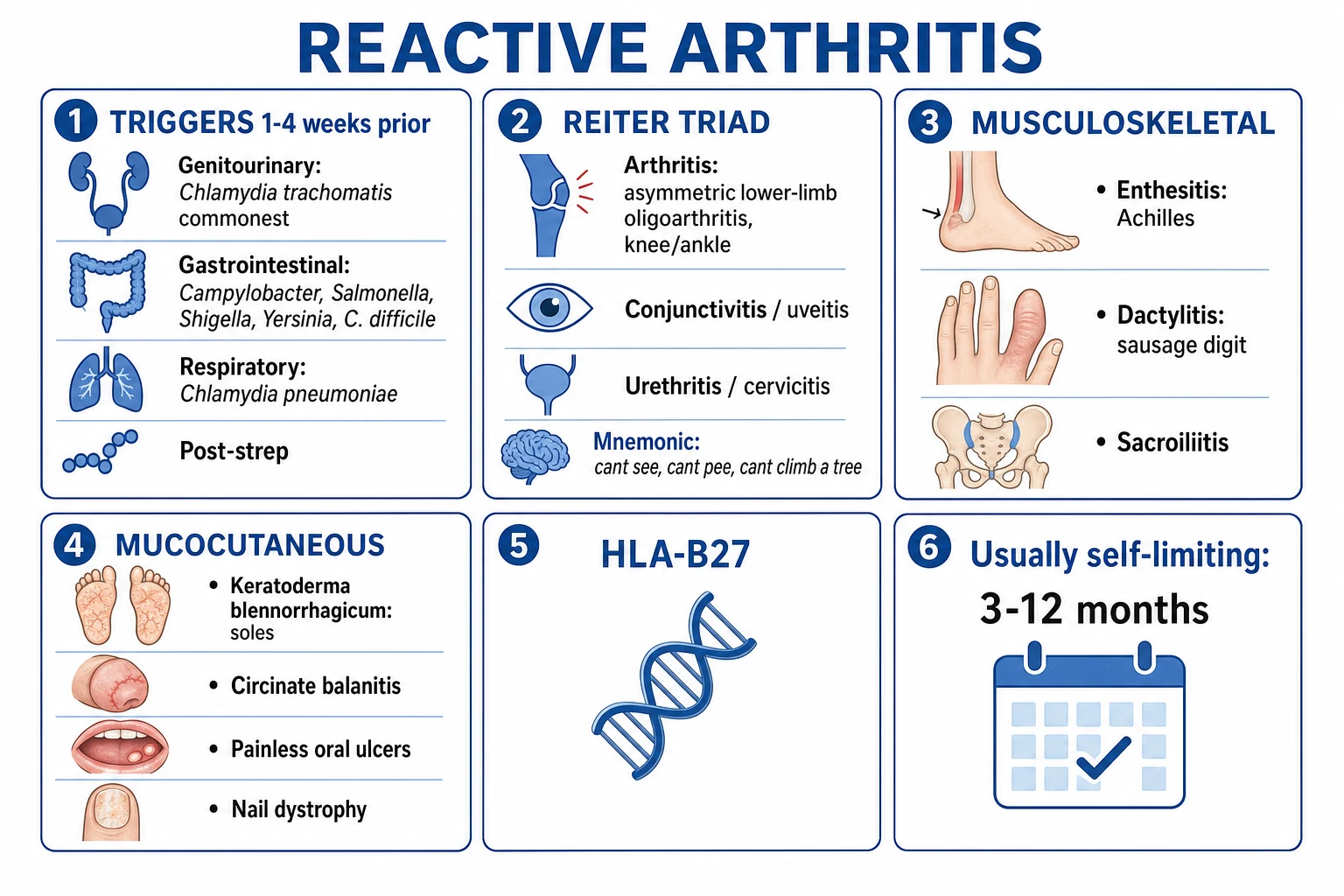
Reactive arthritis (ReA, formerly Reiter syndrome) is a sterile inflammatory arthritis arising 1 to 4 weeks after an extra-articular infection (classically genitourinary — Chlamydia trachomatis; or gastrointestinal — Campylobacter, Salmonella, Shigella, Yersinia, Clostridioides difficile), belonging to the HLA-B27-associated seronegative spondyloarthropathies. The classic triad (Reiter): arthritis (asymmetric, lower-limb oligoarthritis), urethritis/cervicitis, and conjunctivitis/uveitis ('can't see, can't pee, can't climb a tree'). Characteristic features include enthesitis, dactylitis (sausage digit), sacroiliitis, keratoderma blennorrhagicum, circinate balanitis, painless oral ulcers, and nail changes. Usually self-limiting (3 to 12 months); 15 to 30 percent develop chronic or recurrent spondyloarthritis. Diagnosis is clinical, supported by a culture-negative, crystal-negative inflammatory synovial fluid. Management: treat the trigger and sexual contacts, NSAIDs first-line, intra-articular corticosteroids, DMARDs (sulfasalazine) for persistent disease, and TNF inhibitors for refractory cases.
On this page & tools
Your progress
Saved locally on this device.
Exam tags
Red flags
Overview & Definition
Reactive arthritis is the spondyloarthropathy that follows infection at a distant mucosal site — the joints are inflamed but sterile. The causative organism triggers the disease from a distance and is not recoverable from the joint by standard culture, which is the defining pathological and diagnostic feature separating reactive arthritis from septic and gonococcal arthritis. The arthritis typically begins 1 to 4 weeks after a genitourinary or gastrointestinal infection and targets the entheses (tendon and ligament insertions into bone) as much as the synovium, producing the characteristic combination of asymmetric lower-limb oligoarthritis, enthesitis and dactylitis.[1][3]
The eponym Reiter syndrome — named after Hans Reiter, who described a soldier with the triad of urethritis, conjunctivitis and arthritis after a bout of dysentery in 1916 — is now discouraged both because Reiter's medical and ethical record under the Nazi regime has been discredited and because the triad is an incomplete description of the disease. The preferred term reactive arthritis captures the essential concept: an immune-mediated arthritis reactive to a distant infection, encompassing the full spectrum of articular, entheseal, ocular, mucocutaneous and systemic features.[1]
The clinical skill in this disease rests on three linked steps: first, linking the arthritis to the preceding infection (always ask explicitly about diarrhoea and genital symptoms in the preceding 1 to 4 weeks); second, excluding septic and gonococcal arthritis by aspirating any acutely hot joint; and third, staging the treatment — most cases settle with NSAIDs, but the persistent minority need DMARDs and a few need biologics. The full Reiter triad is uncommon in practice; most patients present with an asymmetric oligoarthritis and only some elements of the extra-articular picture.[3][4]
Classification

Reactive arthritis is classified by its triggering infection, because the trigger determines the epidemiology, the sexual-contact and public-health implications, and sometimes the clinical phenotype. The two classical categories are post-genitourinary (post-venereal) and post-enteric (post-dysenteric), with rarer respiratory and post-streptococcal forms recognised separately.[2][4]
Post-genitourinary (SARA)
- Triggered by **Chlamydia trachomatis** (commonest GU cause); rarely Ureaplasma and Mycoplasma genitalium
- **Sexually acquired reactive arthritis (SARA)** — predominantly young men, 5 to 10 times more common in males
- Preceding urethritis or cervicitis may be asymptomatic; **screen and treat sexual contacts**; test for co-infections (HIV, syphilis, gonorrhoea)
- HLA-B27 positivity higher in this form; more severe and chronic disease
Post-enteric (post-dysenteric)
- Triggered by **Campylobacter jejuni, Salmonella, Shigella (especially S. flexneri), Yersinia enterocolitica/pseudotuberculosis, and Clostridioides difficile**
- Equal sex ratio (no male predominance); follows food-borne or water-borne outbreaks
- Diarrhoea usually self-limited before arthritis onset; **stool culture or serology** may help if presentation is delayed
- Outbreaks allow incidence estimation: approximately 1 to 4 percent of HLA-B27-positive individuals after a triggering GI infection
Respiratory / other
- **Chlamydia pneumoniae** respiratory tract infection can trigger ReA (less common)
- Post-streptococcal reactive arthritis is recognised as a **separate entity** from acute rheumatic fever
- Numerous other agents (Q fever, Legionella, Leptospira, Giardia, Cryptosporidium, and viruses including HIV, parvovirus, Hepatitis B/C) have been implicated as rare triggers
Chronic / persistent ReA
- Disease persisting beyond 6 to 12 months, or recurrent flares after remission
- Occurs in **15 to 30 percent**; more likely if **HLA-B27 positive, hip involvement, or repeated triggering infections**
- May evolve into **axial spondyloarthritis or ankylosing spondylitis** over years — long-term follow-up required
- DMARDs and TNF inhibitors are the therapeutic escalation for this subgroup
Reactive arthritis is classified within the seronegative spondyloarthropathies (SpA), sharing the family with ankylosing spondylitis, psoriatic arthritis, enteropathic (IBD-associated) arthritis, and undifferentiated SpA. The ASAS (Assessment of SpondyloArthritis international Society) classification separates axial from peripheral SpA; reactive arthritis is the archetype of peripheral SpA with a known trigger, but may coexist with axial (sacroiliitis, spondylitis) involvement.[9][10]
Epidemiology & Risk Factors
Reactive arthritis most commonly affects young adults aged 20 to 40 years, with an overall incidence estimated at 0.6 to 27 per 100,000 per year (the wide range reflects variable trigger prevalence, genetic background, and reporting). The post-genitourinary form predominates in young men (male-to-female ratio up to 9 to 1 for chlamydia-triggered disease, partly reflecting urethral symptom reporting and chlamydia ascertainment), while the post-enteric form has an equal sex ratio.[1][4]
The single most important host risk factor is HLA-B27 positivity, which is present in 30 to 50 percent of patients with reactive arthritis overall (higher in those with severe, chronic, or axial disease) compared with roughly 4 to 8 percent of the general population. Among HLA-B27-positive individuals exposed to a triggering infection, the estimated risk of developing reactive arthritis is 1 to 4 percent after chlamydia and 1 to 20 percent after certain enteric outbreaks — demonstrating that HLA-B27 is necessary but not sufficient for disease.[1][2]
Reactive arthritis — headline epidemiology
Other recognised risk factors and associations include male sex (for chlamydia-triggered disease), HIV infection (reactive arthritis is more frequent and more severe in HIV, particularly axial and severe forms), a family or personal history of spondyloarthropathy, and bacterial virulence factors — not every strain of a species triggers ReA; for example, only certain Shigella flexneri and Yersinia strains are arthritogenic. Geographical variation in HLA-B27 prevalence (high in Scandinavian and certain Native American populations, low in African and Japanese populations) explains much of the variation in disease frequency worldwide.[2][4]
Pathophysiology
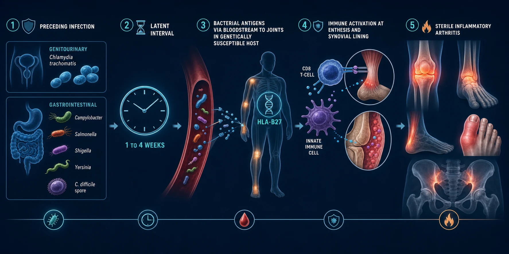
Reactive arthritis is an immune-mediated arthritis in a genetically susceptible host, triggered by bacterial antigens from a distant mucosal infection. The mechanism can be understood in four linked stages.[1][2]
Stage 1 — Mucosal infection at a distant site
The disease begins with a genitourinary or gastrointestinal infection. The triggering organisms share a unifying feature: they are intracellular or facultatively intracellular bacteria that can persist within host cells. Chlamydia trachomatis infects the urethral or cervical epithelium; Campylobacter, Salmonella, Shigella and Yersinia invade the gastrointestinal mucosa. The initial infection may be asymptomatic (especially chlamydial cervicitis and urethritis, which is silent in up to 50 percent of women and 25 percent of men), which is why direct questioning about and laboratory testing for the trigger is essential.[2][4]
Stage 2 — Bacterial antigen dissemination and trafficking to the joint
Bacterial antigens, nucleic acid (DNA and especially rRNA), and lipopolysaccharide are detectable in the synovial fluid, synovial tissue and entheses of affected patients, though viable, culturable organisms are usually absent. For chlamydia-triggered disease, there is strong evidence that C. trachomatis persists in a metabolically active but non-culturable (persistent) form within synovial macrophages, expressing chlamydial heat-shock protein 60 and 16S rRNA. This persistent antigen is the engine driving the ongoing immune response. The organisms or their antigens reach the joint via the bloodstream and possibly via trafficking of infected macrophages to the synovium and entheseal insertion sites.[2]
Stage 3 — The HLA-B27 and innate immune response in a susceptible host
In an HLA-B27-positive host, the immune response to these antigens is dysregulated and excessive. Several mechanisms are implicated: [1]
- Molecular mimicry: HLA-B27 may present bacterial peptides that cross-react with self-peptides at the enthesis and synovium, generating an autoimmune attack on joint tissues.
- Arthritogenic peptide hypothesis: HLA-B27 has a unique peptide-binding groove that preferentially presents certain bacterial-derived peptides to CD8-positive cytotoxic T cells, driving a persistent inflammatory response.
- HLA-B27 misfolding and unfolded protein response: Misfolded HLA-B27 heavy chains accumulate in the endoplasmic reticulum, triggering the unfolded protein stress response and activating the IL-23/IL-17 innate immune axis.
- Free heavy chain / surface homodimer signalling: HLA-B27 homodimers on the cell surface interact with natural killer and Th17 cells, amplifying IL-17 production at entheses. [1]
The IL-23/IL-17 axis is now recognised as the dominant inflammatory pathway in the spondyloarthropathies, including reactive arthritis. Enthesitis — inflammation at the insertion of tendons, ligaments and joint capsules into bone — is the pathological hallmark, explaining the characteristic Achilles tendon and plantar fascia involvement, dactylitis (a sausage digit from combined entheseal and synovial inflammation of a whole digit), and the evolution to sacroiliitis and syndesmophytes in chronic disease.[1][9]
Stage 4 — Sterile inflammatory arthritis with enthesitis
The result is a sterile, culture-negative inflammatory arthritis and enthesitis. The synovium shows a neutrophilic and mononuclear infiltrate; the enthesis shows inflammation at the fibrocartilage-bone interface with erosions, new bone formation (enthesophytes) and periostitis. Because the joint is sterile, cultures are negative and antibiotics treat the trigger and contacts but do not reliably shorten the established arthritis course — a pivotal and frequently-tested point. The disease is self-limiting in most because the immune response eventually clears the antigenic stimulus, but in the HLA-B27-positive minority, the cycle of enthesitis and new bone formation persists, evolving toward chronic spondyloarthritis.[1][6]
REITER — recognise the pattern
REITER
Sterile arthritis 1 to 4 weeks AFTER a GU or GI infection (not during it)
Achilles tendon and plantar fascia insertion pain — the spondyloarthropathy signature
Chlamydia (GU); Campylobacter, Salmonella, Shigella, Yersinia, C. difficile (GI)
Arthritis + urethritis + conjunctivitis ('can't see, can't pee, can't climb a tree')
Keratoderma blennorrhagicum, circinate balanitis, painless oral ulcers, nail changes
Seronegative; HLA-B27 associated; synovial fluid sterile, culture-negative, no crystals
Clinical Presentation
The presentation is acute, arising 1 to 4 weeks after a triggering GU or GI infection. The classic Reiter triad (arthritis plus urethritis plus conjunctivitis) is the exam-stem pattern, but it is complete in only about one third of patients — most have some combination of features rather than the full triad. Examiners test both pattern recognition (the arthritis with a preceding infection) and the specific features.[3][4]
Musculoskeletal features — the spondyloarthropathy signature
The arthritis is characteristically an asymmetric oligoarthritis (two to four joints), predominantly affecting the lower limbs — the knee, ankle, and the small joints of the foot are the commonest targets. The pattern of asymmetric lower-limb oligoarthritis is one of the highest-yield recognition cues in rheumatology. Key features include:[3]
- Asymmetric lower-limb oligoarthritis — knee and ankle effusions are typical; the arthritis is acute and may mimic septic arthritis in a single joint (hence the rule to aspirate).
- Enthesitis — tenderness at tendon and ligament insertions, most characteristically at the Achilles tendon insertion and the plantar fascia origin (calcaneal enthesitis), producing heel pain that is disproportionate to the visible swelling. This is the pathological hallmark of the spondyloarthropathies.
- Dactylitis (sausage digit) — uniform swelling of an entire finger or toe from combined synovitis and enthesitis of all the structures within the digit; a highly characteristic feature that examiners reward.
- Sacroiliitis and spondylitis — low back pain and buttock pain from sacroiliac joint involvement, present in a minority at presentation but more common in chronic disease.
- Non-articular rheumatism — tendinitis, fasciitis, and bursitis may accompany the arthritis. [1]
Extra-articular features — eye, genital, skin, and systemic
The extra-articular features are what distinguish reactive arthritis from a generic inflammatory oligoarthritis and complete the Reiter picture.[1][3]
Ocular
- **Conjunctivitis** — bilateral, mucopurulent, often mild and self-limiting; usually precedes the arthritis
- **Acute anterior uveitis (iritis)** — red eye, pain, photophobia, blurred vision; sight-threatening; needs urgent ophthalmology; more common in HLA-B27-positive, recurrent disease
- Episcleritis and keratitis less commonly
Genitourinary
- **Urethritis or cervicitis** — dysuria, discharge; may be the trigger (Chlamydia) or reactive; often asymptomatic in women
- **Urethritis may be post-dysenteric (sterile inflammatory) rather than infectious**
- **Circinate balanitis** — painless, serpiginous, circinate erosions on the glans penis; highly characteristic
- **Cervicitis** and less commonly salpingitis
Mucocutaneous
- **Keratoderma blennorrhagicum** — pustular, hyperkeratotic, sometimes psoriasiform lesions on the **soles** (and palms); clinically and histologically indistinguishable from pustular psoriasis
- **Circinate balanitis** — as above; on uncircumcised men may appear as moist plaques
- **Painless shallow oral ulcers** — on the palate, buccal mucosa, tongue; non-grouped, transient, easily missed
- **Nail changes** — onycholysis, subungual hyperkeratosis, pitting; overlap with psoriasis
- Balanitis circinata and keratoderma blennorrhagicum are pathognomonic when present
Cardiac (rare, late)
- **Aortic regurgitation** from aortitis of the ascending aorta — late, rare, but classic
- **Carditis and conduction defects** — first-degree atrioventricular block; rarely complete heart block
- Seen mainly in severe, chronic, HLA-B27-positive disease; aortic involvement is a major cause of late mortality
Systemic
- **Fever** (low-grade), fatigue, malaise, weight loss at onset
- **Elevated inflammatory markers** (ESR, CRP); may be markedly raised
- Lymphadenopathy (especially with HIV co-infection)
Atypical presentations
Examiners test the corners deliberately. Atypical presentations include:[4]
- Painless oral ulcers — easily missed because they are non-tender; examine the oral cavity actively.
- Isolated Achilles enthesitis or heel pain without overt arthritis — can be the sole presenting feature; ask about recent infection.
- Acute anterior uveitis as the presenting feature — a red, painful eye may bring the patient to ophthalmology before the arthritis is recognised.
- Reactive arthritis in HIV — can be severe, with aggressive axial and peripheral disease, dactylitis, and keratoderma blennorrhagicum; the skin and joint features are often florid.
- Elderly presentation — less common, may lack the classic extra-articular features, and is more readily misattributed to gout, osteoarthritis, or septic arthritis.
- Asymptomatic triggering infection — especially chlamydial urethritis in women; the absence of a recalled infection does not exclude ReA. [1]
Differential Diagnosis
The pivotal diagnostic steps are pattern recognition (asymmetric lower-limb oligoarthritis with enthesitis/dactylitis and a preceding infection) and excluding septic arthritis by aspirating any acute hot joint. Reactive arthritis is seronegative (RF and ANA negative) and the synovial fluid is sterile, crystal-negative and inflammatory, which separates it from septic, gonococcal and crystal arthropathies. At least three distinguishing features should be considered for each mimic.[1][3]
Reactive arthritis
- Asymmetric lower-limb OLIGOarthritis 1 to 4 weeks after GU/GI infection
- Enthesitis, dactylitis (sausage digit), conjunctivitis/uveitis, urethritis/cervicitis
- Synovial fluid STERILE, culture-negative, WBC 2,000 to 50,000, no crystals
- HLA-B27 associated; RF/ANA negative; NSAIDs first-line; treat trigger plus contacts
Septic arthritis
- Single hot, swollen, very tender joint; severe pain on movement; high fever and rigors
- Synovial WBC usually above 50,000, neutrophilic; POSITIVE Gram stain and culture
- *Staphylococcus aureus* commonest; urgent washout plus intravenous antibiotics; emergency
Gout
- Negatively birefringent NEEDLE-shaped monosodium urate crystals in synovial fluid
- First MTP (podagra) classic; serum urate may be normal during the acute attack
- NSAIDs, colchicine, corticosteroids for the acute attack; allopurinol for chronic urate lowering
Pseudogout (CPPD)
- Positively birefringent RHOMBOID-shaped calcium pyrophosphate crystals
- Knee and wrist; chondrocalcinosis on X-ray; older patients
- Treat the acute attack with NSAIDs, colchicine, or steroids; no urate-lowering equivalent
Gonococcal arthritis
- Young sexually active; migratory polyarthralgia, tenosynovitis, pustular rash on extensor surfaces
- Joint, blood, or NAAT POSITIVE for *Neisseria gonorrhoeae* (true infection, not reactive)
- Ceftriaxone intramuscular or intravenous plus azithromycin/doxycycline; treat contacts
Ankylosing spondylitis
- Chronic inflammatory back pain (insidious onset, under 45 yr, improves with exercise)
- Bilateral sacroiliitis on imaging; HLA-B27 strongly associated; no preceding infection
- NSAIDs first-line; TNF inhibitors or IL-17 inhibitors for refractory disease
Psoriatic arthritis
- Asymmetric oligoarthritis with psoriasis and nail dystrophy (pitting, onycholysis)
- DIP involvement, dactylitis, enthesitis — overlaps with reactive arthritis
- No preceding infection; DMARDs or biologics (TNFi, IL-17i, IL-23i) early
Enteropathic (IBD-associated) arthritis
- Peripheral arthritis mirrors bowel disease activity; axial disease runs independent course
- Co-existing Crohn disease or ulcerative colitis; abdominal symptoms
- Treat the underlying IBD; NSAIDs may worsen bowel disease; biologics effective
Rheumatoid arthritis
- SYMMETRIC small-joint polyarthritis (MCP, PIP, wrists) with morning stiffness over 1 hour
- RF and anti-CCP (ACPA) positive; erosions on X-ray; no enthesitis or preceding infection
- DMARDs (methotrexate first-line); biologics (TNFi, IL-6i, JAKi) for refractory disease
Lyme arthritis
- Late manifestation of *Borrelia burgdorferi* infection; episodic mono- or oligoarthritis, knee commonest
- History of tick exposure or erythema migrans; positive Lyme serology (ELISA plus Western blot)
- Doxycycline or amoxicillin orally; ceftriaxone intravenously for neurological or cardiac disease
The single most dangerous mimic is septic arthritis — a single hot joint with high fever and marked pain on movement must be aspirated before reactive arthritis is assumed, because sepsis can coexist with or mimic reactive disease and delays cause rapid joint destruction. Gonococcal arthritis is a close second and is distinguished by its disseminated pustular rash, tenosynovitis and positive cultures/NAAT — it is a true infection, not a reactive process. Keratoderma blennorrhagicum and pustular psoriasis are clinically and histologically indistinguishable, and the distinguishing clue is the history of a preceding infection and the distribution (soles in ReA; extensor surfaces and scalp in psoriasis).[3][4]
Clinical & Bedside Assessment
The bedside assessment hinges on pattern recognition, a directed history probing the trigger, and a focused examination of joints, entheses, eyes, skin, genitalia and mouth. [1]
History — always ask explicitly about: diarrhoea (timing, blood, duration, travel, food) and genital symptoms (discharge, dysuria, new partners) in the 1 to 4 weeks before the arthritis; a sexual history (number of partners, condom use, prior STIs); travel and food history; eye symptoms (redness, pain, photophobia, blurred vision); skin and mouth (soles, palms, glans penis, oral ulcers); back pain; HIV risk factors; and a family history of spondyloarthropathy, psoriasis, IBD or acute uveitis. The temporal link — arthritis 1 to 4 weeks after a GU or GI infection — is the single most important historical clue and should be elicited actively rather than waited for.[3][4]
Joint and entheseal examination — examine for an asymmetric oligoarthritis (effusions, warmth, synovitis) predominantly in the knees, ankles and feet; test for enthesitis by palpating the Achilles tendon insertion and the plantar fascia origin on the calcaneus (tenderness here is a spondyloarthropathy sign); look for dactylitis (diffuse, sausage-like swelling of a whole digit); examine the sacroiliac joints (direct pressure, sacroiliac stress tests); and assess the spine for reduced mobility if axial symptoms are present. [1]
Ocular examination — inspect for conjunctival injection and discharge (conjunctivitis); assess visual acuity, pupillary light reaction, and slit-lamp findings (cells and flare in the anterior chamber indicate acute anterior uveitis). Any red eye with pain, photophobia or visual change is uveitis, not conjunctivitis, and needs urgent ophthalmology referral. [1]
Mucocutaneous and genital examination — inspect the soles and palms for keratoderma blennorrhagicum (pustular, hyperkeratotic lesions); the glans penis for circinate balanitis (serpiginous, circinate erosions) and the vulva for corresponding lesions; the oral cavity for painless shallow ulcers; and the nails for onycholysis, pitting and subungual hyperkeratosis. Examine the urethral meatus for discharge; perform a genital and pelvic examination as appropriate.[1]
Systemic examination — listen for a murmur (aortic regurgitation of aortitis), assess for fever, examine lymph nodes (generalised lymphadenopathy raises HIV suspicion), and check for abdominal signs of associated or triggering IBD or enteric infection. [1]
Investigations
There is no single diagnostic test for reactive arthritis. The diagnosis is clinical, supported by evidence of a preceding infection, a seronegative autoantibody profile, and a sterile, inflammatory, crystal-negative synovial fluid. Investigations serve three purposes: to confirm the pattern (inflammatory, seronegative SpA), to identify the trigger, and to exclude the dangerous mimics (sepsis, gout, gonococcus).[3][4]
Synovial fluid — the pivotal investigation
Aspirate any acute hot joint. The synovial fluid in reactive arthritis is: [1]
- Culture-negative (sterile) — the defining feature; distinguishes ReA from septic and gonococcal arthritis.
- Crystal-negative — no monosodium urate or calcium pyrophosphate crystals on polarised microscopy; distinguishes ReA from gout and CPPD.
- Inflammatory — white blood cell count typically 2,000 to 50,000 per microlitre, predominantly neutrophilic; distinguishes ReA from osteoarthritis (non-inflammatory, below 2,000) but overlaps with septic arthritis (which is usually above 50,000 and culture-positive). [1]
Gram stain and culture must always be sent even when ReA is suspected, because septic arthritis can coexist and a culture-negative, crystal-negative inflammatory fluid with a compatible clinical story is the pattern that confirms ReA after sepsis is excluded.[4]
Trigger detection
Genitourinary testing
- **Nucleic acid amplification test (NAAT)** for *Chlamydia trachomatis* on first-void urine and urethral or cervical swab — the most sensitive test
- Also test for *Neisseria gonorrhoeae*, *Mycoplasma genitalium*, *Trichomonas vaginalis*, syphilis (serology) and **HIV**
- Screen **all sexual contacts** of the last 60 days if Chlamydia detected; treat empirically
Gastrointestinal testing
- **Stool culture** for *Campylobacter, Salmonella, Shigella, Yersinia* — most useful if diarrhoea is recent or ongoing
- **Serology** for *Yersinia* and *Salmonella* (paired sera, four-fold rise) if presentation is delayed beyond the stool-shedding window
- *Clostridioides difficile* toxin assay if antibiotic-associated diarrhoea preceded the arthritis
Other trigger testing
- **HIV serology** — mandatory in sexually acquired ReA; reactive arthritis is more severe and common in HIV
- Throat swab and **anti-streptolysin O (ASO) titre** if post-streptococcal ReA suspected
- Respiratory *Chlamydia pneumoniae* serology in selected cases (rare)
General laboratory tests
- Inflammatory markers: ESR and CRP are elevated (sometimes markedly) during active disease and trend with disease activity; useful for monitoring response to treatment.
- Full blood count: may show leukocytosis, neutrophilia, thrombocytosis and a normocytic anaemia of chronic inflammation; eosinophilia is not a feature.
- Seronegative profile: rheumatoid factor (RF) negative, antinuclear antibody (ANA) negative, anti-CCP (ACPA) negative — this is the "seronegative" in seronegative spondyloarthropathy. (Low-titre RF may be present in up to 5 percent of healthy older adults and is non-specific.)
- HLA-B27: supportive but not diagnostic — present in 30 to 50 percent of ReA patients (and 4 to 8 percent of the general population). It is most useful in prognostication (HLA-B27 positivity predicts chronic, axial and recurrent disease) and in supporting a clinical diagnosis where the trigger is unclear. It is not a screening test.[1][4]
Imaging
- X-rays are usually normal early in the disease. With chronic or recurrent disease, characteristic changes include sacroiliitis (initially blurring and erosion of the subchondral bone, later sclerosis and, eventually, fusion), enthesophytes and erosions at tendon insertions (Achilles, plantar fascia — "fluffy" or "fuzzy" periostitis), periostitis, and, in the spine, syndesmophytes and squaring of vertebral bodies.
- MRI is more sensitive for early sacroiliitis (bone marrow oedema on short tau inversion recovery sequences) and enthesitis, and may reveal subclinical axial inflammation that predicts progression to axial spondyloarthritis.
- Ultrasound (power Doppler) can detect subclinical enthesitis, synovitis and dactylitis, and guide intra-articular injection.
- Plain films of the pelvis (sacroiliac joints) and an X-ray of the heel (calcaneal enthesophyte) are high-yield classic exam findings in chronic ReA.[1]
Tests for systemic and extra-articular involvement
- Urinalysis — sterile pyuria (reactive urethritis) or evidence of the triggering infection.
- ECG — if cardiac involvement (conduction defects, first-degree AV block) suspected.
- Echocardiogram — if aortic regurgitation or carditis suspected (listen for an early diastolic murmur).
- HIV testing — recommended in all sexually acquired ReA.[4]
Management — Resuscitation & Immediate Actions
Reactive arthritis is rarely a true medical emergency, but several situations demand immediate action before the staged treatment is begun.[3][4]
[1]Immediate symptomatic measures while awaiting investigation results include rest of the inflamed joint(s), ice and elevation for comfort, and an NSAID at full anti-inflammatory dose (provided there are no contraindications — renal impairment, active peptic ulcer disease, anticoagulation, heart failure, aspirin-sensitive asthma). Do not give systemic corticosteroids or DMARDs before the joint is aspirated and sepsis excluded.[3]
Management — Definitive & Stepwise
Management is staged: treat the trigger first, then control symptoms with NSAIDs and local steroids, then escalate to DMARDs and biologics for the persistent minority. The ASAS-EULAR recommendations for spondyloarthritis management, with reactive arthritis as the archetype of peripheral SpA, frame this approach.[5][10]
Step 1 — Treat the trigger and the contacts
Treating the triggering infection addresses the source, prevents onward transmission, and prevents the reproductive complications of untreated chlamydia (pelvic inflammatory disease, infertility, ectopic pregnancy).[6]
Antibiotic regimens for Chlamydia-triggered reactive arthritis
- Chlamydia trachomatis: azithromycin 1 g orally as a single dose, or doxycycline 100 mg orally twice daily for 7 days (14 days for epididymitis or PID overlap). Treat all sexual partners of the preceding 60 days; offer expedited partner therapy where available.
- Gastrointestinal triggers: the triggering enteric infection has usually resolved by the time arthritis appears; antibiotics are not routinely needed for the arthritis, though specific treatment (e.g. for C. difficile with oral vancomycin or fidaxomicin) is guided by the infective picture. Prolonged antibiotics do not improve the arthritis course — a pivotal and frequently-tested point confirmed by meta-analysis and the Carter combination-antibiotic trial.[6][7]
- HIV: if positive, initiate or refer for antiretroviral therapy; this may improve the associated reactive arthritis.
- Screening: all patients with sexually acquired ReA should be screened for HIV, syphilis, gonorrhoea and hepatitis B and C.[4]
Step 2 — First-line symptomatic control
NSAIDs are the cornerstone of first-line therapy for the acute arthritis and enthesitis. Use a full anti-inflammatory dose, continued for the duration of active disease:[3]
NSAID first-line doses for reactive arthritis
- Naproxen 500 mg orally twice daily, or ibuprofen 400 to 800 mg three times daily (maximum 2.4 g per day), or indomethacin 50 mg two to three times daily (75 to 100 mg is the traditional upper daily dose; well-regarded for spondyloarthropathy but with higher CNS and gastrointestinal side effects). Add a proton pump inhibitor (omeprazole 20 mg daily) for patients with risk factors for NSAID gastropathy.
- Intra-articular corticosteroid injection — for an inflamed oligoarticular joint (e.g. a hot knee) that is not adequately controlled by NSAIDs, or where NSAIDs are contraindicated. Triamcinolone acetonide 40 mg (knee) or a proportionate dose for smaller joints, after aspiration and confirmation that the fluid is sterile. Highly effective for mono- or oligoarticular disease.
- Topical corticosteroids — for circinate balanitis, keratoderma blennorrhagicum and oral ulcers (e.g. clobetasol 0.05 percent cream to the soles, triamcinolone dental paste to oral ulcers, mild topical steroid to the glans).
- Uveitis — urgent ophthalmology referral for topical, periocular or systemic corticosteroids and mydriatics (atropine 1 percent or cyclopentolate 1 percent); untreated anterior uveitis threatens sight through synechiae, cataract and glaucoma.
- Physiotherapy — rest during the acute phase, then graded mobilisation and stretching (especially of the Achilles tendon and plantar fascia) to preserve mobility and prevent contracture.[3]
Step 3 — DMARDs for persistent or severe disease
For the 15 to 30 percent with disease persisting beyond 6 to 12 months, polyarticular or axial disease, or recurrent flares, disease-modifying antirheumatic drugs (DMARDs) are added:[3][5]
- Sulfasalazine is the first-line DMARD: start at 500 mg orally twice daily and titrate to 2 to 3 g per day in divided doses over 4 to 8 weeks. Monitor full blood count and liver function (the drug can cause leukopenia, thrombocytopenia and transaminitis); screen for G6PD deficiency before starting (risk of haemolysis). Sulfasalazine has the best evidence of the conventional DMARDs for reactive arthritis and peripheral spondyloarthritis.
- Methotrexate is an alternative or add-on DMARD: 7.5 to 15 mg orally once weekly, titrating to 20 to 25 mg weekly. Supplement with folic acid 5 mg weekly (or 1 mg daily); monitor full blood count, liver function and renal function; screen for and treat latent tuberculosis, hepatitis B and C before starting. Contraindicated in pregnancy (teratogenic).
- Short-course systemic corticosteroids — prednisolone 0.5 mg/kg/day orally, tapering over 2 to 4 weeks, for severe polyarticular flares unresponsive to NSAIDs, or as a bridge while DMARDs take effect. Avoid prolonged use.[5]
Step 4 — Biologics for refractory disease
For the refractory minority — disease unresponsive to NSAIDs and DMARDs, especially with chronic axial spondyloarthritis — TNF inhibitors are the evidence-supported escalation:[5][10]
TNF inhibitor doses for refractory / chronic reactive arthritis
- Adalimumab 40 mg subcutaneously every 2 weeks, etanercept 50 mg subcutaneously weekly, or infliximab 5 mg/kg intravenously at weeks 0, 2 and 6, then every 8 weeks. Screen for latent tuberculosis (interferon-gamma release assay or chest X-ray), hepatitis B and C and HIV before starting; ensure vaccinations (influenza, pneumococcal, hepatitis B, zoster where appropriate) are up to date, and avoid live vaccines on therapy.
- IL-17 inhibitors (secukinumab, ixekizumab) and IL-23 inhibitors (ustekinumab) have a role in psoriatic and axial spondyloarthritis and are increasingly considered for refractory reactive arthritis, though the evidence base for ReA specifically is smaller than for ankylosing spondylitis and psoriatic arthritis.[5][10]
Specific Subtypes & Scenarios
Chlamydia-triggered (sexually acquired) reactive arthritis (SARA)
This is the archetypal reactive arthritis and the most frequently tested subtype. It predominantly affects young men (male-to-female ratio up to 9 to 1, partly reflecting ascertainment), follows an often-asymptomatic chlamydial urethritis or cervicitis by 1 to 4 weeks, and is HLA-B27 associated in a higher proportion than enteric ReA. Management combines treatment of the patient and sexual contacts (azithromycin 1 g single dose or doxycycline 7 to 14 days), a full STI screen including HIV, and the staged NSAID-to-DMARD-to-biologic ladder. The persistent antigen load from viable-but-non-culturable chlamydial forms in the synovium may explain why prolonged combination antibiotics (doxycycline plus rifampicin) showed benefit in the Carter combination-antibiotic trial for chronic chlamydia-induced ReA — a nuanced finding that contrasts with the general meta-analytic conclusion that prolonged antibiotics do not help ReA overall.[2][7]
Post-enteric reactive arthritis
Triggered by Campylobacter jejuni (the commonest enteric trigger in many series), Salmonella enteritidis and typhimurium, Shigella flexneri (particularly arthritogenic; S. sonnei less so), Yersinia enterocolitica and pseudotuberculosis, and Clostridioides difficile. It has an equal sex ratio, often follows food-borne outbreaks (allowing incidence estimation), and the triggering diarrhoea has usually resolved by the time the arthritis appears. Antibiotics are rarely needed for the arthritis (the infection is self-limited); the focus is on NSAIDs and escalation for persistent disease. Yersinia-triggered ReA may be accompanied by erythema nodosum and can mimic IBD.[2][4]
Post-streptococcal reactive arthritis
A separate entity from acute rheumatic fever (ARF). It follows group A streptococcal pharyngitis, lacks the major Jones criteria of ARF (no carditis, no chorea, no erythema marginatum, no subcutaneous nodules), and has a shorter latent interval (1 to 2 weeks rather than 2 to 4 weeks for ARF). It is not prevented by routine penicillin prophylaxis in the way ARF is — though some authorities recommend a period of secondary prophylaxis — and does not have the same cardiac implications. Management is NSAIDs; the role of long-term antibiotic prophylaxis is debated.[8]
Respiratory and other triggers
Chlamydia pneumoniae respiratory infection can trigger ReA, and a growing list of other agents — Q fever (Coxiella burnetii), Legionella, Leptospira, Giardia lamblia, Cryptosporidium, Brucella, and viruses including HIV, parvovirus B19, hepatitis B and C — have been implicated as rare or single-case triggers. These are relevant when the classical GU and GI screens are negative.[2]
HIV-associated reactive arthritis
Reactive arthritis is more common and more severe in HIV-positive patients, particularly with florid skin and joint involvement (severe keratoderma blennorrhagicum, dactylitis, aggressive axial disease). The relationship is complex: HIV does not directly cause ReA, but the immune dysregulation it produces amplifies the spondyloarthropathy phenotype in susceptible individuals. HIV testing is mandatory in any sexually acquired ReA. Management principles are the same, though immunosuppression must be coordinated with antiretroviral therapy; sulfasalazine and TNF inhibitors can be used safely with appropriate monitoring and infectious screening.[4]
Chronic and recurrent reactive arthritis
The 15 to 30 percent with disease beyond 6 to 12 months, or recurrent flares after remission, constitute the chronic/persistent subgroup. Predictors include HLA-B27 positivity, hip involvement, polyarticular disease at onset, and repeated triggering infections. This group may evolve into axial spondyloarthritis or ankylosing spondylitis over years and requires long-term rheumatology follow-up, DMARDs (sulfasalazine first, methotrexate), and TNF inhibitors for refractory disease. The ASAS-EULAR 2022 update frames this as the management of axial spondyloarthritis, where TNF inhibitors (and increasingly IL-17 inhibitors) are the standard biologic escalation after NSAIDs fail.[9][10]
Complications & Pitfalls
Major complications of reactive arthritis — frequency and consequence
The major complications are chronic or recurrent arthritis (the largest burden, affecting up to a third), uveitis (the major sight threat), aortic regurgitation and conduction defects (rare, late, classically in severe HLA-B27-positive disease), and, very rarely, secondary (AA) amyloidosis from years of chronic inflammation. The reproductive complications of untreated chlamydia — pelvic inflammatory disease, tubal infertility, ectopic pregnancy — are preventable by prompt treatment of the patient and contacts. Reactive arthritis in HIV is more severe and demands coordinated rheumatology-HIV care.[1][4]
Classic pitfalls — the recurring ways reactive arthritis is missed or mismanaged: [1]
- Failing to aspirate a hot joint because "it is obviously reactive" — missing septic arthritis, the most dangerous error.
- Ascribing conjunctivitis to the Reiter triad when the red eye is actually uveitis — delaying urgent ophthalmology and risking visual loss.
- Omitting an HIV test in sexually acquired ReA — missing co-infection that changes management and prognosis.
- Forgetting to treat sexual contacts of a chlamydia-triggered case — allowing re-infection and onward transmission.
- Using prolonged antibiotics to treat the arthritis — they eradicate the trigger but do not reliably shorten the arthritis course, and inappropriate prolonged use drives resistance.
- Missing the diagnosis because the triad is incomplete — most patients have only some features; the pattern (asymmetric lower-limb oligoarthritis plus a recent infection) is the key.
- Not examining the soles, palms and glans penis — keratoderma blennorrhagicum and circinate balanitis are pathognomonic when present but easily missed if not actively sought.
- Over-diagnosing ReA when the trigger is dubious — post-streptococcal ReA and undifferentiated SpA can masquerade as ReA; the 1 to 4 week post-infection link is the discriminating historical feature.[1][3]
Exam application bank (NEET-PG / INICET)
One-line answer
Reactive arthritis (ReA, formerly Reiter syndrome) is a sterile inflammatory arthritis arising 1 to 4 weeks after an extra-articular infection (classically genitourinary — Chlamydia trachomatis; or gastrointestinal — Campylobacter, Salmonella, Shigella, Yersinia, Clostridioides difficile), belonging to the HLA-B27-associated seronegative spondyloarthropathies. The classic triad (Reiter): arthritis (asymmetric, lower-limb oligoarthritis), urethritis/cervicitis, and conjunctivitis/uveitis ('can't see, can't pee, can't climb a tree'). Characteristic features include enthesitis, dactylitis (sausage digit), sacroiliitis, keratoderma blennorrhagicum, circinate balanitis, painless oral ulcers, and nail changes. Usually self-limiting (3 to 12 months); 15 to 30 percent develop chronic or recurrent spondyloarthritis. Diagnosis is clinical, supported by a culture-negative, crystal-negative inflamma
Worked stems (answer without another resource)
Stem 1 — Classic presentation. Map symptoms to mechanism; name the first investigation and first treatment step with dose/route if drug therapy is standard. [1]
Stem 2 — Unstable / complicated. List red flags that force immediate resuscitation, theatre, ICU, antidote, or reperfusion — and what you do in the first 15 minutes. [1]
Stem 3 — Atypical group. Elderly, pregnancy, child, or immunocompromised: how presentation and thresholds change. [1]
Stem 4 — Differential trap. Name the three closest mimics and one discriminator for each. [1]
Stem 5 — Disposition. Who goes home with safety-netting, who is admitted, who needs HDU/ICU/theatre, and what follow-up is mandatory. [1]
Rapid viva checklist
- Definition + classification
- Pathophysiology chain
- Bedside signs / criteria
- Score with exact components (if any)
- Emergency bundle
- Definitive therapy with doses
- Complications of disease and of treatment
- Special populations
- Guideline/trial name if classic
- Three exam traps
Coverage self-check
If you cannot answer any stem above from this page alone, re-read the matching section — the page is intended to be self-sufficient for final-prof and NEET-PG/INICET questions on Reactive Arthritis (Reiter Syndrome).
Prognosis & Disposition
Reactive arthritis is usually self-limiting, resolving within 3 to 12 months in the majority of patients. The pivotal prognostic question is who will join the chronic or recurrent 15 to 30 percent, because that subgroup needs long-term DMARD or biologic therapy and rheumatology follow-up.[1][9]
Adverse prognostic factors — the "high-risk profile" for chronicity: [1]
- HLA-B27 positivity — predicts chronic, axial and recurrent disease.
- Hip (coxofemoral) joint involvement at presentation.
- Polyarticular or axial disease at onset.
- Repeated triggering infections — recurrent chlamydia or enteric infections drive recurrent flares.
- Male sex and young age in chlamydia-triggered disease.
- Elevated inflammatory markers that fail to normalise. [1]
Patients with the chronic phenotype may evolve into axial spondyloarthritis or ankylosing spondylitis over years; long-term follow-up is essential to detect this transition and escalate to TNF inhibitors when NSAIDs and DMARDs are insufficient.[9][10]
Disposition — most patients are managed in the ambulatory setting by rheumatology (or general medicine where rheumatology access is limited), with referral to ophthalmology (uveitis), genitourinary medicine / sexual health (STI treatment and contact tracing), physiotherapy (mobilisation, enthesitis management), dermatology (severe skin disease), cardiology (aortic regurgitation, conduction disease), and infectious diseases (HIV or complex enteric infection). Hospital admission is rarely needed unless pain control is impossible, sepsis cannot be confidently excluded, or uveitis is severe.[3]
Monitoring and follow-up — review at 2 to 4 weeks to assess response to NSAIDs and confirm trigger treatment; at 3 months to determine whether the disease is settling or persisting (prompting DMARD escalation); and at 6 to 12 months to confirm remission or to institute DMARD/biologic therapy for chronic disease. Each visit should assess joint and entheseal activity, ocular symptoms, skin and genital features, inflammatory markers (ESR, CRP), and treatment tolerance. Patients with uveitis need ongoing ophthalmology surveillance because recurrent attacks threaten sight. Counsel all patients on safe sex (condoms), food and water hygiene, and prompt treatment of any future triggering infection to reduce the risk of recurrence.[1][3]
Special Populations
HIV-positive patients
Reactive arthritis is more frequent and more severe in HIV, with florid skin and joint features (severe keratoderma blennorrhagicum, aggressive dactylitis and axial disease). HIV testing is mandatory in any sexually acquired ReA. Management is the same in principle (NSAIDs, DMARDs, TNF inhibitors), but immunosuppression must be coordinated with antiretroviral therapy, and infectious screening before biologics (latent TB, hepatitis B and C) is critical given the higher background prevalence.[4]
Women
Chlamydia-triggered ReA is under-diagnosed in women because chlamydial cervicitis is frequently asymptomatic (silent in up to 50 percent). Maintain a low threshold for NAAT testing in a woman with an asymmetric oligoarthritis and any genital symptom or risk factor. The circinate balanitis equivalent (vulvovaginal or cervical lesions) is less obvious and easily missed.[1]
Children and adolescents
Reactive arthritis is uncommon in children; when it occurs, it usually follows a gastrointestinal infection (post-dysenteric, especially Yersinia and Campylobacter). The principles of diagnosis and management are the same, with weight-based dosing for NSAIDs (e.g. naproxen 10 to 15 mg/kg/day in divided doses), antibiotics and DMARDs. Septic arthritis is a more prominent differential in young children with a hot joint, and aspiration is mandatory. Consider juvenile idiopathic arthritis (enthesitis-related arthritis) in the differential of chronic disease.[4]
Pregnancy
Use azithromycin (safe in pregnancy and breastfeeding) rather than doxycycline (contraindicated — tetracycline tooth discolouration and teratogenicity) for chlamydia-triggered ReA in pregnancy. NSAIDs should be avoided after 20 weeks' gestation (oligohydramnios, premature closure of the ductus arteriosus) and especially in the third trimester; paracetamol is the preferred analgesic, and short courses of oral or intra-articular corticosteroid may be used for severe disease. Sulfasalazine is considered compatible with pregnancy (supplement with folic acid 5 mg); methotrexate is absolutely contraindicated. Coordinate care with obstetrics and rheumatology.[3]
Elderly
Reactive arthritis is less common in older adults and more readily misattributed to gout, osteoarthritis, septic arthritis or polymyalgia rheumatica. Weigh NSAID risks carefully (renal impairment, heart failure, peptic ulcer disease, anticoagulation, aspirin-sensitive asthma) and prefer intra-articular corticosteroid injection for oligoarticular disease when NSAIDs are contraindicated. Review the medication list for drug interactions.[3]
Evidence, Guidelines & Regional Differences
The evidence base for reactive arthritis management is anchored by clinical reviews, the antibiotic meta-analyses and trials, and the broader ASAS-EULAR spondyloarthritis framework.[1][10]
The Barber 2013 systematic review and meta-analysis concluded that prolonged antibiotics do not improve the arthritis course of reactive arthritis overall (with a possible signal for chlamydia-triggered disease). The Carter 2010 double-blind, placebo-controlled trial of combination doxycycline plus rifampicin for chronic chlamydia-induced ReA showed significant benefit over placebo — a nuanced finding that highlights the role of persistent chlamydial antigen in a specific subset. The practical synthesis: treat the trigger (and contacts) definitively, but do not use prolonged antibiotics routinely for the arthritis.[6][7]
The Zeng 2020 review summarised the evidence for biologic agents (principally TNF inhibitors) in refractory reactive arthritis, supporting their use in chronic, DMARD-resistant disease, especially with axial involvement — consistent with the broader TNF-inhibitor evidence in ankylosing spondylitis and psoriatic arthritis.[5]
The De Craemer 2022 study demonstrated that peripheral manifestations (arthritis, enthesitis, dactylitis) are major determinants of disease phenotype and outcome in new-onset spondyloarthritis, with reactive arthritis as the archetype — supporting early aggressive management of peripheral disease to prevent chronicity.[9]
The Ramiro 2022 (ASAS-EULAR) recommendations frame the management of the axial component of spondyloarthritis (relevant to chronic ReA): NSAIDs first-line for all patients, TNF inhibitors or IL-17 inhibitors for inadequate responders, and conventional DMARDs for peripheral arthritis (sulfasalazine, methotrexate).[10]
The ASAS-EULAR framework for spondyloarthritis management, with reactive arthritis classified under peripheral SpA with a known trigger, is the international standard. NSAIDs are first-line; sulfasalazine is the conventional DMARD of choice for persistent peripheral disease; TNF inhibitors are the standard biologic for refractory or axial disease. Prolonged antibiotics are not recommended for the arthritis (though the trigger and contacts are treated).[10]
Exam Pearls
- Reactive arthritis = sterile inflammatory arthritis 1 to 4 weeks after a GU (Chlamydia) or GI (Campylobacter, Salmonella, Shigella, Yersinia, C. difficile) infection; HLA-B27 seronegative spondyloarthropathy.
- Reiter triad: arthritis + urethritis + conjunctivitis ("can't see, can't pee, can't climb a tree") — classic but complete in only about one third.
- Pattern: asymmetric LOWER-limb OLIGOarthritis (knee, ankle) plus enthesitis (Achilles, plantar fascia) plus dactylitis (sausage digit).
- Mucocutaneous: keratoderma blennorrhagicum (soles), circinate balanitis (glans penis), painless oral ulcers, nail dystrophy — pathognomonic when present.
- Synovial fluid: STERILE, culture-negative, crystal-negative, inflammatory WBC 2,000 to 50,000 — the key discriminator from septic and crystal arthritis.
- Treat the trigger + contacts (azithromycin 1 g single dose or doxycycline 7 days for Chlamydia); NSAIDs first-line; intra-articular steroid for oligoarthritis; DMARDs (sulfasalazine 2 to 3 g/day) if persistent; TNF inhibitors refractory.
- Red eye with visual change = acute anterior uveitis — urgent ophthalmology, NOT conjunctivitis.
- Prolonged antibiotics do NOT improve the arthritis course (Barber meta-analysis) — they treat the trigger and contacts only; the exception is combination antibiotics for chronic chlamydia-induced ReA (Carter trial).
- Usually self-limiting (3 to 12 months); 15 to 30 percent become chronic — predicted by HLA-B27, hip involvement, polyarticular disease.
- Post-streptococcal ReA is a SEPARATE entity from acute rheumatic fever — no major Jones criteria, shorter latency, uncertain role for penicillin prophylaxis.
- HIV-associated ReA is severe — screen HIV in all sexually acquired ReA.
- Keratoderma blennorrhagicum is clinically indistinguishable from pustular psoriasis — the history of a preceding infection and the spondyloarthropathy pattern distinguish them.
- Aspirate any acute hot joint before assuming reactive — septic arthritis is the most dangerous mimic and can coexist. [1]
References
- [1]Bentaleb I, Abdelghani KB, Rostom S, et al. Reactive Arthritis: Update Curr Clin Microbiol Rep, 2020.PMID 33014690
- [2]Zeidler H, Hudson AP. Reactive Arthritis Update: Spotlight on New and Rare Infectious Agents Implicated as Pathogens Curr Rheumatol Rep, 2021.PMID 34196842
- [3]Jubber A, Moorthy A. Reactive arthritis: a clinical review J R Coll Physicians Edinb, 2021.PMID 34528623
- [4]Schmitt SK. Reactive Arthritis Infect Dis Clin North Am, 2017.PMID 28292540
- [5]Zeng H, Luo B, Zhang Y, Xie Z. Treatment of reactive arthritis with biological agents: a review Biosci Rep, 2020.PMID 32039436
- [6]Barber CE, Kim J, Inman RD, Esdaile JM. Antibiotics for treatment of reactive arthritis: a systematic review and metaanalysis J Rheumatol, 2013.PMID 23588936
- [7]Carter JD, Espinoza LR, Inman RD, et al. Combination antibiotics as a treatment for chronic Chlamydia-induced reactive arthritis: a double-blind, placebo-controlled, prospective trial Arthritis Rheum, 2010.PMID 20155838
- [8]Bawazir Y, Towheed T, Anastassiades T. Post-Streptococcal Reactive Arthritis Curr Rheumatol Rev, 2020.PMID 31393253
- [9]De Craemer AS, Renson T, Deroo L, Van Praet L, et al. Peripheral manifestations are major determinants of disease phenotype and outcome in new onset spondyloarthritis Rheumatology (Oxford), 2022.PMID 34850859
- [10]Ramiro S, Nikiphorou E, Sepriano A, Ortolan A, et al. ASAS-EULAR recommendations for the management of axial spondyloarthritis: 2022 update Ann Rheum Dis, 2023.PMID 36270658