EM · Anaemia and transfusion
Anaemia and blood transfusion in the emergency department
Also known as Symptomatic anaemia · Blood transfusion · Massive transfusion protocol · Transfusion reactions · Patient blood management
Anaemia and blood transfusion — the symptomatic anaemia that crosses the threshold at a haemoglobin around 70 g/L (or 80 g/L in the ischaemic heart disease), the restrictive transfusion strategy proven by TRICC, FOCUS, TRISS and Villanueva, the transfusion reactions (TACO, TRALI, the acute haemolytic, the allergic, the febrile non-haemolytic), and the massive transfusion protocol with its 1-to-1-to-1 ratio and the tranexamic acid. The management — the crossmatch, the consent including the option to decline, the bedside two-identifier check, the monitoring. The differential — the GI bleed, the haemolysis, the marrow failure. ACEM-primary, globally tagged.
On this page & tools
Your progress
Saved locally on this device.
Target exams
Red flags
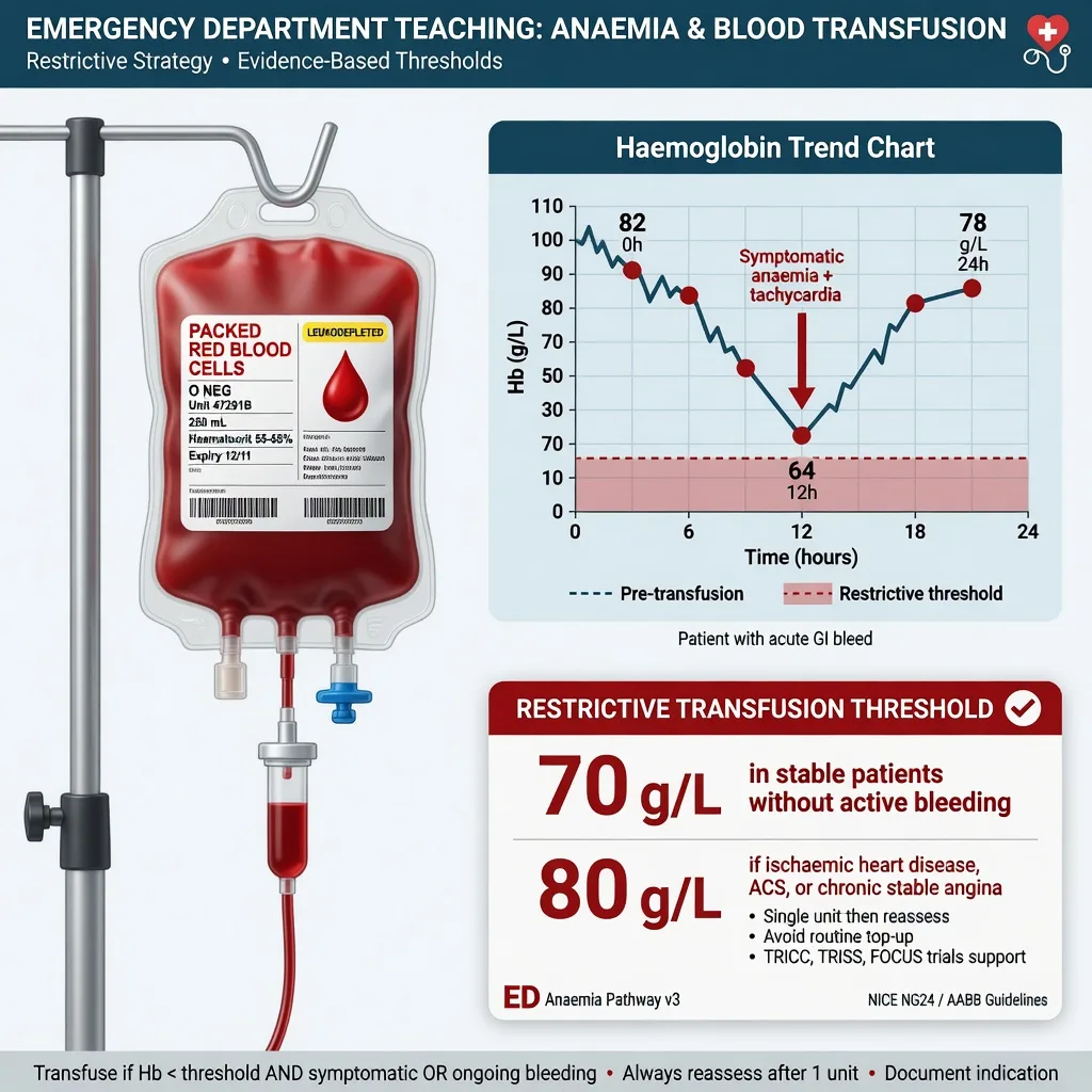
Anaemia is the commonest abnormal blood result in the emergency department, and the blood transfusion that may follow it is one of the commonest and most consequential procedures a trainee performs unsupervised. The Fellowship examiner holds the candidate to four things: the transfusion threshold (and its exceptions), the consent and the pre-transfusion checking, the recognition and management of the transfusion reaction, and the massive haemorrhage protocol. The unifying principle is patient blood management — transfuse only when the benefit is real, give one unit at a time, and remember that the storage lesion blunts the oxygen release so a transfusion above the restrictive threshold is often harm dressed as therapy.[1][2]
Definition and classification
Anaemia is a haemoglobin below the reference range for age and sex (under 130 g/L in men, under 120 g/L in non-pregnant women by the WHO definition). It becomes an emergency department problem when it is symptomatic — dyspnoea, angina, presyncope, or a falling haemoglobin from acute loss — or when it is severe, conventionally under 70 g/L. Transfusion is the intravenous infusion of a blood component: red cells to restore the oxygen-carrying capacity, fresh frozen plasma for coagulopathy, platelets for thrombocytopenia or platelet dysfunction, and cryoprecipitate for the low fibrinogen. [1]
Transfusion is classified by its urgency. The elective or ward transfusion is a single unit given over 90 to 120 minutes to a stable patient at a restrictive threshold. The massive transfusion is the delivery of blood products in a balanced ratio to a bleeding patient — conventionally defined as the loss of one blood volume within 24 hours, or the ongoing need for product in the actively bleeding patient, and operationalised in the emergency department as the activation of the massive haemorrhage protocol. [1]
Epidemiology and risk
Anaemia is present in up to a quarter of emergency department presentations; transfusion follows in a minority. The iatrogenic burden of transfusion is real but now well-characterised: a transfusion reaction occurs in roughly 1 per cent of transfusions, the majority minor (febrile, allergic). The severe reactions sit in the 1-in-10,000 to 1-in-50,000 range — acute haemolytic, TRALI, TACO, anaphylaxis. TACO is now the leading reported cause of transfusion-related death in the haemovigilance databases of the UK (SHOT), the United States and France, having overtaken TRALI as donor-recruitment shifts (male-predominant plasma) drove TRALI down. The takeaway for the candidate is that the dangerous reaction is the common one — the circulatory overload in the elderly recipient of a unit run too fast — not the exotic one. [1]
Pathophysiology — the oxygen-delivery chain and the transfusion trigger
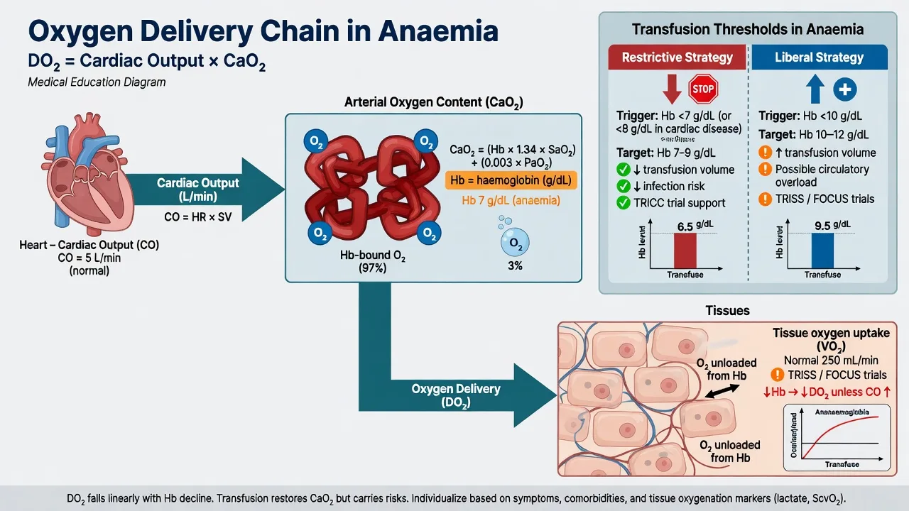
The oxygen delivery to the tissues is the cardiac output multiplied by the arterial oxygen content, and the arterial oxygen content is dominated by the haemoglobin term: (Hb multiplied by 1.34 multiplied by the saturation) plus the small dissolved-oxygen term. The body compensates for a falling haemoglobin by raising the cardiac output (tachycardia, a hyperdynamic stroke volume) and by increasing the oxygen extraction. In the healthy adult this compensation preserves the oxygen delivery until the haemoglobin falls to around 70 g/L, below which the delivery becomes supply-dependent and the lactate rises. [1]
Clinical presentation
Symptomatic anaemia presents with exertional dyspnoea, fatigue, pallor, palpitations, and angina or presyncope in the more severe cases. The chronically anaemic patient — the young woman with menorrhagia at a haemoglobin of 55 g/L — may walk in complaining only of tiredness, because the slow decline allowed the full cardiovascular compensation. The acutely bleeding patient at the same haemoglobin is in class III to IV shock. [1]
Two presentations are the Fellowship traps. First, the elderly or cardiac patient in whom angina or pulmonary oedema is the first sign of a haemoglobin in the 60s — the angina-equivalent of anaemia, where the myocardium cannot compensate because it extracts near-maximally at baseline. Second, the acute bleed where the haemoglobin is normal on arrival — haemodilution lags the bleed by 6 to 12 hours, so the initial haemoglobin reflects the pre-bleed state plus interstitial fluid shift. Assess the shock on the physiology — the heart rate, the blood pressure, the lactate, the base excess — not on the haemoglobin number. [1]
Differential diagnosis — the three mechanisms
The cause of the anaemia frames the workup and the disposition. The reticulocyte count and the mean cell volume partition the differential at the bedside. [1]
Acute blood loss
- GI bleed (upper — haematemesis, melaena; lower — haematochezia), trauma, ruptured ectopic, retroperitoneal
- Normocytic initially; reticulocyte response rises after 3 to 5 days
- Haemoglobin may be NORMAL early — haemodilution lags by 6 to 12 h
- Assess shock on physiology, not the haemoglobin
- Treat the source; transfuse to 70 g/L (80 g/L if IHD)
Haemolysis
- Intravascular (dark urine, low haptoglobin, high LDH, haemoglobinaemia) or extravascular (splenomegaly, jaundice)
- Causes: autoimmune (warm IgG, cold IgM), mechanical (valve, MAHA), infection (malaria), drug, sickle crisis
- Normocytic or macrocytic; high reticulocytes; positive direct antiglobulin test in immune
- Acute severe haemolysis is a transfusion and an immunology emergency
- Group and screen carefully — the autoantibody may mask alloantibodies
Marrow failure / impaired production
- Nutritional (iron deficiency — microcytic; B12 or folate — macrocytic), aplastic, infiltrative (leukaemia, myelofibrosis, metastatic), chronic disease
- Low or inappropriately normal reticulocyte count for the degree of anaemia
- Often pancytopenia if the marrow is the problem — check the white cells and platelets
- The film and the blood count are the first-line discriminators
- Urgent haematology referral; transfuse only if symptomatic or a procedure is planned
The mimics of anaemia are few — the peripheral pallor of poor perfusion, the cyanosis misread as an oxygen problem — but the mimic of the acute bleed is the chief pitfall: a normal haemoglobin in a shocked patient does not exclude a major haemorrhage. [1]
Bedside assessment
The bedside assessment answers three questions: how sick is the patient, where is the blood going (or failing to be made), and is this safe to transfuse. The ABCDE for the unstable; the focused history for the source (melaena, PR examination, menorrhagia, the anticoagulant, the known haematology). The cardiovascular examination for the compensation (a flow murmur, a tachycardia, a gallop in the over-transfused). And, before any unit goes up, the bedside pre-transfusion check: two patient identifiers against the unit label, the unit number, the blood group, the expiry, and the visual inspection of the unit for clots or discoloration. This check prevents the acute haemolytic reaction. [1]
Investigations
The full blood count and film, the reticulocyte count, the mean cell volume and the iron studies, B12 and folate define the production and the deficiency. The haemolysis screen — lactate dehydrogenase, haptoglobin, bilirubin and the direct antiglobulin test — separates the haemolysis. The group and screen establishes the ABO and the RhD group and screens for the clinically significant red-cell antibodies; a crossmatch confirms the compatibility. In the actively bleeding patient, add the coagulation (INR, APTT), the fibrinogen, the ionised calcium (the citrate chelates it) and the venous gas with the lactate. [1]
Transfusion thresholds — the restrictive strategy
The restrictive transfusion strategy is one of the best-evidenced in medicine: transfuse at a haemoglobin of 70 g/L in the stable adult and 80 g/L in the patient with ischaemic heart disease or an acute coronary syndrome. The evidence base is four landmark trials. [1]
The transfusion thresholds and the trials behind them
The TRICC trial (Hebert 1999) randomised 838 critically ill adults to a restrictive threshold of 70 g/L versus a liberal 100 g/L and found no mortality difference and a marked reduction in transfusion; it founded the modern restrictive paradigm.[1] The FOCUS trial (Carson 2011) tested the question specifically in the cardiovascular-disease patient after hip-fracture surgery and found no benefit of a liberal 100 g/L over a restrictive 80 g/L — the exception threshold for the ischaemic heart.[2] The TRISS trial (Holst 2014) extended the restrictive strategy to septic shock (70 versus 90) and again found no benefit of the higher threshold.[3] The Villanueva trial (2013) showed that a restrictive strategy in the acute upper gastrointestinal bleed improved survival and reduced rebleeding.[4]
Immediate management — the transfusion bundle
The elective transfusion of the symptomatic stable patient follows a bundle: the indication, the consent, the check, the unit, and the monitoring. [1]
The consent must include the option to decline. The Jehovah's Witness patient may refuse all major blood components on religious grounds, and that refusal is binding in the competent adult; the discussion explores the alternatives — cell salvage (a continuous circuit may be acceptable), the fractionated products (albumin, clotting factors — a matter of individual conscience), the erythropoietin and the iron, and the acceptance of risk. Document the specific products accepted and declined. A paediatric transfusion to the child of Jehovah's Witness parents who refuses a life-saving transfusion is the subject of an emergency legal order — the emergency-department team treats the child, the court ratifies. [1]
Massive transfusion and the massive haemorrhage protocol
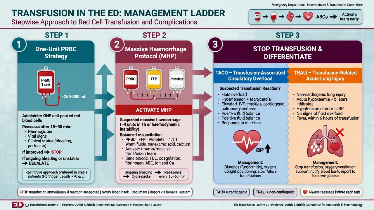
The massive haemorrhage protocol is activated by a single call to the blood bank and the assembly of the team. It delivers blood products in a balanced ratio and abandons the clear-fluid resuscitation that dilutes the clotting factors. [1]
The component targets in the actively bleeding patient are a fibrinogen over 1.5 g/L (cryoprecipitate), a platelet count over 50 (a pool of platelets raises it by 20 to 40), and an INR under 1.5 (fresh frozen plasma at 15 mL per kilogram, typically 4 units in the adult). The viscoelastic test (ROTEM or TEG) at the bedside guides the ratio in real time. [1]
Transfusion reactions — the five to know
Every transfusion candidate must recognise the five reactions and execute the universal first step. The reactions are distinguished by the timing and the bedside features. [1]
TACO (circulatory overload)
- Within 6 hours; the leading cause of transfusion-related death
- Dyspnoea, hypertension, raised JVP, pulmonary oedema, positive fluid balance, raised BNP
- Risk: elderly, renal, cardiac, rapid transfusion
- Management: STOP, sit up, oxygen, furosemide 20 to 40 mg IV, consider NIV
- Prevention: one unit at a time, slow, consider a diuretic in the at-risk
TRALI (acute lung injury)
- Within 6 hours; donor antibody (often multiparous female donor) attacks the recipient leucocytes
- Dyspnoea, hypoxia, HYPOTENSION, bilateral infiltrates, normal cardiac output and no fluid overload
- Two-hit: recipient primed (sepsis, trauma) plus donor antibody
- Management: STOP, supportive — oxygen, NIV or ventilation; diuretics do NOT help and may harm
- Mortality high (5 to 25 per cent) but survivors recover fully in days
Acute haemolytic
- Within minutes of starting the unit; an ABO incompatibility
- Fever, flank pain, hypotension, tachycardia, nausea, haemoglobinuria, DIC, renal failure
- The commonest cause is a CLERICAL error — a wrong-patient sample or unit
- Management: STOP immediately, keep IV open with saline, force the diuresis, return unit and fresh samples to blood bank, repeat the group and the direct antiglobulin test
- Prevention: the two-identifier bedside check
Allergic / anaphylactoid
- Urticaria, pruritus, mild — common; anaphylaxis rare but catastrophic
- Mild urticaria: slow the rate, chlorphenamine 10 mg IV, observe
- Anaphylaxis: hypotension, bronchospasm — typically in the IgA-deficient with anti-IgA
- Management: STOP, adrenaline 500 micrograms intramuscular, fluid, airway; wash the cells next time
- Prevention: washed products for the IgA-deficient
Febrile non-haemolytic
- Fever, rigors, during or up to 4 hours after; the recipient antibodies to donor leucocytes or the cytokine accumulation in storage
- Haemodynamically stable; no haemolysis
- Management: STOP, exclude haemolysis (recheck the sample), paracetamol 1 g, slow the rate, resume if settles
- Prevention: leucodepletion (now standard) reduces the incidence
Subtypes and scenarios
The anticoagulated patient who bleeds is transfused for the symptom and reversed for the agent — the prothrombin complex concentrate for the warfarin, the idarucizumab for the dabigatran, the andexanet for the apixaban or the rivaroxaban, and the Beriplex-equivalent for the life-threatening bleed. The chronic transfusion patient — the thalassaemia, the myelodysplasia — accumulates the iron overload and needs chelation; the candidate should not chase a normal haemoglobin in these patients but transfuse to the symptom threshold. The paediatric transfusion is weight-based — 10 to 15 mL per kilogram of packed red cells raises the haemoglobin by 20 to 30 g/L, given over 2 to 4 hours. The pregnant patient receives CMV-negative, K-negative products; anti-D immunoglobulin is given to the RhD-negative mother after a sensitising event. The immunocompromised patient — the stem-cell transplant, the chemotherapy — receives irradiated products to prevent the transfusion-associated graft-versus-host disease, the rare but uniformly fatal reaction in which the donor T-lymphocytes engraft. [1]
Complications and pitfalls
The complications of transfusion are the reactions above and the infectious risks (now vanishingly rare — HIV and hepatitis C under 1 in a million units in the ANZ and UK blood supplies, but bacterial contamination of platelets remains the commonest infectious threat). The longer-term complications are the iron overload in the chronically transfused and the alloimmunisation. The pitfalls: transfusing above the restrictive threshold; running a unit too fast in the elderly or the cardiac (TACO); missing the bedside check (the haemolytic reaction); restarting a reaction on a guess that it is "just febrile"; resuscitating the bleeder with crystalloid instead of activating the massive haemorrhage protocol; and forgetting the calcium and the warmth in the massive transfusion. [1]
Prognosis and disposition
The outcome is the underlying cause's outcome — the transfusion buys the time and the oxygen delivery, it does not treat the disease. The severely anaemic but stable patient is transfused one unit, rechecked, and admitted for the workup of the cause. The actively bleeding patient is resuscitated to theatre or interventional radiology for the source control. The massive-transfusion patient goes to the intensive care after the haemorrhage control for the correction of the lethal triad. The patient with a transfusion reaction is admitted, investigated, and reported to the haemovigilance system. [1]
Special populations
The ischaemic heart disease patient is the one exception to the 70 g/L threshold — transfuse to 80 g/L, because the myocardium extracts near-maximally at rest and cannot compensate for a falling haemoglobin, so the anaemia precipitates the demand ischaemia; but even here the FOCUS trial supports the restrictive approach over the liberal.[2] The elderly is the TACO-prone patient — transfuse slowly, one unit, consider a diuretic. The renal-disease patient tolerates both the anaemia (chronic, with the erythropoietin deficiency) and the fluid (TACO) poorly — transfuse judiciously. The paediatric patient is weight-based. The pregnant patient is CMV-negative and K-negative; the post-partum haemorrhage triggers its own protocol.
Evidence and regional guidelines
The contemporary framework rests on the restrictive strategy and the massive-haemorrhage protocol. TRICC (Hebert 1999) founded the restrictive paradigm.[1] FOCUS (Carson 2011) confirmed it in the cardiovascular-disease patient and set the 80 g/L threshold for ischaemic heart disease.[2] TRISS (Holst 2014) extended it to septic shock.[3] Villanueva (2013) showed the survival benefit of the restrictive strategy in the upper GI bleed.[4] PROPPR (Holcomb 2015) established the 1:1:1 ratio in the massive transfusion.[5] The Vlaar 2019 consensus redefined TRALI as a two-event syndrome, separating it from TACO.[6]
ANZ practice note. The National Blood Authority's Patient Blood Management guidelines govern the practice: a restrictive threshold of 70 g/L (80 g/L in the ischaemic heart disease), one unit at a time with a recheck, the consent including the option to decline, and the bedside two-identifier check. The massive haemorrhage protocol is hospital-specific but follows the 1:1:1 ratio with the tranexamic acid and the calcium replacement. All blood products in Australia and New Zealand are leucodepleted (reducing the febrile and the CMV transmission) and the irradiated products are reserved for the immunocompromised. The serious transfusion reactions are reported to the Blood Service and the state haemovigilance system. The Jehovah's Witness consent is documented on the specific consent form with the products accepted and declined. [1]
Exam pearls
- 70 stable, 80 ischaemic heart disease — the restrictive strategy (TRICC, FOCUS, TRISS, Villanueva).
- One unit at a time, recheck the haemoglobin — do not transfuse to a number.
- The bedside two-identifier check prevents the acute haemolytic reaction — the commonest cause is the clerical error.
- TACO is the leading cause of the transfusion-related death now — slow, one unit, diurese the elderly and the cardiac.
- TRALI is hypoxia with hypotension and bilateral infiltrates within 6 hours — the diuretic does not help and may harm.
- At any reaction: STOP, saline, return the unit and the samples.
- Massive haemorrhage — 1:1:1, the tranexamic acid within 3 hours, the calcium, the permissive hypotension.
- Consent includes the option to decline — the Jehovah's Witness discussion is a common communication OSCE.
- The haemoglobin is normal in the first 6 to 12 hours of an acute bleed — assess the shock on the physiology. [1]
Model answer — the ischaemic-heart-disease patient with melaena and an Hb of 58 g/L
A 78-year-old woman with the known ischaemic heart disease presents with melaena and a haemoglobin of 58 g/L. The transfusion target is 80 g/L — the ischaemic heart disease threshold — because the myocardium cannot compensate for the falling haemoglobin and a restrictive-70 strategy risks the demand ischaemia in this group (FOCUS supports the 80-over-100, not the 70).[2] The pre-transfusion checks: group and screen, the antibody screen, and the bedside two-identifier check of the patient against the unit, the unit number, the group and the expiry. The consent explains the indication, the common risks (febrile, allergic, TACO) and the rare serious risks (TRALI, haemolytic, infection) and the option to decline. Give one unit of packed red cells over 90 to 120 minutes — slower in this elderly cardiac patient with the TACO risk, consider furosemide 20 to 40 mg — with the observations at baseline and at 15 minutes. Recheck the haemoglobin 15 minutes after the unit. If she develops the dyspnoea, the hypertension and the pulmonary oedema within 6 hours, suspect TACO — STOP, sit up, oxygen, furosemide, consider the NIV — and report it. If the hypoxia comes with the hypotension and the bilateral infiltrates, suspect TRALI — STOP, supportive ventilation, the diuretic does not help.[6]
Exam practice
SAQ — Massive transfusion protocol activation in polytrauma
10 minutes · 10 marks
A 34-year-old man is brought to the trauma bay 25 minutes after a high-speed motorcycle crash. He is pale, diaphoretic and agitated (GCS 13), BP 72/46, HR 132, RR 28, SpO2 94 per cent on 15 L oxygen via a non-rebreather mask. He has a markedly distended abdomen with free fluid on the focused assessment with sonography in trauma (FAST), an open femoral shaft fracture with active external bleeding, and clinical pelvic instability. The initial haemoglobin is 134 g/L, lactate 7.2 mmol/L, INR 1.9, fibrinogen 1.2 g/L, ionised calcium 0.82 mmol/L. The trauma surgeon and the orthopaedic team are en route; the massive haemorrhage protocol has been activated.
SAQ — Acute transfusion reaction: the differentiation at the bedside
10 minutes · 10 marks
A 68-year-old woman with known ischaemic heart disease and a haemoglobin of 58 g/L from melaena is 15 minutes into her first unit of packed red cells when she becomes acutely dyspnoeic. Respiratory rate 32, SpO2 88 per cent on room air, blood pressure 168/96 (baseline 142/82 on arrival), JVP raised at 10 cm, and auscultation reveals bilateral coarse crackles to the mid-zones. She is afebrile, with no urticaria, no rigors and no flank pain. She was given the unit over the standard 90 minutes after the bedside two-identifier check, and her pre-transfusion weight was 62 kg with an eGFR of 38.
Red flags
[1]References
- [1]Hébert PC, Wells G, Blajchman MA, et al. A multicenter, randomized, controlled clinical trial of transfusion requirements in critical care. Transfusion Requirements in Critical Care Investigators, Canadian Critical Care Trials Group N Engl J Med, 1999.PMID 9971864
- [2]Carson JL, Terrin ML, Noveck H, et al. Liberal or restrictive transfusion in high-risk patients after hip surgery N Engl J Med, 2011.PMID 22168590
- [3]Holst LB, Haase N, Wetterslev J, et al. Lower versus higher hemoglobin threshold for transfusion in septic shock N Engl J Med, 2014.PMID 25270275
- [4]Villanueva C, Colomo A, Bosch A, et al. Transfusion strategies for acute upper gastrointestinal bleeding N Engl J Med, 2013.PMID 23281973
- [5]Holcomb JB, Tilley BC, Baraniuk S, et al. Transfusion of plasma, platelets, and red blood cells in a 1:1:1 vs a 1:1:2 ratio and mortality in patients with severe trauma: the PROPPR randomized clinical trial JAMA, 2015.PMID 25647203
- [6]Vlaar APJ, Juffermans NP, Bux J, et al. A consensus redefinition of transfusion-related acute lung injury Transfusion, 2019.PMID 30993745