EM · Haematology and general medical emergencies
Sickle cell crisis
Also known as Vaso-occlusive crisis · Acute chest syndrome · Splenic sequestration crisis · Aplastic crisis · Sickle cell pain crisis
Sickle cell crisis is the umbrella for the acute, life-threatening events of sickle cell disease (HbS disease): the vaso-occlusive pain crisis, acute chest syndrome, splenic sequestration crisis and aplastic crisis. The Fellowship-critical syndromes are acute chest syndrome — fever with a new pulmonary infiltrate and hypoxia, the leading cause of death in sickle cell disease — and the rapidly fatal childhood splenic sequestration. The management backbone is oxygen, cautious isotonic hydration, and prompt opioid analgesia (morphine 0.1 mg/kg intravenously), with exchange transfusion to reduce the HbS fraction below 30 per cent for the severe acute chest syndrome, stroke and refractory vaso-occlusive crisis. The reticulocyte count is the diagnostic discriminator: it is high in the vaso-occlusive, sequestration and haemolytic crises but absent in the parvovirus B19 aplastic crisis. ACEM-primary, globally tagged.
On this page & tools
Your progress
Saved locally on this device.
Target exams
Red flags
Related topics
- Community-acquired pneumonia
- Pulmonary embolism (acute, in the emergency department)
- DKA, HHS and hypoglycaemia
- Sepsis and septic shock — the emergency department approach
- Paediatric sepsis and septic shock (the septic child in the emergency department)
- Acute ischaemic stroke
- Fluid resuscitation in the emergency department
Sickle cell crisis is the umbrella term for the acute, life-threatening events that complicate sickle cell disease (HbS disease), the inherited haemoglobinopathy in which a single amino acid substitution in the β-globin chain allows haemoglobin to polymerise when deoxygenated. The crisis syndromes — the vaso-occlusive pain crisis, acute chest syndrome, splenic sequestration crisis and aplastic crisis — share a common mechanism (sickled erythrocytes obstructing the microvasculature, with haemolysis and ischaemia) but differ in their bedside presentation, their urgency and their specific treatment. The Fellowship candidate must recognise each syndrome from its fingerprint, deliver oxygen, hydration and prompt opioid analgesia as the universal backbone, and escalate to exchange transfusion for the severe acute chest syndrome, stroke and refractory pain.[1][2]

Definition and classification
Sickle cell disease is the autosomal recessive disorder caused by the HBB mutation (glutamate substituted by valine at the sixth position of the β-globin chain, the glu6val substitution on chromosome 11) that produces haemoglobin S. The disease encompasses several genotypes: homozygous HbSS (classic sickle cell anaemia, the most severe), and the compound heterozygotes HbSC disease, HbSβ-thalassaemia and the rarer HbS variants, which run a milder but still crisis-prone course. A "sickle cell crisis" is any acute event driven by sickling, and the emergency physician classifies the presentation by its dominant clinical syndrome, because the syndromes demand different escalation.[1]
The four acute syndromes carry the Fellowship weight. Vaso-occlusive crisis (VOC) is the acute pain crisis — the commonest presentation — in which sickled cells occlude the microvasculature of bone, marrow and viscera, producing severe ischaemic pain without an alternative cause. Acute chest syndrome (ACS) is the new pulmonary infiltrate on the chest radiograph accompanied by a respiratory symptom or sign (fever, cough, dyspnoea, pleuritic chest pain, hypoxia) — it is the leading cause of death in sickle cell disease and demands aggressive treatment.[2] Splenic sequestration crisis is the sudden trapping of blood in an enlarging spleen, producing hypovolaemic shock and a falling haemoglobin, seen in young children before functional asplenia develops. Aplastic crisis is the transient arrest of erythropoiesis, classically triggered by parvovirus B19, in which the haemoglobin falls precipitously and the reticulocyte count — normally the marrow's response to chronic haemolysis — is inappropriately low or absent. Two further acute events sit within the crisis umbrella: stroke (ischaemic, disproportionately affecting children) and priapism (a painful prolonged erection from veno-occlusion).
The four acute crisis syndromes at a glance
Epidemiology and risk factors
Sickle cell disease is the commonest severe monogenic disorder worldwide, with the gene maintained at high frequency in sub-Saharan Africa, the Mediterranean, the Middle East and parts of India where falciparum malaria is or was endemic (the heterozygote carrier, sickle cell trait, confers partial malaria protection). The sickle haplotype and genotype vary by region: HbSS is the predominant and most severe form. In Australia and New Zealand the prevalence is low in the general population but rising through migration from high-prevalence regions, and every metropolitan emergency department will see sickle cell patients; the candidate must not assume the diagnosis is absent because the local population prevalence is low.[1]
The risk of an acute crisis is driven by the triggers that promote sickling. The recognised precipitants are hypoxia (high altitude, air travel, sleep apnoea), dehydration (acidosis and increased plasma viscosity), infection (the commonest precipitant, especially respiratory infection, parvovirus B19, and in children the encapsulated organisms of functional asplenia — pneumococcus, Haemophilus, meningococcus), cold, acidosis, fever, surgery and anaesthesia, pregnancy, and psychological stress. Functional asplenia develops progressively through childhood as repeated splenic infarction autoinfarcts the organ (the "autosplenectomy" of HbSS, usually complete by age five), after which the patient is permanently at risk of overwhelming encapsulated-bacteraemia. The mortality of sickle cell disease has fallen substantially with penicillin prophylaxis, vaccination, hydroxycarbamide and transfusion programmes, but the acute chest syndrome remains the leading cause of death, and stroke remains the leading cause of neurological morbidity in children with the disease.[2]
Pathophysiology
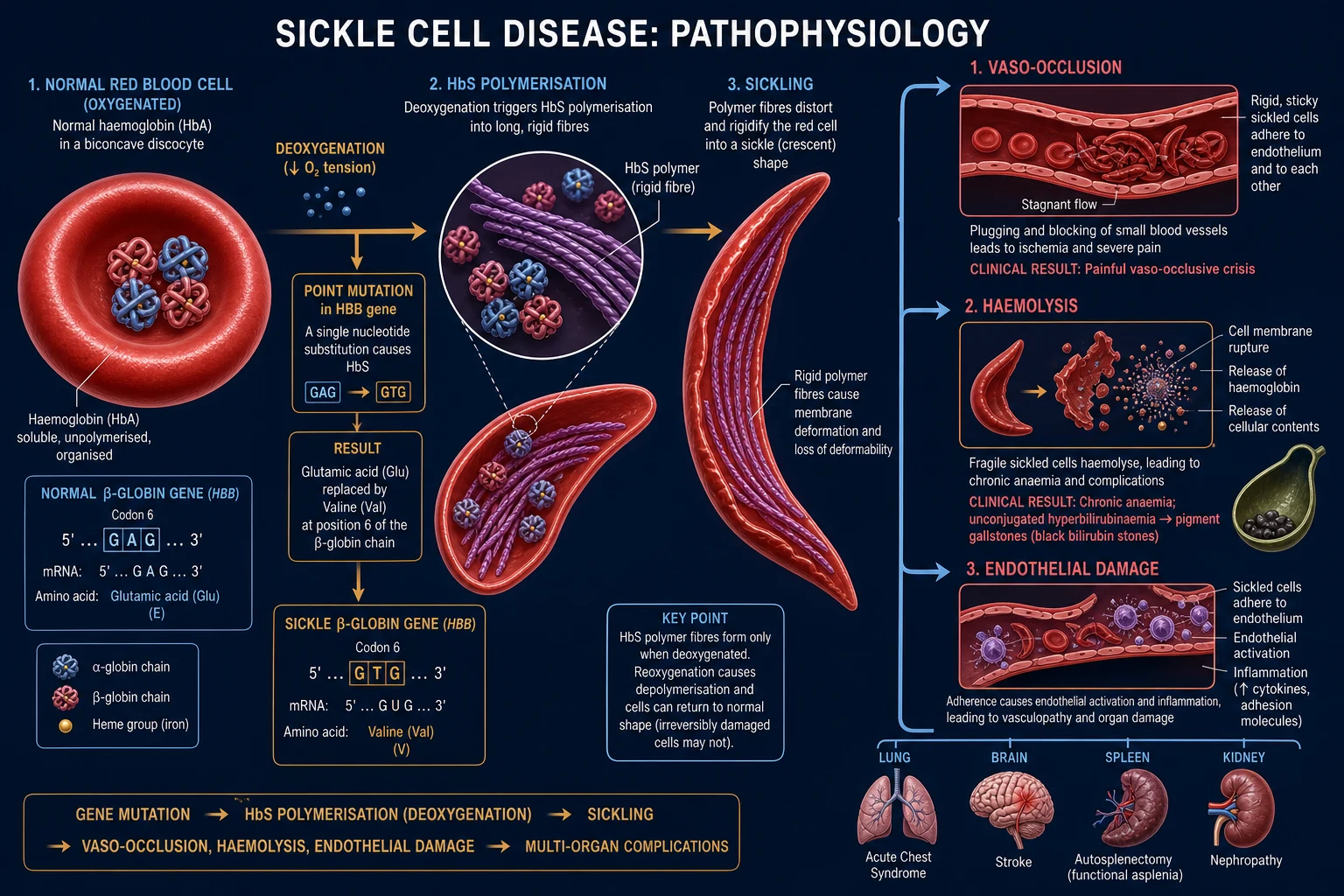
The single molecular event is the polymerisation of deoxygenated haemoglobin S. When oxygen tension falls, the valine at position six of the β-globin chain locks into a hydrophobic pocket on an adjacent haemoglobin molecule, forming rigid polymers that elongate the red cell into the characteristic sickle shape. Sickling is initially reversible on reoxygenation, but repeated cycles produce membrane damage, intracellular dehydration and irreversibly sickled cells. Two downstream consequences dominate the clinical disease.[1]
The first is chronic haemolysis. Sickled cells are rigid and fragile, and are destroyed in the spleen and circulation; the steady-state haemoglobin of HbSS is therefore typically 60 to 90 g/L with a compensatory reticulocytosis. Free haemoglobin released by haemolysis scavenges nitric oxide, depleting the vasodilator that normally opposes pulmonary and systemic vascular tone, and this contributes over years to pulmonary hypertension, leg ulcers and the haemolysis-endothelial dysfunction phenotype. The second is vaso-occlusion. Sickled erythrocytes, along with leucocytes and platelets, adhere to an activated vascular endothelium and obstruct the microvasculature, producing ischaemia, infarction and the severe pain of the crisis, and — when this occurs in the pulmonary, cerebral or splenic circulation — the acute chest syndrome, stroke and splenic sequestration. [1]
[1]The pathophysiological cascade — one mutation to a crisis
1
2
3
4
5
6
Clinical presentation
The clinical presentation depends on the dominant syndrome, and the emergency physician must keep all four in mind for every sickle cell patient, because one syndrome can evolve into another (a vaso-occlusive pain crisis progressing to acute chest syndrome is the classic sequence).[2]
Vaso-occlusive crisis presents with severe, acute pain, usually in the back, chest, long bones, abdomen or joints, and often at sites of previous crises. The pain is typically severe enough to be rated 7 to 10 out of 10, is reproducible, and is frequently accompanied by no objective physical findings — the absence of signs does not exclude the crisis. Tachycardia, hypertension and diaphoresis from pain and adrenergic surge are common; fever may indicate an infective trigger or evolving acute chest syndrome. Abdominal pain from mesenteric sickling can mimic an acute abdomen, and the candidate must distinguish it from surgical pathology. [1]
Acute chest syndrome presents with the new onset of respiratory symptoms — fever, cough, dyspnoea, pleuritic chest pain or wheeze — accompanied by a new pulmonary infiltrate on the chest radiograph and, frequently, hypoxia. The radiographic infiltrate may lag the symptoms by hours to days, so a normal early film does not exclude the diagnosis; a falling oxygen saturation, rising respiratory rate or increasing work of breathing in a sickle cell patient is acute chest syndrome until proven otherwise. The syndrome may be triggered by infection (bacterial, viral including SARS-CoV-2, or atypical), by fat embolism from marrow infarction, by pulmonary infarction, or by hypoventilation and atelectasis from chest-wall pain and splinting.[2]
Splenic sequestration crisis presents in a child under five (before functional asplenia develops) with sudden pallor, lethargy, irritability, abdominal pain and distension, and signs of hypovolaemic shock — tachycardia, prolonged capillary refill and hypotension. The spleen is palpably enlarged and may enlarge further over hours; the haemoglobin drops by 20 g/L or more, and the reticulocyte count is high. The crisis can be fatal within hours from hypovolaemia and severe anaemia. [1]
Aplastic crisis presents with the symptoms of acute, severe anaemia — progressive pallor, fatigue, dyspnoea on exertion, headache and, in severe cases, high-output cardiac failure — over days. There is typically a viral prodrome; parvovirus B19 is the classical trigger. The reticulocyte count is inappropriately low or absent, distinguishing the aplastic crisis from the haemolytic and sequestration crises. [1]
Stroke and priapism are the two further acute events. Stroke in sickle cell disease is predominantly ischaemic, affects children disproportionately, and may present with a focal deficit, seizures, altered conscious level, or (in the silent infarcts) only cognitive decline. Priapism — a prolonged, painful erection from veno-occlusion of the corpora cavernosa — is a urological emergency when it persists beyond four hours; stuttering priapism precedes a major event in many patients. [1]
Vaso-occlusive (pain) crisis — #1 presentation
- The commonest presentation: severe ischaemic pain in the back, chest, abdomen, limbs or joints, often reproducible at sites of prior crises; tachycardia and diaphoresis from the adrenergic surge
- Reticulocyte count HIGH; Hb at the baseline 60 to 90 g/L; no new infiltrate; fever may herald infection or evolving acute chest syndrome
- Morphine 0.1 mg/kg IV every 15 to 20 min then PCA; hydration 1 to 1.5x maintenance; incentive spirometry every 2 h; identify and treat the trigger
- Discharge on oral analgesia once controlled; admit for IV opioids, any ACS feature, or an uncontrolled trigger
Acute chest syndrome — #1 cause of death
- New pulmonary infiltrate PLUS a respiratory symptom (fever, cough, dyspnoea, pleuritic pain) or hypoxia; the infiltrate may lag the symptoms by hours to days
- Triggers: infection, fat embolism from marrow infarction, pulmonary infarction, or hypoventilation and atelectasis from chest-wall pain and splinting
- Oxygen; ceftriaxone + macrolide; bronchodilators; incentive spirometry; HFNC or NIV for hypoxia
- Simple transfusion if moderate; urgent EXCHANGE transfusion (HbS under 30 per cent) if severe — multilobar, worsening hypoxia, rapid progression, falling platelets or neurology
Splenic sequestration crisis
- Children under five (before autoinfarction); sudden pallor, lethargy, abdominal distension and hypovolaemic shock with an ENLARGING SPLEEN — can be fatal within hours
- Hb drop of 20 g/L or more; reticulocyte count HIGH; platelets may fall; the older patient has autoinfarcted the spleen and cannot sequester
- Fluid boluses 10 to 20 mL/kg of 0.9 per cent saline; urgent packed-cell transfusion; transfuse cautiously — sequestered blood re-enters the circulation as the crisis resolves
- Recurrence is common and fatal; after a second event start a chronic transfusion programme and plan splenectomy
Aplastic crisis (parvovirus B19)
- Acute severe anaemia over days with a viral prodrome; pallor, dyspnoea, headache, high-output cardiac failure in the worst case
- Hb low; reticulocyte count INAPPROPRIATELY LOW OR ABSENT — the discriminator; parvovirus B19 IgM or PCR positive
- Transfuse packed red cells until the marrow recovers (1 to 2 weeks); ISOLATE — parvovirus is contagious to pregnant contacts (hydrops fetalis) and the immunocompromised
- IVIG for persistent parvovirus in the immunocompromised patient; monitor the reticulocyte count for recovery
Hyperhaemolysis crisis
- A rapid Hb drop with an exaggerated reticulocytosis and haemolysis markers (LDH high, haptoglobin low); classically triggered by a recent transfusion
- The dangerous paradox — further transfusion ACCELERATES the haemolysis, so transfusing for the low Hb makes it worse
- AVOID further transfusion if possible; corticosteroids and IVIG; supportive care; urgent haematology input
- Distinguish from the aplastic crisis by the reticulocyte count: HIGH in hyperhaemolysis, ABSENT in aplasia
Stroke — especially in children
- Acute focal deficit, seizure or altered conscious level; ischaemic from sickle vasculopathy and large-vessel stenosis, but haemorrhagic moyamoya also occurs
- CT or MRI; transcranial Doppler velocity at or above 200 cm/s screens children at risk; do NOT default to thrombolysis
- Urgent EXCHANGE transfusion to HbS under 30 per cent within hours; consult stroke and haematology before any lytic or thrombectomy
- Chronic transfusion programme for secondary prevention; screen children annually with TCD (STOP)
Priapism
- A painful prolonged erection beyond 4 hours from veno-occlusion of the corpora cavernosa; stuttering episodes precede a major event
- Clinical diagnosis; investigate sickle status if it is the first presentation
- Urology emergency: corporal aspiration and irrigation with intracavernosal phenylephrine; exchange transfusion and a surgical shunt for refractory cases
- Pseudoephedrine or etilefrine for stuttering prophylaxis; optimise the underlying disease with hydroxycarbamide
Differential diagnosis
The crisis syndromes each have mimics that share their presentation, and the candidate must distinguish them at the bedside — the vaso-occlusive abdominal pain from the acute abdomen, the acute chest syndrome from pneumonia and pulmonary embolism, and the aplastic anaemia from diabetic ketoacidosis and from blood loss. The history of sickle cell disease, the haemolysis pattern, the reticulocyte count and the chest radiograph are the discriminators. [1]
Pneumonia (vs ACS)
- Fever, cough, purulent sputum, focal crackles, infiltrate; may be the infective trigger of ACS rather than a separate diagnosis
- Distinguished from ACS only by context — a sickle cell patient with a new infiltrate IS treated as ACS regardless, because the management (oxygen, antibiotics, transfusion for severity) is the same
- Oxygen, antibiotics covering pneumococcus and atypicals (ceftriaxone plus macrolide), fluids; transfuse or exchange-transfuse if hypoxia or worsening
- ACS is treated identically to pneumonia plus the sickle-specific escalation — do not wait to "decide" between them
Pulmonary embolism (vs ACS)
- Pleuritic chest pain, dyspnoea, sudden onset, sometimes leg swelling; sickle cell patients are prothrombotic and PE is genuinely more common
- D-dimer is unhelpful — it is elevated at baseline in sickle cell disease; CTPA if PE is suspected; the ACS infiltrate and haemolysis favour sickle lung
- Anticoagulation for confirmed PE; oxygen and transfusion support; the two may coexist
- A sickle cell patient with pleuritic pain and hypoxia needs an infiltrate-seeking film first, then CTPA if the picture fits PE
Diabetic ketoacidosis (vs aplastic crisis / acute abdomen)
- Polyuria, polydipsia, Kussmaul breathing, ketotic breath; glucose high; can present with abdominal pain that mimics mesenteric VOC
- Hyperglycaemia, ketones above 3 mmol/L, metabolic acidosis with pH below 7.3 distinguish DKA; the reticulocyte count and haemolysis markers are normal
- Fluid, fixed-rate intravenous insulin 0.1 units/kg/h, potassium replacement; not opioids or transfusion
- A sickle cell patient with abdominal pain and acidosis needs a glucose and a venous gas first — DKA and VOC can coexist
Acute abdomen (appendicitis, cholecystitis, pancreatitis)
- Localised peritoneal signs, guarding, rigidity; gallstones are common in sickle cell disease from chronic haemolysis
- Lipase, ultrasound for gallstones, surgical review; the reticulocytosis and pain history favour VOC but do not exclude surgical pathology
- Surgical pathway; continue sickle-cell hydration and analgesia in parallel
- Mesenteric VOC can mimic appendicitis — observe and re-examine; do not operate on an uncomplicated VOC
Investigations and diagnostic targets
The investigations are sent in parallel with the first analgesia and oxygen; they seek the crisis syndrome, its severity and any precipitant. A full blood count shows the baseline anaemia (haemoglobin 60 to 90 g/L in HbSS); an acute fall signals sequestration, aplasia or hyperhaemolysis. The reticulocyte count is the single most discriminating test: it is high in the vaso-occlusive, sequestration and haemolytic crises (the marrow responding appropriately) and inappropriately low or absent in the aplastic crisis. A blood film shows sickled cells, target cells, and (after autosplenectomy) Howell-Jolly bodies. Haemolysis markers — raised lactate dehydrogenase, raised indirect bilirubin, low haptoglobin — confirm haemolysis. A type and screen or crossmatch is sent early because transfusion is frequently required. Blood cultures and a urine culture are sent if the patient is febrile, and a chest radiograph is mandatory to detect the new infiltrate of acute chest syndrome; the film may lag the symptoms, so a clinical suspicion with a normal early film warrants repeat imaging. Venous or arterial blood gas quantifies the hypoxia and the acidosis that drive sickling. An ECG is routine. Lipase excludes pancreatitis in the abdominal-pain presentation; β-hCG is checked in any woman of reproductive age. A haemoglobin electrophoresis quantifies the HbS fraction and confirms the genotype if it is not already documented, but it is not an emergency test.[1][3]
Diagnostic criteria reproduced
Immediate management and resuscitation
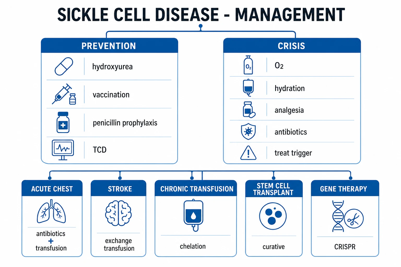
The resuscitation is syndrome-specific but follows a universal backbone: oxygen, hydration, analgesia, with escalation to transfusion for the severe presentations. Secure the airway and give supplemental oxygen to maintain the oxygen saturation at or above 94 per cent — hypoxia is both a consequence of the acute chest syndrome and a driver of further sickling, so it must be corrected promptly. Establish intravenous access, attach cardiac monitoring, and send the investigations in parallel. Treat any hypovolaemia or dehydration with cautious isotonic crystalloid (0.9 per cent saline), because both over- and under-hydration harm: dehydration worsens sickling, but aggressive fluid causes pulmonary oedema that accelerates the acute chest syndrome. A rehydration rate of roughly one to one-and-a-half times maintenance is reasonable in the adult, moderated by the cardiovascular and renal status.[1]
Analgesia must be prompt. The under-treatment of sickle cell pain is the classic and most-cited failure, and the pain of a vaso-occlusive crisis is comparable to surgical pain. The first opioid should be delivered within 30 minutes of triage. The agent of choice in the haemodynamically stable adult is morphine 0.1 mg per kilogram intravenously, repeated every 15 to 20 minutes and titrated to the reported pain score, followed by a scheduled regimen or patient-controlled analgesia. Fentanyl is an alternative, particularly in the patient with renal impairment or a morphine allergy. Meperidine (pethidine) is avoided because its metabolite normeperidine accumulates, lowers the seizure threshold and causes seizures, especially in renal impairment and with repeated dosing. Paracetamol and, where renal function allows, a non-steroidal anti-inflammatory are useful adjuncts.[5][6]
The acute chest syndrome is treated on diagnosis with oxygen, broad-spectrum antibiotics (a third-generation cephalosporin such as ceftriaxone to cover pneumococcus and Haemophilus, plus a macrolide to cover atypicals, since infection is the commonest identified cause), bronchodilators if there is wheeze or a history of asthma, incentive spirometry to reverse the hypoventilation and atelectasis that drive the infiltrate, and respiratory escalation (high-flow nasal cannula or non-invasive ventilation) for hypoxia. Transfusion is the sickle-specific escalation: a simple transfusion for moderate acute chest syndrome, and an exchange transfusion for the severe syndrome (worsening hypoxia, multilobar infiltrate, rapid progression, or a falling haemoglobin), with the aim of reducing the HbS fraction below 30 per cent.[2][3]
The first hour of a suspected sickle cell crisis in one breath
Recognise the syndrome — the history of sickle cell disease, the pattern of the pain, and the respiratory status. Give oxygen to keep the saturation at or above 94 per cent; establish intravenous access; run in 0.9 per cent saline at one to one-and-a-half times maintenance (resuscitate the shocked child faster). Give morphine 0.1 mg per kilogram intravenously as the first opioid, reassess the pain score at 15 minutes and re-titrate; do not use meperidine. Send the full blood count, reticulocyte count, blood film, haemolysis markers, type and screen, blood cultures if febrile, venous gas, chest radiograph, ECG, lipase and β-hCG. If the chest radiograph shows a new infiltrate with respiratory symptoms, treat as acute chest syndrome: add ceftriaxone and a macrolide, incentive spirometry, bronchodilators if wheeze, and escalate to transfusion or exchange transfusion for worsening hypoxia. If the haemoglobin has fallen with a low reticulocyte count, isolate and transfuse for aplastic crisis; if a child is shocked with an enlarging spleen, resuscitate and transfuse urgently for splenic sequestration. Reassess the pain, the saturation and the respiratory rate at least every 30 minutes. [1]
The ED first hour of the sickle cell crisis
0 to 10 minutes — recognise and resuscitate
ABCDE. Oxygen to keep the SpO2 at or above 94 per cent; two large-bore cannulae; full monitoring. Identify the syndrome at the bedside: pain only, respiratory, a shocked child, or a new neurological deficit. Send the bloods in parallel with the first analgesia, not after.
0 to 30 minutes — analgesia first
Morphine 0.1 mg/kg IV, reassess and re-titrate every 15 to 20 minutes; do NOT under-treat and do NOT use meperidine. Layer paracetamol and, if renal function allows, an NSAID. Set up patient-controlled analgesia for the established adult crisis.
0 to 30 minutes — hydration and the panel
0.9 per cent saline at 1 to 1.5x maintenance (10 to 20 mL/kg boluses for the shocked child, repeated). Send the FBC, reticulocyte count, blood film, LDH, bilirubin, haptoglobin, group-and-screen, blood cultures if febrile, venous gas, chest radiograph, ECG, lipase and beta-hCG.
Classify by syndrome
Read the reticulocyte count and the chest film together. VOC: reticulocytes high, no infiltrate. ACS: a new infiltrate plus a respiratory symptom — treat on the clinical picture even if the early film is normal. Sequestration: a child with an Hb drop, reticulocytes high and shock. Aplastic: an Hb drop with reticulocytes absent (parvovirus). Stroke: a focal deficit — exchange transfusion. Priapism: beyond 4 hours — urology.
Syndrome-specific escalation
ACS adds ceftriaxone and a macrolide, bronchodilators, incentive spirometry and respiratory support; a simple transfusion for the moderate case, an exchange transfusion for the severe case. Sequestration: fluid and urgent transfusion. Aplastic: transfuse and isolate. Stroke: urgent exchange transfusion. Priapism: corporal aspiration and intracavernosal phenylephrine.
Reassess every 30 minutes
Re-check the pain score, the SpO2, the respiratory rate, the Hb trend and the neurology. Escalate to high-dependency or intensive care for worsening ACS, a falling saturation, multilobar infiltrates or any neurological deterioration. The transition from VOC to ACS is the classic deterioration — watch for it and repeat the chest film.
Definitive management — the syndrome-specific ladder
Beyond the universal backbone, each syndrome has its own escalation ladder. [1]
Vaso-occlusive crisis. The definitive therapy is opioid analgesia titrated to the pain score, hydration to correct dehydration, and the search for and treatment of any trigger (infection, dehydration, hypoxia). A patient-controlled analgesia (PCA) regimen is preferred for the established crisis in the adult: morphine 1 mg per kilogram per 24 hours as the basal demand with a 20 microgram per kilogram bolus and a 10-minute lockout is a representative regimen, adjusted to the patient's opioid history. The opioid-tolerant chronic-pain patient needs higher doses and a multimodal plan involving the pain service and haematology. Incentive spirometry every two hours while awake is prescribed for every admitted patient, because it prevents the atelectasis that precipitates acute chest syndrome. The patient is transitioned to oral analgesia once the pain is controlled, and the precipitant is treated.[5][6]
Acute chest syndrome. Oxygen, antibiotics (ceftriaxone plus a macrolide), bronchodilators, incentive spirometry and respiratory support form the backbone. Transfusion is the sickle-specific escalation. A simple transfusion is given for moderate acute chest syndrome — a falling haemoglobin, worsening hypoxia or a rising respiratory rate — aiming to raise the haemoglobin toward 100 to 110 g/L without exceeding that ceiling (a higher total haemoglobin raises viscosity and worsens vaso-occlusion). An exchange transfusion (manual or automated erythrocytapheresis) is reserved for the severe syndrome — multilobar infiltrates, rapid progression, oxygen saturation below 90 per cent despite oxygen, or a falling platelet count, worsening anaemia or neurological compromise — and aims to reduce the HbS fraction below 30 per cent while keeping the total haemoglobin below 110 g/L.[2][3]
Splenic sequestration crisis. The child is resuscitated with fluid boluses (10 to 20 mL per kilogram of 0.9 per cent saline) and transfused urgently with packed red cells to restore the circulating volume and haemoglobin. The transfusion is given cautiously because the sequestered blood can re-enter the circulation as the crisis resolves, causing a relative polycythaemia. A paediatric haematologist is involved, and after a recurrence the child is considered for chronic transfusion programme and eventual splenectomy, because recurrence is common and potentially fatal.[1]
Aplastic crisis. The patient is supported with packed red cell transfusion until the marrow recovers — typically one to two weeks — and isolated because parvovirus B19 is contagious, with particular risk to pregnant contacts (hydrops fetalis) and immunocompromised patients. The reticulocyte count is monitored for recovery. Intravenous immunoglobulin is occasionally used in the immunocompromised patient with persistent parvovirus.[1]
Stroke. An acute neurological deficit in a sickle cell patient is a stroke until proven otherwise and is treated with urgent exchange transfusion to reduce the HbS fraction below 30 per cent, ideally within hours of onset — this differs from the thrombolysis pathway of the non-sickle ischaemic stroke and is the reason the genotype must be known early. Intravenous thrombolysis and thrombectomy are considered only after consultation with stroke and haematology, because the mechanism (sickle vasculopathy and large-vessel stenosis) differs from cardioembolic stroke.[4]
Priapism. A prolonged episode beyond four hours is a urological emergency: aspiration of the corpora cavernosa with irrigation and intracavernosal phenylephrine is first-line, with exchange transfusion and urgent urology involvement for refractory cases. [1]
Transfusion in sickle cell crisis — the targets
Disease-modifying and preventive therapies
Beyond the acute episode, three classes of disease-modifying therapy reduce the crisis frequency, the organ damage and the mortality, and the Fellowship candidate should know the landmark evidence behind each. Hydroxycarbamide is the backbone for HbSS and HbS-beta-zero-thalassaemia; crizanlizumab and voxelotor are layered on for refractory disease; and haematopoietic stem cell transplantation is curative for selected patients.[1]
MSH — Charache 1995 (NEJM): hydroxyurea halves the painful crises
New England Journal of Medicine
PMID 7715639
Key finding
A double-blind randomised trial of hydroxyurea versus placebo in 299 adults with at least three painful crises a year. Hydroxyurea reduced the median annual crisis rate from 4.5 to 2.5, halved the acute chest syndrome rate, and reduced the transfusion need. A survival benefit was confirmed at the nine-year follow-up.
Practice change
Hydroxyurea (hydroxycarbamide) is the disease-modifying backbone of HbSS — it raises HbF and reduces crises, ACS and mortality. Offer it to every adult and child over nine months with HbSS or HbS-beta-zero-thalassaemia.
STOP — Adams 1998 (NEJM): transfusion prevents the first stroke in children with an abnormal TCD
New England Journal of Medicine
PMID 9647873
Key finding
A randomised trial of chronic transfusion versus standard care in 130 children with sickle cell anaemia and abnormal transcranial Doppler velocities (at or above 200 cm/s). The trial was stopped early — transfusion cut the stroke rate from 10 per cent to under 1 per cent over three years, a roughly 90 per cent relative reduction.
Practice change
Annual transcranial Doppler screening of children aged 2 to 16 with HbSS, with a chronic transfusion programme for an abnormal result, is the standard of primary stroke prevention. A sickle child with a new TCD abnormality is started on chronic transfusion.
STOP II — Adams 2005 (NEJM): stopping prophylactic transfusion is unsafe
New England Journal of Medicine
PMID 16382063
Key finding
A randomised trial in 79 children whose TCD velocities had normalised on a chronic transfusion programme, comparing continued transfusion with stopping. Stopping transfusion carried a high rate of reversion to an abnormal TCD and of stroke, and the trial was stopped early for this harm.
Practice change
Once a chronic transfusion programme is begun for primary stroke prevention it is NOT safely stopped in childhood, because the risk reverts. This drove the search for a transfusion-sparing alternative, answered by TWiTCH.
TWiTCH — Ware 2016 (Lancet): hydroxycarbamide is non-inferior to chronic transfusion for stroke prevention
Lancet
PMID 26670617
Key finding
A multicentre open-label phase 3 non-inferiority trial in 121 children with sickle cell anaemia on chronic transfusion for an abnormal TCD (and no vasculopathy on MRI/MRA), randomised to continue transfusion or switch to hydroxycarbamide after 30 months. Hydroxycarbamide was non-inferior for the composite of stroke or reversion to an abnormal TCD over three years, with a lower iron load.
Practice change
Children with an abnormal TCD and no pre-existing vasculopathy can be transitioned from chronic transfusion to hydroxycarbamide, sparing the iron overload and alloimmunisation of lifelong transfusion.
SUSTAIN — Ataga 2017 (NEJM): crizanlizumab reduces the pain crises
New England Journal of Medicine
PMID 27959701
Key finding
A phase 2 randomised trial of crizanlizumab (a P-selectin inhibitor) versus placebo in 198 patients with sickle cell disease. The high dose roughly halved the annual crisis rate (1.63 versus 2.98) and increased the proportion of patients who were crisis-free (45.6 per cent versus 32.6 per cent).
Practice change
Crizanlizumab is the first targeted therapy to reduce the vaso-occlusive crisis frequency; it is an adjunct to (not a replacement for) hydroxycarbamide, transfusion and haematopoietic stem cell transplant.
HOPE-A — Vichinsky 2019 (NEJM): voxelotor raises the haemoglobin
New England Journal of Medicine
PMID 31199090
Key finding
A phase 3 randomised trial of voxelotor (an HbS polymerisation inhibitor that stabilises the oxygenated state) versus placebo in 274 patients with sickle cell disease. Voxelotor produced a haemoglobin response (a rise of more than 10 g/L) in 51 per cent versus 6.5 per cent and reduced the markers of haemolysis.
Practice change
Voxelotor addresses the chronic haemolytic anaemia; it is a third disease-modifying option alongside hydroxycarbamide and crizanlizumab, though its effect on clinical events is still being established.
Platt 1994 (NEJM): the natural history and mortality of sickle cell disease
New England Journal of Medicine
PMID 7993409
Key finding
A landmark observational cohort of 3764 patients documenting the median survival (42 years for men, 48 for women with HbSS), the age distribution of death, and the leading causes — the acute chest syndrome, overwhelming encapsulated bacteraemia in the asplenic patient, stroke and renal failure. A high pain-crisis frequency in early life predicted an early death.
Practice change
Defined the natural history that the modern interventions — penicillin prophylaxis, vaccination, hydroxycarbamide, transfusion and stem cell transplant — have since improved, and it remains the benchmark for the untreated prognosis.
Complications and pitfalls
Death from acute chest syndrome remains the leading cause of mortality in sickle cell disease, driven by delay in recognition and escalation; a missed or under-treated stroke causes permanent neurological deficit in a child; and the splenic sequestration crisis kills the shocked child within hours when the diagnosis is not considered. The recurring pitfalls are the inverse of the protocol. The first is under-treatment of pain — the patient labelled "drug-seeking" or given subtherapeutic doses because of unfounded opioid fear; the pain of a vaso-occlusive crisis is severe and the analgesia must be titrated to the reported score. The second is the meperidine (pethidine) prescription, still occasionally used for "sickle pain," accumulating normeperidine and causing seizures. The third is delayed recognition of acute chest syndrome, attributing hypoxia to "pain splinting" or a normal early radiograph while the infiltrate develops. The fourth is aggressive fluid resuscitation causing pulmonary oedema that accelerates the acute chest syndrome — fluids are given to correct dehydration, not to "flush" the sickle cells. The fifth is transfusion to a high total haemoglobin without reducing the HbS fraction, producing hyperviscosity and worsening vaso-occlusion; the ceiling is 110 g/L unless an exchange is reducing the HbS in parallel. The sixth is missing the aplastic crisis by treating a falling haemoglobin as "just sickle anaemia" without checking the reticulocyte count.[1][2][3]
Prognosis and disposition
The disposition is syndrome-driven. A vaso-occlusive pain crisis that is controlled with oral or simple analgesia, with no features of acute chest syndrome and an identified and treated trigger, may be managed as an outpatient with haematology follow-up and a sickle cell action plan; a crisis requiring ongoing intravenous opioids is admitted for analgesia, hydration and incentive spirometry. An acute chest syndrome is admitted — frequently to high-dependency or intensive care for the severe case, because the syndrome can deteriorate rapidly over hours. A splenic sequestration crisis is admitted to a paediatric high-dependency bed after transfusion. An aplastic crisis is admitted for transfusion support and isolation. A stroke is admitted to a stroke or intensive-care unit after exchange transfusion. The long-term outlook has improved markedly with hydroxycarbamide (which raises fetal haemoglobin and reduces crisis frequency, acute chest syndrome and mortality), penicillin prophylaxis and vaccination in the asplenic patient, chronic transfusion programmes for secondary stroke prevention, and, in selected patients, curative haematopoietic stem cell transplantation.[1][4]
Special populations
Children are the group in whom the splenic sequestration crisis and the aplastic crisis present, in whom stroke is disproportionately common, and in whom the dosing is weight-based — morphine 0.1 mg per kilogram intravenously, fluid boluses of 10 to 20 mL per kilogram, and transfusion volumes adjusted to weight. Pregnancy increases the frequency of vaso-occlusive crises and acute chest syndrome and the risk of fetal loss; prophylactic transfusion in pregnancy is debated and reserved for high-risk indications. The functionally asplenic patient (the older child and adult with HbSS, autosplenectomy complete by age five) is permanently at risk of overwhelming encapsulated bacteraemia and must receive penicillin prophylaxis and the full vaccination schedule (pneumococcal, Haemophilus influenzae type b, meningococcal, influenza, and SARS-CoV-2), and any febrile asplenic presentation is treated as a potential sepsis with empirical antibiotics. The opioid-tolerant chronic-pain patient presents a particular challenge: the baseline opioid requirement is high, the crisis pain is real, and a multimodal plan involving the pain service, haematology and the patient's own sickle cell team is essential to avoid both under-treatment and the harm of uncoordinated high-dose opioids.[1][6]
Evidence and regional guidelines
The contemporary framework rests on the NHLBI 2014 evidence-based report (Yawn and colleagues), which sets the universal backbone of oxygen, hydration and analgesia, the syndrome-specific transfusion strategy, the trigger avoidance, and the preventive use of hydroxycarbamide and penicillin prophylaxis.[1] The American Society of Hematology 2020 guidelines operationalise the transfusion strategy (Chou and colleagues) and the prevention and treatment of cerebrovascular disease (DeBaun and colleagues): exchange transfusion to reduce the HbS fraction below 30 per cent for the severe acute chest syndrome and for stroke, simple transfusion for moderate acute chest syndrome, and chronic transfusion for secondary stroke prevention.[3][4] The Vichinsky 2000 New England Journal study of the acute chest syndrome established the causes (infection, fat embolism, pulmonary infarction) and outcomes, and remains the landmark reference for the syndrome.[2] The van Beers 2007 and Telfer 2014 studies underpin the opioid analgesia approach, supporting patient-controlled analgesia and prompt titrated opioid delivery.[5][6] Regional practice is convergent: the British Society for Haematology and NHS sickle cell guidelines mirror the NHLBI and ASH approach; the Australian and New Zealand practice follows the same principles, adapted to a lower local prevalence but with the same haemoglobinopathy-transfusion standards set by the national blood authorities.
ANZ practice note. Sickle cell disease is uncommon in the Indigenous Australian and New Zealand populations but is seen in patients of African, Mediterranean, Middle Eastern and South Asian heritage in every metropolitan centre. The emergency management follows the NHLBI and ASH guidelines: oxygen to a saturation of 94 per cent or above, cautious isotonic rehydration at one to one-and-a-half times maintenance, morphine 0.1 mg per kilogram intravenously titrated to the pain score (avoiding meperidine), and exchange transfusion — coordinated through the hospital transfusion service and the state haemoglobinopathy service — for the severe acute chest syndrome and stroke, targeting an HbS fraction below 30 per cent with the total haemoglobin kept below 110 g/L. The asplenic patient receives penicillin prophylaxis and is managed by the febrile-neutropenia-equivalent pathway for any fever. The Royal Children's Hospital Melbourne and the equivalent paediatric haemoglobinopathy services hold the regional transfusion and transfusion-chelation protocols. [1]
SAQ — The acute chest syndrome complicating a pain crisis
10 minutes · 10 marks
A 23-year-old man with homozygous sickle cell disease is admitted for a vaso-occlusive pain crisis. Twelve hours into the admission he develops a fever of 38.7 degrees, a rising respiratory rate, and an oxygen saturation of 90 per cent on room air. A chest radiograph shows a new right lower lobe infiltrate.
SAQ — The stroke in the child with sickle cell disease
10 minutes · 10 marks
A 7-year-old girl with the sickle cell disease is brought to the emergency department after the sudden onset of the right-sided weakness and the slurred speech. The blood pressure is 105 over 65, the heart rate is 100, and the Glasgow Coma Scale score is 14. A computed tomography shows an acute left middle cerebral artery territory ischaemic stroke.
Exam pearls
- Acute chest syndrome is the leading cause of death in sickle cell disease — any new pulmonary infiltrate with respiratory symptoms in a sickle cell patient is ACS, even if the early film is normal.
- Morphine 0.1 mg/kg intravenously, titrated every 15 to 20 minutes — the pain of a vaso-occlusive crisis is severe and surgical-grade; never under-treat it.
- The reticulocyte count discriminates the crises — high in VOC, sequestration and haemolysis; absent in the parvovirus B19 aplastic crisis. A falling haemoglobin with a low reticulocyte is aplasia until proven otherwise.
- Splenic sequestration is a disease of children under five — sudden pallor, enlarging spleen, Hb drop of 20 g/L or more, and shock; resuscitate with fluid and transfuse urgently. The older patient has autoinfarcted the spleen and cannot sequester.
- Exchange transfusion targets the HbS fraction below 30 per cent for the severe acute chest syndrome, stroke and refractory VOC; keep the total haemoglobin below 110 g/L to avoid hyperviscosity.
- Avoid meperidine (pethidine) — normeperidine accumulates and causes seizures, especially in renal impairment.
- Incentive spirometry every two hours prevents the atelectasis-driven acute chest syndrome in the admitted VOC patient.
- An acute neurological deficit in a sickle cell patient is a stroke — urgent exchange transfusion within hours, not antiplatelet therapy; the mechanism is sickle vasculopathy and large-vessel stenosis, not cardioembolism.
- Triggers to avoid: hypoxia, dehydration, infection, cold, acidosis, fever, surgery, pregnancy — prevent and treat each.
- The functionally asplenic patient needs penicillin prophylaxis, the full vaccination schedule, and empirical antibiotics for any fever.
- The molecular lesion: a single GAG-to-GTG point mutation replaces glutamic acid with valine at position six of the beta-globin chain (glu6val on chromosome 11) — one amino acid, one disease.
- HbF opposes sickling because the gamma-chain lacks the beta-6 valine and cannot enter the polymer; this is why hydroxycarbamide works (it reactivates gamma-globin) and why the high-HbF haplotypes run a milder course.
- Hydroxycarbamide is the disease-modifying backbone of HbSS — the MSH trial halved the crises and the acute chest syndrome and improved survival; offer it to every adult and child over nine months with HbSS or HbS-beta-zero-thalassaemia.
- Transcranial Doppler screens children for stroke risk (TCD velocity at or above 200 cm/s); STOP showed chronic transfusion cuts the first-stroke rate by about 90 per cent, and TWiTCH showed hydroxycarbamide can later replace the transfusion if no vasculopathy has developed.
- Crizanlizumab (anti-P-selectin) and voxelotor (HbS-polymerisation inhibitor) are newer adjuncts — crizanlizumab halves pain crises (SUSTAIN), voxelotor raises the haemoglobin (HOPE); both layer onto, not replace, hydroxycarbamide.
- The hyperhaemolysis paradox: a rapid Hb drop after a recent transfusion, with a HIGH reticulocyte count — further transfusion makes it worse. Treat with steroids and IVIG, and involve haematology before transfusing again.
- Keep the total haemoglobin at or below 110 g/L during transfusion unless an exchange is simultaneously lowering the HbS fraction — a high total Hb raises viscosity and worsens vaso-occlusion.
- Aggressive fluid is harmful, not helpful: fluids correct dehydration, they do not "flush" sickle cells; over-resuscitation causes pulmonary oedema that accelerates the acute chest syndrome.
- Priapism beyond four hours is a urological emergency — corporal aspiration with irrigation and intracavernosal phenylephrine first-line; exchange transfusion and a surgical shunt for refractory cases.
- Every fever in the asplenic sickle patient is sepsis until proven otherwise — blood cultures and empirical ceftriaxone regardless of how well the patient looks; the spleen is autoinfarcted by age five. [1]
Red flags
[1]References
- [1]Yawn BP, Buchanan GR, Afenyi-Annan AN, et al. Management of sickle cell disease: summary of the 2014 evidence-based report by expert panel members JAMA, 2014.PMID 25203083
- [2]Vichinsky EP, Neumayr LD, Earles AN, et al. Causes and outcomes of the acute chest syndrome in sickle cell disease. National Acute Chest Syndrome Study Group N Engl J Med, 2000.PMID 10861320
- [3]Chou ST, Alsawas M, Fasano RM, et al. American Society of Hematology 2020 guidelines for sickle cell disease: transfusion support Blood Adv, 2020.PMID 31985807
- [4]DeBaun MR, Jordan LC, King AA, et al. American Society of Hematology 2020 guidelines for sickle cell disease: prevention, diagnosis, and treatment of cerebrovascular disease in children and adults Blood Adv, 2020.PMID 32298430
- [5]van Beers EJ, van Tuijn CF, Nieuwkerk PT, et al. Patient-controlled analgesia versus continuous infusion of morphine during vaso-occlusive crisis in sickle cell disease, a randomized controlled trial Am J Hematol, 2007.PMID 17617790
- [6]Telfer P, Baheng E, Baiden S, et al. Management of the acute painful crisis in sickle cell disease- a re-evaluation of the use of opioids in adult patients Br J Haematol, 2014.PMID 24750050
- [7]Charache S, Terrin ML, Moore RD, et al. Effect of hydroxyurea on the frequency of painful crises in sickle cell anemia. Investigators of the Multicenter Study of Hydroxyurea in Sickle Cell Anemia N Engl J Med, 1995.PMID 7715639
- [8]Platt OS, Brambilla DJ, Rosse WF, et al. Mortality in sickle cell disease. Life expectancy and risk factors for early death N Engl J Med, 1994.PMID 7993409
- [9]Adams RJ, McKie VC, Hsu L, et al. Prevention of a first stroke by transfusions in children with sickle cell anemia and abnormal results on transcranial Doppler ultrasonography N Engl J Med, 1998.PMID 9647873
- [10]Adams RJ, Brambilla D; Optimizing Primary Stroke Prevention in Sickle Cell Anemia (STOP 2) Trial Investigators. Discontinuing prophylactic transfusions used to prevent stroke in sickle cell disease N Engl J Med, 2005.PMID 16382063
- [11]Ware RE, Davis BR, Schultz WH, et al. Hydroxycarbamide versus chronic transfusion for maintenance of transcranial doppler flow velocities in children with sickle cell anaemia-TCD With Transfusions Changing to Hydroxyurea (TWiTCH): a multicentre, open-label, phase 3, non-inferiority trial Lancet, 2016.PMID 26670617
- [12]Ataga KI, Kutlar A, Kanter J, et al. Crizanlizumab for the Prevention of Pain Crises in Sickle Cell Disease N Engl J Med, 2017.PMID 27959701
- [13]Vichinsky E, Hoppe CC, Ataga KI, et al. A Phase 3 Randomized Trial of Voxelotor in Sickle Cell Disease N Engl J Med, 2019.PMID 31199090