EM · Bowel obstruction
Bowel obstruction
Also known as Small bowel obstruction · Large bowel obstruction · Intestinal obstruction · Sigmoid volvulus · Closed-loop obstruction · Strangulated bowel
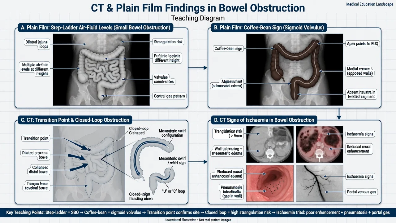
Bowel obstruction — the mechanical blockage of the intestinal lumen, split into the small bowel (the adhesions, the hernia, the malignancy) and the large bowel (the colorectal cancer, the sigmoid volvulus, the diverticular stricture). The colicky abdominal pain, the distension, the vomiting and the constipation or the obstipation; the tinkling bowel sounds early then absent. The plain film shows the dilated loops, the air-fluid levels and the step-ladder pattern of the SBO and the coffee-bean sign of the sigmoid volvulus. The CT defines the transition point, the closed-loop and the ischaemia signs — the bowel wall thickening, the mesenteric oedema, the pneumatosis and the portal venous gas. The management is the NBM, the nasogastric tube for the decompression, the IV fluid resuscitation with the 0.9 per cent saline and the potassium replacement, the surgical referral, and the laparotomy for the strangulation, the perforation or the ischaemia. The morphine 5 mg IV, the ondansetron 4 mg IV and the ceftriaxone 2 g IV for the surgical prophylaxis. ACEM-primary, globally tagged.
On this page & tools
Your progress
Saved locally on this device.
5 MCQs with explanations
Target exams
Red flags
Bowel obstruction is the mechanical blockage of the intestinal lumen that, untreated, progresses from the simple obstruction through the closed-loop and the strangulation to the ischaemia, the perforation and the death. It is one of the commonest surgical presentations to the emergency department and a high-yield Fellowship topic because the candidate must, within the first hour, answer four questions: is this a small bowel or a large bowel obstruction, is it a simple obstruction or a strangulated one, what does the imaging show at the transition point, and does this patient need the theatre now or the conservative decompression first. The two limbs of the gut behave differently — the small bowel obstruction is dominated by the adhesions, the hernia and the malignancy, the large bowel obstruction by the colorectal cancer, the sigmoid volvulus and the diverticular stricture — and the management diverges. The Fellowship candidate who reaches for the surgery in every obstruction, or who delays the laparotomy for the strangulated bowel, fails the station.[1][2]
Definition and classification
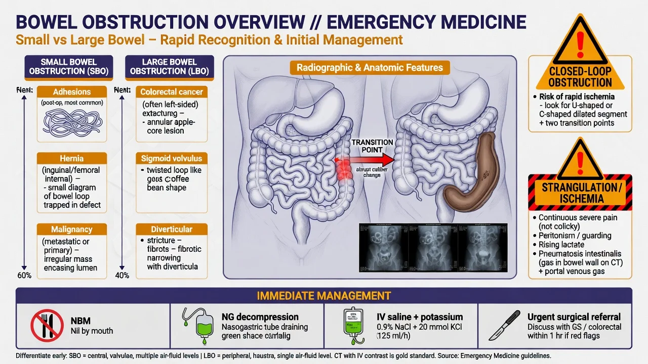
Bowel obstruction is the partial or the complete interruption of the normal antegrade transit of the intestinal contents. The obstruction may be mechanical (a physical barrier in the lumen, the wall or the outside of the gut) or functional (the paralysed or the dysmotile gut with no physical block — the paralytic ileus and the pseudo-obstruction). The Fellowship station is built around the mechanical obstruction, but the functional mimics must be distinguished because the management is the opposite.[1]
The mechanical obstruction is classified along three axes that determine the urgency and the management: [1]
| The axis | The subtypes | The significance |
|---|---|---|
| The site | The small bowel (proximal to the ileocaecal valve), the large bowel (distal to the valve) | Determines the cause, the imaging and the distension pattern |
| The completeness | The complete (no gas or stool past the obstruction) versus the partial | The complete obstruction strangulates sooner; the partial may be managed conservatively |
| The blood supply | The simple (the blood supply intact), the strangulated (the vascular compromise), the closed-loop (both ends occluded) | The strangulated and the closed-loop are the surgical emergencies |
The classification at a glance
Epidemiology and risk
The small bowel obstruction accounts for the majority of the mechanical obstructions and is the commonest reason for the surgical admission for the small bowel pathology. The lifetime risk is around 4 per cent, and the patient profile is the one who has had the previous abdominal surgery — the adhesions cause 60 to 75 per cent of the small bowel obstructions in the developed world, and the figure is the single most useful piece of the history. The other causes of the small bowel obstruction, in the descending order of the frequency, are the malignancy (the primary small bowel cancer, the metastatic, the peritoneal carcinomatosis), the hernia (the inguinal, the femoral, the incisional, the paraumbilical), the Crohn stricture, the intussusception (the child, or the adult with the lead point), the gallstone ileus (the elderly woman) and the bezoar. [1]
The large bowel obstruction is less common than the small bowel and carries a different demographic. The mechanical large bowel obstruction is caused by the malignancy in 50 to 60 per cent of the cases — the colorectal cancer, most often in the sigmoid or the rectosigmoid — and by the volvulus in 10 to 15 per cent (the sigmoid volvulus in the elderly, the institutionalised or the psychiatric patient; the caecal volvulus in the younger mobile caecum), by the diverticular stricture, and rarely by the intussusception or the faecal impaction. The sigmoid volvulus has a regional pattern — it is the commonest cause of the large bowel obstruction in parts of Africa, Asia and South America, and the rarer cause in the Anglophone West, but the Fellowship candidate in any region must recognise it.[1]
Pathophysiology — the cascade to the strangulation
The obstructed bowel accumulates the swallowed air and the secreted fluid proximal to the block, and the peristalsis against the obstruction produces the colicky pain and the distension. The proximal bowel dilates; the distal bowel collapses. The fluid and the gas accumulation is enormous — the adult small bowel secretes 7 to 9 litres per day, and most of this is normally reabsorbed; the obstructed loop sequesters it into the lumen, the third space, and the patient develops the intravascular depletion, the hypokalaemia, the metabolic alkalosis from the vomiting and the prerenal acute kidney injury. This is the physiological basis of the fluid resuscitation with the isotonic saline and the potassium replacement. [1]
The transition from the simple to the strangulated obstruction is the central event the emergency physician is watching for. The clinical signs are the continuous (not colicky) severe pain, the tachycardia out of proportion to the dehydration, the fever, the peritonism, and the rising lactate. The imaging signs on the CT are the bowel wall thickening, the mesenteric oedema and the engorgement, the abnormal wall enhancement (the decreased or the absent), the pneumatosis intestinalis (the gas in the bowel wall), the portal venous gas, and the ascites. The presence of any of these in the obstructed patient changes the management from the conservative to the urgent laparotomy.[3]
Clinical presentation
The four classical symptoms are the colicky abdominal pain, the vomiting, the abdominal distension and the constipation progressing to the obstipation. The character and the timing of each maps onto the site and the severity of the obstruction. [1]
The colicky pain is the first symptom and is the peristalsis against the obstruction. The colic of the small bowel obstruction is the central or the periumbilical cramping that comes in waves every few minutes; the colic of the large bowel obstruction is the lower abdominal cramping that comes at longer intervals. The pain that becomes continuous, severe and localised is the danger sign — it signals the peritonism and the strangulation, and the obstruction is no longer simple. [1]
The vomiting is early and the bilious in the high small bowel obstruction (the proximal jejunum), the faeculent in the distal small bowel obstruction, and late and the faeculent or absent in the large bowel obstruction. The earlier and the more profuse the vomiting, the more proximal the obstruction and the more rapid the dehydration and the electrolyte disturbance. The distension is the hallmark of the distal obstruction — marked in the large bowel and the distal small bowel, minimal in the high small bowel. The constipation (the failure to pass stool) and the obstipation (the failure to pass stool or flatus) develop later; the complete obstruction produces the obstipation, the partial may continue to pass small amounts. [1]
The examination follows the inspection, the auscultation, the palpation, the percussion sequence. The inspection shows the distension (the pattern of the distension is informative — the central ladder-pattern of the SBO, the peripheral distension of the LBO) and the old surgical scars (the clue to the adhesions) and the visible hernia (the incarcerated mass in the groin). The auscultation hears the tinkling, the high-pitched or the rushing bowel sounds early as the hyperactive bowel fights the obstruction, then the absent bowel sounds late as the ileus of the ischaemia or the peritonitis sets in. The palpation assesses the tenderness (the diffuse, the colicky discomfort is expected; the localised, the continuous tenderness with the guarding is the peritonism and the strangulation) and the masses (the hernia, the palpable tumour, the distended caecum of the closed-loop). The digital rectal examination may reveal the empty rectum of the complete obstruction, the mass of the rectal cancer, or the impacted faeces. [1]
[1]Differential diagnosis
The bowel obstruction is one cause of the abdominal pain, the distension and the vomiting, and the Fellowship candidate must distinguish the mechanical obstruction from its functional and the inflammatory mimics, because the management diverges sharply.[1]
Small bowel obstruction
- The previous surgery (adhesions), the hernia, the malignancy; the central colicky pain, the early bilious vomiting, the moderate distension
- The dilated small bowel loops over 3 cm with the air-fluid levels and the step-ladder pattern on the plain film; the transition point on the CT
- The NBM, the NG tube, the IV fluid resuscitation, the surgical referral; the laparotomy for the strangulation
- The commonest cause is the adhesions; the strangulation mandates the surgery
Large bowel obstruction
- The older patient; the colorectal cancer, the sigmoid volvulus, the diverticular stricture; the marked distension, the late faeculent vomiting
- The dilated colon over 6 cm (over 9 cm for the caecum) on the plain film; the coffee-bean sign of the volvulus; the transition point and the cause on the CT
- The NBM, the NG tube, the fluids, the surgical referral; the endoscopic decompression for the uncomplicated sigmoid volvulus
- The malignancy is the commonest cause; the volvulus may be decompressed endoscopically
Paralytic ileus
- The postoperative patient, the electrolyte disturbance (the hypokalaemia), the opioid use, the retroperitoneal haematoma, the peritonitis
- The dilated loops of the small and the large bowel with NO transition point on the CT; the absent bowel sounds
- The conservative management — the NG tube, the IV fluids, the correction of the electrolytes, the stop the opioids
- A functional obstruction; surgery does not help and is avoided
Pseudo-obstruction (Ogilvie)
- The elderly, the bedbound, the postoperative, the critically ill; the massive caecal dilatation without a mechanical cause
- The CT shows the dilated colon with NO transition point and NO mechanical cause; the exclusion diagnosis
- The neostigmine 2 mg IV (the cautious, the cardiac-monitored); the colonoscopic decompression; the surgery for the peritonitis
- The caecum over 9 to 12 cm risks the perforation; the surgery for the ischaemic or the perforated
Gastroenteritis
- The acute-onset cramps, the profuse diarrhoea and the vomiting; the infectious contact; the self-limiting course
- The normal or the mildly dilated loops; the no transition point; the resolution over the 24 to 72 hours
- The supportive care — the fluids, the antiemetics; the no surgery
- The cramps are diffuse and non-localising; the distension is minimal
Mesenteric ischaemia
- The elderly vascular patient; the severe pain out of proportion to the examination; the metabolic acidosis, the raised lactate
- The CT angiography shows the mesenteric artery occlusion or the superior mesenteric vein thrombosis; the pneumatosis late
- The urgent revascularisation or the embolectomy; the laparotomy for the necrotic bowel; the anticoagulation
- The pain-severity mismatch is the hallmark; the mortality is high without the early diagnosis
The other considerations include the acute pancreatitis (the epigastric pain radiating to the back, the raised amylase and lipase), the leaking or ruptured abdominal aortic aneurysm (the elderly man, the back and abdominal pain, the pulsatile mass — kill before the obstruction kills), the gastric outlet obstruction (the succussion splash, the large gastric residuum, no colic), the retroperitoneal haematoma (the anticoagulated patient, the ileus), and in the child the intussusception (the sausage-shaped mass, the redcurrant-jelly stool). [1]
[1]Bedside assessment
The assessment begins with the ABCDE and the simultaneous search for the signs of the strangulation. The airway and the breathing — the patient who is vomiting the faeculent material is at the risk of the aspiration, and the elderly comorbid patient with the hypovolaemia may be the respiratory-compromised; give the high-flow oxygen and have the suction ready. The circulation — the tachycardia, the hypotension, the cool peripheries, the prolonged capillary refill and the oliguria flag the severe dehydration (the third-space losses) or the sepsis of the strangulation; the intravenous access and the fluid resuscitation proceed in parallel with the assessment. The disability — the drowsiness is the dehydration, the sepsis or the electrolyte disturbance (the hyponatraemia). The exposure — the full abdominal examination with the inspection of the groins (the hernia) and the digital rectal examination. [1]
The history establishes the previous abdominal surgery (the adhesions), the known cancer (the recurrence or the peritoneal disease), the known hernia, the change in the bowel habit and the bleeding per rectum (the colorectal cancer), the comorbidities (the cardiac, the renal — they govern the fluid resuscitation), the medications (the opioids, the anticholinergics — the ileus; the anticoagulants — the haematoma), and the onset, the character, the progression of the symptoms (the continuous pain and the peritonism are the red flags). [1]
Investigations
The investigations confirm the obstruction, define the site, the cause and the transition point, and search for the ischaemia. [1]
The blood panel includes the full blood count (the leukocytosis and the neutrophilia of the strangulation; the anaemia of the malignancy), the urea and electrolytes (the dehydration raises the urea; the vomiting causes the hypokalaemia, the hypochloraemia and the metabolic alkalosis; the acute kidney injury), the liver function tests (the derangement is the sepsis or the biliary pathology), the lactate (the rising lactate over 2 mmol per litre is the perfusion failure and the ischaemia — the single most useful blood test for the strangulation), the C-reactive protein (the rising trend supports the ischaemia), the group and save or the crossmatch (for the surgery), the amylase or the lipase (to exclude the pancreatitis), and the venous blood gas (the pH, the base deficit, the lactate). The beta-hCG is mandatory in every woman of the reproductive age — the ectopic pregnancy and the ovarian torsion mimic the obstruction, and the adnexal mass may cause the obstruction itself. [1]
The plain abdominal radiograph
The erect chest radiograph and the supine and the erect abdominal films are the traditional first-line investigations, though they are increasingly replaced by the CT. The chest film looks for the free air under the diaphragm (the perforation) and the chest pathology. The abdominal film looks for the dilated small bowel loops over 3 cm (the central, the ladder-pattern, the valvulae conniventes that cross the full width of the lumen — the "stacked coins" or the step-ladder appearance), the dilated large bowel over 6 cm (the peripheral, the haustra that do not cross the full width, the sacculations), the air-fluid levels on the erect film (the step-ladder pattern of the SBO), the absent rectal gas (the complete obstruction), and the specific signs: the coffee-bean sign and the absence of haustra on the left side of the sigmoid volvulus, the bird's-beak or the bird-of-prey sign at the site of the volvulus on the contrast enema, and the air in the biliary tree with the small bowel obstruction of the gallstone ileus (Rigler triad: the air in the biliary tree, the small bowel obstruction, the ectopic gallstone). [1]
[1]The CT of the abdomen and the pelvis
The CT with the intravenous contrast is the investigation of choice for the bowel obstruction — it confirms the obstruction, localises the transition point, identifies the cause, and demonstrates the ischaemia and the closed-loop with the sensitivity for the strangulation around 80 to 90 per cent.[3]
The CT signs of the obstruction: the dilated bowel proximal to the obstruction (the small bowel over 3 cm, the large bowel over 6 cm, the caecum over 9 cm) and the collapsed bowel distal to it; the transition point — the abrupt change from the dilated to the collapsed bowel, the single most useful sign; and the cause at the transition point (the adhesive band, the hernia, the tumour, the volvulus, the intussusception, the gallstone). [1]
The CT signs of the ischaemia and the strangulation — the high-risk features that mandate the surgery:[3]
| The CT sign | The meaning |
|---|---|
| The bowel wall thickening (over 3 mm) | The oedema and the congestion of the venous obstruction |
| The decreased or the absent wall enhancement | The arterial compromise, the ischaemia, the necrosis |
| The mesenteric oedema and the engorgement (the clouding) | The venous and the lymphatic obstruction |
| The whirl sign (the mesenteric twisting) | The closed-loop, the volvulus |
| The pneumatosis intestinalis (the gas in the bowel wall) | The transmural ischaemia, the late sign |
| The portal venous gas | The gas-forming organisms in the ischaemic wall, the very late and the ominous sign |
| The ascites | The transudation from the ischaemic or the obstructed loop |
| The closed-loop configuration (the U- or the C-shaped loop) | The segment occluded at both ends, the high strangulation risk |
The CT signs and the surgery
The contrast follow-through (the water-soluble)
The water-soluble contrast follow-through (the gastrografin challenge) has a role in the adhesive small bowel obstruction without the signs of the strangulation: the 50 to 100 mL of the water-soluble contrast given via the NG tube, with the repeat film at 4 to 8 hours. The contrast reaching the colon within 24 hours predicts the resolution with the sensitivity over 90 per cent; the failure to reach the colon predicts the need for the surgery. The gastrografin has the osmotic effect that may itself help the resolution of the adhesive obstruction, and the meta-analysis supports its use in the selected patient.[2]
Immediate management — the resuscitation and the decompression
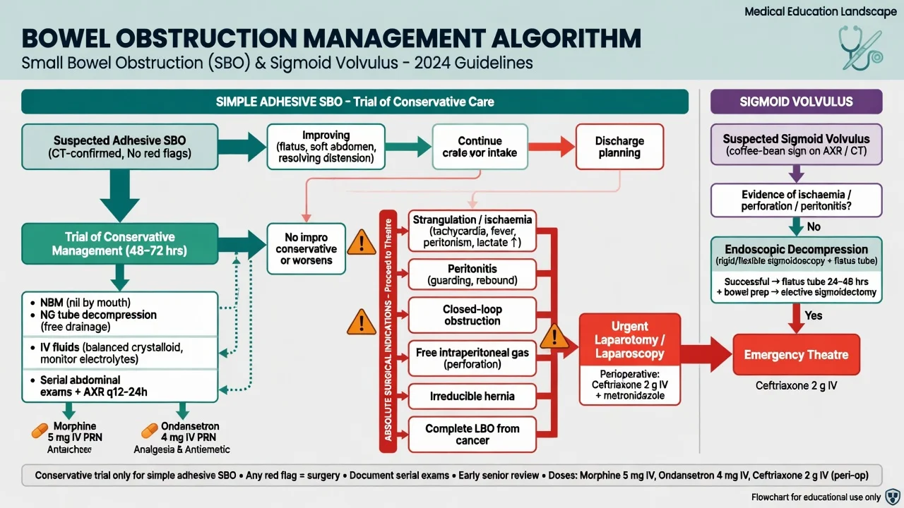
The management of the bowel obstruction begins with the resuscitation and the decompression, proceeds in parallel with the imaging and the surgical referral, and the goal is the restoration of the perfusion and the intravascular volume, the relief of the obstruction, and the prevention or the treatment of the strangulation. [1]
The ABCDE and the intravenous access — the two large-bore cannulae, the monitoring (the pulse, the blood pressure, the oxygen saturation, the urine output via the catheter). The fluid resuscitation targets the correction of the dehydration and the electrolyte disturbance. The 0.9 per cent saline (the normal saline) is the standard initial crystalloid — the 1 to 2 litres over the first hour in the dehydrated adult, then the titrated to the clinical response and the urine output (the target 0.5 mL per kilogram per hour). The Hartmann or the balanced crystalloid is the alternative that avoids the hyperchloraemic acidosis of the large-volume saline. The potassium replacement (the potassium chloride 20 to 40 mmol per litre of the fluid, the cautious in the renal impairment and the cardiac monitoring above 10 mmol per hour) corrects the hypokalaemia that drives the ileus if left uncorrected. The blood transfusion for the anaemia of the malignancy or the bleeding. [1]
The analgesia — the morphine 5 mg intravenously, titrated (the 0.1 mg per kilogram, the adult 5 to 10 mg, the repeat at 5 to 10 minute intervals to the pain), is the standard opioid and does not mask the physical signs or delay the diagnosis — the systematic reviews support the generous analgesia. The ondansetron 4 mg intravenously for the nausea and the vomiting. The paracetamol 1 g intravenously for the baseline analgesia and the fever. Avoid the NSAIDs in the dehydrated or the renal-impaired patient — the acute kidney injury. [1]
The nasogastric tube — the large-bore (the 14 to 18 French) NG tube on the free drainage, with the intermittent aspiration, decompresses the stomach and the proximal bowel, relieves the vomiting, reduces the aspiration risk, and allows the measurement of the gastric residuum and the delivery of the contrast. The NG tube is placed in nearly every obstructed patient. The urinary catheter for the urine-output monitoring. The NBM (the nil by mouth) — the patient takes nothing by mouth; the oral free water is restricted. [1]
The antibiotics — the ceftriaxone 2 g intravenously once daily plus the metronidazole 500 mg intravenously every 8 hours — are given for the suspected strangulation, the ischaemia, the perforation, or the preoperative cover, and cover the enteric Gram-negatives and the anaerobes. The broad-spectrum escalation (the piperacillin-tazobactam 4.5 g intravenously every 8 hours, or the meropenem 1 g intravenously every 8 hours) for the established sepsis or the perforation.[1]
The drug doses for the bowel obstruction
Definitive management — the conservative versus the surgical
The definitive management splits at the bedside into the conservative (the decompression and the watchful waiting) and the surgical (the laparotomy or the laparoscopy). The decision is made jointly with the surgeon and hinges on the cause, the completeness, the presence of the strangulation, and the response to the initial decompression.[1][2]
The conservative management
The conservative (the non-operative) management is appropriate for the adhesive small bowel obstruction without the signs of the strangulation, the early postoperative obstruction, the partial obstruction, the Crohn stricture, and some of the carcinomatosis. The management is the NBM, the NG tube on the free drainage, the IV fluid resuscitation with the correction of the electrolytes, the serial abdominal examinations (the every-2-to-4-hourly), the serial bloods (the lactate, the WCC, the CRP), and the repeat imaging if the deterioration. The adhesive small bowel obstruction resolves with the conservative management in 65 to 80 per cent of the cases, most within the first 48 to 72 hours. The water-soluble contrast follow-through (the gastrografin challenge) at 24 to 48 hours both predicts the resolution and may hasten it. The surgery is indicated if the patient fails the conservative management (the no resolution by 3 to 5 days), develops the signs of the strangulation, or shows the high-risk features on the imaging.[2]
When to abandon the conservative management and operate
The conservative management of the adhesive SBO is abandoned and the surgery is undertaken if any of the following develop: the continuous severe pain or the new peritonism; the haemodynamic instability or the rising lactate; the fever or the rising leukocytosis; the CT signs of the ischaemia (the decreased wall enhancement, the mesenteric oedema, the pneumatosis, the portal venous gas); the closed-loop configuration; or the failure to resolve by 3 to 5 days of the conservative management. The strangulated bowel is operated on at the time of the diagnosis — there is no trial of the conservative management. [1]
The surgical management
The surgical management is indicated at the time of the diagnosis for the strangulated or the ischaemic obstruction, the perforation, the peritonitis, the closed-loop obstruction that cannot be decompressed endoscopically, the complete obstruction that fails the conservative management, and the obstruction caused by the surgically correctable lesion (the hernia, the tumour). The exploratory laparotomy (the midline incision) is the standard approach for the emergency obstruction; the laparoscopy is increasingly used for the adhesive obstruction in the experienced hands and offers the faster recovery and the fewer wound complications. The operation releases the adhesion (the adhesiolysis), reduces and repairs the hernia, resects the necrotic or the perforated bowel (the primary anastomosis or the stoma depending on the contamination and the patient), resects the tumour (the primary or the staged), or detorses and fixes the volvulus. [1]
The sigmoid volvulus is the exception to the surgery-first rule for the obstruction. The uncomplicated sigmoid volvulus (no peritonitis, no ischaemia) is decompressed endoscopically — the flexible sigmoidoscope is passed to the twist, the flatus tube is left in situ for 24 to 72 hours, and the detorsion is achieved in 70 to 90 per cent of the cases. The endoscopic decompression is the bridge to the elective sigmoid colectomy (the recurrence rate is high without the surgery). The sigmoid volvulus with the peritonitis, the ischaemia, the gangrene or the failed decompression goes straight to the theatre for the sigmoid colectomy.[1]
The colorectal cancer causing the large bowel obstruction may be managed with the emergency surgery (the Hartmann procedure for the left-sided, the resection and the primary anastomosis for the right-sided) or, in the selected centre, with the endoscopic colonic stenting as the bridge to the elective surgery or the palliation. The colonic stent relieves the obstruction, allows the bowel preparation and the staging, and the definitive surgery is undertaken electively — but the perforation rate is real and the stenting is the specialist procedure.[1]
Subtypes and scenarios
The adhesive small bowel obstruction is the commonest scenario — the previous surgery (the open or the laparoscopic), the colicky central pain, the distension, the CT transition point without a mass, and the trial of the conservative management with the high resolution rate. The strangulated hernia — the incarcerated groin or the incisional hernia with the tender irreducible mass and the obstruction — is the surgical emergency; the reduction is NOT attempted in the tender or the overlying-skin-changed hernia (the risk of the reduction en masse of the ischaemic bowel). The closed-loop small bowel obstruction (the internal hernia, the adhesion band) has the C-shaped or the U-shaped dilated loop on the CT with the mesenteric oedema and the high strangulation risk — operate. [1]
The sigmoid volvulus — the elderly, the institutionalised, the nursing-home or the psychiatric patient — presents with the massive distension, the colicky pain, the vomiting and the obstipation, often a few days into the course. The plain film shows the massive dilation of the sigmoid loop arising from the pelvis (the coffee-bean sign, the apex pointing to the right upper quadrant), and the CT shows the whirl sign at the mesenteric root. The endoscopic detorsion first; the surgery for the peritonitis or the failed detorsion. The caecal volvulus — the younger patient with the mobile caecum — presents with the small bowel obstruction picture (the caecum twists and the ileum obstructs) and is managed surgically (the caecopexy or the right hemicolectomy); the endoscopic decompression is not effective for the caecal volvulus. [1]
The gallstone ileus — the elderly woman with the intermittent, the recurrent obstruction — has the Rigler triad on the imaging (the air in the biliary tree, the small bowel obstruction, the ectopic gallstone in the terminal ileum). The management is the surgery (the enterolithotomy — the stone removal via the enterotomy; the cholecystectomy is staged or omitted in the elderly). The intussusception in the adult (unlike the child) almost always has a pathological lead point (the tumour, the polyp) and is managed with the resection. [1]
Complications and pitfalls
The complications of the obstruction itself are the strangulation and the ischaemic necrosis, the perforation (the most common at the caecum in the closed-loop large bowel obstruction — the law of Laplace: the wall tension is greatest at the widest part), the peritonitis and the sepsis, the fluid and the electrolyte disturbance (the hypokalaemia, the metabolic alkalosis, the acute kidney injury), the aspiration of the gastric contents (the leading cause of the death in the obstructed patient who is vomiting), and the short bowel syndrome after the extensive resection. [1]
The complications of the surgery are the anastomotic leak, the intra-abdominal abscess, the wound infection, the bleeding, the enterotomy (the inadvertent, especially in the dense adhesions), the stoma complications, and the adhesions (the recurrence of the obstruction). The complications of the colonoscopy for the volvulus are the perforation (the ischaemic segment is fragile) and the failed detorsion. [1]
[1]The pitfalls in the management are the failure to recognise the strangulation (the continuous pain, the lactate, the CT signs are the keys), the over-reliance on the conservative management in the high-risk patient, the failure to correct the electrolytes (the hypokalaemia perpetuates the ileus after the obstruction is relieved), the aspiration of the vomiting patient (the airway protection, the NG tube before the induction), the missed gallstone ileus (look for the biliary gas), the missed sigmoid volvulus in the demented or the institutionalised patient who cannot give the history, and the misdiagnosis of the pseudo-obstruction (Ogilvie) as the mechanical obstruction and the unnecessary laparotomy. [1]
Prognosis and disposition
The prognosis of the simple adhesive small bowel obstruction managed conservatively is excellent — the resolution in 65 to 80 per cent, the mortality under 2 per cent. The mortality of the small bowel obstruction rises to 5 to 10 per cent when the surgery is required and to 10 to 20 per cent or higher with the strangulation, the necrosis, or the perforation — the mortality doubles for every 24 hours of the delay to the surgery in the strangulated obstruction. The large bowel obstruction has a higher overall mortality (the 10 to 20 per cent) because of the older age, the comorbidity and the underlying malignancy. The sigmoid volvulus has the mortality of 5 to 10 per cent for the uncomplicated and 20 to 50 per cent for the gangrenous. [1]
The disposition: the patient with the suspected obstruction is admitted under the surgical team. The patient with the strangulation, the ischaemia, the perforation or the peritonitis goes to the theatre emergently. The patient with the adhesive SBO and no signs of the strangulation is admitted for the conservative management with the serial examinations and the surgery if the deterioration or the failure to resolve. The patient with the uncomplicated sigmoid volvulus is admitted for the endoscopic decompression and the elective surgery. The patient with the pseudo-obstruction is admitted for the neostigmine or the colonoscopic decompression. The patient with the resolved obstruction is discharged with the safety-net advice — to return if the pain worsens, the vomiting recurs, the distension increases, or the fever develops. [1]
Special populations
The elderly patient with the obstruction is more likely to have the malignancy, the volvulus, the diverticular stricture and the comorbidity; the signs may be muted, the delay is common, the strangulation is more frequent, and the mortality is higher — have a low threshold for the CT and the early surgical referral. The postoperative patient with the obstruction in the first two weeks after the surgery is most often the early postoperative ileus (the conservative, the resolution in most); the early postoperative mechanical obstruction (the adhesion, the internal hernia) is less common but must be distinguished — the CT is the key, and the conservative management is often successful. The pregnant patient with the obstruction has the added risk of the maternal and the fetal compromise, and the imaging is the ultrasound first, the MRI second, the CT with the counselling if the diagnosis cannot be made otherwise; the adhesions and the volvulus (the caecal, more common in the pregnancy) are the common causes. The paediatric patient with the obstruction has the intussusception (under 2 years, the sausage-shaped mass, the redcurrant-jelly stool, the ultrasound target sign, the pneumatic reduction), the hernia (the incarcerated inguinal), the malrotation with the volvulus (the bilious vomiting, the emergency), and the Hirschsprung disease. The anticoagulated patient — the spontaneous intramural haematoma, the conservative management, the anticoagulation reversal. [1]
Evidence and regional guidelines
The contemporary framework rests on the World Society of Emergency Surgery (WSES) guidelines for the diagnosis and the management of the bowel obstruction, including the 2019 narrative review for all physicians and the 2021 position paper on the small bowel obstruction in the virgin abdomen.[1][2] The WSES endorses the risk-stratified approach: the CT for the definitive diagnosis and the identification of the ischaemia; the conservative management for the adhesive SBO without the strangulation, with the water-soluble contrast follow-through at 24 to 48 hours; the early surgery for the strangulation, the closed-loop, the ischaemia and the peritonitis; and the endoscopic decompression first for the uncomplicated sigmoid volvulus. The role of the CT in the risk stratification of the SBO is supported by the contemporary evidence on the transition point, the closed-loop and the ischaemia signs.[3]
ANZ practice note. The Royal Australasian College of Surgeons and the ACEM endorse the CT-first approach to the suspected bowel obstruction, the conservative management of the adhesive SBO with the serial examinations and the gastrografin challenge, and the early surgery for the strangulation. The sigmoid volvulus is decompressed endoscopically first by the surgical or the gastroenterology team, with the elective sigmoid colectomy to prevent the recurrence. The pseudo-obstruction (Ogilvie) is managed with the neostigmine (the cautious, the atropine available for the bradycardia) or the colonoscopic decompression. The surgical registrar is the senior decision-maker; the disposition and the timing of the surgery are the joint decisions. [1]
Model answer — the ED workup of the suspected bowel obstruction
A 68-year-old man with the previous appendicectomy and the two days of the central colicky pain, the vomiting, the distension and the obstipation. ABCDE: the stable airway, the high-flow oxygen, the two large-bore cannulae, the IV 0.9 per cent saline 1 litre over the first hour then the titrated, the potassium chloride 20 mmol per litre in the fluid (the K 3.0 on the bloods), the morphine 5 mg IV titrated for the pain, the ondansetron 4 mg IV, the ceftriaxone 2 g IV plus the metronidazole 500 mg IV. The NG tube on the free drainage, the urinary catheter, the NBM. The investigations — the FBC, the UandE, the lactate (1.8), the CRP, the group and save, the amylase, the beta-hCG if applicable, the venous gas. The CT abdomen and pelvis with the IV contrast — the dilated small bowel loops to the transition point in the right iliac fossa, no signs of the ischaemia, the diagnosis of the adhesive SBO. The management — the admission under the surgeons, the conservative management (the NBM, the NG tube, the fluids, the serial examinations and the lactate), the gastrografin challenge at 24 to 48 hours, the surgery if the deterioration or the failure to resolve. The safety-net — the return if the pain worsens, the fever, the peritonism. [1]
Exam practice
SAQ — Strangulated adhesive small bowel obstruction with CT signs of ischaemia
10 minutes · 10 marks
A 64-year-old man presents to the emergency department with three days of central colicky abdominal pain, vomiting and distension that over the last eight hours has become constant and severe. He had an open appendicectomy at age 20. On arrival he is pale and diaphoretic: temperature 38.2, HR 124, BP 96/58, RR 24, SpO2 96 per cent on room air. The abdomen is distended with generalised tenderness, involuntary guarding and absent bowel sounds. Venous gas: pH 7.30, lactate 3.8 mmol/L, bicarbonate 18. WCC 18.2, CRP 210. The CT abdomen with IV contrast shows dilated small bowel loops to a transition point in the right iliac fossa, with reduced wall enhancement, mesenteric oedema, a closed-loop configuration and pneumatosis intestinalis.
SAQ — Malignant large bowel obstruction with caecal dilatation and perforation risk
10 minutes · 10 marks
A 76-year-old woman presents to the emergency department with four days of worsening lower abdominal colicky pain, progressive distension and absolute constipation, with vomiting over the last 24 hours. She has lost 8 kg over three months and her stools have become narrower. On arrival she is volume-depleted: HR 108, BP 102/64, RR 22, afebrile. The abdomen is markedly distended and tympanitic with tinkling bowel sounds and mild diffuse tenderness but no peritonism. The CT abdomen with IV contrast shows a circumferential enhancing mass at the rectosigmoid junction with proximal colonic dilatation to 11 cm at the caecum, no pneumatosis and no free gas.
Exam pearls
- The four classical symptoms are the colicky pain, the vomiting, the distension and the constipation or the obstipation — the timing and the character map onto the site.
- The small bowel obstruction is the adhesions, the hernia and the malignancy; the large bowel obstruction is the colorectal cancer, the sigmoid volvulus and the diverticular stricture.
- The transition from the simple to the strangulated obstruction is the central event — the continuous pain, the lactate over 2, the peritonism and the CT signs of the ischaemia are the triggers for the surgery.
- The CT with the IV contrast is the definitive investigation — it shows the transition point, the closed-loop and the ischaemia signs (the decreased wall enhancement, the mesenteric oedema, the pneumatosis, the portal venous gas).
- The sigmoid volvulus is decompressed endoscopically first; the caecal volvulus is managed surgically; the pseudo-obstruction (Ogilvie) is the neostigmine or the colonoscopic decompression — none of these is the surgery.
- The adhesive SBO resolves with the conservative management in 65 to 80 per cent — the gastrografin challenge at 24 to 48 hours predicts the resolution.
- The caecum is the most common site of the perforation in the closed-loop LBO (the law of Laplace) — the caecum over 9 to 12 cm is the high risk.
- The gallstone ileus has the Rigler triad — the biliary gas, the SBO, the ectopic gallstone in the terminal ileum.
- The fluid resuscitation is the 0.9 per cent saline with the potassium replacement; correct the hypokalaemia or the ileus persists. [1]
Red flags
[1]References
- [1]Catena F, De Simone B, Coccolini F, et al. Bowel obstruction: a narrative review for all physicians World J Emerg Surg, 2019.PMID 31168315
- [2]Amara Y, Barsoum A, Bello B, et al. Diagnosis and management of small bowel obstruction in virgin abdomen: a WSES position paper World J Emerg Surg, 2021.PMID 34217331
- [3]Huang Z, et al. Computed tomography for diagnosis and risk stratification of small bowel obstruction: a systematic review and meta-analysis Eur J Radiol, 2026.PMID 42308707