EM · Acute appendicitis
Acute appendicitis
Also known as Acute appendicitis · Appendiceal obstruction · Perforated appendix · Appendiceal abscess
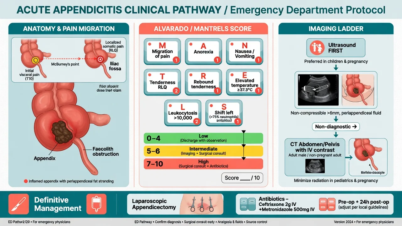
Acute appendicitis — the obstruction of the appendiceal lumen (the faecolith, the lymphoid hyperplasia) driving the mucus accumulation, the bacterial translocation, the ischaemia and the perforation; the central pain migrating to the right iliac fossa over 12 to 24 hours with the anorexia, the nausea, the low-grade fever and the McBurney point tenderness (the Rovsing, the psoas, the obturator signs); the Alvarado (MANTRELS) and the Appendicitis Inflammatory Response (AIR) scores; the ultrasound first in the child and the young woman, the CT in the adult male and the atypical; the surgical referral, the laparoscopic appendicectomy first-line, the ceftriaxone 2 g IV plus the metronidazole 500 mg IV as the perioperative prophylaxis. ACEM-primary, globally tagged.
On this page & tools
Your progress
Saved locally on this device.
5 MCQs with explanations
Target exams
Red flags
Acute appendicitis is the obstruction of the appendiceal lumen that progresses, over 12 to 36 hours, from a mucosal inflammation through wall ischaemia to perforation and peritonitis. It is the most common acute surgical abdomen worldwide, and the Fellowship candidate must know the pathophysiological cascade (the obstruction, the mucus accumulation, the bacterial translocation, the ischaemia, the perforation), the clinical picture (the central pain that migrates to the right iliac fossa, the anorexia, the McBurney point tenderness and the named peritoneal signs), the two scores that risk-stratify the patient at the bedside (the Alvarado and the Appendicitis Inflammatory Response score), the imaging strategy (the ultrasound first in the child and the pregnant or young woman, the CT in the adult male and the atypical), and the management (the surgical referral, the laparoscopic appendicectomy first-line, the ceftriaxone and the metronidazole as the perioperative prophylaxis).[1][2]
Definition and epidemiology
Acute appendicitis is the acute inflammation of the vermiform appendix caused by the obstruction of the narrow appendiceal lumen. It is the most common general surgical emergency, with a lifetime risk of approximately 7 to 8 per cent, and the peak incidence falls in the second and third decades, between the ages of 10 and 30. Men are affected slightly more often than women (ratio approximately 1.4 to 1), but the diagnosis is more frequently missed in women because the pelvic pathology — the ovarian cyst, the torsion, the PID, the ectopic — mimics it. The perforation rate at presentation is 20 to 30 per cent and is highest at the extremes of age — under 5 and over 65 — where the atypical presentation and the delayed presentation drive the missed diagnosis and the higher mortality (under 5 perforation rate over 50 per cent). The familial clustering and the low-fibre diet are weak risk factors; the strongest predictor of perforation is the duration of symptoms before the surgery.[1]
[1]Pathophysiology
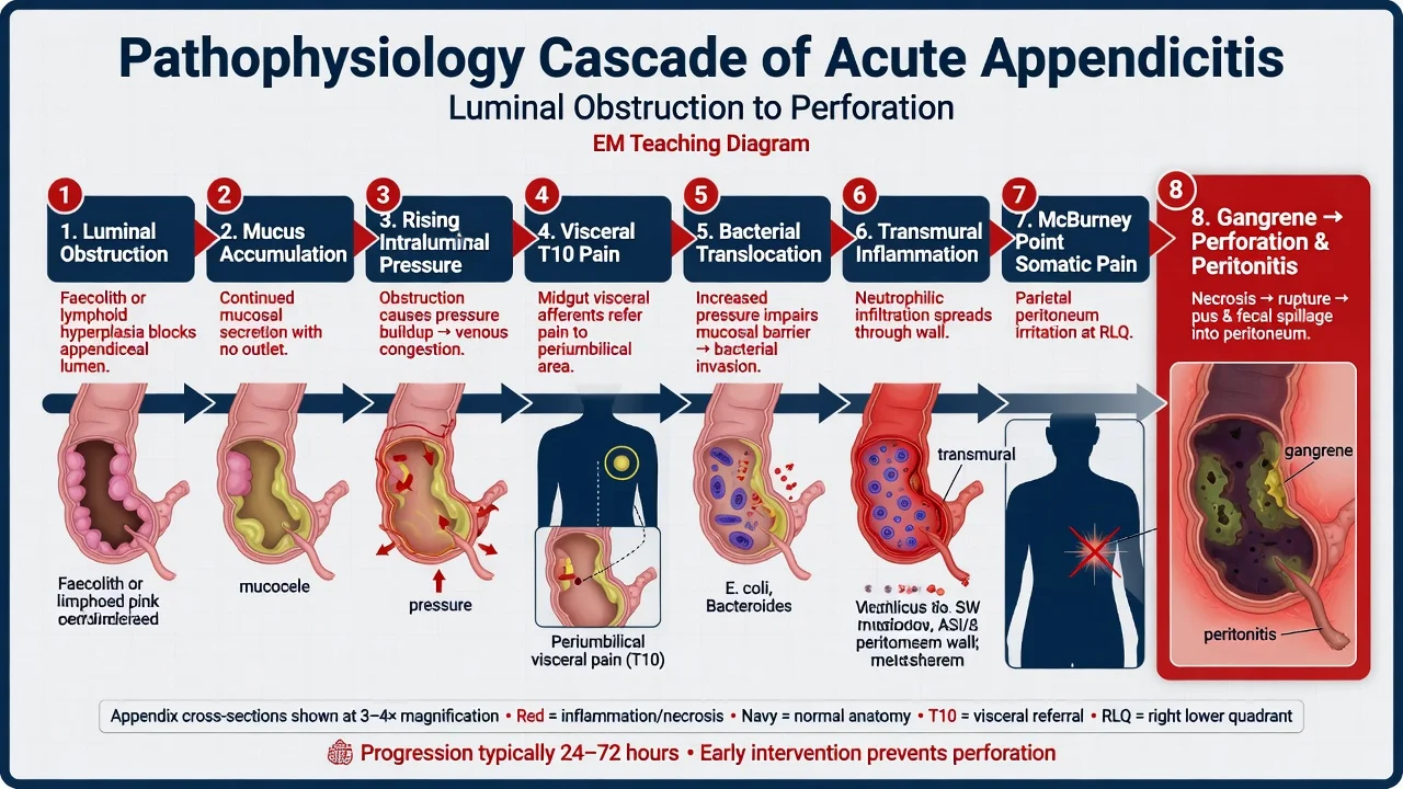
The initiating event is the obstruction of the appendiceal lumen. In the adult the commonest cause is the faecolith (the hardened inspissated faecal material, sometimes calcified as the appendicolith), and in the child and the adolescent the commonest cause is the lymphoid hyperplasia of the submucosal lymphoid follicles, often triggered by a viral or bacterial gastroenteritis. The rarer causes are the tumours (the carcinoid, the adenocarcinoma), the foreign body (the seeds, the pinworm), the stricture from the previous inflammation, and the parasitic load (the Ascaris) in the endemic region. [1]
Once the lumen is obstructed, the mucus secreted by the appendiceal mucosa continues to accumulate and the intraluminal pressure rises. The distended appendix stimulates the visceral afferent fibres of the midgut (the T10 dermatome), and the patient perceives a dull, poorly localised, central periumbilical pain — the visceral pain that is the first symptom and that precedes the localisation. As the pressure rises further, the lymphatic drainage, then the venous drainage, and finally the arterial supply are compromised; the mucosa becomes ischaemic and ulcerates, the luminal bacteria (the Escherichia coli, the Bacteroides, the Klebsiella, the Enterococcus) translocate across the ischaemic wall, and the inflammation becomes transmural. When the full-thickness inflammation reaches the parietal peritoneum, the somatic afferent fibres localise the pain to the right iliac fossa over the appendix — the somatic pain at the McBurney point. The ischaemic wall then gangrenes and the appendix perforates, releasing the purulent or the faecal contents into the peritoneal cavity and producing the generalised peritonitis, the sepsis and, untreated, the death.[2]
The pathophysiological cascade — obstruction to perforation
1
2
3
4
5
6
7
8
Clinical presentation
The classic presentation unfolds in a predictable sequence. The first symptom is the periumbilical abdominal pain that over 12 to 24 hours migrates to the right iliac fossa — the migration is the hallmark. The pain is continuous, moderate to severe, and worsened by the movement, the cough and the deep inspiration, so the patient lies still. The anorexia is almost universal — a patient with a normal appetite rarely has the appendicitis — and it is followed by the nausea and a single or a few episodes of the vomiting. Profuse or the bilious vomiting suggests another diagnosis (the obstruction, the gastroenteritis). The low-grade fever (around 37.5 to 38°C) develops after the pain; the high fever and the rigors suggest the perforation or another diagnosis (the pyelonephritis, the cholangitis). [1]
The examination shows the tenderness at the McBurney point (the junction of the lateral and the middle thirds of the line from the umbilicus to the anterior superior iliac spine — the surface marking of the base of the appendix) and the localised peritonism with the guarding and the rigidity. The named signs reflect the inflammation of the adjacent structures: the Rovsing sign (the palpation of the left iliac fossa produces the pain in the right iliac fossa — the indirect rebound from the peritoneal irritation); the psoas sign (the pain on the active extension of the right hip against resistance, or on the passive hyperextension — the retrocaecal appendix irritating the iliopsoas); the obturator sign (the pain on the passive internal rotation of the flexed right hip — the pelvic appendix irritating the obturator internus). The digital rectal examination may reveal the right-sided tenderness in the pelvic appendix but is not routinely required to make the diagnosis. [1]
The diagnostic signs and their meaning
The symptom sequence — the order matters
Differential diagnosis — the right iliac fossa pain
The right iliac fossa pain has a long and dangerous differential. The Fellowship candidate must distinguish the surgical (the appendicitis, the ectopic, the torsion, the intussusception) from the medical (the gastroenteritis, the mesenteric adenitis, the pyelonephritis), and must never anchor on the common diagnosis without excluding the lethal mimic. [1]
Acute appendicitis
- The migratory pain, the anorexia, the low-grade fever, the McBurney tenderness and the peritonism
- The raised WCC and CRP; the inflamed appendix on the US or the CT; the Alvarado 7 or above
- The surgical referral, the laparoscopic appendicectomy, the ceftriaxone and the metronidazole
- The most common acute surgical abdomen; the perforation risk rises with the duration of the symptoms
Ectopic pregnancy
- The woman of reproductive age; the missed period, the vaginal bleeding, the shoulder-tip pain
- The positive beta-hCG; the empty uterus with the free fluid or the adnexal mass on the TVS
- The methotrexate for the stable unruptured; the laparoscopic salpingectomy for the ruptured
- The leading cause of the first-trimester maternal death — exclude with the beta-hCG in every woman
Ovarian torsion or cyst
- The sudden severe unilateral pain; the torsion may have the intermittent colicky history
- The negative beta-hCG; the enlarged ovary with the absent venous Doppler flow on the US
- The urgent surgical or gynaecological referral; the detorsion and the salpingo-oophorectomy
- The ischaemic ovary must be salvaged within 6 hours
Ureteric colic
- The loin-to-groin pain; the writhing patient who cannot get comfortable; the haematuria
- The normal WCC; the CT KUB shows the stone; the absence of the peritonism
- The analgesia (the diclofenac 75 mg IM, the morphine), the hydration, the alpha-blocker
- The normal abdomen on examination; the pain is out of proportion to the physical findings
Mesenteric adenitis
- The child or the adolescent; the recent viral illness; the pain that does not migrate
- The normal or mildly raised WCC; the enlarged mesenteric nodes on the US or the CT
- The supportive care; the analgesia and the hydration
- The most common mimic of the appendicitis in the child
Pyelonephritis
- The flank pain, the high fever, the rigors, the dysuria and the frequency
- The positive urinalysis (the leucocytes, the nitrites); the bacteraemia; the raised WCC
- The ceftriaxone 2 g IV; the fluids; the admission for the septic
- The right-sided pyelonephritis may masquerade as the appendicitis
The other considerations include the Meckel diverticulitis, the Crohn terminal ileitis, the intussusception (the child under 2 with the redcurrant-jelly stool), the right-sided diverticulitis (more common in the Asian population), the gallstone ileus, the perforated peptic ulcer (the fluid tracks down the right paracolic gutter — the Vallecha sign), the mesenteric ischaemia (the pain out of proportion to the examination), the testicular torsion (the referred pain — examine the scrotum in every male), the epiploic appendagitis (the self-limiting inflammation of the appendage epiploica), and in the older patient the caecal carcinoma and the sigmoid volvulus. [1]
Bedside assessment
The assessment begins with the ABCDE — the airway, the breathing, the circulation (the heart rate, the blood pressure, the capillary refill, the peripheral perfusion), the disability, and the exposure. The shocked patient with the tachycardia, the hypotension and the cool peripheries has the perforation and the septic peritonitis until proven otherwise — the resuscitation and the surgical referral proceed in parallel. The history establishes the pain onset, the character, the radiation, the migration, the associated symptoms (the anorexia, the nausea, the vomiting, the fever, the dysuria, the bowel habit), the last menstrual period in every woman, and the past surgical and the gynaecological history. The abdominal examination assesses the inspection (the distension, the old scars), the auscultation (the bowel sounds — absent or the tinkling in the ileus of the peritonitis), the palpation (the tenderness, the guarding, the rigidity, the rebound, the masses), and the named appendiceal signs. The scrotal examination is mandatory in the male with the lower abdominal pain — the testicular torsion refers the pain to the abdomen. The pelvic examination is reserved for the woman in whom the gynaecological cause is suspected and the bedside ultrasound is not diagnostic. [1]
Investigations and the scores
The full blood count shows the leukocytosis (the WCC over 10 × 10⁹/L) and the neutrophilia (the shift to the left, the polymorphs over 75 per cent). The CRP is raised (over 10 mg per litre) and the combination of a normal WCC and a normal CRP makes the appendicitis unlikely (the negative predictive value over 95 per cent). The urea and electrolytes assess the dehydration and the renal function; the liver function tests and the amylase or lipase exclude the biliary and the pancreatic pathology; the beta-hCG (the urine, then the serum if equivocal) is mandatory in every woman of reproductive age. The urinalysis may show the leucocytes and the blood from the irritated bladder by the pelvic appendix — but the frank infection suggests the urinary tract source. The venous blood gas gives the lactate and the base deficit for the occult sepsis; a lactate over 2 mmol per litre with the tachycardia signals the perfusion failure. [1]
The Alvarado score
The Alvarado score (MANTRELS) is the bedside score derived from the symptoms, the signs and the bloods that stratifies the probability of the appendicitis. It is the most widely taught score and a favourite of the Fellowship examiner.[1]
| The component | The mnemonic | The points |
|---|---|---|
| The migration of the pain | Migration | 1 |
| The anorexia | Anorexia | 1 |
| The nausea or the vomiting | Nausea | 1 |
| The tenderness in the right iliac fossa | Tenderness | 2 |
| The rebound tenderness | Rebound | 1 |
| The elevated temperature | Elevated temperature | 1 |
| The leukocytosis | Leukocytosis | 2 |
| The shift to the left (the neutrophilia) | Shift | 1 |
| The total | MANTRELS | 10 |
The interpretation: the score of 1 to 4 makes the appendicitis unlikely (discharge or observe); the score of 5 to 6 is the equivocal case (the imaging and the observation); the score of 7 to 8 makes the appendicitis probable (the surgical referral); the score of 9 to 10 makes the appendicitis very probable (the theatre). The limitation is the modest sensitivity (around 70 per cent) — the high score supports the diagnosis but a low score does not exclude it, and the score performs poorly in the female patient and at the extremes of age. [1]
MANTRELS
The Appendicitis Inflammatory Response score
The Appendicitis Inflammatory Response (AIR) score was developed to correct the weakness of the Alvarado by weighting the inflammatory response (the CRP, the polymorphs) and the degree of the peritonism. It outperforms the Alvarado in the meta-analysis and is the preferred score in the contemporary European and the ANZ practice.[2][3]
| The component | The points |
|---|---|
| The vomiting | 1 |
| The pain in the right iliac fossa | 1 |
| The rebound tenderness — light | 1 |
| The rebound tenderness — medium | 2 |
| The rebound tenderness — strong | 3 |
| The temperature 38.5°C or above | 1 |
| The WCC 9.0 to 12.9 × 10⁹/L | 1 |
| The WCC 13.0 × 10⁹/L or above | 2 |
| The CRP 10 to 49 mg/L | 1 |
| The CRP 50 mg/L or above | 2 |
| The polymorphs 70 to 84 per cent | 1 |
| The polymorphs 85 per cent or above | 2 |
| The total | 0 to 12 |
The interpretation: the score of 0 to 4 is the low risk (the discharge with the safety-net, the negative appendicitis rate under 10 per cent); the score of 5 to 8 is the intermediate risk (the imaging — the US or the CT — and the surgical review); the score of 9 to 12 is the high risk (the surgical referral for the appendicectomy, the positive appendicitis rate over 90 per cent).[3]
Alvarado (MANTRELS)
- Eight components; the maximum 10; the migration, the anorexia, the nausea, the RIF tenderness (2), the rebound, the fever, the WCC (2), the shift
- Thresholds: 1–4 unlikely, 5–6 equivocal (scan), 7–8 probable (refer), 9–10 very probable (theatre)
- Strengths: simple, no CRP, taught everywhere, the examiner favourite; rapid at the bedside
- Weaknesses: no CRP weighting; sensitivity ~70%; poorer in women (gynaecological mimic) and at the extremes of age
AIR score
- Nine components; the maximum 12; the vomiting, the RIF pain, the graded rebound (1–3), the fever, the graded WCC (1–2), the graded CRP (1–2), the graded polymorphs (1–2)
- Thresholds: 0–4 low (discharge, NPV >90%), 5–8 intermediate (scan + review), 9–12 high (operate, PPV >90%)
- Strengths: weights the CRP and the polymorphs; outperforms the Alvarado in meta-analysis; the preferred score in Europe and ANZ
- Weaknesses: needs the CRP (the bloods); the graded rebound is subjective; not validated as widely in the very young and the very old
Imaging strategy
The imaging is stratified by the patient and the pre-test probability. [1]
The ultrasound is the first-line investigation in the child, the young woman and the pregnant patient, where the avoidance of the ionising radiation is paramount. The ultrasound shows the non-compressible blind-ending loop over 6 mm in diameter, the wall thickening, the peri-appendiceal fluid, and occasionally the faecolith. The limitation is the operator dependence and the body habitus — the retrocaecal appendix and the obese patient are difficult to visualise, and the sensitivity of the ultrasound is around 75 to 85 per cent (the specificity over 90 per cent). A negative ultrasound does not exclude the appendicitis. [1]
The CT of the abdomen and the pelvis with the intravenous contrast is the first-line investigation in the adult male and the atypical presentation, and the problem-solving investigation after an equivocal ultrasound. The CT shows the inflamed appendix (the diameter over 6 mm, the wall thickening, the peri-appendiceal fat stranding), the faecolith, the abscess, the free air or the free fluid. The CT has the sensitivity and the specificity over 95 per cent, and the modern low-dose protocol (the sub-millisievert) makes the radiation dose negligible. In the pregnancy, the magnetic resonance imaging is the second-line investigation after an equivocal ultrasound — it avoids the radiation and is highly accurate.[2]
Ultrasound (first-line)
- The child, the young woman, the pregnant patient — the radiation-free option; the graded compression technique
- Non-compressible appendix >6 mm, the wall thickening >2 mm, the peri-appendiceal fluid, the faecolith; sensitivity 75–85%, specificity >90%
- Strengths: no radiation, the bedside, the repeatable; the simultaneous pelvic scan for the gynaecological cause
- Weaknesses: operator-dependent; the retrocaecal appendix and the obese habitus are missed; a negative scan does not exclude
CT (gold standard)
- The adult male, the atypical, the problem-solving after the equivocal US; the IV contrast portal-venous phase
- Appendix >6 mm, the wall thickening, the peri-appendiceal fat stranding, the appendicolith, the abscess, the free air; sensitivity and specificity >95%
- Strengths: the highest accuracy; identifies the alternative diagnoses; the modern low-dose (sub-mSv) protocol; not operator-dependent
- Weaknesses: the ionising radiation (mitigated by the low dose); the contrast nephropathy; the incidentalomas; the cost
MRI (pregnancy)
- The pregnant patient after the equivocal US — the radiation-free, the gadolinium-free, the second-line; the T2 single-shot sequences
- Appendix >6 mm, the wall thickening, the peri-appendiceal fluid; the accuracy comparable to the CT
- Strengths: no radiation, no gadolinium needed; the accurate; the simultaneous assessment of the gynaecological cause
- Weaknesses: the availability, the time, the cost; the patient tolerance in the supine position; the claustrophobia
ANZ practice note. The Royal Australasian College of Surgeons and the ACEM endorse the risk-stratified imaging pathway: the AIR score first, the ultrasound for the intermediate-risk child and the young woman, the CT for the intermediate-risk adult male and the atypical case, and the direct surgical referral for the high-risk patient. The routine CT for every right iliac fossa pain is not endorsed — it delays the surgery and the radiation in the young is not justified. The surgical registrar is the senior decision-maker. [1]
Immediate management — the resuscitation and the analgesia
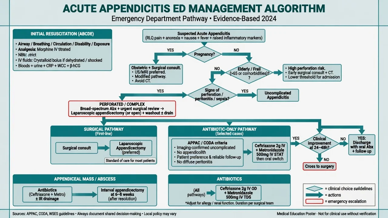
The management begins with the resuscitation of the septic or the shocked patient and the symptom control, and proceeds in parallel with the surgical referral. The intravenous access (the one or the two large-bore cannulae), the fluid resuscitation (the balanced crystalloid — the Hartmann or the normal saline — in the 10 mL per kilogram boluses to restore the perfusion), the oxygen for the shocked or the hypoxic patient, and the monitoring (the pulse, the blood pressure, the oxygen saturation, the urine output) are the foundation. The analgesia is generous and does not obscure the diagnosis — the morphine 0.1 mg per kilogram intravenously (the typical adult dose 5 to 10 mg) titrated to the pain, with the ondansetron 4 mg intravenously for the nausea and the vomiting, and the paracetamol 1 g intravenously for the fever and the baseline analgesia. The controversial topic of the opioid and the masking of the physical signs is settled — the systematic reviews show no adverse effect on the diagnostic accuracy.[2]
The antibiotics are started once the diagnosis is made or strongly suspected (the high-risk score, the imaging-confirmed appendicitis, the perforation with the peritonitis) and the cultures are taken. The empirical regimen covers the enteric Gram-negatives and the anaerobes: the ceftriaxone 2 g intravenously once daily plus the metronidazole 500 mg intravenously every 8 hours is the standard perioperative prophylaxis and the empirical therapy; the piperacillin-tazobactam 4.5 g intravenaneously every 8 hours or the meropenem 1 g intravenously every 8 hours for the severe sepsis or the healthcare-associated infection. The antibiotics are continued as the prophylaxis for the uncomplicated appendicectomy (the single preoperative dose), and extended for the 4 to 5 days in the perforation and the peritonitis. [1]
The drug doses for the appendicitis
Definitive management — the appendicectomy
The surgical referral is made as soon as the diagnosis is made or strongly suspected — the emergency physician does not wait for the morning, the registrar or the formal imaging if the patient is high-risk. The laparoscopic appendicectomy is the first-line operation for the uncomplicated appendicitis — it has the lower wound infection rate, the shorter hospital stay, the faster return to the normal activity, and the better visualisation of the whole abdomen if the appendix is normal (the diagnostic laparoscopy). The open appendicectomy (the Lanz or the Gridiron incision at the McBurney point) is reserved for the patient with the extensive previous surgery, the contraindication to the pneumoperitoneum, or the confirmed complicated case where the laparoscopy is converted. [1]
The appendiceal mass (the inflamed appendix matted to the omentum and the bowel) and the appendiceal abscess in the delayed presentation (the symptoms over 3 to 5 days) are often managed conservatively — the antibiotics (the ceftriaxone and the metronidazole), the percutaneous drainage of the abscess under the imaging guidance, and the interval appendicectomy after 6 to 8 weeks. The immediate surgery in the mass has the higher complication rate (the enterotomy, the fistula, the bleeding) and is avoided unless the patient fails the conservative management. [1]
The antibiotic-first (the conservative) management of the uncomplicated appendicitis is an emerging and evidence-supported option — the APPAC and the CODA trials showed the antibiotics cure approximately 70 to 80 per cent at one year, with the 20 to 30 per cent recurrence and the need for the appendicectomy. It is an option for the patient who declines or cannot have the surgery, but it is not yet the standard of care for the otherwise fit patient, and the Fellowship candidate should present it as an option rather than the default.[2]
The trials behind the antibiotic-first option
APPAC
Multicentre open-label randomised controlled non-inferiority trial, Finland, 530 adults with CT-confirmed uncomplicated acute appendicitis
Population: Adults with CT-confirmed uncomplicated appendicitis (no perforation, no abscess, no appendicolith on the CT)
Comparator: Standard open appendicectomy
Key finding
Antibiotics resolved the appendicitis in 73 per cent at 1 year; the appendectomy arm had the resolution rate of essentially 100 per cent — the non-inferiority was NOT met (the difference 27 per cent exceeded the margin). The 27 per cent of the antibiotic group had the recurrence and went on to the surgery.
Practice change
Antibiotic therapy is effective but not non-inferior to the appendicectomy — approximately three-quarters of the selected patients avoid the surgery at 1 year, but a quarter recur. It is an acceptable option for the informed patient who declines the surgery, not the default.
CODA
Pragmatic randomised non-inferiority trial, USA, 1552 adults with imaging-confirmed appendicitis (including the complicated cases)
Population: Adults with the imaging-confirmed appendicitis, including the appendicolith-bearing and the complicated — broader than the APPAC
Comparator: Appendectomy (the laparoscopic or the open)
Key finding
Antibiotics were non-inferior on the health status at 30 days; 29 per cent of the antibiotic group had the appendectomy within 90 days. The appendicolith subgroup had the higher complication rate (20 versus 8 per cent) and the higher recurrence.
Practice change
Antibiotics are a reasonable first treatment for the imaging-confirmed appendicitis, but the appendicolith identifies the higher-risk patient in whom the surgery may be preferable. Long-term (4-year) follow-up showed the persistent recurrence and the comparable overall outcomes.
Subtypes and scenarios
The perforated appendicitis presents with the severe generalised pain, the high fever, the tachycardia, the rigid abdomen and the sepsis — the management is the aggressive resuscitation, the broad-spectrum antibiotics and the urgent surgery. The retrocaecal appendix produces the flank or the back pain and the muted abdominal signs — the psoas sign is positive and the diagnosis is frequently delayed. The pelvic appendix produces the lower abdominal and the suprapubic pain, the urinary frequency, the diarrhoea, and the tenderness on the digital rectal examination — the obturator sign is positive. The post-ileal appendix (behind the terminal ileum) is the rare variant that produces the vomiting and the central abdominal pain late into the course. [1]
The appendicitis in the pregnancy is the most common non-obstetric surgical emergency, and the diagnosis is difficult because the gravid uterus displaces the appendix upwards and to the right (the pain may localise to the right upper quadrant in the third trimester), the nausea and the mild leukocytosis are normal in the pregnancy, and the delay in the surgery risks the fetal loss and the preterm labour. The ultrasound first, the MRI second, no CT is the imaging pathway, and the surgery is not delayed for the pregnancy — the perforated appendicitis has the fetal mortality up to 30 per cent. [1]
[1] [1] [1]Complications and pitfalls
The complications of the appendicitis itself are the perforation (the 20 to 30 per cent rate at the presentation, the highest in the extremes of age), the appendiceal abscess (the walled-off perforation, the managed with the drainage and the antibiotics), the pylephlebitis (the septic thrombophlebitis of the portal vein with the liver abscesses, from the Bacteroides bacteraemia), the intestinal obstruction (from the paralytic ileus of the peritonitis or the adhesions), and the sepsis and the multi-organ failure in the untreated case. The complications of the appendicectomy are the wound infection (the commonest, the reduced by the laparoscopic approach and the prophylactic antibiotics), the intra-abdominal abscess, the bleeding, the enterotomy, the stump appendicitis (the inflammation of the residual appendiceal stump after the incomplete appendicectomy), and the faecal fistula. [1]
[1]The pitfalls in the diagnosis are the over-reliance on a single investigation (the WCC and the CRP may be normal early; the ultrasound may miss the retrocaecal appendix), the failure to exclude the ectopic pregnancy in the woman of the reproductive age (the beta-hCG is non-negotiable), the failure to examine the scrotum in the male (the testicular torsion), the anchoring on the common diagnosis in the elderly (the caecal carcinoma, the ischaemic bowel, the sigmoid volvulus), and the failure to recognise the atypical presentation at the extremes of age and in the diabetic, the immunosuppressed and the pregnant patient. [1]
Prognosis and disposition
The prognosis of the uncomplicated appendicitis treated with the timely appendicectomy is excellent — the mortality is under 0.1 per cent and the recovery is 1 to 2 weeks for the laparoscopic approach and 2 to 4 weeks for the open approach. The perforation raises the mortality to 1 to 3 per cent, the wound infection rate to 15 per cent and the intra-abdominal abscess rate to 10 per cent. The disposition: the high-risk patient (the AIR 9 to 12, the imaging-confirmed appendicitis, the peritonitis) is admitted under the surgeons for the surgery; the intermediate-risk patient (the AIR 5 to 8, the equivocal imaging) is admitted for the observation, the serial examination and the surgical review; the low-risk patient (the AIR 0 to 4, the normal bloods and the imaging) is discharged with the safety-net advice — to return if the pain worsens, the fever develops, the vomiting persists, or the pain migrates to the right iliac fossa. [1]
Special populations
The paediatric patient (the under 5) presents atypically — the diffuse abdominal pain, the irritability, the refusal to walk, the vomiting and the diarrhoea — and the perforation rate is over 50 per cent at the presentation because the diagnosis is delayed. The ultrasound first, the surgery without the delay, and the awareness that the appendicitis is the most common surgical emergency in the child over 2. The elderly patient has the muted pain, the lower fever, the less pronounced leukocytosis and the higher perforation rate — and the caecal carcinoma must be excluded (the follow-up colonoscopy after the recovery). The pregnant patient — discussed above — needs the ultrasound or the MRI, the early surgical referral, and the surgery without the delay. The immunosuppressed and the diabetic patient has the muted signs and the higher perforation rate. [1]
Child (under 5)
- The diffuse pain, the irritability, the refusal to walk, the vomiting and the diarrhoea — the atypical picture is the rule
- The perforation rate over 50 per cent at the presentation; the lymphoid hyperplasia is the cause
- The ultrasound first; the surgery without the delay; the general surgical and the paediatric joint care
- The most common surgical emergency in the child over 2; the missed diagnosis drives the mortality
Elderly (over 65)
- The muted pain, the lower fever, the less pronounced leukocytosis; the presentation is delayed
- The faecolith is the cause; the perforation rate 50 to 70 per cent; the caecal carcinoma must be excluded
- The CT first (the atypical); the surgery; the follow-up colonoscopy after the recovery
- The mortality rises fivefold with the perforation; the ACEM candidate must have a low threshold for the imaging
Pregnant
- The pain may localise to the right upper quadrant in the third trimester (the gravid uterus displaces the appendix)
- The nausea and the mild leukocytosis are normal in the pregnancy — the CRP is the discriminator
- The ultrasound first, the MRI second, no CT; the early surgical referral; the left-lateral tilt for the venous return
- The perforated appendicitis has the fetal mortality up to 30 per cent and the preterm labour — the surgery is not delayed
Immunosuppressed / diabetic
- The muted signs, the blunted fever, the neuropathy may mask the peritonism; the higher perforation rate
- The CT for the atypical; the broader antibiotic cover for the healthcare-associated and the resistant organisms
- The aggressive resuscitation, the early surgery; the awareness of the delayed presentation
- The neutropenic patient may have no fever and no leukocytosis at all — the imaging is decisive
Evidence and regional guidelines
The contemporary framework is the World Society of Emergency Surgery (WSES) guidelines for the diagnosis and the treatment of the acute appendicitis, the American College of Surgeons (ACS) guidelines, and the NICE guidelines (the United Kingdom). The WSES grades the appendicitis into the uncomplicated (the inflamed appendix, no perforation) and the complicated (the perforation, the abscess, the peritonitis), and endorses the risk-stratified approach (the AIR or the Alvarado score, the selective imaging, the laparoscopic appendicectomy for the uncomplicated, the conservative management for the mass and the abscess).[2][3]
Model answer — the ED workup of the suspected appendicitis
The patient with the suspected appendicitis is assessed with the ABCDE, the history (the migration of the pain, the anorexia, the nausea, the fever), the examination (the McBurney tenderness, the Rovsing, the psoas, the obturator signs), the beta-hCG in every woman, the WCC and the CRP, and the AIR score. The intermediate-risk patient gets the ultrasound (the child, the young woman, the pregnant) or the CT (the adult male, the atypical). The management is the analgesia (the morphine 0.1 mg per kilogram IV), the antiemetic (the ondansetron 4 mg IV), the fluids (the Hartmann), the antibiotics (the ceftriaxone 2 g IV plus the metronidazole 500 mg IV), and the surgical referral for the laparoscopic appendicectomy. The disposition is determined by the score and the imaging, with the safety-net advice for the discharged patient.
The ED workup of the suspected appendicitis
1
2
3
4
5
6
7
8
Exam pearls
- The migration of the pain from the centre to the right iliac fossa over 12 to 24 hours is the single most discriminating feature — over 80 per cent sensitive.
- The Alvarado (MANTRELS) — the score of 7 or above is the surgical referral; the score of 5 to 6 is the imaging; the score of 4 or below is the discharge with the safety-net.
- The AIR score outperforms the Alvarado — it weights the CRP and the polymorphs; the 9 to 12 is the high risk, the 5 to 8 is the intermediate, the 0 to 4 is the low.
- The beta-hCG in every woman of the reproductive age — non-negotiable; the ectopic is the lethal mimic.
- The ultrasound first in the child, the young woman and the pregnant; the CT in the adult male and the atypical; the MRI second-line in the pregnancy.
- The ceftriaxone 2 g IV plus the metronidazole 500 mg IV — the enteric Gram-negative and the anaerobic cover; the perioperative prophylaxis.
- The laparoscopic appendicectomy first-line; the conservative management (the antibiotics, the drainage, the interval appendicectomy) for the mass and the abscess.
- The stump appendicitis — the previous appendicectomy does not exclude the appendicitis.
- The atypical presentation at the extremes of age, in the diabetic, the immunosuppressed and the pregnant — the diagnosis is missed and the perforation is more common.
- The faecolith (the adult) and the lymphoid hyperplasia (the child) are the causes of the obstruction — the child perforates faster because the lumen is narrowest and the omentum is short.
- The dual innervation explains the migration — the visceral T10 pain (the periumbilical, dull, poorly localised) gives way to the somatic localised right iliac fossa pain as the parietal peritoneum is reached.
- The McBurney point — one-third of the line from the ASIS to the umbilicus; the tenderness is the most constant sign. The Rovsing, the psoas and the obturator signs localise the appendix and confirm the peritonism.
- The non-compressible appendix over 6 mm on the ultrasound is diagnostic — but up to 40 per cent of asymptomatic adults have the appendix over 6 mm; the symptoms and the fat stranding must concur.
- The analgesia does not mask the diagnosis — the morphine is safe and humane; the systematic reviews confirm no adverse effect on the diagnostic accuracy.
- The APPAC and the CODA trials — the antibiotics cure 70 to 80 per cent at one year but are not non-inferior to the appendectomy; the appendicolith predicts the failure and the higher recurrence.[4][5]
- The appendicolith on the imaging — the higher perforation risk, the higher recurrence after the antibiotics, and the relative contraindication to the conservative management.
- The appendiceal neoplasm (the carcinoid, the adenocarcinoma, the mucinous neoplasm) is found in 1 to 2 per cent of the appendicectomy specimens — the antibiotic-first path forfeits the histopathology; counsel the patient.
- The pylephlebitis — the septic portal thrombophlebitis with the liver abscesses from the Bacteroides; the swinging fever and the jaundice days after the appendicectomy warrant the liver ultrasound and the portal-vein Doppler.
- The persistent fever after the appendicectomy — the intra-abdominal abscess until proven otherwise; the CT and the drainage.
Exam practice
SAQ — Classic appendicitis: the Alvarado and the AIR scores in a young man
10 minutes · 10 marks
A 24-year-old man presents with a 16-hour history of central abdominal pain that migrated to the right iliac fossa 4 hours ago. He is anorexic, nauseated, and vomited once. T 38.6 degrees C, HR 102, BP 122/74. There is McBurney point tenderness with voluntary guarding, rebound tenderness, and a positive Rovsing sign. WCC 15.8 x 10^9/L with 88 per cent neutrophils, CRP 55 mg/L, urine beta-hCG negative.
SAQ — Perforated appendicitis with septic shock in the elderly diabetic
10 minutes · 10 marks
A 68-year-old man with type 2 diabetes presents 48 hours after the onset of abdominal pain, now with severe generalised pain, rigors, and confusion. T 39.2 degrees C, HR 128, BP 86/48 (MAP 60), RR 24, SpO2 95 per cent on room air. The abdomen is rigid and diffusely tender with absent bowel sounds. WCC 22.5 x 10^9/L, lactate 4.2 mmol/L, CRP 180 mg/L, creatinine 165 micromol/L. The CT shows a perforated appendix with free intraperitoneal air and widespread purulent fluid. The qSOFA is 3.
Red flags
[1]References
- [1]Alvarado A. A practical score for the early diagnosis of acute appendicitis Ann Emerg Med, 1986.PMID 3963537
- [2]Andersson RE. Diagnostic value of the appendicitis inflammatory response (AIR) score. A systematic review and meta-analysis World J Emerg Surg, 2025.PMID 39923099
- [3]Andersson M, Andersson RE, Ripa T, et al. Validation of the Appendicitis Inflammatory Response (AIR) Score World J Surg, 2021.PMID 33825049
- [4]Salminen P, Paajanen H, Rautio T, Nordström P, Aarnio M, Rantanen T, et al. Antibiotic Therapy vs Appendectomy for Treatment of Uncomplicated Acute Appendicitis: The APPAC Randomized Clinical Trial JAMA, 2015.PMID 26080338
- [5]CODA Collaborative, Flum DR, Davidson GH, Monsell SE, Fischkoff KM, et al. A Randomized Trial Comparing Antibiotics with Appendectomy for Appendicitis N Engl J Med, 2020.PMID 33017106
- [6]Doria AS, Moineddin R, Kellenberger CJ, Epelman M, Beyene J, Babyn PS, et al. US or CT for Diagnosis of Appendicitis in Children and Adults? A Meta-Analysis Radiology, 2006.PMID 16928974