EM · Acute pancreatitis
Acute pancreatitis
Also known as Acute pancreatitis · Severe acute pancreatitis · Gallstone pancreatitis · Alcoholic pancreatitis
Acute pancreatitis — severe epigastric pain radiating straight through to the back with vomiting, a lipase 3 times the upper limit of normal, gallstones and alcohol as the dominant causes (GET SMASHED), the Ranson and Glasgow severity scores, aggressive but goal-directed fluid resuscitation, morphine analgesia, early enteral feeding, antibiotics reserved for infected necrosis, and ERCP for the obstructive biliary case. ACEM-primary, globally tagged.
On this page & tools
Your progress
Saved locally on this device.
8 MCQs with explanations
Target exams
Red flags
Related topics
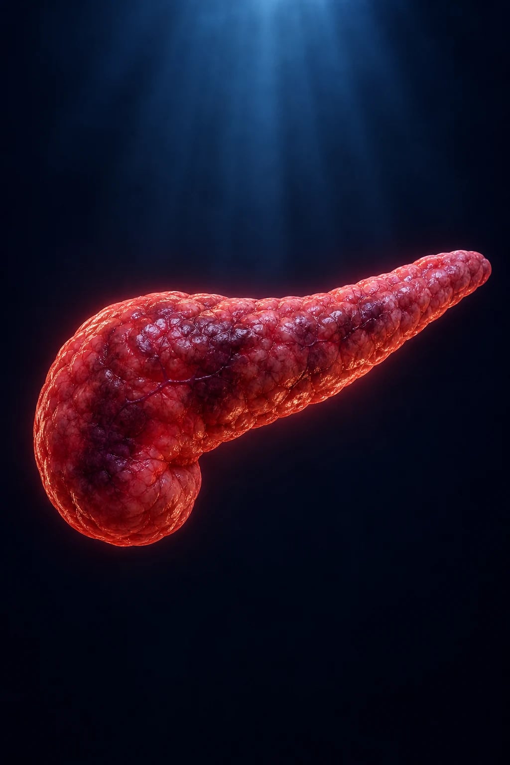
Acute pancreatitis is an acute inflammatory process of the pancreas with variable involvement of other regional tissues and remote organ systems, ranging from a mild self-limiting illness to a lethal, multi-organ disease driven by a systemic inflammatory response. The Fellowship candidate must make the diagnosis at the bedside from two of three features (the pain, the lipase and the imaging), grade the severity early, resuscitate aggressively but with discipline, and resist the two classic errors — withholding enteral feeding and giving prophylactic antibiotics.[1][2]
Definition and classification
Acute pancreatitis is defined by two of three criteria: (1) characteristic acute epigastric pain radiating to the back; (2) a serum lipase (or amylase) at least three times the upper limit of normal; and (3) characteristic findings on contrast-enhanced CT or MRI. Imaging is not required when the first two are present. The 2012 Revised Atlanta classification stratifies severity into three grades at 48 hours: mild (no organ failure and no local or systemic complications — the majority), moderately severe (transient organ failure resolving within 48 hours, or local complications such as peripancreatic fluid collections and necrosis, or an exacerbation of comorbidity, but without persistent organ failure), and severe (persistent organ failure beyond 48 hours — single or multi-organ).[2] Organ failure is defined by a modified Marshall score, and the distinction between transient and persistent organ failure is the single most important prognostic fork in the disease.
Epidemiology and risk — the causes
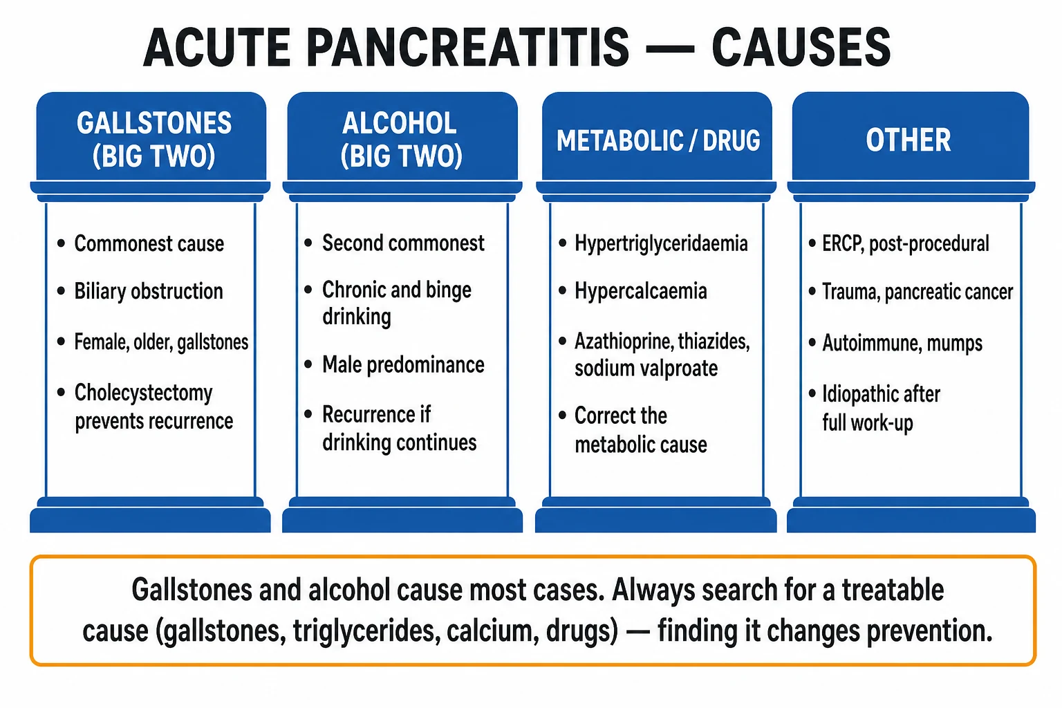
Acute pancreatitis is common and rising, with an annual incidence of about 13 to 45 per 100 000. Gallstones and alcohol together account for about 70 per cent of cases: gallstones (including microlithiasis and biliary sludge) cause roughly 40 per cent by transiently obstructing the ampulla of Vater, and alcohol causes about 30 per cent through direct acinar toxicity and protein-plug obstruction of small ducts. The remainder follow ERCP, hypertriglyceridaemia, hypercalcaemia, drugs and a long list of less common causes. The causes are remembered by the mnemonic below. [1]
GET SMASHED — the causes of acute pancreatitis
GET SMASHED
The commonest cause; microlithiasis and biliary sludge included
The second commonest; direct acinar toxicity and protein plugs
Blunt pancreatic injury; also post-ERCP and post-operative
And other drugs — azathioprine, thiazides, sodium valproate, didanosine
Viral (mumps, Coxsackie); ampullary or pancreatic tumour obstruction
Autoimmune pancreatitis, often with raised IgG4
Rare; also snake envenomation
Triglycerides above 11 mmol/L (about 1000 mg/dL)
Post-procedural; embolic or ischaemic
Didanosine, azathioprine, thiazides, sulphonamides, oestrogens
Pathophysiology
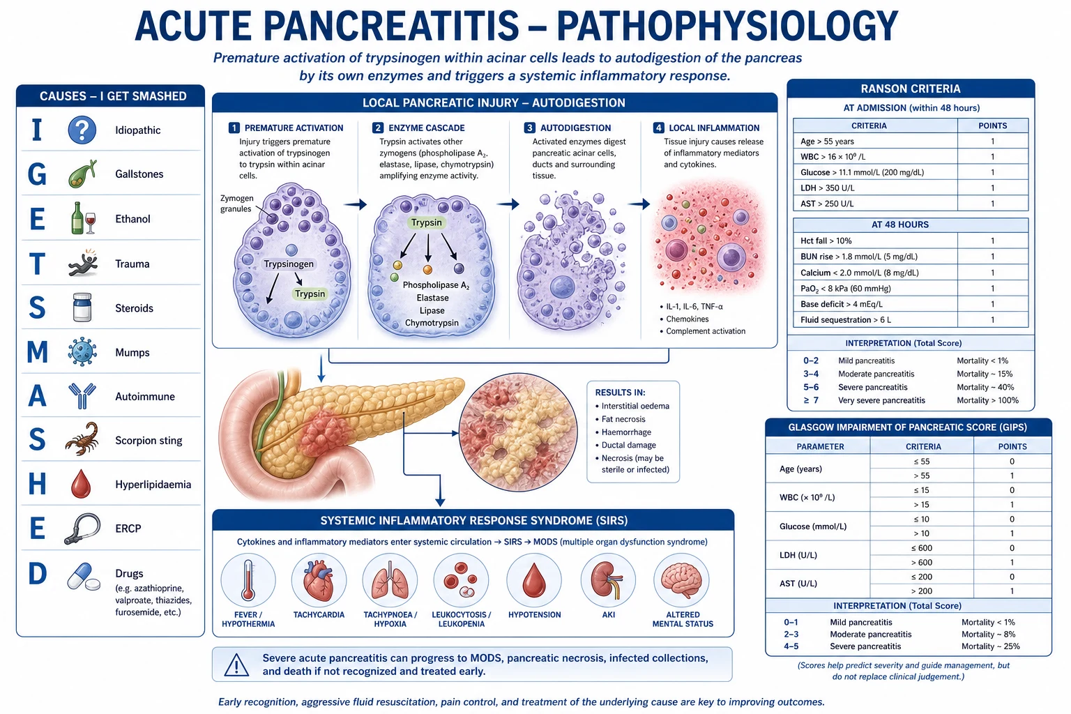
The unifying event is the premature intracellular activation of trypsinogen to trypsin within the pancreatic acinar cell, escaping the protective compartmentalisation of digestive enzymes. Once trypsin is active inside the cell it activates a cascade of other zymogens (phospholipase A2, elastase, proteases), the cell autodigests, and the release of cytokines (interleukin-6, tumour necrosis factor) drives a marked systemic inflammatory response syndrome (SIRS). Gallstones cause this by transiently obstructing the shared biliopancreatic duct and raising intraductal pressure; alcohol causes it by direct toxic injury to acinar cells and by precipitating protein plugs in the small ducts. The local result is oedema, peripancreatic fat stranding and, in severe cases, pancreatic and peripancreatic necrosis. The systemic result is capillary leak, third-space fluid loss, hypovolaemia and, in the worst cases, the multi-organ failure that defines severe disease. SIRS and a compensatory anti-inflammatory response alternate over the first weeks, governing the risk of organ failure and later infection of necrotic tissue. [1]
Clinical presentation
The presentation is acute severe epigastric pain that radiates straight through to the back, is often eased a little by sitting forward, and is accompanied by nausea and vomiting. The pain reaches its maximum within minutes to hours, distinguishing it from the colicky waxing-and-waning pain of biliary colic. Low-grade fever and tachycardia are common from SIRS rather than infection in the early phase. Jaundice suggests an obstructive biliary cause — a gallstone impacted at the ampulla, or evolving cholangitis — and mandates urgent biliary imaging and consideration of ERCP. Examination reveals epigastric tenderness and guarding, but typically without the board-like rigidity of a perforated viscus. Two rare but feared signs mark severe haemorrhagic pancreatitis: Cullen sign (periumbilical bruising) and Grey Turner sign (flank bruising), from retroperitoneal blood tracking along fascial planes — their absence does not exclude severe disease. Hypoxia, oliguria, confusion and hypotension indicate established organ failure and an immediate intensive-care referral. [1]
Differential diagnosis
The epigastric pain of pancreatitis overlaps with several lethal mimics, and the first task is to exclude a perforation, a vascular catastrophe, an inferior myocardial infarction and mesenteric ischaemia before attributing the picture to the pancreas. [1]
Acute pancreatitis
- Epigastric pain radiating to the back, vomiting, SIRS
- Lipase 3 times the upper limit of normal
- Gallstones on ultrasound or an alcohol history
- CT shows peripancreatic fat stranding ± necrosis
Perforated peptic ulcer
- Sudden severe epigastric pain becoming generalized
- Rigid, board-like abdomen; absent liver dullness
- Erect chest X-ray or CT shows free gas
- Lipase normal or mildly raised; sudden onset is the clue
Inferior myocardial infarction
- Epigastric pain, nausea, diaphoresis; may mimic pancreatitis
- ECG shows inferior ST elevation (II, III, aVF)
- Lipase normal; troponin rises
- Always do an ECG in epigastric pain
Mesenteric ischaemia
- Severe pain out of proportion to the examination
- Metabolic acidosis, raised lactate, bloody diarrhoea
- Risk factors: atrial fibrillation, vascular disease
- CT angiography is diagnostic; lipase normal
Ruptured abdominal aortic aneurysm
- Sudden back or abdominal pain, syncope, hypotension
- Pulsatile mass in the older male smoker
- Bedside ultrasound confirms the aneurysm
- Unstable → theatre, not CT; lipase normal
Biliary colic / cholecystitis
- Right upper quadrant pain after fatty food, colicky
- Murphy sign positive in cholecystitis; low-grade fever
- Ultrasound shows stones ± wall thickening
- Lipase normal; pancreatitis coexists if the stone obstructs
Bedside assessment
Assess airway, breathing and circulation first — the patient with severe pancreatitis is hypovolaemic from massive third-space losses and may be hypoxic from early ARDS. Establish intravenous access, give oxygen, attach monitoring, and treat pain and vomiting early. Take a focused history of the pain onset, alcohol intake, recent ERCP, gallstones, lipid disorders and drugs (thiazides, azathioprine, sodium valproate, didanosine, oestrogens). Examine for jaundice (biliary obstruction), epigastric tenderness and guarding, the rare periumbilical or flank bruising of haemorrhagic pancreatitis, and any signs of chronic liver disease. Screen for organ failure explicitly — oxygen saturation, urine output, blood pressure, conscious level — because finding it changes the disposition from ward to intensive care. Re-examine and re-assess repeatedly over the first 48 hours, because the grade of severity is determined at 48 hours and the picture can deteriorate quickly. [1]
Investigations
The diagnosis needs two of three criteria. Send a serum lipase — at three times the upper limit of normal it is both sensitive and specific, and it stays elevated longer than amylase (which rises faster but falls over three to five days). A normal lipase at 48 hours makes pancreatitis unlikely; a markedly raised lipase with a compatible story is diagnostic, and imaging is not required to confirm. A full blood count, urea and electrolytes, liver function tests, glucose, calcium, C-reactive protein, triglycerides and a venous or arterial blood gas are mandatory: these both grade severity (the Ranson and Glasgow criteria) and screen for organ failure (hypoxia, renal impairment, acidosis, hypocalcaemia). An ERCP-related cause is suggested by the history. An abdominal ultrasound is performed early in every patient to look for gallstones, biliary sludge and bile-duct dilation, even though bowel gas often limits the pancreatic view. A contrast-enhanced CT is not routine on day one — the diagnosis is clinical and biochemical — but is indicated when the diagnosis is uncertain, when there is organ failure or a deteriorating course, or after 48 to 72 hours in severe disease to assess for necrosis. Early CT (within 48 hours) can underestimate necrosis because perfusion has not yet declared itself. An ECG and troponin exclude an inferior myocardial infarction, and an erect chest X-ray may reveal a perforation. [1]
[1]Severity scoring
No single score is perfect, but a combination stratifies severity and flags the patient who needs intensive care. The Ranson criteria are scored at admission (5 criteria) and at 48 hours (6 criteria); a score of 3 or more indicates severe disease and a score above 6 carries a high mortality. The modified Glasgow (Imrie) score collects eight data points within 48 hours and, like Ranson, signals severe disease at 3 or more. The APACHE II is the most accurate continuous score and is used in intensive care, calculated on admission and repeated. The bedside BISAP is a simple five-point score that predicts severe disease from admission data. [1]
Ranson criteria — the admission and 48-hour scores
The BISAP score (one point each: BUN over 25, Impaired mental status, SIRS, Age over 60, Pleural effusion) identifies severe disease from admission data and is easily calculated in the emergency department. The modified Glasgow (Imrie) score awards one point each within 48 hours for age over 55, white cell count over 15, glucose over 10, urea over 16, PaO2 under 8, calcium under 2, albumin under 32 and the liver enzyme AST over 100. The APACHE II is calculated on a wider physiological dataset and is the most accurate but the most laborious; many units use it to confirm the Ranson or Glasgow prediction. Whichever is used, the operational point is the same: a severity score of 3 or more, organ failure, or a CRP over 150 at 48 hours identifies the severe patient who needs intensive care.[2]
Immediate management — resuscitation
Resuscitation is the priority and the first 12 to 24 hours of fluid therapy determine the course. Give oxygen, establish large-bore intravenous access, insert a urinary catheter in the unwell patient to monitor output, and treat pain and vomiting. The cornerstone is goal-directed intravenous fluid resuscitation: give a bolus and then titrate to a target of urine output about 0.5 to 1 mL per kilogram per hour and a normalising haematocrit, with frequent reassessment. Lactated Ringer solution is preferred to normal saline by the international guidelines on the basis of reduced systemic inflammation, though the difference is modest. The WATERFALL trial is the key evidence: aggressive fluid resuscitation (a bolus followed by a high rate) caused more fluid overload than a moderate regimen without improving outcomes, so the modern standard is goal-directed, not a blanket high rate.[4]
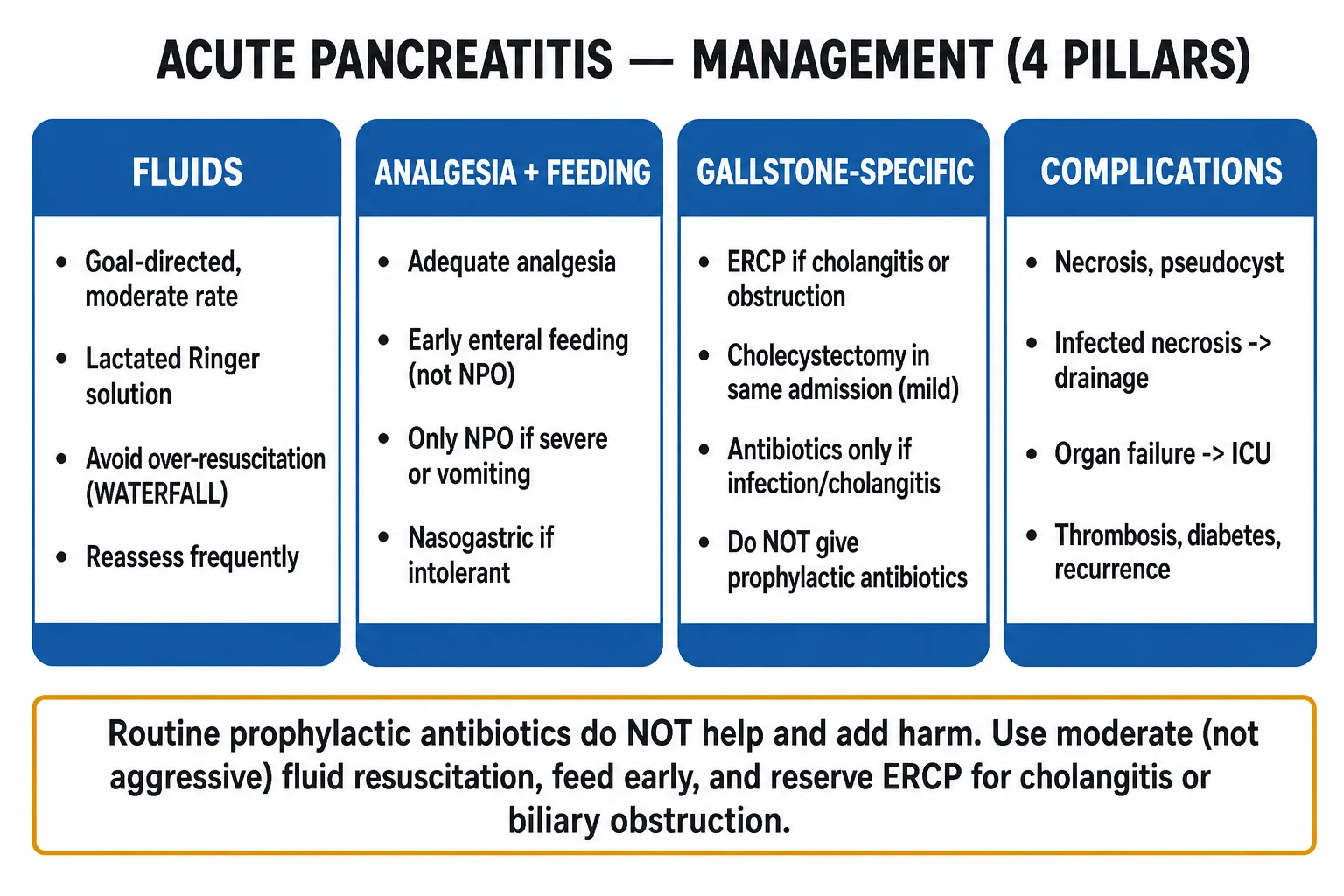
Analgesia: give morphine 5 mg intravenously, titrated to effect and repeated as required. The old dogma that morphine causes sphincter of Oddi spasm and worsens pancreatitis is not supported by clinical evidence, and morphine is the standard opioid; fentanyl 25 to 50 micrograms intravenously is an alternative. Antiemesis: give ondansetron 4 mg intravenously for nausea and vomiting. Early enteral feeding within 24 to 48 hours is now the standard of care — it reduces infective complications, length of stay and mortality compared with traditional prolonged fasting — and is tolerated in mild pancreatitis; in severe disease, nasojejunal feeding is used if gastric feeding is not tolerated, with parenteral nutrition reserved for those who cannot be enterally fed.[1][3]
[1]Definitive management and disposition
Definitive management follows the severity. Mild pancreatitis is managed with fluids, analgesia and early feeding on a general ward, and usually resolves in three to seven days; the gallbladder is removed during the index admission (a laparoscopic cholecystectomy) to prevent recurrence, which is highest in the first weeks. Severe pancreatitis (persistent organ failure) is managed in intensive care with invasive monitoring, organ support (ventilation for ARDS, renal replacement therapy for renal failure) and continued fluid and electrolyte correction. ERCP with sphincterotomy within 24 to 72 hours is indicated for cholangitis or persistent biliary obstruction (a stone in the common bile duct with jaundice or dilated ducts) — it relieves the obstruction and removes the stone, and is not indicated for pancreatitis without obstruction. Infected necrosis is the key late complication: suspected when the patient develops fever, sepsis or a rising inflammatory marker after the first week, and confirmed by gas within necrotic tissue on CT or by fine-needle aspiration. Infected necrosis is treated with antibiotics and minimally invasive necrosectomy — carbapenems or the combination of ciprofloxacin 400 mg intravenously twice daily plus metronidazole 500 mg intravenously three times daily penetrate necrotic tissue well, and necrosectomy is delayed by several weeks where possible to allow demarcation (a step-up approach from percutaneous drainage to minimally invasive retroperitoneal necrosectomy).[3]
[1]A pseudocyst (a walled-off collection of pancreatic secretions, mature at four or more weeks) is observed if asymptomatic and drained (endoscopically, percutaneously or surgically) if it is symptomatic, infected or growing. A peripancreatic fluid collection in the first four weeks is managed expectantly. Symptomatic walled-off necrosis is drained, ideally endoscopically. Disposition: mild disease to the ward with a plan for cholecystectomy; moderately severe and severe disease to a high-dependency or intensive-care unit; any deterioration in organ failure is an intensive-care referral. [1]
Complications and pitfalls
The complications are local and systemic. Local: peripancreatic fluid collection, pancreatic and peripancreatic necrosis (sterile then potentially infected), walled-off necrosis, pseudocyst, gastroduodenal obstruction, haemorrhage, and splenic or portal vein thrombosis. Systemic: the systemic inflammatory response drives ARDS, acute kidney injury and renal failure, shock, disseminated intravascular coagulation (DIC), hypocalcaemia, hyperglycaemia and hypomagnesaemia. Infected necrosis is the dominant late cause of death. The pitfalls are the inverse of good care: giving prophylactic antibiotics (no benefit, real harm); withholding enteral feeding (worsens outcome and gut-barrier function); over-resuscitating with fluid (pulmonary oedema, abdominal compartment syndrome); under-resuscitating (persistent hypovolaemia drives necrosis); delaying ERCP in true cholangitis; performing early necrosectomy (sterile necrosis is managed conservatively, and even infected necrosis is delayed to allow demarcation); and missing a lethal mimic (the inferior MI, the perforation, the mesenteric ischaemia or the ruptured aneurysm) by attributing epigastric pain to the pancreas on a raised lipase alone. [1]
Prognosis and disposition
Mortality tracks severity: mild pancreatitis carries a mortality of about 1 to 3 per cent, severe pancreatitis (persistent organ failure, pancreatic necrosis) carries 10 to 30 per cent, and infected necrosis pushes it higher. The Ranson and Glasgow scores and the APACHE II predict severity; a Ranson score above 6 carries a mortality approaching 40 per cent. Persistent organ failure beyond 48 hours is the dominant determinant of death. Mild cases are discharged once pain and the inflammatory markers settle, with a cholecystectomy arranged (during the index admission in gallstone disease). Severe cases follow a prolonged intensive-care and rehabilitation course. Recurrence is common in alcohol-related disease (continued drinking) and in unremoved gallbladders; alcohol cessation counselling and a cholecystectomy are the two most effective secondary-prevention measures. [1]
Special populations
Pregnancy: acute pancreatitis in pregnancy is most often gallstone-related; management is unchanged (fluids, analgesia, early feeding) and cholecystectomy is safe in the second trimester if needed. Severe hypertriglyceridaemia in pregnancy can precipitate pancreatitis and is treated with fluid, analgesia and, in extreme cases, plasmapheresis. The elderly present atypically with confusion or hypotension and tolerate fluid overload poorly — goal-directed resuscitation and early organ-support are essential. Post-ERCP pancreatitis is usually mild and self-limiting; rectal diclofenac or indomethacin and a prophylactic pancreatic stent reduce its incidence in high-risk patients. Children are rare; the causes include trauma, biliary anomaly, drugs and metabolic disease, and the management is the same. [1]
Evidence and regional guidelines
The contemporary framework is the 2013 American College of Gastroenterology guideline and the IAP/APA evidence-based guidelines, both built on the 2012 Revised Atlanta classification.[1][2][3] The three practice-defining changes of the modern era are: early enteral feeding (replacing prolonged fasting), no prophylactic antibiotics, and goal-directed fluid resuscitation. The WATERFALL trial (2022) established that aggressive resuscitation causes fluid overload without benefit, settling the long-standing question of how much fluid to give.[4] Necrosectomy has shifted from early open surgery to a delayed, minimally invasive step-up approach, which reduces complications and death.
ANZ practice note. Australasian management follows the same evidence base. Gallstone pancreatitis with cholangitis or persistent obstruction is referred for urgent ERCP at the local endoscopy service; otherwise the patient has a cholecystectomy during the index or an early readmission. Severe pancreatitis is managed in intensive care, and infected necrosis follows a multidisciplinary surgical, gastroenterology and intensive-care pathway with a minimally invasive step-up approach. Alcohol-related disease is paired with an alcohol-harm brief intervention and referral before discharge. [1]
Exam pearls
- Diagnosis needs two of three: the pain, the lipase at three times the upper limit of normal, and the imaging. Imaging is NOT required when the first two are present.
- Causes — GET SMASHED: Gallstones and Ethanol are two thirds; the rest is trauma, steroids, mumps/malignancy, autoimmune, scorpion, hypercalcaemia/hypertriglyceridaemia, ERCP, drugs.
- Severity at 48 hours: mild (no organ failure), moderately severe (transient organ failure or local complications), severe (persistent organ failure). The fork that decides intensive care.
- Ranson: 5 criteria at admission, 6 at 48 hours; 3 or more is severe. Glasgow (Imrie) and BISAP are the bedside alternatives; APACHE II is the intensive-care standard.
- No prophylactic antibiotics — they do not prevent infected necrosis. Antibiotics are for infected necrosis (carbapenems, or ciprofloxacin plus metronidazole) or cholangitis.
- Early enteral feeding within 24 to 48 hours — not fasting. It reduces infection and mortality.
- Goal-directed fluids, not aggressive — WATERFALL showed harm from over-resuscitation. Target urine output and a falling haematocrit.
- ERCP is for cholangitis or persistent biliary obstruction, not for pancreatitis without obstruction.
- Morphine 5 mg IV is standard analgesia; the sphincter of Oddi concern is not clinically relevant.
- Infected necrosis is suspected after the first week with fever and sepsis; treat with antibiotics and a delayed, minimally invasive step-up necrosectomy. [1]
Exam practice
SAQ — Severe gallstone pancreatitis with SIRS and biliary obstruction
12 minutes · 10 marks
A 58-year-old obese woman presents with eight hours of severe epigastric pain radiating straight through to the back, repeated vomiting and jaundice. She has known gallstones. On arrival she is in pain and distressed: T 38.4, HR 124, BP 92/58, RR 28, SpO2 92 per cent on room air, GCS 15. The abdomen is tender with voluntary guarding in the epigastrium. Lipase 1240 U/L (ULN 60), ALT 220, bilirubin 68 micromol/L, glucose 13.2, LDH 420, calcium 2.05. ABG: pH 7.28, PaO2 7.6 kPa, lactate 3.1. Ultrasound shows multiple gallstones and a common bile duct of 9 mm.
SAQ — Infected pancreatic necrosis in the third week of severe pancreatitis
12 minutes · 10 marks
A 49-year-old man with severe alcoholic pancreatitis (admission Ranson 5, BISAP 3) is now on day 16 of his intensive-care stay. He had been improving on nasojejunal feeding. Over the past 48 hours he has spiked temperatures to 39.1, become tachycardic (HR 128) and hypotensive (BP 88/52 on noradrenaline 0.25 microgram/kg/min). WCC 22, CRP 280, lactate 4.2. Contrast-enhanced CT shows 60 per cent pancreatic and peripancreatic necrosis with multiple gas bubbles within the necrotic tissue.
Red flags
[1]Severity scoring — the four scores in detail
No severity score is perfect, and none should be acted upon in isolation, but together they answer two exam questions that recur every year: which patient is going to deteriorate? and which patient needs intensive care now? The four scores you must be able to reproduce are Ranson, the modified Glasgow (Imrie), APACHE II and BISAP, and you should be able to quote at least one fully in the viva. [1]
Ranson criteria
Ranson is the classic two-timepoint score — half the criteria are collected at admission and half at 48 hours. It was originally derived from alcoholic pancreatitis and modified for gallstone disease; the values below are the common examination form. [1]
Ranson criteria — five at admission, six at 48 hours
Ranson score — mortality bands
Modified Glasgow (Imrie) score
The Glasgow score collects eight data points all within 48 hours, unlike Ranson which splits them. It is the easier bedside score and the most common viva target. A score of 3 or more indicates severe pancreatitis. The mnemonic is PANCREAS — but be warned the eight letters map unevenly to eight criteria; most candidates memorise the list directly. [1]
Modified Glasgow (Imrie) — eight points within 48 hours, 3 or more is severe
BISAP — the admission-only score
BISAP is a simple five-point score calculated entirely from admission data, which makes it the most practical emergency-department tool. One point is awarded for each of: BUN over 25 mg/dL (8.9 mmol/L), Impaired mental status (Glasgow Coma Scale under 15), SIRS (two of four), Age over 60, and Pleural effusion on imaging. A score of 3 or more identifies severe disease with a sensitivity and specificity comparable to APACHE II at a fraction of the effort. [1]
[1]APACHE II — the intensive-care standard
APACHE II is a continuous physiological score (12 variables, including temperature, mean arterial pressure, oxygenation, pH, sodium, potassium, creatinine, haematocrit, white cell count, Glasgow Coma Scale, age and chronic health) calculated on admission and repeated. A score over 8 predicts severe pancreatitis. It is the most accurate single score but is laborious and best suited to the intensive-care setting, where it is run routinely by the unit. In the Fellowship exam it is enough to know that APACHE II is the most accurate, is continuous, and that a threshold of 8 is used. [1]
Ranson
- Two timepoints: admission + 48 hours
- Alcoholic-pancreatitis derived; gallstone variant exists
- Score of 3 or more is severe
- Predicts mortality in bands (0–2 ~1%, 5–6 ~40%)
- Cannot be completed on day one
Glasgow (Imrie)
- Single timepoint within 48 hours
- Eight criteria — age, WCC, glucose, urea, PaO2, albumin, calcium, LDH
- Score of 3 or more is severe
- The commonest bedside viva target
- Albumin replaces AST; LDH over 600
BISAP
- Admission only — the ED score
- Five points: BUN, Impaired mental status, SIRS, Age, Pleural effusion
- Score of 3 or more is severe
- Comparable accuracy to APACHE II at a fraction of the effort
- Use it to triage before 48 hours
APACHE II
- Continuous; 12 physiological variables
- Most accurate single score
- Threshold of 8 predicts severe disease
- Intensive-care standard, repeated daily
- Laborious — not a front-door score
CRP > 150 at 48 h
- A single high-yield biomarker, not a full score
- Predicts necrosis and severe disease
- Cheap and universally available
- Use as an adjunct, not a replacement
- Normal in the first 24 hours — useless early
Local complications — the Atlanta terminology
The 2012 Revised Atlanta classification gave the local complications precise names tied to the contents (fluid vs necrosis) and the timepoint (under or over four weeks) and the presence of a wall. The Fellowship candidate must use these terms correctly — examiners mark down "pancreatic abscess" and "phlegmon", which are obsolete. [1]
Acute peripancreatic fluid collection
- First four weeks; no necrosis
- Homogeneous fluid, no wall
- Usually sterile and self-resolving
- Mild pancreatitis
- No intervention; observe
Acute necrotic collection
- First four weeks; necrosis present
- Heterogeneous: fluid plus non-liquefied tissue
- May become infected after the first week
- Moderately severe or severe pancreatitis
- Sterile → conservative; infected → antibiotics + drainage
Pseudocyst
- After four weeks; no necrosis
- Homogeneous fluid enclosed by a fibrous wall
- Follows a fluid collection that has matured
- Often asymptomatic
- Drain if symptomatic, infected or enlarging
Walled-off necrosis
- After four weeks; necrosis present
- Heterogeneous contents enclosed by a mature wall
- The mature counterpart of an acute necrotic collection
- Often causes gastric-outlet obstruction
- Drain endoscopically; do not debride early
Infected necrosis
- Necrosis seeded by gut organisms
- Suspected with fever and sepsis after week one
- Gas in necrotic tissue on CT is pathognomonic
- Confirmed by fine-needle aspiration
- Carbapenems or ciprofloxacin + metronidazole, then minimally invasive necrosectomy
Systemic complications and organ failure
The systemic inflammatory response drives the organ failures that define severe disease and that kill the patient in the first week. Organ failure is graded by a modified Marshall score (respiratory, cardiovascular, renal), and persistence beyond 48 hours is the single criterion that escalates a case from moderately severe to severe. [1]
ARDS / hypoxaemia
- Capillary leak floods the alveoli; SIRS in the lung
- PaO2 under 8 kPa is a Ranson/Glasgow criterion
- Pleural effusions (especially left-sided) predict severity in BISAP
- Treat with lung-protective ventilation
- The commonest cause of early death
Acute kidney injury
- Hypovolaemia and SIRS-mediated renal vasoconstriction
- Rising urea despite fluids is a severity marker
- May progress to renal replacement therapy
- Goal-directed fluids are renal-protective
- Monitor urine output hourly with a catheter
DIC and coagulopathy
- Consumptive coagulopathy from disseminated inflammation
- Consumes calcium (saponification) and factors
- Low calcium is a severity marker, not a cause to replace
- Monitor platelets, fibrinogen, INR and D-dimer
- Supportive — treat the underlying pancreatitis
Shock
- Massive third-space loss plus vasodilation
- Hypotension is a feature of severe disease
- Resuscitate with goal-directed crystalloid
- Vasopressors if fluid-refractory
- Marks persistent organ failure
Metabolic derangement
- Hypocalcaemia from fat saponification
- Hyperglycaemia from impaired insulin release
- Hypomagnesaemia accompanies hypocalcaemia
- Acidosis from shock and renal failure
- Correct the cause, not just the number
Vascular thrombosis
- Splenic vein thrombosis — gastric varices
- Portal vein thrombosis — portal hypertension
- From peripancreatic inflammation and compression
- May cause gastrointestinal bleeding
- Anticoagulation decisions are individualised
ED management — the first six hours, step by step
The resuscitation delivered in the first six hours determines whether the disease stays mild or progresses to necrosis. The sequence below is the practical order at the bedside. [1]
The first six hours — targets and reassessment
Gallstone pancreatitis — the biliary pathway
Gallstone (including microlithiasis and sludge) pancreatitis is the commonest single cause, and the biliary case is the one most likely to need a procedure in the emergency department or within days. The decision tree turns on whether there is cholangitis or persistent biliary obstruction — both are ERCP indications — versus a stone that has already passed, which needs a cholecystectomy instead. [1]
Stone already passed
- No jaundice, improving LFTs, no duct dilation
- Lipase may be very high then falls
- No ERCP needed
- Laparoscopic cholecystectomy in the index admission
- Low risk of recurrence once the gallbladder is out
Persistent obstruction
- Jaundice, dilated common bile duct on ultrasound
- Persistently abnormal or rising liver enzymes
- ERCP with sphincterotomy within 24–72 hours
- Cholecystectomy afterwards in the same admission
- Risk of cholangitis if not relieved
Ascending cholangitis
- Charcot triad: fever, jaundice, RUQ pain
- Reynolds pentad adds hypotension and confusion
- Urgent ERCP within 24 hours — a source-control emergency
- Broad-spectrum antibiotics in addition
- The clearest ERCP indication in pancreatitis
Cholecystectomy timing
- Same-admission in mild gallstone pancreatitis
- Recurrence is highest in the first weeks if delayed
- Severe/necrotising disease — delay cholecystectomy until recovery
- Cholecystectomy does not treat the pancreatitis itself
- Consider intraoperative cholangiography for the duct
Infected necrosis — the late killer
Infected necrosis is the dominant late cause of death in severe pancreatitis, typically declaring itself in the second to third week as the patient develops fever, worsening sepsis and a rising inflammatory marker after an initial improvement. The diagnosis is suspected clinically and confirmed by gas within necrotic tissue on contrast-enhanced CT or by positive culture on fine-needle aspiration. [1]
Landmark evidence you must cite
WATERFALL (2022)
- de-Madaria et al., NEJM
- Aggressive vs moderate fluid resuscitation
- Aggressive arm caused more fluid overload
- No outcome benefit from aggressive fluids
- Settled the modern goal-directed standard
PANTER (2010)
- van Santvoort et al., NEJM
- Minimally invasive step-up vs open necrosectomy
- Step-up halved the composite complication/death endpoint
- Fewer fistulas and less new-onset diabetes
- Established the step-up approach
PROPATRIA (2008)
- Besselink et al., Lancet
- Probiotic prophylaxis in predicted severe disease
- Increased mortality (16% vs 6%)
- Increased bowel ischaemia
- A landmark trial of harm — probiotics are NOT given
Revised Atlanta (2013)
- Banks et al., Gut
- Standardised severity: mild, moderately severe, severe
- Named the local complications precisely
- Retired "abscess" and "phlegmon"
- The classification the whole field uses
Additional red flags
[1]References
- [1]Tenner S, Baillie J, DeWitt J, Vege SS. American College of Gastroenterology guideline: management of acute pancreatitis Am J Gastroenterol, 2013.PMID 23896955
- [2]Banks PA, Bollen TL, Dervenis C, et al. Classification of acute pancreatitis--2012: revision of the Atlanta classification and definitions by international consensus Gut, 2013.PMID 23100216
- [3]Working Group IAP/APA Acute Pancreatitis Guidelines. IAP/APA evidence-based guidelines for the management of acute pancreatitis Pancreatology, 2013.PMID 24054878
- [4]de-Madaria E, Buxbaum JL, Maisonneuve P, et al. Aggressive or Moderate Fluid Resuscitation in Acute Pancreatitis N Engl J Med, 2022.PMID 36103415
- [5]Besselink MG, van Santvoort HC, Buskens E, et al. Probiotic prophylaxis in predicted severe acute pancreatitis: a randomised, double-blind, placebo-controlled trial Lancet, 2008.PMID 18279948
- [6]van Santvoort HC, Besselink MG, Bakker OJ, et al. A step-up approach or open necrosectomy for necrotizing pancreatitis N Engl J Med, 2010.PMID 20410514