EM · Procedural & diagnostic ED skills
Central and arterial line insertion in the emergency department
Also known as Central venous catheterisation · CVC insertion · Internal jugular cannulation · Arterial line · Arterial cannulation · Seldinger technique
Central and arterial line insertion in the ED — the indications (central venous access for vasopressors, TPN, long-term therapy, massive transfusion; arterial access for continuous blood pressure monitoring and arterial blood gas sampling), the site selection (ultrasound-guided internal jugular as first-line, subclavian for long-term ambulatory access, femoral for the crashing or coagulopathic patient), the Seldinger technique, the complications (pneumothorax with post-procedure CXR, arterial puncture, central-line-associated bloodstream infection, venous air embolism, arterial thrombosis and hand ischaemia), and the differential access options (PICC, intraosseous, midline). The radial arterial line with the modified Allen test and femoral arterial access are covered. ACEM-primary, globally tagged.
On this page & tools
Your progress
Saved locally on this device.
Target exams
Red flags
Central and arterial line insertion are the two access procedures that the Fellowship candidate is expected to perform, teach and defend under examination. A central venous catheter (CVC) delivers high-concentration vasoactive drugs, total parenteral nutrition, long-term intravenous therapy and large-volume resuscitation; an arterial line provides beat-to-beat blood pressure monitoring and reliable arterial blood gas sampling in the haemodynamically unstable or the critically ill. Both share the Seldinger technique — needle, guidewire, dilator, catheter — and the same hard rules of sterility, ultrasound guidance and post-procedure verification. The examiner will press three things: the site selection (internal jugular first-line, subclavian when long-term and ambulatory, femoral when crashing or coagulopathic), the complications and their immediate rescue (pneumothorax, arterial puncture, central-line-associated bloodstream infection, venous air embolism, arterial thrombosis), and the rationale for choosing one access route over the alternatives.[1][4]
Definition and classification
A central venous catheter is a catheter whose tip lies in a great vein at the cavoatrial junction, advanced through a peripheral central puncture. It is classified by site (internal jugular, subclavian, femoral), by intended duration (short-term non-tunnelled for the ED and ICU; long-term tunnelled such as the Hickman or the Port-a-Cath, placed outside the ED), by lumen count (single, double, triple lumen, or the large-bore introducer sheath for massive transfusion and pulmonary artery catheter access), and by coating (standard versus antimicrobial-impregnated, used when the infection risk is high or the dwell time prolonged).[1][6]
An arterial line is a catheter placed in a peripheral or central artery for continuous invasive pressure monitoring and sampling. The common sites are the radial (first-line, superficial, dual circulation, compressible), the femoral (central, for the crashing patient and when the upper limbs are unavailable), and, less commonly, the brachial, the axillary and the dorsalis pedis. The arterial catheter is connected to a flush transducer system that displays a continuous waveform — and it is the waveform, not the number alone, that the candidate must be able to read. [1]
Indications
The indications for a central line are the conditions in which peripheral access is insufficient, unsafe or impossible. A CVC is indicated for the administration of vasoactive drugs (noradrenaline, adrenaline, vasopressin) that extravasate catastrophically in a peripheral vein; for total parenteral nutrition, which is hypertonic and scleroses peripheral veins; for long-term or frequent intravenous access (chemotherapy, repeated antibiotics, transplant); for large-bore resuscitation in massive haemorrhage (the introducer sheath, or large-bore catheters for rapid transfusion); for transvenous pacing and pulmonary artery catheterisation; when peripheral access cannot be obtained in the shocked patient; and for specific therapies (haemodialysis, plasma exchange).[1][6]
The indications for an arterial line are the need for continuous beat-to-beat blood pressure monitoring in the haemodynamically unstable (the shocked, the critically ill, the patient on a high-dose vasopressor, the patient under general anaesthesia for a major procedure) and the need for repeated arterial blood gas sampling in the respiratory-failure patient on a ventilator. The arterial line is preferred over the non-invasive cuff whenever the blood pressure is labile, the vasoactive infusion is being titrated, or the patient is being actively resuscitated and the trend matters more than the snapshot.[7]
Contraindications
There are few absolute contraindications. The absolute ones are infection or burn over the intended insertion site, an ipsilateral arteriovenous fistula (for that limb), and trauma to the limb or the territory it drains. For a CVC, coagulopathy is a relative contraindication that shifts the site rather than forbids the procedure: a coagulopathic patient (INR above 1.5, platelets below 50) should be cannulated at a compressible site (the femoral, or the ultrasound-guided internal jugular) rather than at the non-compressible subclavian, and the coagulopathy is corrected when feasible.[1][4]
The relative contraindications shape site selection. The subclavian route is avoided in the coagulopathic patient (the vessels are not compressible), in the patient who cannot tolerate a pneumothorax (the severe chronic lung disease, the single-lung patient, the patient about to receive positive-pressure ventilation for the opposite pneumothorax risk), and in children, in whom the small vessels and the proximity to the pleura raise the complication rate. The femoral route is avoided when the infection risk is high (the contaminated groin, the incontinent patient) or when the patient is ambulatory and the line is to dwell for long. The internal jugular has the fewest relative contraindications and is therefore the first-line site.[4][6]
For an arterial line, the modified Allen test and the clinical assessment of dual circulation govern the radial site: a patient with an abnormal Allen test, with known radial artery occlusion, with Raynaud or a vasospastic syndrome, with an ipsilateral AV fistula, or with severe peripheral vascular disease should not be cannulated at the radial and should be moved to the femoral or the alternative site.[7]
Relevant anatomy and landmarks
The internal jugular vein runs within the carotid sheath, deep to the sternocleidomastoid, lateral to the carotid artery and medial to the internal jugular as it descends. Its surface landmark is the triangle formed by the two heads of sternocleidomastoid and the clavicle (the lesser supraclavicular fossa); the vein lies at the apex of this triangle, lateral to the carotid pulsation. Ultrasound shows the vein as a compressible, thin-walled structure lateral to the round, pulsatile, non-compressible carotid. The right internal jugular is preferred because it offers a straight path to the superior vena cava, the apex of the lung is lower on the right, and the thoracic duct is on the left.[5]
The subclavian vein runs beneath the clavicle, anterior to the subclavian artery and the pleura, from the outer border of the first rib to the medial end of the clavicle where it joins the internal jugular to form the brachiocephalic. The landmark approach is an infraclavicular insertion at the junction of the medial third and the middle third of the clavicle, with the needle aimed at the suprasternal notch in the plane just beneath the clavicle. The vein lies immediately anterior to the apex of the lung, which is why the pneumothorax rate is the highest of the three sites.[1][6]
The femoral vein lies within the femoral sheath, medial to the femoral artery (the mnemonic NAVEL — nerve, artery, vein, empty space, lymphatics — from lateral to medial, just below the inguinal ligament). It is the largest target, is compressible, and has no risk of pneumothorax — the ideal site for the crashing patient and the coagulopathic patient. The needle enters two fingerbreadths below the inguinal ligament, medial to the femoral pulsation, and is aimed cephalad.[1]
The radial artery is the most common arterial site. It runs along the lateral aspect of the wrist, between the radial styloid and the flexor carpi radialis tendon, and is palpated at the wrist crease. It is superficial, compressible, and supplied through the dual circulation of the radial and the ulnar arteries, with the deep palmar arch usually dominant from the radial and the superficial palmar arch dominant from the ulnar — the anatomy that makes the Allen test meaningful.[7]
Equipment
The central line kit contains the catheter (a multi-lumen catheter for most indications, or a large-bore introducer sheath for rapid transfusion and pacing), the introducer needle (18 or 16 gauge), the guidewire (a J-tipped flexible wire, 45 to 60 cm), the scalpel (number 11 blade), the dilator, a syringe and the local anaesthetic, a connector, and the securing device. The modern kit often includes a sterile ultrasound probe cover, sterile gel, and the catheter securing suture. The arterial line kit contains a 20 gauge cannula (over-needle or the integrated guidewire type), the transducer set, the flush bag (heparinised saline at 300 mmHg delivering 3 mL per hour), the connecting tubing and the pressure bag.[1][7]
The standard line kits — the contents
The full monitoring and resuscitation set must be in the bay before the procedure begins: continuous ECG (the guidewire can provoke arrhythmia), pulse oximetry, the non-invasive blood pressure, oxygen, suction, and the resuscitation drugs. The patient is positioned, the ultrasound is confirmed to image the target vessel before draping, and the sterile field is established.[4]
Patient preparation, consent and positioning
Consent is obtained when the patient is able: the indication, the alternative of a peripheral line, the specific risks (pneumothorax for the upper-body sites, arterial puncture, infection, bleeding, the rare air embolism) and the post-procedure chest X-ray are explained. In the emergency, the procedure is performed under the emergency exception and the consent documented after.[4]
Positioning changes success by site. For the internal jugular the patient is placed flat, in 15 to 30 degrees of head-down Trendelenburg to distend the vein and reduce the air embolism risk, with the head turned 30 to 45 degrees away from the side. For the subclavian the same head-down position is used, often with a rolled towel between the scapulae to let the shoulders fall back and open the angle between clavicle and first rib. For the femoral the patient is supine in reverse Trendelenburg if anything, the leg slightly abducted and externally rotated. For the radial arterial line the wrist is supinated and dorsiflexed over a rolled towel, exposing the artery.[1][7]
The coagulation is checked (platelets, INR) and the coagulopathy is corrected or accommodated by site selection. The skin is prepped with chlorhexidine or povidone-iodine, the full sterile barrier is used (cap, mask, sterile gown, sterile gloves, full body drape) — the full barrier is the single most effective CLABSI prevention step and is non-negotiable for every central line.[2][3]
Differential diagnosis — the access options, distinguished
The first decision is not which central site but whether a central line is the right access at all. The Fellowship candidate must distinguish the central line from the alternatives — the peripherally inserted central catheter (PICC), the midline, the intraosseous (IO) device, and the peripheral cannula — and choose the access that fits the indication, the dwell time and the patient.[1][6]
Central venous catheter (CVC)
- Tip at cavoatrial junction; internal jugular first-line, subclavian for ambulatory long-term, femoral for the crashing or coagulopathic
- For vasopressors, TPN, long-term therapy, massive transfusion, transvenous pacing, PA catheter
- Risks: pneumothorax (IJ/SC), CLABSI, arterial puncture, air embolism; needs post-procedure CXR for IJ/SC
- Higher infection and complication burden than PICC; first-line for sick inpatient access
Peripherally inserted central catheter (PICC)
- Tip at cavoatrial junction like a CVC, but inserted via an upper-extremity vein (basilic, brachial, cephalic); no risk of pneumothorax
- For longer-term therapy (weeks), antibiotics, chemotherapy; placed by a trained team, often under US
- Lower CLABSI rate than a non-tunnelled CVC; not ideal for high-flow resuscitation or rapid massive transfusion
- Preferred when central access is needed for weeks, the patient is stable, and bedside ED CVC is not required urgently
Midline catheter
- Tip in the axillary or subclavian vein, not central; placed via an upper-arm vein under US
- For therapy of 1 to 4 weeks — antibiotics, fluids; no central access for vesicant vasopressors
- Lower thrombosis and infection than a CVC; cannot deliver TPN or high-risk vesicants centrally
- The choice for stable patients needing medium-duration access without the CVC complication burden
Intraosseous (IO) access
- Drill-placed cannula into the medullary cavity (proximal humerus, proximal tibia, sternum); first-line for the arrested or the peri-arrest patient
- For rapid resuscitation when peripheral access fails; anything deliverable into a CVC can be given through an IO
- Painful in the conscious patient; replaced by a definitive line once the patient is stable
- The crash access of choice — every drug, every fluid, every blood product, by a route faster than a CVC
Peripheral intravenous cannula
- The default for the stable patient; rapid, low-risk, no central requirement
- Adequate for most fluids and many drugs; peripheral vasopressors acceptable for short periods with a large cannula and close monitoring
- Inadequate for TPN, long-term access, high-dose vasopressors or massive rapid transfusion
- Always attempt first when feasible; a CVC without a clear indication is an avoidable harm
The discriminating questions are three. Is the access central or peripheral? — TPN, high-dose vasopressors and pulmonary artery access require central; the rest may not. How long is the dwell? — a few hours (CVC), weeks (PICC, midline), or the minutes of an arrest (IO). How sick is the patient? — the crashing patient gets the femoral CVC or the IO; the stable patient gets the PICC or the midline and avoids the CVC complication burden.[1][6]
Why ultrasound guidance is the standard
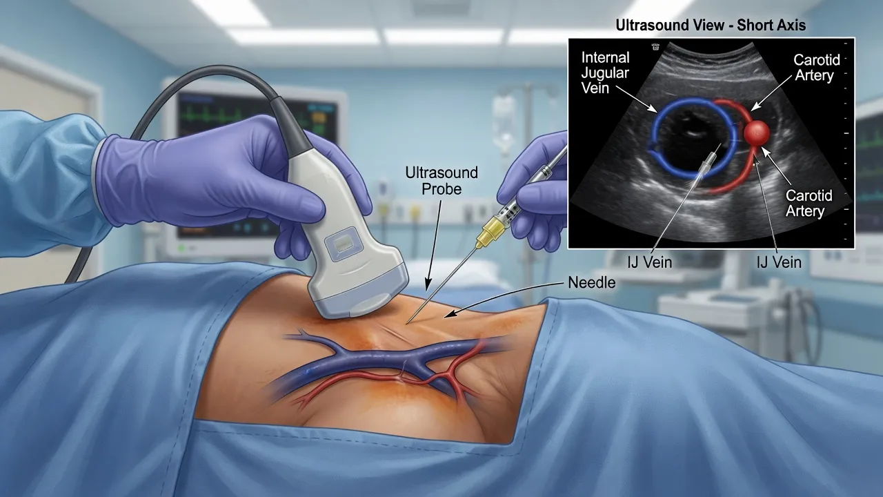
Ultrasound guidance is the standard of care for central venous catheterisation and the single evidence-based change that has improved the safety of the procedure. The Cochrane review of the internal jugular vein found that real-time two-dimensional ultrasound, compared with the landmark technique, increased the success rate, reduced the number of attempts, and reduced the rate of arterial puncture and haematoma.[5] The candidate must describe the technique: a high-frequency linear probe in a sterile cover, held transverse to the vessel to identify the vein (compressible, thin-walled) and the artery (round, pulsatile, non-compressible), then rotated to the longitudinal view to confirm needle passage within the lumen, and the wire visualised within the vein before the dilator is passed.[4][5]
The same logic now applies to the arterial line — the ultrasound-guided radial arterial cannulation improves the first-pass success rate, especially in the hypotensive or the oedematous patient in whom the pulse is impalpable.[7]
Stepwise technique — the Seldinger sequence
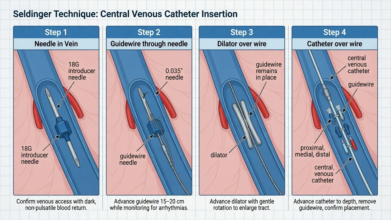
The Seldinger technique is the universal sequence for central and arterial access: needle into the vessel, guidewire through the needle, needle removed, skin nicked, dilator over the wire, dilator removed, catheter over the wire, wire removed, catheter secured. The discipline is that the wire is always controlled (one hand holds the wire at all times), the needle is never reinserted over a wire that has already passed (it can shear the wire), and the catheter is never advanced until the wire is confirmed in the vessel. [1]
The ultrasound-guided right internal jugular Seldinger insertion, in order
- Prepare and position. Head-down 15 to 30 degrees, head turned 30 to 45 degrees to the left. Full sterile barrier (cap, mask, gown, gloves, full drape). Confirm the ultrasound images the vein before draping.
- Scout the vessel. Place the covered probe transverse at the apex of the sternocleidomastoid triangle. Identify the compressible internal jugular vein lateral to the non-compressible carotid artery. Confirm compressibility and patency.
- Anaesthetise. Infiltrate 1 per cent lidocaine (maximum 3 mg/kg plain, 7 mg/kg with adrenaline) subcutaneously and along the needle tract, taking care to keep the needle out of the vessel.
- Cannulate the vein under ultrasound. Advance the 18-gauge introducer needle attached to a syringe, in-plane or out-of-plane, watching the needle tip enter the vein lumen. Aspirate dark, non-pulsatile venous blood to confirm. Transduce or float the wire to confirm.
- Pass the guidewire. Thread the J-tipped guidewire through the needle, advancing smoothly. The wire should pass with no resistance; if it catches, withdraw slightly and reorient. Never advance against resistance. Watch for ectopy on the ECG — the wire too deep irritates the right atrium; withdraw 1 to 2 cm.
- Remove the needle, holding the wire. One hand always controls the wire. Nick the skin at the wire entry with a number 11 blade.
- Dilate. Pass the dilator over the wire with a firm, controlled, rotating motion, advancing only the short distance needed to create the tract. Hold the wire at its distal end to avoid wire shearing or loss.
- Pass the catheter. Remove the dilator. Thread the catheter over the wire to the measured depth (right internal jugular about 12 to 15 cm to the cavoatrial junction). Remove the wire.
- Aspirate and flush all lumens. Confirm venous blood return. Secure the catheter with sutures or the securing device.
- Confirm. Ultrasound confirms the wire and the catheter in the vein; the post-procedure chest X-ray confirms the tip at the cavoatrial junction and excludes a pneumothorax. [1]
The subclavian and femoral routes follow the same Seldinger sequence with different puncture geometry. The subclavian is approached infraclavicularly at the junction of the medial and middle thirds of the clavicle, the needle aimed at the suprasternal notch just deep to the clavicle, with the needle kept flat to avoid the pleura. The femoral is approached two fingerbreadths below the inguinal ligament, medial to the femoral pulsation, the needle aimed cephalad at 45 degrees; ultrasound confirmation of the vein medial to the artery is now standard.[1][6]
Arterial line technique — the radial and the Allen test
The radial arterial line begins with the modified Allen test. The patient makes a fist; the operator compresses both the radial and the ulnar arteries at the wrist until the hand blanches; the patient opens the hand; the operator releases the ulnar artery. If colour returns to the palm within 5 to 15 seconds, the ulnar circulation is adequate and the radial artery can be cannulated safely; if colour returns slowly or not at all, the ulnar supply is insufficient and the radial should not be cannulated (a positive, abnormal Allen test mandates an alternative site). The Allen test is supplemented by pulse oximetry or plethysmography of the thumb during ulnar compression, and by direct visualisation with ultrasound.[7]
The ultrasound-guided radial arterial line, in order
- Position. Wrist supinated, dorsiflexed over a rolled towel, secured. Confirm the radial pulse and the modified Allen test (ulnar refill under 15 seconds).
- Scout. Place the covered high-frequency probe transverse at the wrist crease. Identify the radial artery (round, pulsatile, non-compressible) lateral to the vein.
- Anaesthetise. Infiltrate 1 per cent lidocaine (without adrenaline) subcutaneously over the puncture site, a small volume to avoid distortion.
- Cannulate. Advance the 20-gauge over-needle cannula at a 30-degree angle, in-plane, watching the tip enter the artery lumen. Watch for the flashback of bright pulsatile blood.
- Thread and secure. Once the flash is steady, lower the angle and advance the cannula 1 to 2 mm further, then slide the catheter off the needle into the artery. Remove the needle, confirm pulsatile flow, attach the transducer set, secure the cannula, and zero the transducer at the level of the heart. [1]
The femoral arterial line is the alternative when the upper limbs are unavailable or the patient is being resuscitated and the upper-body arteries are vasoconstricted. The technique mirrors the femoral venous approach but lateral to the vein (the artery is lateral, the vein medial); the ultrasound confirms the position. The femoral site is not compressible for the cannulated patient who is ambulatory, but it is compressible for haemostasis after removal, and it is the site of choice for the arterial line during cardiac arrest and the resuscitation of the shocked patient.[7]
Drug doses and local anaesthesia
Local anaesthesia is the only pharmacology of the procedure. Lidocaine 1 per cent is infiltrated subcutaneously and along the tract: the maximum dose is 3 mg/kg plain (about 20 mL for a 70 kg adult) and 7 mg/kg with adrenaline (about 50 mL for a 70 kg adult). The dose is calculated before drawing up, and the injection is aspirated to avoid intravascular delivery. For the arterial line, a small subcutaneous volume keeps the field undistorted. Sedation is rarely required for a central line in the ED; when the patient is agitated or unable to cooperate, a low-dose short-acting agent (midazolam 1 to 2 mg intravenously, fentanyl 25 to 50 mcg intravenously, titrated) is given with full monitoring, recognising that any sedation in the critically ill raises the aspiration and the airway risk.[4]
Complications
The complications divide into the immediate mechanical, the infectious, and the delayed, and each has a defined recognition and rescue.[1]
The complications and the immediate response
Pneumothorax is the pneumothorax of the apex, produced by the needle or the wire nicking the pleura. The risk is highest with the subclavian (2 to 10 per cent), lower with the internal jugular (1 to 5 per cent, lower still with ultrasound), and zero with the femoral. The recognition is the post-procedure chest X-ray, mandatory after every internal jugular or subclavian line; an apical pneumothorax may be small and occult on the immediate film, and any symptomatic patient or any patient about to receive positive-pressure ventilation must be re-evaluated, because a small occult pneumothorax can convert to a tension pneumothorax under positive pressure. The management is the standard — oxygen, observation for the small, aspiration or chest tube for the symptomatic, and the finger thoracostomy or needle decompression for the tension.[1][6]
Arterial puncture is the accidental cannulation of the adjacent artery — the carotid with the internal jugular, the subclavian with the subclavian, the femoral with the femoral. With the finder needle, the management is withdrawal and firm direct pressure for 5 to 10 minutes (the carotid and the femoral are compressible; the subclavian is not, which is one reason it is avoided in the coagulopathic). With a large-bore dilator or catheter, the rule is different: the catheter should be left in place, the patient referred to vascular surgery or interventional radiology, and the catheter removed in a controlled environment — removing it in the ED risks catastrophic bleeding or embolic stroke from dislodged thrombus. Ultrasound guidance reduces the arterial puncture rate at every site.[1][5]
Venous air embolism is rare but lethal: as little as 0.5 to 1 mL per kilogram of air can obstruct the right ventricular outflow and the pulmonary circulation. It occurs when the needle or the catheter is open to air while the intrathoracic pressure is negative — the inspiratory effort of the awake patient, or the disconnection during catheter exchange. The recognition is the sudden cough, the desaturation, the mill-wheel murmur over the precordium, and the cardiovascular collapse. The management is immediate: place the patient in the head-down left lateral position (to trap the air in the right atrium and away from the pulmonary outflow), give 100 per cent oxygen (to denitrogenate and reduce the air bubble), aspirate the air through the central line if a catheter is in place, perform cardiopulmonary resuscitation if arrest occurs, and consider hyperbaric therapy for the neurologic sequelae. The prevention is the procedural discipline — the needle is occluded at every stage, the patient is head-down, and the catheter is clamped or flushed during exchanges.[1][4]
Central-line-associated bloodstream infection (CLABSI) is the catheter contaminated by skin flora at insertion or by hub contamination during use, producing a bloodstream infection. The CDC and the Michigan Keystone intervention established that CLABSI is largely preventable through a bundle: hand hygiene before and after the procedure, full sterile barrier (cap, mask, gown, gloves, full drape), chlorhexidine skin preparation (2 per cent), avoidance of the femoral route in adults when possible, daily review of the necessity of the line with prompt removal, and the use of antimicrobial-impregnated catheters when the dwell time is long or the risk is high. The Michigan intervention reduced CLABSI by two-thirds across the ICUs studied, and the bundle is the standard the Fellowship candidate must recite.[2][3]
Arterial thrombosis and hand ischaemia complicate the radial arterial line, especially in the patient with an abnormal Allen test, a prolonged dwell, high-dose vasopressors, hypoperfusion or peripheral vascular disease. The recognition is the pale, painful, cold hand with poor capillary refill or the loss of the pulse distal to the cannula. The management is immediate removal of the catheter, assessment of the hand perfusion (Doppler, capillary refill), and vascular review if ischaemia persists. The femoral arterial line carries a lower thrombosis rate but a higher risk of retroperitoneal haemorrhage if the puncture is above the inguinal ligament.[7]
Pitfalls and practical tips
The pitfalls invert the discipline. Cannulating without ultrasound when ultrasound is available — the avoidable arterial puncture and the failed attempt. Forgetting the post-procedure chest X-ray after an upper-body line — the missed apical pneumothorax that becomes a tension pneumothorax under positive pressure. Reinserting the needle over an indwelling wire — the wire shearing that sends a fragment into the circulation. Removing a large-bore catheter from the carotid in the ED — the avoidable exsanguination or stroke. Leaving the line in longer than necessary — the preventable CLABSI. Skipping the full sterile barrier — the single most preventable infection step. Cannulating the radial artery despite an abnormal Allen test — the avoidable hand ischaemia. Failing to control the wire at all times — the lost guidewire, a surgical emergency.[1][3]
The practical tips are the mirror. Ultrasound for every central line and the radial arterial line. The post-procedure chest X-ray for every internal jugular and subclavian line. The wire always in one hand. The needle never re-advanced over a wire. The finder-needle arterial puncture managed by pressure, the large-bore by leaving the catheter in and calling for help. The full sterile barrier and the CLABSI bundle for every central line. The Allen test before every radial arterial line. The line reviewed daily and removed the moment it is no longer needed.[2][7]
Post-procedure care and disposition
After the central line is placed, the immediate checks are the ultrasound confirmation of the catheter in the vein, the aspiration and flush of every lumen, the secure fixation, and the post-procedure chest X-ray for every internal jugular and subclavian line. The chest X-ray confirms two things: the catheter tip at the cavoatrial junction (too high risks thrombosis and poor flow; too deep risks atrial arrhythmia and perforation) and the absence of a pneumothorax. The femoral line does not require a chest X-ray. The arterial line is confirmed by the pulsatile waveform on the transducer and the level set at the heart.[1][6]
The disposition is the underlying condition that required the line — the shocked patient to the intensive care unit, the long-term therapy patient to the ward. The dressing is applied, the date of insertion recorded, and the daily line review scheduled. The handover to the receiving team includes the indication, the site, the date and time of insertion, the confirmation method, and the planned review date for removal. The line is removed as soon as it is no longer required, because the CLABSI risk rises with the dwell time.[3]
Special populations
The coagulopathic patient (INR above 1.5, platelets below 50, on a direct oral anticoagulant or therapeutic heparin) is cannulated at a compressible site — the femoral or the ultrasound-guided internal jugular — never the subclavian, where a bleeding complication cannot be controlled by pressure. The coagulopathy is corrected when feasible and when it does not delay an urgent line, with the understanding that the femoral and the ultrasound-guided internal jugular are sufficiently safe even in the uncorrected coagulopathy for the patient who needs the line now.[1][4]
The obese patient needs the ultrasound for every site; the landmark technique is unreliable, the internal jugular is deeper and the femoral artery is harder to palpate. The paediatric patient is cannulated with size-appropriate catheters, the femoral route is common in the resuscitation of the sick child, and the intraosseous device is the immediate access when the peripheral or central access is delayed. The trauma patient in a cervical collar is managed with the ultrasound-guided internal jugular (in-line stabilisation maintained) or the femoral; the subclavian is avoided in the patient with a suspected thoracic or cervical injury. The shocked or arrested patient is managed with the femoral CVC, the intraosseous device, or both — the fastest access wins in the crash.[1][7]
Evidence and the regional guidelines
The contemporary framework is built on the CDC 2011 guidelines for the prevention of intravascular catheter-related infections, which codified the CLABSI bundle that the Michigan Keystone study demonstrated could reduce bloodstream infections by two-thirds.[2][3] The ASA practice guidelines for central venous access established ultrasound guidance as the standard for the internal jugular and recommended the site selection that balances the pneumothorax, the infection and the compression risk.[4] The Cochrane review of the internal jugular vein confirmed the superiority of ultrasound over the landmark technique for success, attempts and arterial puncture.[5] The ESPEN guidelines on central venous catheters set out the long-term access, the site selection and the management of the complications.[6] The contemporary arterial-line guidance confirms ultrasound as the standard for the radial cannulation, especially in the hypotensive or the oedematous patient.[7]
ANZ practice note. In Australian and New Zealand emergency departments, the central line is inserted under ultrasound guidance with the full sterile barrier and the CLABSI bundle, the right internal jugular as the first-line site, the femoral as the site for the crashing or the coagulopathic patient, and the subclavian reserved for the ambulatory long-term patient. The post-procedure chest X-ray is mandatory for every upper-body line. The arterial line is the standard for the shocked patient on a vasopressor and for the respiratory-failure patient needing serial blood gases, with the radial site first-line and the modified Allen test performed before cannulation. The intraosseous device is the immediate access in the arrested and the peri-arrest patient. The ACEM and the ANZICS promote the structured central-line training, the CLABSI bundle, and the daily line review as the quality standard. [1]
Exam pearls
- Ultrasound is the standard for the internal jugular and the radial arterial line — the Cochrane evidence for fewer attempts and fewer arterial punctures is the answer the examiner wants.
- Site selection is the recurring SAQ — internal jugular first-line, subclavian for the ambulatory long-term, femoral for the crashing and the coagulopathic; never subclavian in the coagulopathic or in the patient who cannot tolerate a pneumothorax.
- The post-procedure chest X-ray is mandatory for every internal jugular and subclavian line; the femoral line does not need one.
- The Seldinger sequence is recited step-by-step — needle, wire, needle out, skin nick, dilator, catheter — with the wire always controlled and never re-advanced over the needle.
- The CLABSI bundle is recited as the prevention answer — hand hygiene, full barrier, chlorhexidine, femoral avoided, daily review, early removal.
- The venous air embolism rescue is head-down left lateral, 100 per cent oxygen, aspirate via the line — the rare complication with a defined answer.
- The large-bore carotid catheter is left in and the vascular or the interventional radiology team is called — the one situation where removing the line is the wrong move.
- The Allen test precedes every radial arterial line; an abnormal test moves the site to the femoral. [1]
SAQs — exam practice
SAQ — Ultrasound-guided right internal jugular central line in septic shock
10 minutes · 10 marks
A 64-year-old man with septic shock from a urinary source has a MAP of 60 mmHg despite 2 L of crystalloid, and requires a noradrenaline infusion for ongoing hypotension. Peripheral access is poor. You elect to insert a right internal jugular central venous catheter under ultrasound guidance.
SAQ — Arterial line insertion: the complications and the hand that has gone cold
10 minutes · 10 marks
A 68-year-old woman on a noradrenaline infusion for septic shock has a left radial arterial line inserted without difficulty. Two hours later the nurse notes that the left hand is pale, cold and painful with sluggish capillary refill of 5 seconds. The right radial pulse and the right hand are normal.
Red flags
[1]References
- [1]McGee DC, Gould MK. Preventing complications of central venous catheterization N Engl J Med, 2003.PMID 12646670
- [2]Pronovost P, Needham D, Berenholtz S, et al. An intervention to decrease catheter-related bloodstream infections in the ICU N Engl J Med, 2006.PMID 17192537
- [3]O'Grady NP, Alexander M, Burns LA, et al.; Healthcare Infection Control Practices Advisory Committee. Guidelines for the prevention of intravascular catheter-related infections Clin Infect Dis, 2011.PMID 21460264
- [4]American Society of Anesthesiologists Task Force on Central Venous Access. Practice guidelines for central venous access: a report by the American Society of Anesthesiologists Task Force on Central Venous Access Anesthesiology, 2012.PMID 22307320
- [5]Brass P, Hellmich M, Kolodziej L, Schick G, Smith AF. Ultrasound guidance versus anatomical landmarks for internal jugular vein catheterization Cochrane Database Syst Rev, 2015.PMID 25575244
- [6]Pittiruti M, Hamilton H, Biffi R, MacFie J, Pertkiewicz M; ESPEN. ESPEN Guidelines on Parenteral Nutrition: central venous catheters (access, care, diagnosis and therapy of complications) Clin Nutr, 2009.PMID 19464090
- [7]Wang A, Rennie S, Margetts S, Arntfield R. Better With Ultrasound: Arterial Line Placement Chest, 2020.PMID 31634448