EM · Procedural & diagnostic ED skills
Procedural sedation in the emergency department
Also known as Procedural sedation and analgesia · PSA · Conscious sedation · Dissociative sedation · ED sedation
Procedural sedation in the ED — the indications (fracture reduction, dislocation reduction, abscess drainage, cardioversion, paediatric procedures), the pre-sedation assessment (fasting status, ASA physical status, the airway, the Mallampati), the monitoring (ECG, pulse oximetry, capnography, BP), the drugs (propofol 1 to 2 mg/kg, ketamine 1 to 2 mg/kg IV or 4 to 5 mg/kg IM, midazolam 0.05 to 0.1 mg/kg, fentanyl 1 to 2 mcg/kg), the antagonists (flumazenil 200 mcg, naloxone 400 mcg), the rescue airway, and the complications (hypoventilation, hypotension, hypoxia, emesis, laryngospasm). ACEM-primary, globally tagged.
On this page & tools
Your progress
Saved locally on this device.
8 MCQs with explanations
Target exams
Red flags
Related topics
- The primary survey (ABCDE) — the trauma assessment framework
- Upper airway obstruction in the emergency department
- Paediatric trauma — the modified approach
- Acute severe asthma
- Tachyarrhythmias in the emergency department
- Bradyarrhythmias and atrioventricular block in the emergency department
- Wound assessment and management
Procedural sedation and analgesia (PSA) in the emergency department is the controlled, intentional depression of consciousness so that a painful or distressing procedure can be performed, while the patient's airway reflexes, spontaneous ventilation and haemodynamics are preserved to the greatest extent possible. It sits between the conscious, oriented patient and general anaesthesia, and the Fellowship candidate must treat it with anaesthetic-grade discipline: a structured pre-sedation assessment, the right monitoring (capnography is the standard of care), a defined drug plan with the antagonists drawn up, and a rescue airway within arm's reach. The two questions an examiner will press are whether the patient needs sedation at all, and whether the sedationist can rescue the airway if the drug goes deeper than intended.[1][3]
Definition and the targets of sedation
Procedural sedation delivers analgesia (the relief of pain), anxiolysis (the relief of fear) and sedation (the lowering of awareness) in a single, titrated encounter so that a short, painful procedure can be completed safely. The American Society of Anesthesiologists defines four depths — minimal sedation (anxiolysis, the patient responds to voice), moderate sedation (the patient responds to purposeful touch or verbal stimulus, the airway and ventilation are maintained), deep sedation (the patient responds only to repeated or painful stimulus, the airway may need support, spontaneous ventilation may be inadequate) and dissociative sedation (a cataleptic state induced by ketamine in which the airway reflexes, tone and spontaneous breathing are preserved).[4] The ED target for most reductions and cardioversions is deep or dissociative sedation; the candidate must recognise that depth is a continuum, that every agent can cross unexpectedly from moderate to deep, and that the team must be equipped to manage the deepest end of that continuum regardless of the depth intended.
Indications — when to sedate
The indications are any short, painful or distressing ED procedure for which analgesia alone is inadequate. The common scenarios are the fracture reduction (the paediatric distal forearm, the displaced Colles, the angulated tibia-fibula), the joint dislocation reduction (the anterior shoulder, the prosthetic or native hip, the patella, the temporomandibular joint), the abscess incision and drainage (especially the large, deep or loculated abscess, or any abscess in a child who cannot cooperate), the electrical or pharmacological cardioversion of the unstable tachyarrhythmia, the pacemaker or defibrillator placement, and the paediatric procedures — the facial laceration repair, the foreign body removal, the burn dressing change, the lumbar puncture and the orthopaedic manipulation. Thoracostomy and chest-tube insertion in the awake patient, dental and ophthalmic procedures, and the reduction of a paraphimosis or a priapism are further indications. The unifying test is that the procedure is brief, the pain is intense, and the patient cannot tolerate it awake.[1]
Contraindications
There are few absolute contraindications to ED procedural sedation: the haemodynamically unstable patient who needs the procedure (who should instead be managed in theatre or with anaesthetic support), the patient who cannot protect the airway or who needs a definitive airway regardless, the known allergy to the chosen agent, and the patient whose airway is so difficult that an unanticipated deep plane would be unrecoverable in the ED. The relative contraindications are the recognised predictors of adverse events — a high ASA class (III with caution, IV and above with anaesthetic involvement), a difficult airway (the Mallampati III or IV, the short thyromental distance, the limited mouth opening, the obesity, the obstructive sleep apnoea), a recent meal (a full stomach, which raises the aspiration risk but must not delay an urgent procedure), the severe chronic obstructive or restrictive lung disease, the upper respiratory tract infection in a child receiving ketamine (a predictor of laryngospasm), and the elderly or the haemodynamically compromised patient for whom propofol is poorly tolerated. The decision is always a risk-benefit judgement: urgency overrides the relative contraindication, with the airway managed as for a full stomach.[1]
Differential diagnosis — does this patient need sedation at all?
The first differential in procedural sedation is not a disease but a decision: the patient who appears to need sedation may be better served by analgesia alone, which carries no airway risk. The Fellowship candidate must justify sedation against the alternatives before reaching for a syringe. [1]
Procedural sedation (deep or dissociative)
- For the brief, intensely painful, non-tolerable procedure
- Needs the full monitoring set and the rescue airway
- Risk of hypoventilation, hypotension, hypoxia, emesis, laryngospasm
- The patient must be fasted, ASA-assessed and airway-assessed
Regional anaesthesia (nerve blocks)
- The fascia iliaca block for the hip or femoral shaft fracture; the forearm blocks for hand procedures
- Analgesia and motor block without central depression
- The airway, ventilation and consciousness are fully preserved
- The preferred answer when a block fits the procedure
Local anaesthesia (with or without opioid)
- For the small abscess, the simple laceration, the wound exploration
- No airway risk; titrate to the local-anaesthetic maximum dose
- May be inadequate for the large, deep or loculated abscess
- Combine with nitrous oxide or a sub-dissociative ketamine infusion to extend coverage
Nitrous oxide (Entonox, 50:50)
- Self-administered, mild analgesia with anxiolysis, offset within minutes
- Useful alone for the brief dressing change or as an adjunct
- Contraindicated in the pneumothorax and the bowel obstruction (a closed gas space)
- Inadequate as a sole agent for the fracture or the dislocation reduction
Sub-dissociative ketamine (0.1 to 0.3 mg/kg IV)
- Analgesia via the NMDA receptor below the dissociative threshold
- Useful for the painful dressing change or the transport of the burns patient
- Airway reflexes preserved; less emergence than the dissociative dose
- A bridge between pure analgesia and full procedural sedation
The key distinction the examiner rewards is sedation versus analgesia: regional blocks, local infiltration, nitrous and sub-dissociative ketamine can often achieve the goal without ever depressing the central nervous system, and when they can, they are the safer answer. [1]
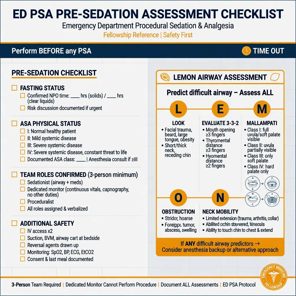
The pre-sedation assessment
The pre-sedation assessment is the single most examinable block of the topic, and it is built from four elements — the fasting status, the ASA physical status, the airway assessment and the focused history and examination. [1]
The ASA physical status classification
The airway assessment screens for the patient in whom an unanticipated deep plane would be unrecoverable. The Mallampati class is scored with the mouth open and the tongue protruded: class I (the full palate, uvula and tonsillar pillars visible), class II (the uvula visible), class III (only the soft and hard palate visible) and class IV (only the hard palate visible) — class III and IV flag a difficult airway. This is combined into the LEMON assessment: Look externally (the receding mandible, the beard, the enlarged tongue), Evaluate the 3-3-2 (mouth opening, hyoid-to-chin and hyoid-to-thyroid distances each of three fingerbreadths), Mallampati, Obstruction (the stridor, the infection, the tumour) and Neck mobility. The focused history captures the obstructive sleep apnoea, the previous anaesthetic difficulty, the drug allergy, the comorbidity and the current medications (the anticoagulant that complicates any airway intervention), and the examination confirms the oxygenation, the blood pressure and the focused system relevant to the procedure. [1]
LEMON — the difficult-airway screen
LEMON
Receding mandible, beard, large tongue, abnormal facies
Mouth opening, hyoid–mentum, hyoid–thyroid (3 fingerbreadths each)
Class III or IV flags a difficult airway
Stridor, infection, tumour, foreign body
Limited extension (cervical collar, arthritis)
Personnel, the equipment and the rescue airway
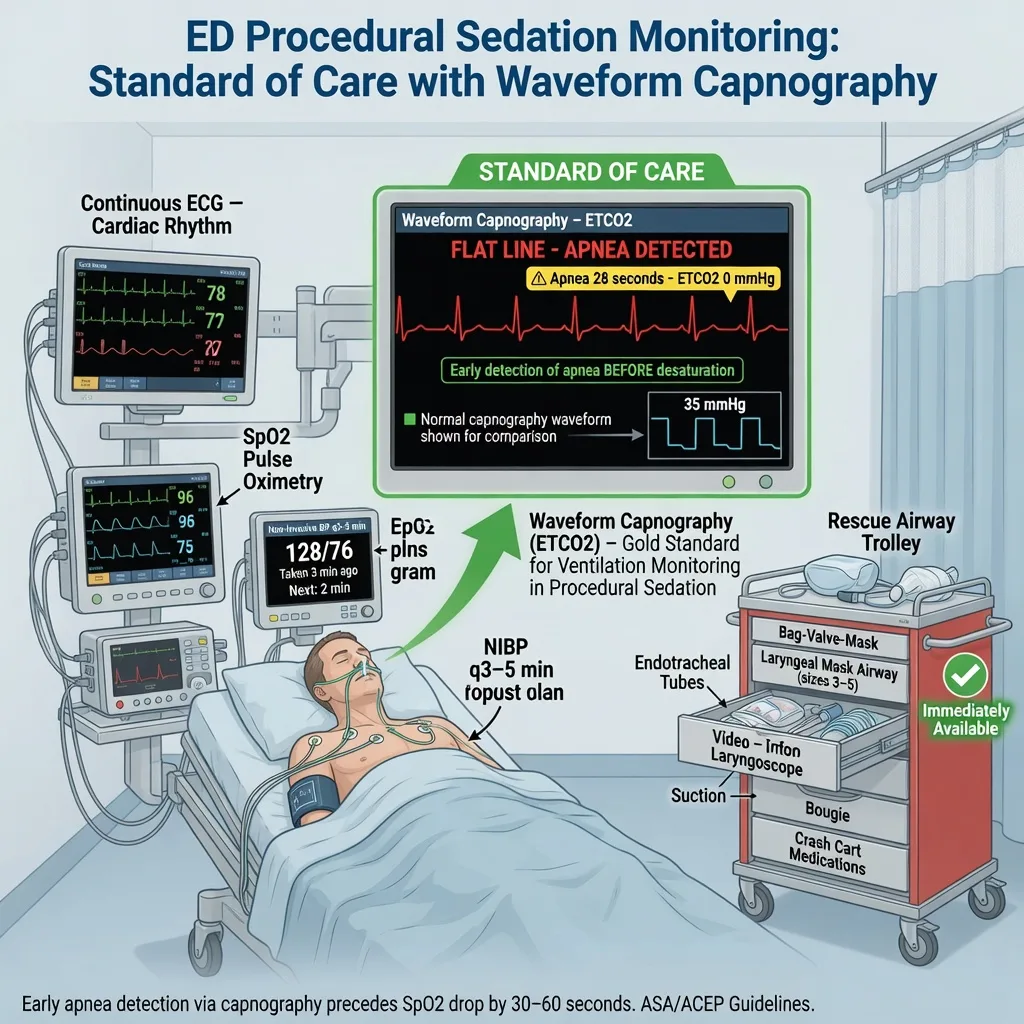
Safe procedural sedation needs the right people, the right equipment and a rescue airway within arm's reach. The personnel are the sedationist (the clinician who chooses and titrates the drug and watches the depth), the dedicated monitor (a second clinician whose only task is to watch the patient, the airway and the monitors, and who does not assist the procedure), and the proceduralist (the clinician who performs the intervention) — the three-person team, all trained in advanced life support and airway rescue. The equipment is the suction (on and within reach), the high-flow oxygen with a bag-valve-mask and the oral and nasal airways, the full monitoring (continuous ECG, pulse oximetry and capnography, with the non-invasive blood pressure cycling every three to five minutes), the resuscitation drugs including the drawn-up reversal agents, and the rescue airway trolley — the supraglottic airway, the laryngoscope and the endotracheal tubes, and the surgical airway set. The non-negotiable rule is that the team must be able to rescue the airway if the intended moderate sedation drifts to deep, and to intubate if it drifts to apnoea. [1]
Monitoring — the four parameters and why capnography is the standard
The four monitored parameters are the continuous ECG (essential for the cardioversion, and a baseline for all), the continuous pulse oximetry (the oxygenation), the waveform capnography (the ventilation) and the non-invasive blood pressure cycled every three to five minutes (the haemodynamics), supplemented by the continuous level of consciousness. Capnography is the standard of care and the single most examinable monitor: it displays the end-tidal CO2 and its waveform, and it detects apnoea and hypoventilation within a single breath — minutes before the pulse oximetry falls, and especially on supplemental oxygen, where the saturation can remain falsely reassuring while the patient stops breathing.[3][4] The capnography flags the partial airway obstruction (the rising, skewed waveform), the complete apnoea (the flat line) and the hypoventilation (the rising end-tidal value), and it gives the team the early window to intervene with jaw thrust, airway opening or ventilation before desaturation occurs. The pulse oximetry is a late and lagging signal in the sedated patient on oxygen; relying on it alone is the classic error.
The monitoring minimum
The pharmacology and the drug doses
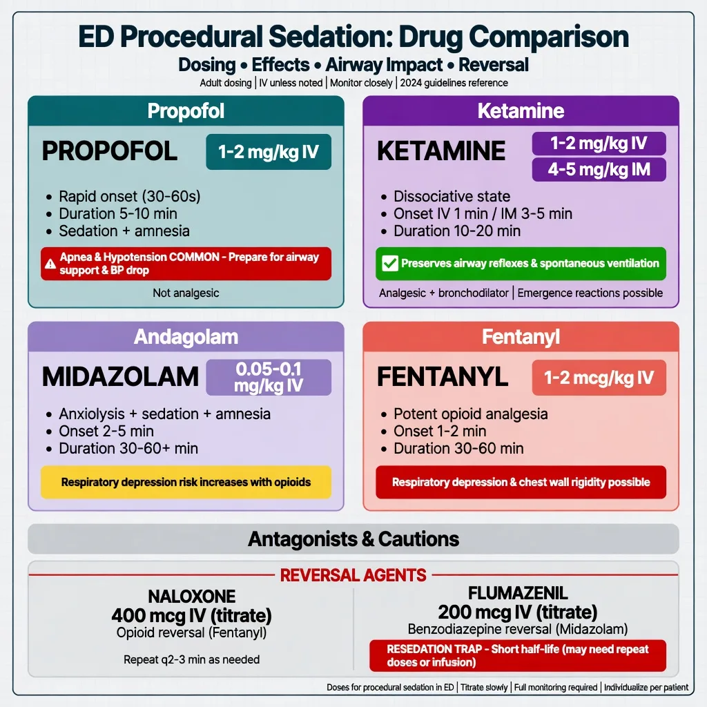
The pharmacology is built around four agents and their combinations, each chosen for the procedure, the patient and the desired depth. The doses are stated precisely and titrated in aliquots to the effect. [1]
The ED procedural-sedation drugs
Propofol (1 to 2 mg/kg intravenously, in aliquots of 0.5 to 1 mg/kg, half the dose in the elderly and the haemodynamically compromised) is the agent for the brief, intensely painful procedure that needs deep sedation — the shoulder reduction, the cardioversion. Its onset is within 30 to 60 seconds and its offset within 5 to 10 minutes, which makes it ideal for the short reduction. Its penalties are the apnoea (it blunts ventilation in a dose-related way), the hypotension (it is a vasodilator and a myocardial depressant, poorly tolerated in the hypovolaemic), the pain on injection and the lack of analgesia — it is almost always paired with a low-dose opioid (fentanyl 0.5 to 1 mcg/kg) so that the propofol dose can be kept low.[3]
Ketamine (1 to 2 mg/kg intravenously, or 4 to 5 mg/kg intramuscularly when no intravenous access can be secured, as is common in the frightened child) is a dissociative agent that produces a cataleptic state while preserving the airway reflexes, the muscle tone, the blood pressure and the spontaneous ventilation — the safest profile in the child and in the haemodynamically unstable patient. Its onset is 1 minute intravenously and 5 minutes intramuscularly, with offset over 10 to 20 minutes. Its penalties are the emesis (reduced by an antiemetic such as ondansetron), the hypersalivation, the laryngospasm (rare but most common in the young child with a recent upper respiratory tract infection or asthma), and the emergence phenomena (the confusion, the nightmares and the hallucinations, more frequent in the adult). An antisialogogue (glycopyrrolate) and an antiemetic are reasonable adjuncts.[1][2]
Midazolam (0.05 to 0.1 mg/kg intravenously, titrated in 0.5 to 1 mg aliquots in the adult) provides anxiolysis, sedation and amnesia with a slower onset and a longer offset (30 to 60 minutes) than propofol. It is reversible with flumazenil but its cumulative effect, its synergy with opioids and its paradoxical agitation in the child make it a second-line ED agent. Fentanyl (1 to 2 mcg/kg intravenously, titrated in 25 to 50 mcg aliquots) is the analgesic partner, with a short offset; rapid high doses cause the chest-wall rigidity. The combinations to know are ketofol (ketamine and propofol in a 1:1 mixture, which reduces the emesis and the hypotension of each alone) and the dangerous trio — the benzodiazepine plus the opioid plus the propofol — whose respiratory synergy is respiratory arrest when given carelessly together. [1]
The antagonists — the reversal agents
The reversal agents are drawn up and ready before the first aliquot, because their role is to rescue the over-sedated patient. Naloxone (400 mcg intravenously, titrated in 40 to 100 mcg aliquots to the respiratory rate, not to the full consciousness — the target is a respiratory rate above 10 to 12 per minute, which avoids the acute withdrawal, the agitation and the pulmonary oedema of the over-titrated reversal) reverses the opioids; it may be given intramuscularly if no access exists. Its half-life (30 to 80 minutes) is shorter than most of the opioids it reverses, and the patient who wakes will resedate — the trap is the apparent recovery followed by the recurrent apnoea, so the observation must extend beyond the wake-up.[1]
Flumazenil (200 mcg intravenously, repeated every 1 to 2 minutes to a maximum of 1 mg in the acute overdose, or 3 mg over the encounter) reverses the benzodiazepines. Its danger is the seizure: in the chronic benzodiazepine user, the mixed overdose and the patient with a proconvulsant co-ingestant, flumazenil can precipitate a refractory seizure, and it is avoided in those settings. Like naloxone, its half-life (40 to 80 minutes) is shorter than the midazolam it reverses, and resedation is the trap. The clinical rule for both is that the reversal buys time and confirms the diagnosis, but the patient must be observed for the resedation window, not discharged at the wake-up. [1]
Stepwise technique — the structured encounter
The encounter is run to a checklist so that no element is missed under time pressure. [1]
The procedural-sedation checklist, in order
- Assess and consent — the indication, the ASA class, the airway (Mallampati, LEMON), the fasting, the focused history; explain the procedure, the sedation and the risks, and obtain consent (or document the emergency exception).
- Assemble the team — the sedationist, the dedicated monitor, the proceduralist; all trained in airway rescue.
- Prepare the equipment — suction on, oxygen and bag-valve-mask, oral and nasal airways, the full monitoring (ECG, SpO2, capnography, NIBP), the drawn-up reversal agents, the rescue airway trolley.
- Secure intravenous access and preoxygenase — 100 per cent oxygen for three minutes, building the apnoeic reserve.
- Administer the agent in aliquots — titrate to the desired depth; never bolus the full calculated dose; combine an analgesic (fentanyl or ketamine) with a sedative when the procedure is painful.
- Perform the procedure once the depth is adequate; the dedicated monitor watches the airway and the capnography throughout.
- Recover — continuous monitoring until the patient returns to the baseline mental status, with the resedation window respected for any reversal given. [1]
The two structural errors are to allow the dedicated monitor to assist the procedure (the airway is then unwatched) and to bolus the full calculated dose (the patient who needs the lower dose is over-sedated). Titration to effect is the rule. [1]
Complications
The complications are the predictable consequences of central depression, and each has a defined rescue. [1]
Hypoventilation and apnoea are the commonest complications, most frequent with propofol and with the opioid-benzodiazepine combinations. The capnography shows the falling end-tidal and the slowing rate before the saturation falls; the rescue is the airway-opening manoeuvre (the chin lift, the jaw thrust, the oral or nasal airway), the bag-valve-mask ventilation, and the reversal agent if the depression is persistent or the agent is long-acting. Hypoxia is the downstream consequence of the hypoventilation, and it is prevented by the preoxygenation and the supplemental oxygen and treated by supporting the ventilation. Hypotension follows the propofol and the midazolam (the vasodilation and the myocardial depression); it is treated by the intravenous fluid, the reduced further dose, and the vasopressor in the refractory case — ketamine is the preferred agent when the blood pressure is already precarious. Emesis and aspiration complicate the ketamine sedation and the full stomach; the prevention is the left lateral position and the suction, and the management is the airway protection and the suction if it occurs. Laryngospasm is the rarest but the most feared complication of ketamine, seen in the young child with a recent upper respiratory infection or asthma; it presents as the inability to ventilate against a closed glottis, and the rescue is the 100 per cent oxygen, the jaw thrust, the Larson manoeuvre (the firm bilateral pressure at the laryngospasm notch, behind the ear lobe), the positive-pressure ventilation, and a paralysing dose of succinylcholine (1 to 2 mg/kg intravenously, or 4 mg/kg intramuscularly) if it is refractory.[1]
Pitfalls and the practical tips
The pitfalls are the inverse of the structure. Skipping the airway assessment — the difficult airway that becomes an unrecoverable airway when the drug goes deep. Relying on the pulse oximetry alone — the late, lagging signal that misses the apnoea the capnography would have shown. Co-administering the benzodiazepine, the opioid and the propofol without titration — the synergy that ends in respiratory arrest. Bolusing the full calculated dose — the over-sedation of the patient who needed the lower dose. Forgetting the resedation window — the reversal agent that wears off before the reversed drug. Under-dosing the analgesia — the patient who is sedated but still in pain, who moves through the procedure and is harder to sedate safely. Sedating without a rescue airway — the team that cannot intubate when it must. Using ketamine in the adult without warning of the emergence phenomena — the distressing recovery that a quiet environment and a small benzodiazepine dose can blunt. The practical tips are the opposite: capnography on every patient, a drawn-up reversal for every opioid and benzodiazepine, an analgesic partner for every painful procedure, the dedicated monitor watching only the airway, and the rescue airway trolley in the bay. [1]
Post-sedation recovery and the disposition
The recovery continues the monitoring until the patient returns to the baseline mental status, with the vital signs stable and the airway unprotected (the patient can maintain it). The structured recovery uses a discharge score (the modified Aldrete or the post-anaesthesia recovery score), and the criteria are the return to the baseline consciousness, the stable vital signs, the absence of the hypoxia and the airway compromise, and — for the discharge home — the ability to tolerate oral fluids, the safe ambulation, and a competent adult escort. The observation time depends on the agent: the propofol recovery is short (the offset is 5 to 10 minutes, and the discharge can follow within 30 to 60 minutes of the baseline return), while the ketamine recovery is longer (the offset is 10 to 20 minutes, and the emergence phenomena may extend the observation). The reversal agents extend the observation window — the resedation risk mandates that the patient is observed for at least two hours beyond the wake-up. The disposition is the discharge home with the escort and the written instructions, the ward admission for the patient with the residual sedation or the comorbidity, or the higher level of care for the patient with the airway or haemodynamic compromise that has not resolved. [1]
Special populations
The child is the population in whom ketamine is the first-line agent — it preserves the airway reflexes, the tone and the haemodynamics, and the intramuscular route (4 to 5 mg/kg) solves the problem of the frightened child with no intravenous access. The weight-based dosing, the antisialogogue and the antiemetic considerations, the recognition that the young child with a recent upper respiratory infection is at the higher laryngospasm risk, and the staffing by clinicians experienced in the paediatric airway are the modifications.[1][2] The elderly patient needs the reduced doses (the propofol at 0.5 to 1 mg/kg), carries the higher ASA class, the polypharmacy and the slower recovery, and is the population in whom the propofol-induced hypotension is the least tolerated. The haemodynamically unstable patient is the one in whom propofol is avoided and ketamine is preferred, and in whom the anaesthetic support is sought early. The pregnant patient is sedated in the left lateral position with the fetal monitoring as indicated, with ketamine as the preferred agent and the avoidance of the benzodiazepine in the first trimester. The chronic obstructive lung disease patient and the obstructive sleep apnoea patient are the ones in whom the hypoventilation and the post-obstructive recovery are anticipated, and in whom the capnography and the preoxygenation are the most important.
Evidence and the regional guidelines
The contemporary framework is built on the ACEM policy on procedural sedation (ANZ), the ACEP clinical policy on procedural sedation and analgesia (US), and the ASA standards, all of which mandate the structured pre-sedation assessment, the dedicated monitoring with capnography, the trained team and the rescue capability. The ketamine safety data come from the large individual-patient meta-analyses that defined the predictors of the airway and respiratory adverse events and of the emesis and recovery agitation.[1][2] The capnography evidence is built on the studies that demonstrated the earlier detection of the apnoea and the subclinical hypoventilation during the propofol sedation.[3] The current reviews reinforce capnography as the vital sign that closes the gap between the oximetry and the true ventilation in the sedated child.[4]
ANZ practice note. The ACEM policy on procedural sedation defines the minimum: a structured pre-sedation assessment (the indication, the fasting, the ASA class and the airway), the trained team of three (the sedationist, the dedicated monitor and the proceduralist), the continuous monitoring with the waveform capnography on every patient, the drawn-up reversal agents, and the rescue airway capability in the bay. The capnography is the standard, the NPO status does not delay the urgent procedure, and the ketamine is the first-line agent in the child. [1]
Exam pearls
- Capnography is the standard of care — it detects apnoea breath-by-breath, before the oximetry falls; the pulse oximetry alone is the classic error.
- NPO status does not delay the urgent procedure — the urgency determines the timing, and a full stomach shifts the airway plan, not the decision to sedate.
- Never co-administer the benzodiazepine, the opioid and the propofol without titration — the respiratory synergy is the commonest cause of the arrest.
- Ketamine preserves the airway and the blood pressure — the first line in the child and the unstable; its penalties are the emesis, the laryngospasm and the emergence.
- Propofol causes the apnoea and the hypotension — ideal for the brief deep sedation, poorly tolerated in the hypovolaemic and the elderly.
- The reversal agents wear off before the reversed drugs — the resedation window is the trap; observe beyond the wake-up.
- Laryngospasm under ketamine — the 100 per cent oxygen, the jaw thrust, the Larson manoeuvre, the positive pressure, and the succinylcholine if refractory.
- Titrate in aliquots to the effect, never bolus the calculated dose — the patient who needed the lower dose is the one who arrests. [1]
Exam practice
SAQ — Ketamine dissociative sedation for a paediatric forearm fracture reduction
10 minutes · 10 marks
A 4-year-old, 16 kg boy presents to the emergency department 90 minutes after a fall onto an outstretched hand. He has a displaced, angulated distal-third both-bone forearm fracture with impending skin compromise. He is distressed and agitated, intravenous access cannot be secured after two attempts, and he last ate a sandwich 2 hours ago. The decision is made to perform a closed reduction under procedural sedation in the resuscitation bay.
SAQ — Capnography-detected apnoea during propofol sedation for a shoulder reduction
10 minutes · 10 marks
A 62-year-old, 80 kg man requires closed reduction of an anterior shoulder dislocation. He has been fasting for 6 hours, is ASA class II with well-controlled hypertension, and gives consent. Preoxygenation is applied; full monitoring is attached including the waveform capnography. Propofol 1 mg/kg is given, followed by a further 0.5 mg/kg, and the proceduralist begins the reduction. Two minutes later the capnography waveform flattens to a straight line, but the SpO2 remains at 99 per cent on the supplemental oxygen and the blood pressure is 118/72.
Red flags
[1]References
- [1]Green SM, Roback MG, Krauss B, et al. Predictors of airway and respiratory adverse events with ketamine sedation in the emergency department: an individual-patient data meta-analysis of 8,282 children Ann Emerg Med, 2009.PMID 19201064
- [2]Green SM, Roback MG, Krauss B, et al. Predictors of emesis and recovery agitation with emergency department ketamine sedation: an individual-patient data meta-analysis of 8,282 children Ann Emerg Med, 2009.PMID 19501426
- [3]Deitch K, Miner J, Chudnofsky CR, Dominici P, Latta D. Does end tidal CO2 monitoring during emergency department procedural sedation and analgesia with propofol decrease the incidence of hypoxic events? A randomized, controlled trial Ann Emerg Med, 2010.PMID 19783324
- [4]Rath S, Nimbalkar A, Phatak A, Desai M, Nimbalkar S. Seeing beyond the numbers: capnography as a vital tool in pediatric emergency care Int J Emerg Med, 2026.PMID 41507776