EM · Electrolyte emergencies — potassium and sodium
Electrolyte emergencies — potassium and sodium
Also known as Hyperkalaemia · Hyponatraemia · Osmotic demyelination syndrome · Hypokalaemia · Hypernatraemia · Diabetes insipidus
Electrolyte emergencies — potassium and sodium. Hyperkalaemia (a serum potassium over 6.5 mmol/L, or any level with ECG change) is a time-critical arrhythmia risk treated up an antidote ladder of membrane stabilisation (calcium chloride 10 mL of 10 per cent IV), potassium shift (insulin 10 units with 50 mL of 50 per cent dextrose IV, salbutamol 10 to 20 mg nebulised, sodium bicarbonate 50 mL of 8.4 per cent IV) and potassium removal (patiromer, sodium zirconium, dialysis). Hypokalaemia (potassium under 3.5 mmol/L) shows U waves and a prolonged QU interval, precipitates torsades de pointes and potentiates digoxin toxicity, and is replaced intravenously at no faster than 10 mmol per hour peripherally and 20 mmol per hour centrally, with the magnesium corrected first. Hyponatraemia (sodium under 125 mmol/L) splits into acute — cerebral oedema and seizure, treated with 3 per cent hypertonic saline 100 mL IV bolus — and chronic, corrected slowly (under 8 mmol/L in 24 hours to avoid osmotic demyelination). Hypernatraemia (sodium over 145 mmol/L) is always a water-deficit problem, corrected at no faster than 0.5 mmol/L per hour with free water (oral or 5 per cent dextrose) and desmopressin for central diabetes insipidus. The ECG changes of hyperkalaemia (peaked T waves, widened QRS, sine wave, VF arrest), the pseudohyperkalaemia trap, and the corrected sodium for hyperglycaemia. ACEM-primary, globally tagged.
On this page & tools
Your progress
Saved locally on this device.
5 MCQs with explanations
Target exams
Red flags
Electrolyte emergencies are the two disturbances that kill the patient between the blood-gas machine and the bed — hyperkalaemia through ventricular arrhythmia, and hyponatraemia through cerebral oedema or its over-correction. The Fellowship candidate must read the ECG and the sodium, treat the membrane and the brain, and respect the one rule that most candidates forget: the speed of correction is the disease. Hyperkalaemia is corrected within minutes; chronic hyponatraemia is corrected over days. The candidate who treats both as the same emergency fails the station and harms the patient. This topic covers the antidote ladder for hyperkalaemia, the hypertonic-saline and correction-rate logic for hyponatraemia, the ECG progression, the pseudohyperkalaemia trap, and the osmotic-demyelination pitfall.[1][2][3][4]
Definition and classification
Hyperkalaemia is a serum potassium concentration above the normal range, graded by severity because the grade drives the urgency. Mild hyperkalaemia is 5.5 to 5.9 mmol per litre, moderate is 6.0 to 6.4, and severe is 6.5 mmol per litre or above — or any level with ECG change. The distinction between moderate and severe matters less than the ECG: a patient with a potassium of 6.0 and a widened QRS is at the same risk of arrest as one with a potassium of 7.5, and both are treated identically with calcium first. [1]
Hyponatraemia is a serum sodium below 135 mmol per litre, and it is graded by both the absolute level and the duration of onset. Moderate hyponatraemia is 125 to 130 mmol per litre; severe/profound is below 125 mmol per litre. The more important split is by the time course: acute hyponatraemia develops within 48 hours (the marathon runner, the post-operative patient on hypotonic fluids, the MDMA user with polydipsia), while chronic hyponatraemia has been present for longer than 48 hours and the brain has had time to adapt by extruding osmolytes. This single distinction decides whether to correct fast or slow.[3][4]
Severity thresholds
Epidemiology and risk
Hyperkalaemia is common in the emergency department, seen in roughly 1 to 3 per cent of all presentations and in up to 10 per cent of those with acute kidney injury or advanced chronic kidney disease. The risk is concentrated in the renal patient, the heart-failure patient on a renin–angiotensin blocker and a potassium-sparing diuretic, and the diabetic with ketoacidosis. Mortality is high when severe: an unrecognised potassium over 6.5 with ECG change can degenerate to ventricular fibrillation within minutes, and hyperkalaemia is one of the reversible causes of cardiac arrest (the fifth H). [1]
Hyponatraemia is the commonest electrolyte disturbance in hospitalised patients, found in 15 to 30 per cent of inpatients. The emergency presentation is the symptomatic case — seizure, confusion, coma — and the risk is bimodal: the young endurance athlete and the MDMA user with acute water intoxication, and the elderly woman on a thiazide diuretic or after surgery with hypotonic fluids. Mortality from acute symptomatic hyponatraemia can reach 30 to 50 per cent from cerebral herniation, while the morbidity of over-correction (osmotic demyelination) is permanent and devastating. [1]
Pathophysiology — the membrane and the brain
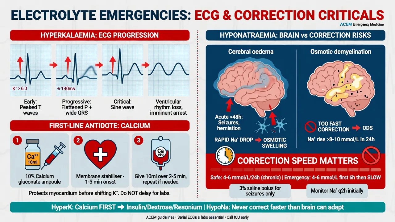
Potassium governs the resting membrane potential of the cardiac myocyte. The Nernst equation gives the equilibrium potential for potassium as approximately negative 90 millivolts, and because the resting membrane sits close to this value, a rise in extracellular potassium depolarises the membrane (makes it less negative). The consequence is biphasic and is the key to the ECG progression: an initial increase in excitability produces the peaked T waves and a shortened QT, but as the potassium rises further the sustained depolarisation inactivates the fast sodium channels, slowing conduction through the atria, the atrioventricular node and the ventricles — PR prolongation, loss of P waves, QRS widening, and finally a sine wave that degenerates into ventricular fibrillation or asystole. Calcium does not lower the potassium; it raises the threshold potential, restoring the gap between resting and threshold potentials and so restoring membrane excitability — which is why calcium is given first, before anything that shifts the potassium.[1][2]
[1]Sodium governs serum osmolality, and the brain sits inside a fixed box. In acute hyponatraemia the fall in extracellular osmolality pulls water into the brain faster than astrocytes can extrude it; cerebral oedema builds, intracranial pressure rises, and the patient herniates. In chronic hyponatraemia the brain has adapted: over 48 hours astrocytes extrude organic osmolytes (myo-inositol, glutamate, taurine) and shrink back toward normal volume, so the patient tolerates a sodium that would kill an acute case. The danger of rapid correction now reverses: if the sodium is raised too quickly, the adapted, osmolyte-depleted brain cannot regain its osmolytes fast enough, water is pulled out, and astrocytes undergo apoptosis — the osmotic demyelination syndrome, classically in the pons, presenting days later with quadriparesis, pseudobulbar palsy and a locked-in state. This is why the duration of onset dictates the correction rate.[3][4]
Clinical presentation
Hyperkalaemia is often clinically silent until it arrests the patient, which is why the ECG and the blood gas are the presentation, not the history. When symptoms exist they are non-specific — muscle weakness, paraesthesia, and a heavy feeling in the chest. The history that matters is the cause: renal failure, heart failure on a renin–angiotensin blocker or potassium-sparing diuretic, newly diagnosed diabetes with acidosis, a tissue-breakdown state (rhabdomyolysis, tumour lysis, burns), Addisonian crisis, or a recently taken trimethoprim or heparin course. [1]
Hyponatraemia presents with cerebral dysfunction: headache, nausea and malaise at the milder end; confusion, agitation and falls in the middle; and seizure and coma at the severe end. The acute case presents dramatically — a collapsed marathon runner, a post-operative patient seizing — while the chronic case presents subtly with falls, confusion or a seizure on a background of thiazide use, heart failure, cirrhosis or SIADH. The onset time, the volume status (dry, euvolaemic, or oedematous) and the recent fluid and drug history are the three questions that frame the management. [1]
Differential diagnosis
The differentials here are not rival diseases but rival mechanisms, and missing the right one leads to the wrong treatment — dialysing a haemolysed sample, or rapidly correcting a chronic sodium. [1]
True hyperkalaemia
- Renal failure (AKI/CKD), RAAS inhibitors, K-sparing diuretics, NSAIDs, trimethoprim, heparin
- Tissue breakdown — rhabdomyolysis, tumour lysis, burns, haemolysis
- Acidosis, DKA, adrenal insufficiency
- ECG changes present; repeat confirms; treat the ladder
Pseudohyperkalaemia
- Haemolysed or old sample, fist clenching during venesection, severe thrombocytosis/leukaemia
- No ECG change, no symptoms, no risk factors
- Repeat a free-flowing sample, preferably venous gas
- Do NOT treat with the ladder or dialysis — recheck first
Hypovolaemic hyponatraemia
- Renal and GI losses, diuretics (thiazide), burns, third-spacing
- Dry mucosae, low JVP, tachycardia, oliguria; high urea
- Urine sodium under 20 mmol/L (extrarenal); fractional excretion of sodium under 1%
- Normal saline restores volume; ADH switches off; sodium corrects
SIADH (euvolaemic)
- Inappropriately concentrated urine with low serum osmolality
- Euvolaemic exam; low urea and uric acid; urine sodium over 40, urine osmolality over 100
- Causes — CNS disease, lung disease, malignancy (small-cell), drugs (SSRIs, carbamazepine)
- Fluid restrict; hypertonic saline only if symptomatic
Pseudo-/hypertonic hyponatraemia
- Hyperglycaemia — sodium falls by ~2 mmol/L per 5.6 mmol/L glucose rise
- Correct the sodium: add 2 per 100 mg/dL glucose above normal
- Hyperlipidaemia/hyperproteinaemia — isotonic pseudo; normal osmolality
- Treat the glucose or the underlying disorder, not the sodium
Bedside assessment
Assess and treat in parallel — the ECG never waits for the blood gas. Secure the airway and breathing, attach a cardiac monitor, establish intravenous access, and obtain a 12-lead ECG at once in any suspected hyperkalaemia, because the ECG, not the laboratory value, sets the first drug. Examine for the cause and the volume status: the conscious level (alert, drowsy, or unresponsive), the hydration (dry mucousae, jugular venous pressure, oedema), and the precipitant (a fistula or recent dialysis in the renal patient, the drug list, the post-operative fluid chart). For the hyponatraemic patient, document the time of onset if known (acute versus chronic), look for the signs of cerebral oedema (headache, vomiting, decreasing conscious level, Cushing response), and assess the fluid compartments to classify the sodium disturbance. [1]
Take blood for urea and electrolytes (with a paired bicarbonate and a venous gas to confirm the potassium rapidly), a full blood count, glucose, calcium, magnesium, thyroid function and a cortisol if adrenal insufficiency is plausible, creatine kinase for rhabdomyolysis, and a beta-human chorionic gonadotropin in women. Send a urine sample for sodium, potassium and osmolality once the diagnosis of hyponatraemia is confirmed, to classify the mechanism. Obtain a venous blood gas for an immediate potassium — it agrees with the laboratory within 0.3 mmol per litre and is the fastest way to act.[1][2]
Investigations and the targets
The 12-lead ECG is the single most important investigation in hyperkalaemia, and its progression is reproduced because it drives the antidote ladder. The earliest change is the tall, peaked (tented) T wave with a narrow base and a shortened QT interval. As the potassium rises, PR prolongation and then loss of the P wave appear as atrial conduction fails. The QRS widens as ventricular conduction slows, the T wave merges with the QRS to form a sine wave, and the rhythm degenerates into ventricular fibrillation or asystole. Bradyarrhythmias and conduction blocks (first-degree block, bundle branch block, complete heart block) are common, and hyperkalaemia is a reversible cause of cardiac arrest. Any of these changes is an indication for calcium immediately.[2]
The serum potassium confirms the severity but should never delay calcium when there is ECG change. A venous blood gas potassium agrees closely with the laboratory and is available within minutes. Pseudohyperkalaemia is excluded by repeating the sample from a free-flowing vein without fist clenching, and by checking the platelet and white-cell count (severe thrombocytosis or leucocytosis leaks potassium in vitro). [1]
For hyponatraemia the framework is osmolality then volume. Measure serum osmolality: a true (hypotonic) hyponatraemia has osmolality below 275 mOsm per kilogram; a normal or high osmolality means pseudo- or hypertonic hyponatraemia. Then classify the volume status against the urine osmolality and urine sodium: hypovolaemic (low urine sodium, urine concentrated), euvolaemic / SIADH (urine sodium over 40, urine osmolality over 100), or hypervolaemic (heart failure, cirrhosis, nephrotic). Calculate the corrected sodium when glucose is high — sodium falls by approximately 2 mmol per litre for every 5.6 mmol per litre (100 mg per decilitre) of glucose above normal. A chest radiograph, thyroid function and a morning cortisol complete the work-up for chronic hyponatraemia.[3]
Immediate management — the hyperkalaemia antidote ladder
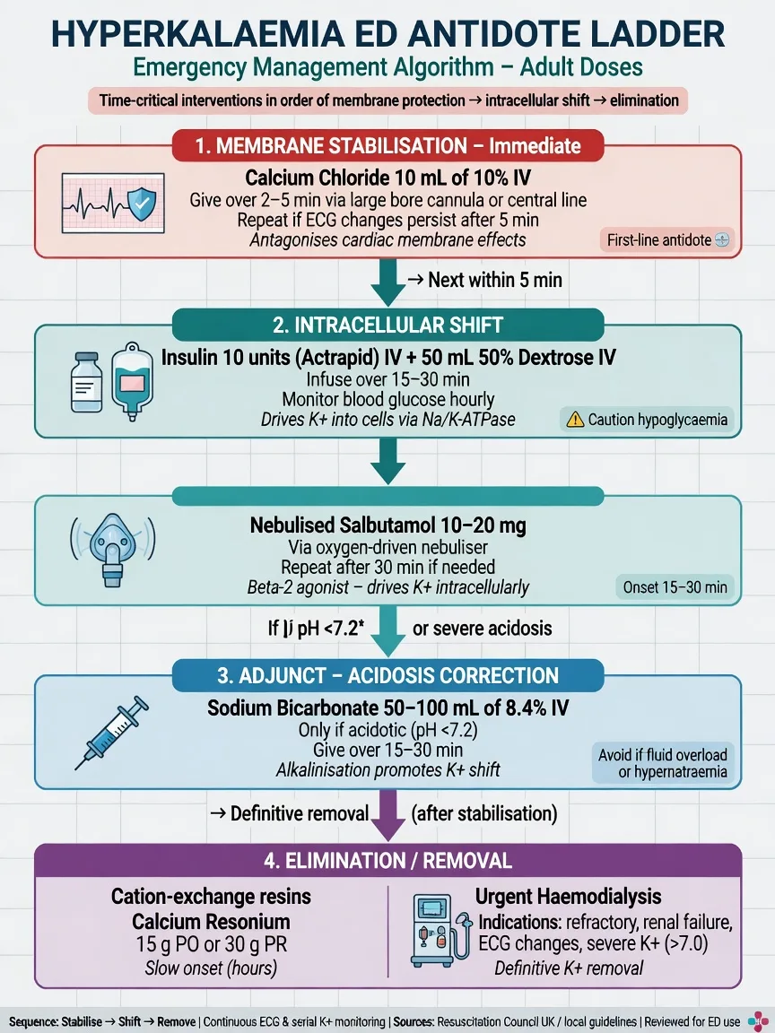
Treat severe hyperkalaemia — potassium over 6.5 mmol per litre, or any level with ECG change — as a peri-arrest emergency. The ladder has three layers, applied together in the symptomatic patient: stabilise the membrane, shift the potassium intracellularly, then remove it from the body.[1][2]
The hyperkalaemia antidote ladder
Step 1 — stabilise the membrane with calcium. Give calcium chloride 10 mL of 10 per cent IV (6.8 mmol of calcium) over two to five minutes through a large central line where possible, or calcium gluconate 10 mL of 10 per cent IV through a peripheral line (gluconate is safer peripherally as it is less vesicant if extravasated). Calcium works within minutes and lasts 30 to 60 minutes; repeat the dose if the ECG changes persist or recur. Calcium does not lower the potassium — it raises the threshold potential and restores excitability — so it must always be paired with a shifting agent. Do not give calcium in the same line as a bicarbonate infusion (it precipitates as calcium carbonate).[2]
Step 2 — shift the potassium into the cells. Give insulin 10 units of soluble (Actrapid) insulin in 50 mL of 50 per cent dextrose IV over 15 to 20 minutes; this lowers the potassium by roughly 1 mmol per litre within 15 to 30 minutes, and the effect lasts four to six hours. Monitor the blood glucose closely (at baseline, 30 minutes, one hour, two hours, and then hourly) because hypoglycaemia is the commonest and most dangerous adverse effect, occurring in up to a third of patients — a 50 per cent dextrose load is given precisely to prevent it, and additional dextrose may be needed. Add salbutamol 10 to 20 mg nebulised (or 5 micrograms per kilogram intravenously in the ventilated or arrest patient), which drives potassium into cells via beta-2 stimulation; it is synergistic with insulin and adds roughly another 0.5 to 1 mmol per litre fall. Use salbutamol with caution in ischaemic heart disease (tachycardia, ischaemia) and in pregnancy. Sodium bicarbonate 50 mL of 8.4 per cent IV shifts potassium intracellularly and is most effective when the patient is acidotic — in the non-acidotic patient its potassium-lowering effect is modest, and it is not a substitute for insulin-dextrose.[1][2]
[1]Step 3 — remove potassium from the body. The shifting agents are temporary (four to six hours); unless the cause is reversible and renal function intact, the potassium will rebound. Patiromer and sodium zirconium are the modern gastrointestinal cation-exchange agents that bind potassium in the gut over hours to days and are suited to the subacute removal. Older resins (sodium polystyrene sulfonate) are slower and carry a bowel-necrosis risk. Haemodialysis is the definitive removal for the refractory case, the dialysis-dependent patient, or the severe hyperkalaemic arrest. Loop diuretics (frusemide 40 mg IV) help the patient with residual renal function excrete potassium. [1]
[1]Definitive management — the hyponatraemia strategy
Hyponatraemia management is governed by two questions answered in order: is the patient symptomatic, and is the onset acute or chronic? The answer decides whether to give hypertonic saline within minutes or to correct over days.[3][4]
Hyponatraemia — symptom-driven correction
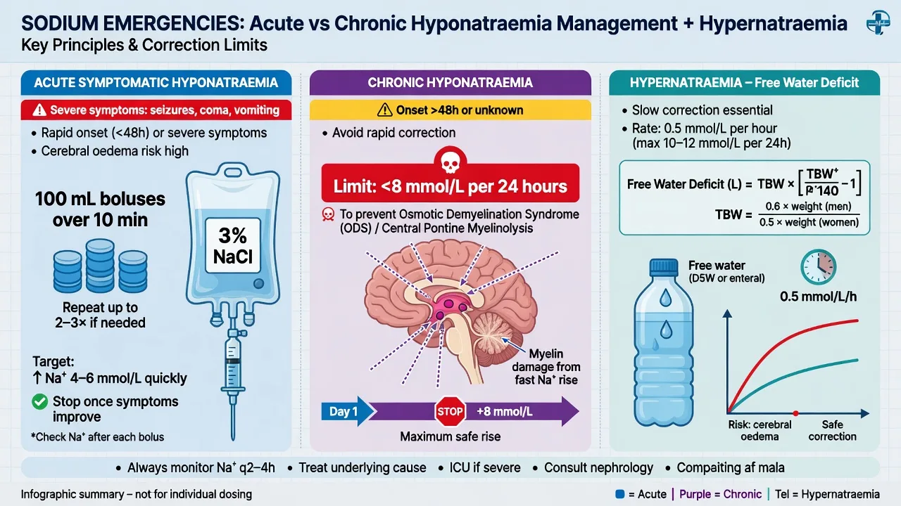
Acute symptomatic hyponatraemia (sodium under 125 mmol per litre with seizure, coma, or severe cerebral oedema) is a cerebral-herniation risk and is treated immediately with 3 per cent (hypertonic) saline 100 mL IV as a bolus over 10 minutes, repeated up to three times until the symptoms resolve or the sodium rises by 4 to 6 mmol per litre. The aim of the bolus is not to normalise the sodium but to raise it just enough (4 to 6 mmol per litre) to relieve cerebral oedema and stop the seizure — a small, tightly-controlled rise reverses the brain swelling because water follows the sodium out of the neurone. Measure the sodium after each bolus. A continuous infusion of 3 per cent saline (0.5 to 2 mL per kilogram per hour) can follow, with two-hourly sodium checks to hold the rise within the safe ceiling. Stop the moment symptoms resolve.[4]
[1]Chronic hyponatraemia is corrected slowly because the brain has adapted. The safe ceiling is a rise of no more than 8 mmol per litre in any 24-hour period (some guidelines allow 10 to 12 mmol per litre in 24 hours as an absolute maximum), and patients at high risk of osmotic demyelination — alcoholism, malnutrition, hypokalaemia, advanced liver disease, women on thiazides — are corrected even more slowly, often 4 to 6 mmol per litre in 24 hours. Check the sodium every two to four hours during active correction. The correction is achieved by treating the cause: fluid restriction and a high-protein/salt diet for SIADH (with oral urea, demeclocycline or a vasopressin V2-receptor antagonist for resistant cases); normal saline for the hypovolaemic patient (which switches off antidiuretic hormone and lets the sodium correct itself); and fluid and sodium restriction plus treatment of the underlying disorder for the hypervolaemic patient. Over-correction is managed by re-lowering the sodium with desmopressin and free water, ideally in consultation with renal and endocrine teams.[3]
[1]Hypokalaemia — the mirror emergency
If hyperkalaemia is the arrhythmia of excess, hypokalaemia (a serum potassium below 3.5 mmol per litre) is the arrhythmia of depletion — and it is the one Fellowship candidates under-call, because the patient is rarely on a renal ward. Severe hypokalaemia (below 2.5 mmol per litre, or any level with ECG change) potentiates digoxin toxicity, precipitates torsades de pointes, converts an acute coronary syndrome into a ventricular fibrillation arrest, and — in the unwary — drives a refractory ventricular fibrillation that will not terminate until the potassium is given. The mirror logic of the topic is that hyperkalaemia is corrected within minutes and hypokalaemia within hours: potassium cannot be pushed into a vein faster than 10 mmol per litre per hour peripherally without inducing phlebitis and a paradoxical arrhythmia from transient local hyperkalaemia.[6][7]
Hypokalaemia severity and risk
Pathophysiology — the repolarisation current
Potassium sets the resting membrane potential, and the low intracellular-to-extracellular ratio in hypokalaemia hyperpolarises the cell (makes the resting potential more negative), which initially suppresses conduction. The dominant clinical effect, however, is on repolarisation: hypokalaemia prolongs the action-potential duration, lengthens the relative refractory period, and generates early after-depolarisations — the substrate for torsades de pointes. The ECG signature is the reverse of hyperkalaemia: flattening then inversion of the T wave, ST-segment depression, the appearance of a U wave (a positive deflection after the T wave, best seen in V2 to V4), a prolonged QU interval, ventricular ectopics, and — at the severe end — torsades de pointes, ventricular tachycardia, or ventricular fibrillation. The digoxin effect is potentiated because potassium competes with digoxin for the sodium-potassium ATPase; a falling potassium unbinds digoxin and converts a therapeutic level into a toxic one.[7]
[1]Causes — the three buckets
Hypokalaemia is always one of three mechanisms: inadequate intake (rare alone), extrarenal loss (the gut and the skin), or renal loss (the tubule, driven by diuretics, magnesium depletion, or acid–base shift). The single best discriminator between renal and extrarenal loss is the urine potassium (or the transtubular potassium gradient, TTKG): a urine potassium over 20 mmol per litre (or a TTKG over 3) in the face of hypokalaemia points to renal wasting; below that, the kidney is appropriately conserving and the loss is extrarenal.[6]
Extrarenal (GI/skin) loss
- Vomiting, diarrhoea, villous adenoma, laxative abuse, high-output stoma
- Profuse sweating, prolonged heat; burns
- Urine K under 20 mmol/L — the kidney conserves correctly
- Replace the potassium and the magnesium; treat the cause
Renal loss — diuretic/drug
- Loop and thiazide diuretics (the commonest), carbonic anhydrase inhibitors
- Cisplatin, aminoglycosides, amphotericin B, corticosteroids
- Renal tubular acidosis, Bartter and Gitelman syndromes
- Urine K over 20 mmol/L; replete, re-check Mg, review the drug chart
Intracellular shift (redistribution)
- Alkalosis (each 0.1 pH rise drops K by ~0.4 mmol/L), insulin, beta-2 agonists
- Refeeding syndrome, thyrotoxic periodic paralysis, familial periodic paralysis
- Hypokalaemia with total-body stores near-normal; treat the trigger
- Refeeding — replace K, Mg, phosphate slowly with thiamine cover
The magnesium trap
- Hypomagnesaemia causes renal potassium wasting via ROMK channel disinhibition
- A hypokalaemia that does not correct is almost always a low magnesium
- Check and replace magnesium (MgSO4 2 g IV over 1 hour) FIRST or alongside the K
- Correct Mg, then the K will hold — the single most-tested pitfall
Immediate management — potassium replacement
The route, the rate, and the monitoring depend on the severity and the ECG. Oral potassium (chloride 40 to 80 mmol per day in divided doses) is preferred for the mild and the moderate case with a functioning gut — it is safer, the gut absorbs it slowly, and it repletes the intracellular store over days. Intravenous potassium chloride is reserved for the severe (below 2.5 mmol per litre), the symptomatic, the ECG-changed, the digoxin-toxic, and the patient who cannot absorb orally. The cardinal rule of IV potassium is the rate limit: no faster than 10 mmol per hour through a peripheral line (in 100 to 250 mL of normal saline over at least one hour) and no faster than 20 mmol per hour through a central line with continuous cardiac monitoring, because a faster rate produces a transient local hyperkalaemia that can precipitate ventricular fibrillation. Never give undiluted potassium as a bolus — it is a lethal injection.[6][7]
Severe hypokalaemia — the ED replacement sequence
0 min — recognise and monitor
Cardiac monitor on every patient with a potassium below 2.5 or any ECG change. The 12-lead for the U wave, the flat T, the ST depression, the QT/QU prolongation, the ectopics. Two large-bore cannulae. Check the magnesium, the phosphate, the calcium, the acid–base.
0–10 min — correct the magnesium first
If the magnesium is low (below 0.7 mmol per litre) give magnesium sulphate 2 g IV over 1 hour (4 g in the torsades patient, more cautiously in renal failure). Without magnesium the potassium will not hold. This is the single step candidates forget.
10–60 min — peripheral IV potassium
Potassium chloride 10 mmol in 100 mL of normal saline over 1 hour through a peripheral line (NO faster than 10 mmol/h). Add 10 mmol pre-mixed into each litre of maintenance fluid. Recheck the potassium at 2 to 4 hours. Warn the patient of the infusion-site pain — phlebitis is the limiting toxicity.
Central-line fast infusion (if severe/periarrest)
If the potassium is below 2.0, or the patient is in torsades or VF, insert a central line and give potassium chloride 20 mmol in 100 mL over 1 hour with continuous cardiac monitoring (NO faster than 20 mmol/h). Treat the torsades with IV magnesium sulphate 2 g first, over 1 to 2 minutes in the arrest.
Identify and treat the cause
Stop the offending diuretic or laxative; correct the alkalosis and the volume deficit; screen for digoxin (stop it, give Fab if toxic); replace the phosphate in refeeding; look for the villous adenoma, the hyperaldosteronism, the renal tubular acidosis. Recheck the potassium at 4, 8, 12 hours and the next morning.
Hypernatraemia — the slow-correction sodium
If hyponatraemia kills by cerebral oedema, hypernatraemia (a serum sodium above 145 mmol per litre) kills by cerebral shrinkage — and by the cerebral oedema that follows if you correct it too fast. Hypernatraemia is always, at its core, a water problem: a relative deficit of water to sodium, from a failure of water intake (the obtunded, the intubated, the elderly), a failure of water conservation (diabetes insipidus), or an excess of sodium (salt tablets, hypertonic saline, sodium bicarbonate, salt poisoning). The patient who cannot drink to thirst is the patient at risk — the infant, the demented, the intubated, the nursing-home resident — and the mortality of severe hypernatraemia in hospital exceeds 40 per cent. The management is free water, given slowly: correct at no more than 0.5 mmol per litre per hour (10 to 12 mmol per litre in 24 hours) in the chronic case to avoid the cerebral oedema of over-rapid correction.[5]
Hypernatraemia — the water-deficit emergency
Pathophysiology — the shrinking brain
Sodium sets the serum osmolality, and hypernatraemia is a hyperosmolar state that pulls water out of every cell, including the brain. In the acute case (within hours) the brain shrinks, the bridging veins stretch and tear, and the patient develops subdural haemorrhage, subarachnoid haemorrhage, or intracerebral haemorrhage — a catastrophic and often missed complication of severe acute hypernatraemia. In the chronic case (over 48 hours) the brain adapts by accumulating idiogenic osmoles (glutamate, taurine, myo-inositol) to hold onto water and restore its volume, exactly as it adapts to hyponatraemia by losing osmoles. The danger now reverses: if the sodium is lowered too fast, the adapted, osmolyte-laden brain takes on water faster than it can shed the osmoles, swells, and cerebral oedema results — seizures, coma, herniation. This is the mirror image of the osmotic-demyelination injury of hyponatraemia, and it is why chronic hypernatraemia, like chronic hyponatraemia, is corrected slowly.[5]
[1]Causes — the three mechanisms
Hypernatraemia is always one of three mechanisms: water loss in excess of sodium loss (extrarenal or renal, with diabetes insipidus the prototype), pure water loss (insensible, fever, burns), or sodium gain (iatrogenic or accidental). The discriminator is the urine osmolality and the volume status: a concentrated urine (over 700 mOsm per kilogram) with hypovolaemia means the hypothalamic–pituitary–renal axis is intact and the kidney is conserving water — the loss is extrarenal or DI-with-intact-vasopressin-less. A dilute urine (under 300 mOsm per kilogram) in the face of serum hyperosmolality is diabetes insipidus — central (no vasopressin) or nephrogenic (unresponsive kidney).[5]
Water loss — extrarenal
- Insensible: fever, heat, tachypnoea, burns; the intubated patient with dry gases
- Vomiting, osmotic diarrhoea, lactulose
- Urine osmolality high (over 700) — kidney conserving water correctly
- Replace with free water (oral or 5% dextrose); correct slowly
Water loss — diabetes insipidus
- Central: pituitary surgery, trauma, tumour, Sheehan, idiopathic — NO vasopressin
- Nephrogenic: lithium (the classic), hypercalcaemia, hypokalaemia, congenital
- Urine osmolality low (under 300) despite serum hyperosmolality; urine dilute
- Central → desmopressin; nephrogenic → remove the cause, thiazide + low-solute diet
Sodium gain (iatrogenic/accidental)
- Hypertonic saline (3% boluses), sodium bicarbonate (cardiac arrest, TCA overdose)
- Salt tablet excess, sea-water drowning, deliberate salt poisoning (child abuse)
- High urine sodium and high urine osmolality; euvolaemic or hypervolaemic
- Stop the source; loop diuretic plus 5% dextrose; dialysis for the extreme
Inadequate water intake
- The obtunded, the intubated, the infant, the nursing-home resident
- Hypothalamic lesion destroying the thirst centre (rare but classic)
- Urine osmolality maximal — the kidney does all it can
- Replace free water; secure an adequate intake before discharge
Immediate management — free water, slowly
The treatment of hypernatraemia is water, given by the safest route at a controlled rate. Oral or nasogastric water is the safest and the preferred route in the alert patient — the gut corrects the sodium more gently than any IV fluid. In the patient who cannot drink, 5 per cent dextrose is the free-water intravenous solution of choice (0.45 per cent saline is an alternative that adds some sodium); avoid 0.9 per cent saline alone, which adds sodium to a sodium-excess state. Calculate the free-water deficit (0.6 × weight in kilograms × (sodium/140 − 1)), and replace half over 24 hours and the remainder over the next 24 to 48 hours, checking the sodium every 2 to 4 hours to hold the correction under 0.5 mmol per litre per hour (10 to 12 mmol per litre in 24 hours). In the central diabetes insipidus patient, give desmopressin (1 to 2 micrograms subcutaneously or 10 to 40 micrograms intranasally) and the kidney will re-concentrate the urine; in nephrogenic DI, remove the cause (lithium, hypercalcaemia) and use a thiazide diuretic with a low-solute diet (paradoxically, the mild volume depletion improves proximal water reabsorption).[5]
Severe hypernatraemia — the slow-correction sequence
0 min — assess and resuscitate
ABCDE. The conscious level, the volume status (most are hypovolaemic), the temperature (fever drives insensible loss), the cause (post-op pituitary, lithium, intubated and under-hydrated, salt exposure). Send the sodium, the glucose, the calcium, the urea and electrolytes; attach cardiac monitoring. Calculate the serum osmolality (2 × Na + glucose + urea).
0–30 min — restore the circulation
If hypovolaemic, give 0.9 per cent saline boluses (250 to 500 mL) to restore the perfusion first — treat shock before the slow water correction. Once perfused, switch to free water. NEVER correct the sodium with saline alone beyond the resuscitation.
30 min — calculate the water deficit
Free-water deficit = 0.6 × weight (kg) × (Na/140 − 1). Replace half over 24 h, the rest over 48 to 72 h. Choose the route: oral or NG water (safest) if alert; 5% dextrose IV if not (2 to 3 mL/kg/h drops the Na by ~0.5 mmol/L/h).
Lock the correction rate
Target a fall of no more than 0.5 mmol per litre per hour (10 to 12 mmol per litre in 24 hours) in the chronic case. Check the sodium every 2 to 4 hours and titrate the free-water rate. Faster correction risks cerebral oedema and seizure — the mirror of the osmotic-demyelination risk.
Treat the specific cause
Central DI: desmopressin 1 to 2 micrograms SC. Nephrogenic DI: stop the lithium, correct the hypercalcaemia and hypokalaemia, add a thiazide plus a low-solute diet. Sodium gain: stop the source, give a loop diuretic with 5% dextrose, consider dialysis. Recheck the sodium at 4, 8, 12 hours and daily.
The over-correction injuries — osmotic demyelination and cerebral oedema
The two sodium-disturbance injuries are mirror images of each other and the Fellowship candidate must hold both at once. Correcting chronic hyponatraemia too fast (over 8 to 10 mmol per litre in 24 hours) strips the adapted, osmolyte-depleted brain of its protective osmolyte gradient and produces osmotic demyelination syndrome — astrocyte apoptosis, classically in the basis pontis, presenting days later with dysarthria, dysphagia, quadriparesis, a locked-in state, and (in the extrapontine form) a movement disorder. Correcting chronic hypernatraemia too fast (over 0.5 mmol per litre per hour) floods the adapted, osmolyte-laden brain with water and produces acute cerebral oedema — seizure, coma, herniation, and death. Both are largely irreversible once established, which is why prevention — the locked correction rate, the frequent sodium check, and the willingness to re-lower with desmopressin and free water — is everything.[12][13][16]
[1] [1]Hyperkalaemic cardiac arrest
Hyperkalaemia is a recognised reversible cause of cardiac arrest — the fifth H in the Hs and Ts — and is suspected in any arrest in a renal patient, a dialysis patient, or one with a known potassium-raising drug. Give calcium chloride 10 mL of 10 per cent IV early in the arrest, plus insulin-dextrose (10 units in 50 mL of 50 per cent) and sodium bicarbonate 50 mL of 8.4 per cent IV by bolus, and check a venous gas potassium immediately. The arrest rhythm is commonly asystole, a slow broad-complex bradycardia, or a sine-wave ventricular fibrillation resistant to defibrillation until the potassium is lowered. Arrange emergency haemodialysis for the refractory hyperkalaemic arrest in the dialysis-dependent patient. [1]
Subtypes and special scenarios
The dialysis patient with hyperkalaemia is the single commonest presentation — typically a non-attendee or one with an intercurrent illness. Treat with the full ladder, then arrange urgent haemodialysis; the shifting agents buy time, only dialysis removes the potassium definitively. Addisonian crisis presents with hyperkalaemia, hyponatraemia, hypotension and hypoglycaemia — give parenteral hydrocortisone (100 mg IV) alongside resuscitation, and the electrolytes correct as the steroid replaces the aldosterone effect. Tumour lysis syndrome, seen after chemotherapy for a high-grade lymphoma or leukaemia, produces acute hyperkalaemia with hyperphosphataemia, hypocalcaemia and acute kidney injury — give rasburicase or allopurinol, aggressive hydration, and dialysis if needed. Exercise-induced hyperkalaemia in the elite athlete is mild and self-limiting and needs no treatment. [1]
The endurance athlete with exercise-associated hyponatraemia is the classic acute case — collapse or seizure after a marathon, having drunk excessive hypotonic fluid. Treat as acute symptomatic hyponatraemia with hypertonic saline. The post-operative patient (often a young woman after gynaecological surgery) given hypotonic fluids or with pain/stress-driven antidiuretic hormone is the other acute case, and is the one most likely to die of cerebral herniation — suspect it in any post-operative patient with nausea, confusion or seizure. The MDMA user develops acute hyponatraemia from polydipsia and syndrome of inappropriate antidiuretic hormone. The thiazide patient — typically an older woman — develops subacute hyponatraemia and is at high risk of osmotic demyelination if over-corrected. [1]
Complications and pitfalls
The complications are cardiac and cerebral. Hyperkalaemia causes the arrhythmia spectrum — bradycardia, conduction block, ventricular fibrillation, asystole — and is reversible if treated early. Hypoglycaemia from insulin-dextrose is the commonest treatment-related harm and is prevented by the 50 per cent dextrose load and glucose monitoring. Hyperkalaemia rebound occurs at four to six hours if the cause is unaddressed and no removal agent is given. Hyponatraemia causes cerebral oedema, herniation and death in the acute case, and osmotic demyelination in the over-corrected chronic case — the latter presenting days later with dysarthria, dysphagia, quadriparesis and a locked-in state, and is largely irreversible. The pitfalls mirror the management: treating a haemolysed pseudohyperkalaemia; giving insulin-dextrose without calcium when there is ECG change; failing to monitor glucose after insulin; correcting a chronic sodium faster than 8 mmol per litre in 24 hours; using hypotonic fluids in a post-operative patient; and forgetting to repeat the sodium to hold the correction rate within the ceiling. [1]
Prognosis and disposition
Severe hyperkalaemia with ECG change is a peri-arrest emergency managed in the resuscitation bay with continuous cardiac monitoring; the patient who stabilises is admitted to a monitored bed, and the dialysis-dependent patient goes to dialysis. Mortality tracks the cause and the speed of treatment. Symptomatic hyponatraemia is admitted to a high-dependency or intensive-care bed for two-hourly sodium monitoring during correction; the asymptomatic chronic case is admitted to a general ward for slow correction and work-up. The osmotic-demyelination risk dictates that the sodium is checked frequently and the rate held within the ceiling. The underlying cause — renal failure, a potassium-raising drug, SIADH, heart failure — is treated before discharge, and the offending drug is reviewed or stopped. [1]
Special populations
The renal and dialysis patient is the highest-risk group for hyperkalaemia and is managed with the full ladder and early dialysis. The elderly accumulate potassium-raising drugs (renin–angiotensin blockers, potassium-sparing diuretics, NSAIDs) and tolerate arrhythmia poorly; they also dominate the thiazide-induced hyponatraemia cohort and are at high risk of osmotic demyelination, so their sodium is corrected most cautiously. The pregnant patient with pre-eclampsia may receive magnesium sulfate, which potentiates the membrane effects of hyperkalaemia, and the sodium is managed with the obstetric team. The child is dosed by weight (calcium 0.1 to 0.2 mL per kilogram of 10 per cent chloride, insulin 0.1 unit per kilogram with 0.5 g per kilogram of dextrose) and is at particular risk of hypoglycaemia after insulin. The endurance athlete and the MDMA user present with acute water-intoxication hyponatraemia and are treated as acute symptomatic cases with hypertonic saline. [1]
Fellowship viva pearls — the high-yield answers
[1]Evidence and regional guidelines
The contemporary hyperkalaemia framework follows the UK Renal Association hyperkalaemia guideline and the European Resuscitation Council guidance, both of which codify the three-step ladder of membrane stabilisation (calcium), intracellular shift (insulin-dextrose, salbutamol, bicarbonate) and removal (resins, dialysis). The evidence for the shifting agents is summarised in a recent emergency-care review and an evidence-based narrative review.[1][2] For hyponatraemia, the Adrogué–Madias review (JAMA 2022) and the European Clinical Practice Guideline set the classification by osmolality and volume, the 4-to-6 mmol per litre bolus target for acute symptomatic disease, and the 8 mmol per litre per 24-hour ceiling for chronic correction to prevent osmotic demyelination; an updated hospital-setting review covers the bolus regimen.[3][4]
The landmark trials and reviews — hyperkalaemia
Allon 1989 — nebulised albuterol for acute hyperkalaemia (Ann Intern Med)
Annals of Internal Medicine
PMID 2919849
Key finding
A randomised placebo-controlled trial of nebulised albuterol in haemodialysis patients with hyperkalaemia. Albuterol (10 to 20 mg nebulised) lowered the potassium by approximately 1 mmol per litre within 30 minutes, with a peak effect at 60 to 90 minutes. The 20 mg dose was more reliably effective than the lower dose; tachycardia was the principal adverse effect.
Practice change
Nebulised salbutamol (albuterol) 10 to 20 mg is a validated intracellular-shift agent for the hyperkalaemia ladder, synergistic with insulin-dextrose. Use with caution in ischaemic heart disease.
Allon & Copkney 1990 — albuterol and insulin for hyperkalaemia (Kidney Int)
Kidney International
PMID 2266671
Key finding
A comparative study of albuterol versus insulin-dextrose versus the combination in haemodialysis patients. The combination lowered potassium more than either agent alone, and approximately a third of patients responded incompletely to insulin alone — supporting dual shift-agent therapy in the severe case.
Practice change
Pair insulin-dextrose with a beta-2 agonist in the severe or refractory case — the agents are synergistic and a single agent is often insufficient.
Weir 2015 — patiromer for hyperkalaemia on RAAS inhibitors (OPAL-HK, NEJM)
New England Journal of Medicine
PMID 25415805
Key finding
A randomised withdrawal trial of 237 patients with chronic kidney disease and hyperkalaemia on renin–angiotensin–aldosterone system inhibitors. Patiromer (a non-absorbed potassium-binding polymer) lowered the serum potassium significantly within the first week, and withdrawal was followed by a recurrent rise. Mild hypomagnesaemia and constipation were the principal adverse effects.
Practice change
Patiromer is a validated gastrointestinal potassium-removal agent for the subacute and outpatient setting — modern replacement for the older resins that carried a bowel-necrosis risk.
Mahoney 2005 — emergency interventions for hyperkalaemia (Cochrane)
Cochrane Database of Systematic Reviews
PMID 15846652
Key finding
A Cochrane systematic review of randomised and quasi-randomised trials of emergency interventions for acute hyperkalaemia. The reviewers found a paucity of high-quality trial evidence for the classic ladder; insulin-dextrose and beta-2 agonists were the best-supported shift agents, and the evidence for calcium as a membrane stabiliser rested largely on animal and mechanistic data rather than human outcome trials.
Practice change
The three-step ladder is the international standard despite limited outcome-grade evidence — the membrane-stabilising role of calcium is mechanistically robust and universally recommended.
Long 2018 — controversies in the management of hyperkalaemia (J Emerg Med)
Journal of Emergency Medicine
PMID 29731287
Key finding
A narrative review synthesising the evidence and the controversies of the emergency hyperkalaemia ladder — the optimal calcium salt and dose, the insulin-dose debate (5 versus 10 units) and its hypoglycaemia burden, the role of bicarbonate in the non-acidotic patient, and the indications for dialysis. The authors emphasise that the ECG, not the number, sets the first drug, and that hypoglycaemia after insulin is the commonest and most under-recognised harm.
Practice change
Apply the full ladder in parallel for the symptomatic patient, monitor the glucose aggressively for several hours after insulin, and arrange dialysis early in the refractory or renal-failure case.
Kovesdy 2015 — management of hyperkalaemia update (Am J Med)
American Journal of Medicine
PMID 26093176
Key finding
An update for the internist on the modern hyperkalaemia management, including the newer gastrointestinal cation-exchange agents (patiromer and sodium zirconium cyclosilicate) that allow chronic potassium control and the continuation of renin–angiotensin–aldosterone system inhibitors in heart failure and chronic kidney disease, where previously these agents would have been stopped.
Practice change
The new binders transform the outpatient and chronic management of hyperkalaemia, but the acute ED ladder — calcium, insulin-dextrose, salbutamol, dialysis — is unchanged for the emergency.
The landmark trials and reviews — sodium disorders
Adrogué & Madias 2000 — hypernatremia (NEJM)
New England Journal of Medicine
PMID 10816188
Key finding
The definitive mechanistic and clinical review of hypernatraemia, defining the three mechanisms (water loss, pure water loss, sodium gain), the free-water-deficit formula and the half-over-24-hour replacement rule, the central-versus-nephrogenic diabetes insipidus distinction, and the correction ceiling of 0.5 mmol per litre per hour to avoid the cerebral oedema of over-rapid correction.
Practice change
The reference framework for every hypernatraemia case: water deficit calculation, slow free-water replacement, and the locked correction rate. Hypernatraemia is always a water problem.
Spasovski 2014 — European clinical practice guideline on hyponatraemia (Eur J Endocrinol)
European Journal of Endocrinology
PMID 24569125
Key finding
A multidisciplinary European clinical practice guideline on the diagnosis and treatment of hyponatraemia, codifying the osmolality-then-volume classification, the hypertonic-saline regimen for severe symptomatic disease (a 150 mL 3 per cent bolus, repeatable), the 10 mmol per litre per 24-hour correction ceiling, and the risk-stratified management of SIADH.
Practice change
The European standard for the classification and the rate-locked correction of hyponatraemia — the osmolality-then-volume algorithm and the correction ceiling that the Fellowship candidate must reproduce.
Sterns 2010 — treatment of hyponatraemia (Curr Opin Nephrol Hypertens)
Current Opinion in Nephrology and Hypertension
PMID 20539224
Key finding
A critical review of the treatment of hyponatraemia that quantified the osmotic-demyelination risk of over-rapid correction, refined the 8 mmol per litre per 24-hour ceiling, and articulated the high-risk phenotype (the alcoholic, the malnourished, the hypokalaemic, the woman on a thiazide) that must be corrected even more slowly.
Practice change
The correction ceiling and the high-risk phenotype are Sterns' contribution — name them in the viva and hold the rate in the patient.
Sterns 2016 — complications and management of hyponatraemia (Curr Opin Nephrol Hypertens)
Current Opinion in Nephrology and Hypertension
PMID 26735146
Key finding
An updated review of the complications of hyponatraemia and its treatment, with the desmopressin-and-free-water re-lowering strategy for the over-corrected patient — the salvage manoeuvre to prevent the osmotic-demyelination lesion within the first 24 to 48 hours.
Practice change
Over-correction is not irreversible at the moment it is recognised — re-lower the sodium with desmopressin and free water, in consultation with renal and endocrine, within the first 48 hours.
Ayus 2019 — misconceptions and barriers to hypertonic saline (Front Med)
Frontiers in Medicine
PMID 30931308
Key finding
A review dismantling the persistent misconceptions that delay hypertonic saline for hyponatraemic encephalopathy — the fear of over-correction, the reliance on continuous infusion over bolus, and the under-recognition of the acute symptomatic case. The authors advocate the small-repeated-bolus regimen (100 mL of 3 per cent over 10 minutes) that is titratable and carries a low over-correction risk.
Practice change
Do not delay 3 per cent saline for the seizure or coma of acute hyponatraemia — the 100 mL bolus is titratable, repeatable to a 4 to 6 mmol per litre rise, and the risk of NOT treating (herniation) dwarfs the risk of treating.
Singh 2014 — central pontine and extrapontine myelinolysis (Eur J Neurol)
European Journal of Neurology
PMID 25220878
Key finding
A systematic review of the osmotic demyelination syndrome — central pontine and extrapontine myelinolysis — defining the clinical spectrum (the classic locked-in quadriparesis and the extrapontine movement disorders), the over-rapid-correction aetiology, and the largely irreversible outcome once established. Mortality and severe morbidity remain high despite supportive care.
Practice change
Osmotic demyelination is a preventable, largely irreversible injury — prevention is the locked correction rate and the willingness to re-lower an over-corrected sodium. No treatment reverses it once it occurs.
The landmark reviews — hypokalaemia
Unwin 2011 — pathophysiology and management of hypokalaemia (Nat Rev Nephrol)
Nature Reviews Nephrology
PMID 21278718
Key finding
A comprehensive clinical review of hypokalaemia, defining the renal-versus-extrarenal-loss discrimination (the urine potassium and the transtubular potassium gradient), the magnesium-potassium coupling (the ROMK channel and the refractory-hypokalaemia mechanism), and the rate-limited intravenous replacement strategy (10 mmol per hour peripheral, 20 mmol per hour central).
Practice change
The reference framework for hypokalaemia: classify the loss by the urine potassium, replace magnesium first, and never exceed the IV rate limit. The magnesium-first rule is the most-tested pearl.
Gennari 2002 — disorders of potassium homeostasis (Crit Care Clin)
Critical Care Clinics
PMID 12053834
Key finding
A definitive critical-care review of potassium homeostasis covering both hypo- and hyperkalaemia, the Nernst-equation membrane-potential logic, the renal handling of potassium, and the cellular shift mechanisms — the scientific basis for the calcium-membrane-stabilisation and the insulin-shift approaches.
Practice change
The mechanistic underpinning of the ladder: calcium raises the threshold potential (does not lower potassium), insulin and beta-2 agonists drive potassium into cells, and the kidney is the route of removal. Master the mechanism to defend the management.
ANZ practice note. The hyperkalaemia ladder follows the UK Renal Association and European Resuscitation Council framework via local renal and resuscitation protocols: calcium chloride 10 mL of 10 per cent IV is first when there is ECG change, insulin 10 units with 50 mL of 50 per cent dextrose is the core shift agent with glucose monitoring, salbutamol 10 to 20 mg nebulised and sodium bicarbonate 50 mL of 8.4 per cent are add-ons, and haemodialysis is the definitive removal in the renal patient. For hyponatraemia, acute symptomatic disease (seizure or coma, sodium under 125 mmol per litre) receives 3 per cent hypertonic saline 100 mL IV as a bolus over 10 minutes, repeated up to three times to a 4 to 6 mmol per litre rise; chronic hyponatraemia is corrected at no more than 8 mmol per litre in 24 hours, with two-hourly sodium checks and renal–endocrine input for the over-corrected case. [1]
Exam pearls
- Severity: potassium 6.5 mmol per litre or above is severe — but any potassium with ECG change is treated as severe. The ECG, not the number, sets the first drug.
- ECG progression: peaked T waves (early) → PR prolongation and loss of P waves → widened QRS → sine wave → ventricular fibrillation or asystole. Calcium first, always.
- Calcium does not lower potassium — it stabilises the membrane by raising the threshold potential. Pair it always with a shifting agent.
- Insulin-dextrose: 10 units of Actrapid in 50 mL of 50 per cent dextrose IV. Monitor glucose — hypoglycaemia is the commonest harm.
- Salbutamol: 10 to 20 mg nebulised, synergistic with insulin; caution in ischaemic heart disease.
- Acute symptomatic hyponatraemia: 3 per cent saline 100 mL IV over 10 minutes, repeat up to three; aim for a 4 to 6 mmol per litre rise, not normalisation.
- Correction ceiling: under 8 mmol per litre in 24 hours for chronic hyponatraemia; over-correction causes osmotic demyelination (pons, days later, locked-in state).
- Corrected sodium: add 2 mmol per litre for every 5.6 mmol per litre (100 mg per decilitre) of glucose above normal.
- Pseudohyperkalaemia: repeat the sample before dialysis — haemolysis, fist clenching, severe thrombocytosis.
- Hypokalaemia ECG: U waves (V2 to V4), flattening/inversion of the T, ST depression, prolonged QU, torsades de pointes. Below 2.5 mmol per litre is severe.
- IV potassium rate limit: 10 mmol per hour peripheral, 20 mmol per hour central, always diluted and with a cardiac monitor. Undiluted potassium as a bolus is a lethal injection.
- The magnesium-first rule: refractory hypokalaemia (potassium that will not rise) is hypomagnesaemia — give magnesium sulphate 2 g IV first or the potassium will not hold.
- Hypokalaemia and digoxin: a falling potassium unbinds digoxin and converts a therapeutic level into a toxic one (bidirectional VT, SVT with block). Replete the potassium and check the digoxin level.
- Hypernatraemia is always a water problem: free-water deficit = 0.6 × weight × (Na/140 − 1); replace half over 24 hours, the rest over 48 to 72 hours.
- Hypernatraemia correction ceiling: no faster than 0.5 mmol per litre per hour (10 to 12 mmol per litre in 24 hours) — faster causes cerebral oedema, the mirror of the osmotic-demyelination injury of hyponatraemia.
- Diabetes insipidus: a dilute urine (under 300 mOsm per kilogram) despite serum hyperosmolality. Central — give desmopressin 1 to 2 micrograms SC. Nephrogenic — stop the lithium, a thiazide and a low-solute diet.
- The over-correction rescue: if the chronic sodium moves faster than the ceiling, re-lower with desmopressin and free water within the first 24 to 48 hours — the window to prevent osmotic demyelination.
- The five high-risk-for-ODS phenotypes: alcoholic, malnourished, hypokalaemic, advanced liver disease, woman on a thiazide — correct these even more slowly (4 to 6 mmol per litre in 24 hours).
Model answer — severe hypokalaemia with torsades
Cardiac monitor on; the 12-lead for the U waves and the prolonged QU. Give IV magnesium sulphate 2 g over 1 to 2 minutes (the torsades antidote) FIRST, then potassium chloride 10 mmol in 100 mL of normal saline over 1 hour through a peripheral line, repeated to a serum potassium of 4.0 to 4.5 mmol per litre. Insert a central line if the potassium is below 2.0 and give 20 mmol per hour with continuous monitoring. Correct the magnesium fully (2 to 4 g), stop any diuretic or laxative, screen for digoxin toxicity, and look for the cause (diuretic, vomiting, refeeding, renal tubular acidosis). Recheck the potassium at 2, 4 and 8 hours.[1]Model answer — severe hypernatraemia from diabetes insipidus
ABCDE. Confirm the volume status and resuscitate with 0.9 per cent saline if hypovolaemic. Calculate the water deficit (0.6 × weight × Na/140 − 1) and replace half over 24 hours with 5 per cent dextrose (or oral water if alert), targeting a fall of 0.5 mmol per litre per hour. Give desmopressin 1 to 2 micrograms SC if the DI is central (the urine will re-concentrate). Check the sodium every 2 to 4 hours and titrate to the ceiling. Identify the cause — pituitary surgery, trauma, tumour for central; lithium, hypercalcaemia, hypokalaemia for nephrogenic. Treat the nephrogenic with a thiazide and a low-solute diet if desmopressin fails.
Model answer — severe hyperkalaemia with ECG change
Exam practice
SAQ — Severe hyperkalaemia with ECG change in the dialysis patient
10 minutes · 10 marks
A 64-year-old man with end-stage kidney disease on haemodialysis (three sessions weekly, last session four days ago after missing his Friday slot) is brought to the resuscitation bay by ambulance with two generalised tonic-clonic seizures at home followed by a broad-complex bradycardia. He takes ramipril 10 mg daily, spironolactone 25 mg daily and a calcium-based phosphate binder. On arrival he is post-ictal, GCS 12, BP 96/58, HR 38 with a broad-complex rhythm on the monitor. The 12-lead ECG shows absent P waves, a QRS of 168 ms, peaked T waves merging into a sine-wave morphology. The venous gas returns within three minutes: potassium 8.1 mmol per litre, pH 7.18, bicarbonate 12, glucose 6.4, haemoglobin 96, calcium (ionised) 0.92 mmol per litre. The troponin is unremarkable.
SAQ — Acute symptomatic hyponatraemia with seizure in the post-operative patient
10 minutes · 10 marks
A previously well 38-year-old woman is brought to the emergency department 22 hours after an uneventful laparoscopic hysterectomy, discharged the same morning. Her husband reports two days of nausea, then today two generalised tonic-clonic seizures at home. She takes no regular medication other than the post-operative oxycodone and paracetamol. On arrival she is post-ictal, GCS 11 (E3V3M5), temperature 36.6, HR 96, BP 118/72, RR 16, SpO2 98 per cent on room air, with no focal neurology and no signs of dehydration or overload. The venous gas shows sodium 116 mmol per litre, glucose 5.8, normal anion gap. Serum osmolality 248 mOsm per kilogram, urine osmolality 410 mOsm per kilogram, urine sodium 62 mmol per litre. The CT brain shows no haemorrhage or herniation. She received 3 litres of 5 per cent dextrose and 1 litre of 0.45 per cent saline intra- and post-operatively.
Red flags
[1]References
- [1]Geldermann N, Lewis P, Boyle T, et al. Acute hyperkalaemia in emergency care: evidence-based approaches Emerg Med J, 2026.PMID 41506858
- [2]Lemoine L, Chauvin K, Delerme S, et al. An Evidence-Based Narrative Review of the Emergency Department Management of Acute Hyperkalemia J Emerg Med, 2021.PMID 33423833
- [3]Adrogué HJ, Madias NE. Diagnosis and Management of Hyponatremia: A Review JAMA, 2022.PMID 35852524
- [4]Prince R, MacKenzie K, Sherwood R, et al. The treatment of acute symptomatic hyponatraemia in the hospital setting Best Pract Res Clin Endocrinol Metab, 2026.PMID 41402221
- [5]Adrogué HJ, Madias NE. Hypernatremia N Engl J Med, 2000.PMID 10816188
- [6]Unwin RJ, Luft FC, Shirley DG. Pathophysiology and management of hypokalemia: a clinical perspective Nat Rev Nephrol, 2011.PMID 21278718
- [7]Gennari FJ. Disorders of potassium homeostasis. Hypokalemia and hyperkalemia Crit Care Clin, 2002.PMID 12053834
- [8]Allon M, Dunlay R, Copkney C. Nebulized albuterol for acute hyperkalemia in patients on hemodialysis Ann Intern Med, 1989.PMID 2919849
- [9]Allon M, Copkney C. Albuterol and insulin for treatment of hyperkalemia in hemodialysis patients Kidney Int, 1990.PMID 2266671
- [10]Weir MR, Bakris GL, Bushinsky DA, et al. Patiromer in patients with kidney disease and hyperkalemia receiving RAAS inhibitors N Engl J Med, 2015.PMID 25415805
- [11]Long B, Warix JA, Koyfman A. Controversies in Management of Hyperkalemia J Emerg Med, 2018.PMID 29731287
- [12]Sterns RH. Treatment of hyponatremia Curr Opin Nephrol Hypertens, 2010.PMID 20539224
- [13]Singh TD, Fugate JE, Rabinstein AA. Central pontine and extrapontine myelinolysis: a systematic review Eur J Neurol, 2014.PMID 25220878
- [14]Ayus JC, Caputo DG, Ballesteros MG, Moritz ML. Misconceptions and Barriers to the Use of Hypertonic Saline to Treat Hyponatremic Encephalopathy Front Med (Lausanne), 2019.PMID 30931308
- [15]Spasovski G, Vanholder R, Allolio B, et al. Clinical practice guideline on diagnosis and treatment of hyponatraemia Eur J Endocrinol, 2014.PMID 24569125
- [16]Sterns RH. Complications and management of hyponatremia Curr Opin Nephrol Hypertens, 2016.PMID 26735146
- [17]Mahoney BA, Smith WAD, Lo DS, Tsoi K, Tonelli M, Clase CM. Emergency interventions for hyperkalaemia Cochrane Database Syst Rev, 2005.PMID 15846652
- [18]Kovesdy CP. Management of Hyperkalemia: An Update for the Internist Am J Med, 2015.PMID 26093176