EM · Imaging choice and radiation risk
Imaging choice and radiation risk in the emergency department
Also known as Modality selection · Radiation dose and ALARA · Appropriate imaging · Contrast risk and nephrogenic systemic fibrosis · Imaging in pregnancy · CT versus MRI versus ultrasound · Contrast-associated acute kidney injury · Choosing Wisely imaging
Imaging choice and radiation risk — the structured selection of the modality (X-ray, CT, MRI, ultrasound) that answers the clinical question at the lowest risk, governed by ALARA and the linear-no-threshold model. The effective doses (CXR 0.02 mSv, CT head 2 mSv, CT abdomen 10 mSv against a 2–3 mSv/yr background); the stochastic (cancer) and deterministic (skin erythema, cataract, infertility) effects; the four-modality differential with speed, cost, sensitivity and operator-dependence; the pregnancy pathway (ultrasound first, MRI second, CT last, never withhold a life-saving image on dose alone); the contrast risks — iodinated contrast-associated AKI and gadolinium-associated nephrogenic systemic fibrosis with the group I/II/III agent stratification; the appropriateness criteria and the Choosing Wisely do-not list. ACEM-primary, globally tagged.
On this page & tools
Your progress
Saved locally on this device.
Target exams
Red flags
Every imaging request the emergency clinician makes is a balance of three forces: does the image answer the clinical question, what does it cost the patient, and what does it cost to delay? The four core modalities — plain X-ray, computed tomography (CT), magnetic resonance imaging (MRI) and ultrasound (US) — differ in their physical principle, their radiation burden, their sensitivity and their suitability for the unstable, the pregnant and the renally impaired patient. The Fellowship candidate must know the effective doses (the catalogued numbers), the ALARA principle, the contrast risks (iodinated and gadolinium), and the appropriateness logic that separates a high-yield image from an avoidable one.[1][2]
Why the topic matters — radiation in context
Medical radiation is now the dominant non-background source of human exposure, and CT accounts for the majority of it. CT utilisation in emergency departments has risen roughly tenfold over two decades, and that rise tracks a measurable increase in the population's cumulative effective dose.[2] The lifetime attributable cancer risk from any single CT is small, but it is real, additive, and highest in the young and in those scanned repeatedly. The counterweight is that an avoidable miss from the wrong modality — or no imaging — is frequently fatal. Imaging choice is therefore a risk-risk trade: radiation and contrast risk on one side, missed-diagnosis risk on the other, mediated by the clinical question.
Pathophysiology — how radiation and the modalities work
Ionising radiation (X-ray, CT, fluoroscopy, nuclear medicine) deposits energy in tissue and breaks DNA. Two effect types govern the risk: [1]
[1]The non-ionising modalities operate by different physics. MRI uses a strong static magnetic field, switched gradient fields and radiofrequency pulses to align and then perturb hydrogen protons; the signal as they relax gives the image. It delivers no ionising radiation but carries the ferromagnetic-projectile, implant-malfunction, and peripheral-nerve-stimulation hazards. Ultrasound uses pulsed high-frequency sound waves reflected at tissue interfaces; it is real-time, dynamic, operator-dependent and carries no radiation. Thermal and mechanical indices govern its bio-effects, both kept low for diagnostic scans. [1]
The four modalities — the modality differential
Plain X-ray
- Ionising — effective dose 0.02 mSv for a CXR, up to 0.7 mSv for a lumbar spine
- Fast, cheap, widely available, portable
- Low sensitivity for soft tissue and early disease; high specificity when abnormal
- Best for fractures, CXR, abdominal free air, foreign bodies
CT
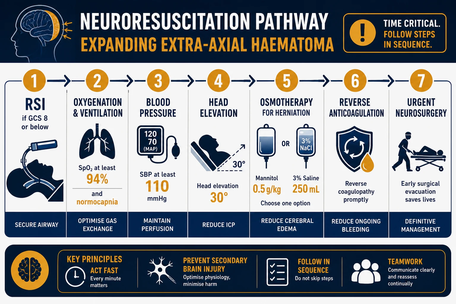
- Ionising — 2 mSv (head), 7 mSv (chest), 10 mSv (abdo/pelvis), higher for CT pulmonary angiogram
- Fast (seconds), high spatial resolution, sensitive to acute haemorrhage, fractures and most solid-organ injury
- Higher dose than X-ray; often needs iodinated contrast; requires transfer to the scanner
- Best for major trauma, stroke, dissection, CTPA, ureteric colic, complex fractures
MRI
- No ionising radiation — radiofrequency and magnetic field
- Superior soft-tissue contrast — tendon, ligament, cord, brain parenchyma, marrow
- Slow (20 to 60 min), claustrophobic, motion-sensitive, ferromagnetic and implant hazards
- Best for spinal cord compression, stroke (diffusion), tendon/ligament injury, septic arthritis, occult fracture
Ultrasound
- No ionising radiation — sound waves; real-time and dynamic
- Bedside, repeatable, cheap, no contrast; can image the unstable patient in resus
- Operator-dependent; limited by body habitus and bowel gas; poor for bone and lung parenchyma
- Best for FAST, AAA, biliary, renal obstruction, DVT, ectopic, cardiac POCUS, vascular access
The effective dose catalog — the numbers to know
The effective dose (in millisieverts, mSv) is the radiation quantity that lets different studies and body regions be compared. The reference catalog, first compiled by Mettler and updated through 2020, is the standard set of numbers.[1][2] Natural background radiation averages 2 to 3 mSv per year — the intuitive unit for explaining dose to patients.
Representative effective doses (mSv) — and the background equivalent
The pregnancy and deterministic thresholds
Differential diagnosis — appropriate versus inappropriate imaging
The examinable differential is not disease-vs-disease but high-yield-versus-low-yield imaging, decided by whether the result will change management, and by the patient's risk profile. [1]
Appropriate imaging
- Answers a defined clinical question that will change a decision (operate, thrombolyse, transfer, discharge)
- Risk-stratified — e.g. Wells score for PE, Canadian CT-head and C-spine rules, PERC rule, Ottawa ankle/knee
- Endorsed by the ACR Appropriateness Criteria or the Choosing Wisely lists
- Modality and dose adjusted for age, pregnancy, renal function and allergy
Inappropriate imaging
- "Just to be sure" — the result will not change management; defensive medicine
- Repeat of a recent study without a new clinical question
- Imaging the low-risk patient against a validated rule (e.g. CT head when the Canadian rule is negative)
- CT in the young for a self-limiting complaint, accumulating lifelong dose
The wrong modality
- A non-contrast CT for suspected aortic dissection — the contrast is mandatory or the test fails
- US for a suspected ureteric stone obscured by bowel gas — low-dose CT KUB is better
- CT for tendon/ligament/cord pathology — MRI is the answer
- X-ray for occult scaphoid or stress fracture — early films are normal; MRI or repeat film
The unstable patient
- CT requires transfer to an unmonitored, distant tube — the deteriorating patient is imaged at the bedside
- FAST, POCUS, mobile CXR and a pelvic X-ray are performed in resus
- A life-saving image (e.g. CTPA in arrest, CT for suspected dissection) is performed with full escort, monitoring and resuscitation drugs
Bedside assessment — the questions to answer before requesting
Five questions precede every request. What is the clinical question, and will the answer change management? If yes, proceed; if not, reconsider. What modality answers it best (bone, soft tissue, real-time, function, vascular)? Is the patient stable enough to travel — the deteriorating patient either is imaged at the bedside or travels with an escort, monitoring and the resuscitation kit. What are the patient-specific risks — pregnancy status, renal function (eGFR), prior contrast reaction, metformin, MRI safety (pacemaker, cochlear implant, ferromagnetic metal), body habitus, breath-hold capacity, agitation and claustrophobia. What is the lowest dose that answers the question — ALARA applied to the CT parameters, the field of view and the use of shielding. [1]
Management — the modality-choice algorithm and the contrast protocol
The algorithm runs in order: clinical question → modality → patient-risk adjustment → ALARA on dose → shield where possible → document the rationale. Stabilise before you image. CT is an unmonitored, distant, noisy tube; an intubated patient is fully controlled before transfer, and the unstable patient travels with a trained escort, full monitoring and the resuscitation drugs. Imaging never precedes the ABCDE primary survey. [1]
The contrast decision — iodinated and gadolinium, with doses
Iodinated contrast (CT). Typical intravenous dose is 1 mL/kg of an iodinated agent at 300 to 370 mg of iodine per mL (commonly 50 to 100 mL for a CTPA, 80 to 120 mL for an abdomen/pelvis). The risk of post-contrast acute kidney injury (PC-AKI) is real but historically overstated for intravenous contrast at typical ED doses; the contemporary incidence is low above an eGFR of 30 mL/min/1.73 m². For the patient at risk, withhold nephrotoxins and give 1 mL/kg/h of isotonic intravenous fluid (normal saline) for several hours pre- and post-contrast; oral hydration is acceptable in the low-risk outpatient.[4] Metformin alone is not a reason to withhold contrast — stop it only for 48 hours after contrast in a patient with an eGFR below 30, or if intra-arterial high-dose contrast is used.
Gadolinium (MRI). Standard dose 0.1 mmol/kg intravenously. The risk is nephrogenic systemic fibrosis (NSF), a devastating fibrosing disorder of skin and viscera seen with the older group I (linear, high-risk) agents in severe renal impairment. With the modern group II (macrocyclic, low-risk) agents (gadoterate, gadobutrol, gadoteridol), the consensus is that the NSF risk is negligible at an eGFR of 30 or above and very low even below it; the ACR-NKF consensus allows group II use in any patient in whom the benefit justifies it, with dialysis the same day for eGFR below 30.[3]
Imaging in pregnancy
Pregnancy concentrates every consideration: the radiation risk applies to the fetus, the contrast risk applies to both, and the default shifts to the non-ionising modalities. The governing principle is ALARA applied to the mother-fetus unit, with no image withheld on dose grounds alone when the question is life-critical.[5]
The order is ultrasound first (appendicitis, ectopic, biliary, AAA, DVT, FAST), MRI second (non-contrast where possible — acute appendicitis in pregnancy, suspected adnexal torsion, spinal cord compression), and CT last, reserved for the limb- or life-threatening problem the first two cannot answer (trauma with suspected solid-organ injury, pulmonary embolism when V/Q is equivocal, suspected dissection). Iodinated contrast crosses the placenta in small amounts and is generally regarded as safe in pregnancy; gadolinium is reserved for the indispensable study because it crosses the placenta and is excreted by the fetal kidney. Shielding the pelvis with lead is standard for non-pelvic X-ray and CT, and the CT parameters are reduced to keep the fetal dose as low as compatible with a diagnostic study. A single CT abdomen delivers a fetal dose well below the 100 mGy deterministic threshold; the stochastic cancer risk is small relative to the risk of a missed ectopic, appendicitis, PE or dissection. [1]
Contrast safety — iodinated AKI and gadolinium NSF
The contrast risks are the examinable safety core. Post-contrast acute kidney injury (PC-AKI) is the rise in creatinine within 48 to 72 hours of iodinated contrast. Historical uncontrolled series inflated the apparent incidence; modern matched data show that the incremental risk from intravenous contrast at ED doses is small, and the genuine risk concentrates in the patient with an eGFR below 30, sepsis, heart failure, diabetes, multiple myeloma or concurrent nephrotoxins. Prevention is intravenous isotonic saline at 1 mL/kg/h for several hours around the study for the high-risk patient, withholding nephrotoxins, and metformin held for 48 hours only when the eGFR is below 30 (it does not cause the AKI; the concern is lactic acidosis if AKI supervenes).[4]
Nephrogenic systemic fibrosis (NSF) is the gadolinium-associated fibrosis of skin, subcutaneous tissue, muscle and viscera, seen predominantly with the group I (linear, high-risk) agents (gadodiamide, gadopentetate, gadoversetamide) in patients with severe renal failure or on dialysis. The condition is largely irreversible and may be fatal. The response was the move to group II (macrocyclic, low-risk) agents and the contemporary ACR–National Kidney Foundation consensus that group II gadolinium has a negligible NSF risk at an eGFR of 30 or above, and a very low risk even below it, such that the benefit-risk balance usually favours giving the contrast when the study is needed. Group I agents are avoided; dialysis is performed the same day for the patient on dialysis or with an eGFR below 30.[3]
Special populations
The pregnant patient is managed with the ultrasound-first, MRI-second, CT-last pathway above; iodinated contrast is generally safe, gadolinium is reserved for the indispensable study, and no life-saving image is withheld on dose grounds. The paediatric patient has a higher tissue sensitivity (more dividing cells and a longer life over which a stochastic cancer may appear), so paediatric CT uses age- and size-adjusted low-dose protocols (the Image Gently principle), prefers ultrasound and MRI where feasible, and shields the gonads. The renally impaired patient is risk-stratified by eGFR — iodinated contrast is given with hydration for the high-risk patient, and group II gadolinium is the default for MRI. The elderly patient accumulates dose over a lifetime of imaging but has a lower stochastic life-expectancy risk; the metformin and polypharmacy interactions are the practical concern. The agitated, intubated or claustrophobic patient needs sedation or anaesthesia for MRI, and a controlled airway before CT transfer. [1]
ALARA — the three pillars of radiation protection
The acronym ALARA — As Low As Reasonably Achievable — encodes the operational doctrine of every imaging department and is derived directly from the linear-no-threshold model. It is not an absolute prohibition on radiation; it is a requirement that every exposure be justified by benefit, optimised to the lowest dose that answers the question, and subject to dose limitation where regulation applies. The International Commission on Radiological Protection (ICRP) articulates these as the three pillars — justification, optimisation, and dose limitation — and the Fellowship candidate must be able to name and define each. [1]
[1]The practical tools that translate ALARA into daily ED practice are the diagnostic reference level (DRL) — a locally or nationally set benchmark dose for a standard examination above which the protocol is audited and revised — and the image gently and image wisely campaigns that promote paediatric and adult dose-reduction protocols. The Fellowship candidate should know that the DRL is set at the 75th percentile of the dose distribution across a representative sample of scanners, and that exceeding it triggers a review of protocol parameters, not necessarily a clinical error. [1]
Pregnancy imaging rules — the 10-day rule, the 28-day rule and universal pregnancy testing
The radiation risk in pregnancy is governed by two principles: the fetus must not receive a deterministic dose, and the stochastic risk must be justified by the clinical need. The historical framework was the 10-day rule — restricting elective radiological examinations involving ionising radiation in women of reproductive age to the first 10 days after the onset of menstruation, on the assumption that the woman is least likely to be pregnant in this window. The 10-day rule has been largely superseded by the 28-day rule (the study may proceed if the woman's last menstrual period was within 28 days, reflecting that organogenesis does not begin until after day 14 and that the all-or-nothing period precedes implantation) and by the contemporary practice of universal serum or urine beta-hCG testing for every woman of reproductive age before any CT that delivers a pelvic dose. [1]
The 10-day rule (historical)
- Restrict elective ionising-radiation studies to days 1 to 10 of the menstrual cycle — when pregnancy is least likely
- Largely superseded but still cited in fellowship exams as the classical framework
- Applies to elective, non-urgent imaging — never to an emergency
The 28-day rule (contemporary)
- An examination may proceed if the LMP was within 28 days, provided the study is justified
- Based on the all-or-nothing principle (pre-implantation) and the absence of organogenesis before day 14
- Standard for most elective imaging in the UK and ANZ
Universal beta-hCG
- A serum or urine beta-hCG is checked for every woman of reproductive age before a CT that delivers a pelvic dose
- Removes the guesswork from the menstrual-history approach
- Does not apply when the imaging is immediately life-saving — never delay the emergency CT for a pregnancy test
Contrast hypersensitivity — the premedication protocol and the breakthrough reaction
Acute contrast reactions are classified as allergic-like (urticaria, bronchospasm, laryngeal oedema, anaphylaxis) or physiological (nausea, vomiting, vasovagal). The modern low-osmolar non-ionic iodinated agents (e.g. iohexol, iopamidol, iodixanol) have reduced the incidence of mild reactions to 0.2 to 0.7 per cent and of severe reactions to 0.02 to 0.04 per cent, a roughly fivefold improvement over the older high-osmolar ionic agents. A prior allergic-like reaction remains the strongest predictor of a repeat reaction; the standard response is premedication and contrast substitution. [1]
The standard corticosteroid-antihistamine premedication regimen
The classical 13-hour regimen (for elective imaging):
- Methylprednisolone 32 mg orally at 12 hours and again at 2 hours before contrast (total 64 mg), OR prednisolone 50 mg orally at 13, 7 and 1 hour before.
- Diphenhydramine 50 mg intravenously or orally at 1 hour before contrast (optional H2 blocker — ranitidine is no longer available in many markets; famotidine is the substitute).
- Plus switching to a different low-osmolar non-ionic agent from the one that caused the original reaction. [1]
The emergency (accelerated) regimen (for urgent imaging where 12 hours is not available):
- Hydrocortisone 200 mg intravenously or methylprednisolone 40 mg intravenously at 1 hour before, then every 4 hours until imaging is complete.
- Diphenhydramine 50 mg intravenously at 1 hour before.
- Acknowledge that the accelerated regimen is less effective — breakthrough reactions occur in approximately 10 per cent of premedicated patients versus 1 per cent with the full 13-hour course. [1]
The immediate reaction (life-saving image, no time for premedication):
- Proceed with contrast after informed consent; use a different low-osmolar agent; have full anaphylaxis kit, adrenaline and airway equipment at the bedside.
The contemporary evidence on premedication is nuanced. The randomised study by Davenport and colleagues showed that switching the contrast agent is at least as effective as — and possibly superior to — steroid pretreatment for preventing breakthrough allergic-like reactions at repeat CT, and that the two strategies combined did not outperform either alone.[8] The practical implication is that for the patient with a prior mild-to-moderate reaction, a different non-ionic low-osmolar agent may be sufficient and avoids the hypoglycaemia, immunosuppression and logistical delay of steroid premedication.
Gadolinium brain deposition — the contemporary caveat
Beyond nephrogenic systemic fibrosis, a second gadolinium concern emerged in 2014: T1-shortening signal hyperintensity in the dentate nucleus and globus pallidus on unenhanced MRI, observed in patients who had received multiple doses of gadolinium-based contrast agents, particularly the linear (group I and group III) agents. Kanda and colleagues demonstrated that this deposition is real, dose-dependent, and persistent.[7] The clinical consequence of brain deposition remains uncertain — no clear neurocognitive or neurologic harm has been attributed to it — but the European Medicines Agency and regulatory bodies worldwide have restricted or withdrawn the linear agents. The practical ED consequence is that macrocyclic (group II) agents (gadoterate, gadobutrol, gadoteridol) are now the default for all gadolinium-enhanced MRI, not only in renal impairment.
The gadolinium agent groups — the examinable comparison
Group I (linear, high risk)
- Gadodiamide (Omniscan), gadopentetate dimeglumine (Magnevist), gadoversetamide (OptiMARK)
- Highest NSF risk — largely withdrawn from the market
- Associated with brain deposition — restricted by the EMA
- Avoid in all patients unless no alternative exists
Group II (macrocyclic, low risk)
- Gadoterate meglumine (Dotarem), gadobutrol (Gadovist), gadoteridol (ProHance)
- Negligible NSF risk at eGFR 30 or above; very low risk even below
- Lower brain deposition than group I agents
- The default gadolinium class for every MRI in contemporary practice
Group III (linear, intermediate)
- Gadoxetate disodium (Eovist/Primovist) — hepatobiliary-specific
- Low NSF risk (hepatic as well as renal excretion)
- Used for focal liver lesion characterisation
- Brain deposition risk intermediate between groups I and II
The landmark evidence — imaging-safety trials
PRESERVE — Weisbord et al, NEJM 2018
New England Journal of Medicine
PMID 29130810
Key finding
A double-blind randomised factorial trial of 5177 patients with stage 3 to 4 chronic kidney disease (eGFR 15 to 44) undergoing angiography, comparing intravenous sodium bicarbonate versus sodium chloride, and oral N-acetylcysteine versus placebo, for the prevention of contrast-associated AKI. No combination reduced the risk of death, dialysis, persistent kidney impairment, or contrast-associated AKI. The trial was stopped early for futility.
Practice change
Sodium bicarbonate and N-acetylcysteine offer no benefit over isotonic saline for the prevention of contrast-associated AKI in the high-risk patient. Normal saline at 1 mL/kg/h is the evidence-based hydration strategy; bicarbonate and NAC can be abandoned for routine intravenous contrast prophylaxis.
Kanda 2016 — brain gadolinium deposition (Jpn J Radiol)
Japanese Journal of Radiology
PMID 26608061
Key finding
A comprehensive review and pooled analysis demonstrating dose-dependent T1-shortening signal hyperintensity in the dentate nucleus and globus pallidus of patients receiving multiple doses of linear gadolinium-based contrast agents. The deposition was persistent on unenhanced MRI, correlated with cumulative gadolinium dose, and was more prominent with linear than with macrocyclic agents.
Practice change
Gadolinium deposits in the brain after repeated exposure, particularly with linear agents. No clinical consequence has been demonstrated, but the precautionary principle has driven the universal shift to macrocyclic group II agents and the withdrawal or restriction of linear agents.
Davenport 2021 — steroid pretreatment versus contrast substitution (Radiology)
Radiology
PMID 34342504
Key finding
A blinded randomised controlled trial of patients with a prior allergic-like contrast reaction undergoing repeat CT, comparing oral steroid pretreatment against switching to a different low-osmolar non-ionic contrast agent. Contrast substitution was at least as effective as steroid pretreatment at preventing breakthrough reactions, and the two combined did not outperform either alone.
Practice change
For the patient with a prior mild-to-moderate contrast reaction, simply switching to a different non-ionic agent is evidence-based and avoids the delay, cost and side effects of corticosteroid premedication. Premedication is still recommended for prior severe reactions and for patients who cannot be switched.
The imaging-request algorithm — the step-by-step pathway
The ED imaging-request decision pathway — from clinical question to the scanner
Clinical question first — what will the image change? If the answer is nothing, do not image. Apply the validated rule (Canadian CT-head, C-spine, Ottawa ankle/knee, PERC, Wells) to justify the request.
Choose the modality — bone and free air to X-ray; acute haemorrhage, fractures, solid organ and vascular to CT; tendon, ligament, cord and marrow to MRI; dynamic, bedside and unstable to ultrasound.
Assess patient stability — the deteriorating patient is imaged at the bedside (FAST, POCUS, mobile CXR, pelvis X-ray). CT requires escort, monitoring and resuscitation drugs; intubate first if the airway or breathing is threatened.
Screen the patient-specific risks — pregnancy status (beta-hCG for every woman of reproductive age before pelvic CT), eGFR before iodinated contrast or gadolinium, prior contrast reaction, metformin use, MRI safety (pacemaker, cochlear implant, ferromagnetic metal), body habitus and breath-hold capacity.
Apply ALARA — request the lowest-dose protocol that answers the question; use low-dose paediatric settings; narrow the field of view; use iterative reconstruction; shield non-targeted tissue.
Choose the contrast strategy — non-ionic low-osmolar iodinated agent for CT; group II macrocyclic gadolinium for MRI; hydrate the high-risk patient at 1 mL/kg/h normal saline; premedicate or substitute for prior reaction; no bicarbonate or NAC needed.
Document the rationale and communicate the result — the request must state the clinical question and the expected change in management; the result is reviewed by a clinician trained to interpret it, not read and dismissed by the untrained.
Clinical pearls — the Fellowship viva cards
[1]Pitfalls and missed diagnoses
The recurring errors are: withholding a life-saving image (suspected SAH, PE, dissection, ruptured AAA, appendicitis in pregnancy) on radiation-dose grounds alone — the deterministic threshold is rarely reached by a single diagnostic CT, but a missed diagnosis is frequently fatal; sending the unstable patient to CT unescorted — stabilise, escort with monitoring and resuscitation drugs, or image at the bedside; ordering the wrong modality (non-contrast CT for suspected dissection, US for the bowel-gas-obscured ureteric stone, X-ray for the occult scaphoid or early stress fracture); reading a scan you are not trained to read and being falsely reassured; not checking the eGFR before gadolinium or high-dose iodinated contrast; not screening MRI safety for pacemakers, cochlear implants and ferromagnetic metal; and the "just-in-case" CT in the young, accumulating lifelong dose for a self-limiting complaint. The cumulatively irradiated patient and the wrongly-imaged patient are the two avoidable harms. [1]
Evidence and regional guidelines
The contemporary framework rests on the ICRP linear-no-threshold model and the ALARA principle (every exposure carries a non-zero stochastic risk, so the dose is kept as low as reasonably achievable), the Mettler effective-dose catalog as the reference set of numbers, the ACR Appropriateness Criteria for the modality-of-choice by indication, and the Choosing Wisely campaign (endorsed by ACEM and the RACP) for the do-not-image lists. The ACR–NKF 2021 consensus governs gadolinium in renal impairment and the group I/II/III stratification.[3] The Canadian Association of Radiologists 2024 guideline governs imaging in the pregnant trauma patient and is the most accessible ANZ-relevant reference.[5]
ANZ practice note. Imaging choice is governed by ALARA, the ACR Appropriateness Criteria and the Choosing Wisely ACEM lists. The eGFR is checked before gadolinium; group II macrocyclic agents are the default; group I are avoided. The pregnant patient is imaged ultrasound-first, MRI-second, CT-last, with pelvis shielding for non-pelvic studies and dose-reduction protocols. No life-saving image is withheld on radiation-dose grounds; the unstable patient is imaged at the bedside or escorted with full monitoring to CT. [1]
Exam pearls
- Effective doses: CXR 0.02 mSv, CT head 2 mSv, CT chest 7 mSv, CT abdomen 10 mSv, background 2 to 3 mSv/yr.
- Stochastic (no threshold, probability rises with dose — cancer) vs deterministic (threshold must be exceeded — skin erythema, cataract, infertility). ALARA derives from the linear-no-threshold model.
- Pregnancy: ultrasound first, MRI second, CT last; never withhold a life-saving image on dose grounds; the deterministic fetal threshold is 100 mGy, far above a single diagnostic CT.
- Gadolinium: group I (linear, high NSF risk) avoid; group II (macrocyclic, low risk) usable at eGFR 30 or above; dialysis the same day if eGFR below 30.
- Iodinated contrast: PC-AKI risk is real but historically overstated for IV ED doses; hydrate the high-risk patient at 1 mL/kg/h; metformin alone is NOT a contraindication — stop 48 h only if eGFR below 30.
- Stabilise before you image: the unstable patient is imaged at the bedside (FAST, POCUS, mobile X-ray) or escorted to CT with full monitoring and resuscitation drugs.
- The request that cannot state what the image will change should not be done — a third of CTs are avoidable and the dose is additive and lifelong.
- ALARA = As Low As Reasonably Achievable — the three ICRP pillars are justification (benefit outweighs harm), optimisation (lowest dose that answers the question), and dose limitation (occupational 20 mSv/yr, public 1 mSv/yr).
- The 10-day rule (elective imaging in the first 10 days of the cycle) is largely superseded by the 28-day rule (proceed if LMP within 28 days) and by universal beta-hCG testing — but is still cited as the classical framework.
- Shielding in pregnancy: 0.5 mm lead-equivalent for gonads/thyroid/breasts in non-targeted fields; shield the pelvis for non-pelvic studies but NEVER shield within the CT field of view (degrades the image and forces a repeat at higher dose).
- PRESERVE (Weisbord, NEJM 2018): sodium bicarbonate and N-acetylcysteine offer NO benefit over isotonic saline for contrast-AKI prophylaxis. The evidence-based hydration strategy is normal saline at 1 mL/kg/h — bicarbonate and NAC can be abandoned.
- Contrast premedication: the full 13-hour regimen is methylprednisolone 32 mg PO at 12 h and 2 h before, plus diphenhydramine 50 mg. The accelerated regimen is hydrocortisone 200 mg IV + diphenhydramine at 1 h before — less effective (10% breakthrough) but pragmatic for urgent imaging. Switching the contrast agent is at least as effective as steroid pretreatment for prior mild reactions.
- Gadolinium brain deposition: repeated linear agents cause dose-dependent T1 hyperintensity in the dentate nucleus and globus pallidus (Kanda 2016) — no clinical harm proven, but the precautionary principle drives the universal shift to group II macrocyclic agents.
- The diagnostic reference level (DRL) is set at the 75th percentile of the dose distribution — exceeding it triggers a protocol audit, not necessarily a clinical error.
- Paediatric CT: use Image Gently low-dose protocols (age and size adjusted), prefer US/MRI, shield the gonads — the lifetime attributable cancer risk is an order of magnitude higher than in the elderly for the same scan. [1]
Exam practice
SAQ — Cauda equina syndrome in a patient with a defibrillator: CT versus MRI and the modality decision
10 minutes · 10 marks
A 45-year-old man presents with the acute onset of severe lower back pain radiating to both legs, progressive bilateral leg weakness over six hours, urinary retention and saddle anaesthesia. He has a known cardiac resynchronisation therapy defibrillator (CRT-D) implanted two years ago for ischaemic cardiomyopathy. He is alert and haemodynamically stable. Examination confirms the dense bilateral leg weakness (MRC grade 3), the saddle anaesthesia and a palpable distended bladder.
SAQ — Suspected appendicitis at 26 weeks gestation: the imaging pathway and the radiation risk in pregnancy
10 minutes · 10 marks
A 27-year-old woman (G2P1) at 26 weeks gestation presents with 18 hours of progressively worsening central abdominal pain that has localised to the right lower quadrant over the last four hours, with anorexia, nausea and two episodes of vomiting. T 38.4, HR 108, BP 108/66, RR 20, SpO2 98 per cent on room air. She is tender in the right lower quadrant with voluntary guarding and a positive Rovsing sign. WCC 15.2 with neutrophilia, CRP 48, urine beta-hCG positive. She is anxious about the radiation to her baby and asks you to avoid a CT.
Red flags
[1]References
- [1]Mettler FA Jr, Huda W, Yoshizumi TT, Mahesh M. Effective doses in radiology and diagnostic nuclear medicine: a catalog Radiology, 2008.PMID 18566177
- [2]Mettler FA Jr. Patient Exposure from Radiologic and Nuclear Medicine Procedures in the United States: Procedure Volume and Effective Dose for the Period 2006-2016 Radiology, 2020.PMID 32181730
- [3]Weinreb JC, Rodby RA, Yee J, et al. Use of Intravenous Gadolinium-based Contrast Media in Patients with Kidney Disease: Consensus Statements from the American College of Radiology and the National Kidney Foundation Radiology, 2021.PMID 33170103
- [4]Rudnick MR, Leonberg-Yoo AK, Literng R, et al. Fluid administration strategies for the prevention of contrast-associated acute kidney injury Curr Opin Nephrol Hypertens, 2022.PMID 35894275
- [5]Qamar SR, Gupta T, Kattan S, et al. CETARS/CAR Practice Guideline on Imaging the Pregnant Trauma Patient Can Assoc Radiol J, 2024.PMID 38813997
- [6]Weisbord SD, Gallagher M, Jneid H, et al. Outcomes after Angiography with Sodium Bicarbonate and Acetylcysteine N Engl J Med, 2018.PMID 29130810
- [7]Kanda T, Osawa M, Oba H, Toyoda K, Kotoku S, Haruyama T, Takeshita K, Furuta S. Brain gadolinium deposition after administration of gadolinium-based contrast agents Jpn J Radiol, 2016.PMID 26608061
- [8]Davenport MS, Asch DJ, Knoepp JD, Cohan RH, Khalatbari S, Ellis JH. Prevention of Allergic-like Reactions at Repeat CT: Steroid Pretreatment versus Contrast Material Substitution Radiology, 2021.PMID 34342504