EM · Procedural & diagnostic ED skills
Focused Assessment with Sonography in Trauma (FAST and E-FAST)
Also known as FAST · Focused Assessment with Sonography in Trauma · E-FAST · Extended FAST · Trauma ultrasound
The FAST (Focused Assessment with Sonography in Trauma): the four views (the Morrison pouch or hepatorenal recess, the splenorenal recess, the pouch of Douglas or rectovesical space, and the pericardial or subxiphoid view), the E-FAST extension (bilateral chest for pneumothorax with lung sliding and the lung point, plus the IVC for volume status), the sensitivity of 60 to 70 per cent for free fluid rising with the repeat scan, the specificity of around 95 per cent, the role in the hypotensive trauma patient (the positive FAST equals the operating theatre, not the CT), the technique with the curvilinear probe, the depth and the gain, and the pitfalls (the false positive from ascites or fat, the false negative from pelvic-only or retroperitoneal bleeding). ACEM-primary, globally tagged.
On this page & tools
Your progress
Saved locally on this device.
5 MCQs with explanations
Target exams
Red flags
Related topics
- The primary survey (ABCDE) — the trauma assessment framework
- Major trauma resuscitation — the team-based systematic approach
- Damage control resuscitation in trauma
- Chest trauma (the immediately and potentially life-threatening injuries)
- Pneumothorax (including tension pneumothorax)
- Abdominal aortic aneurysm (ruptured and intact)
- Point-of-care ultrasound: biliary and renal (and the incidental AAA)
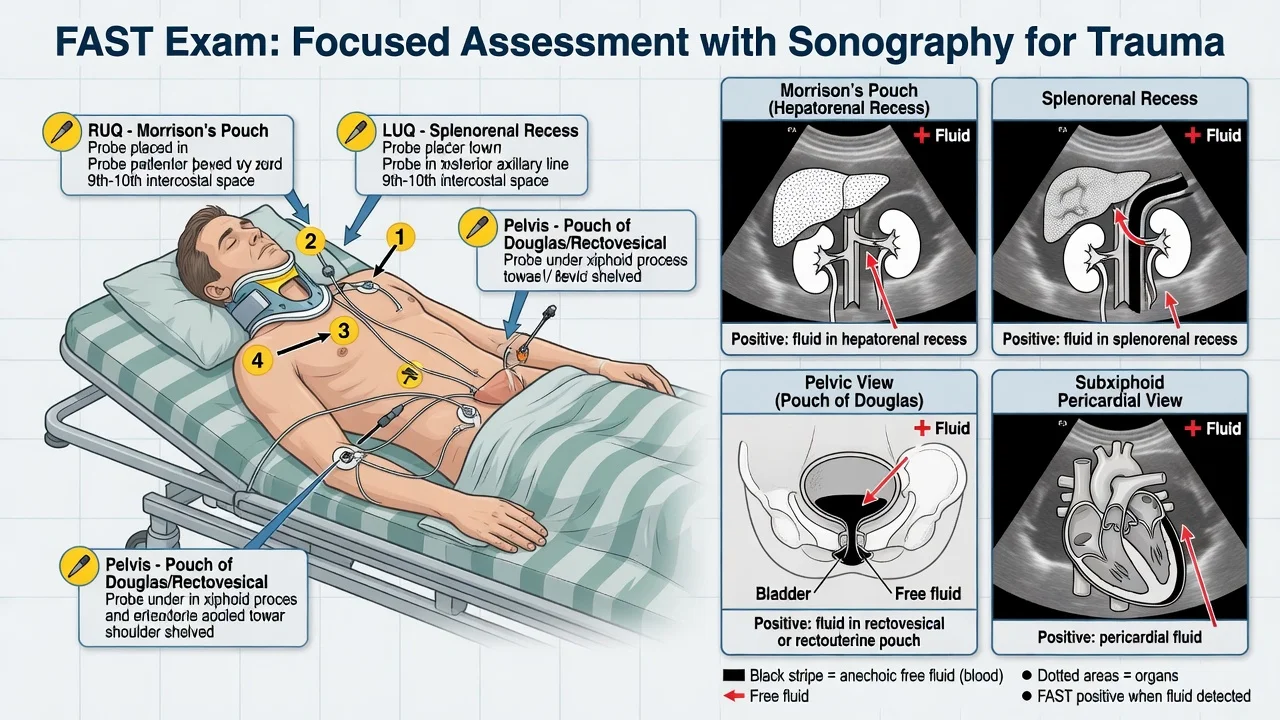
The Focused Assessment with Sonography in Trauma (FAST) is a bedside ultrasound performed by the treating clinician during the primary survey to detect free fluid — in trauma, haemorrhage — in the four most dependent recesses of the torso and around the heart. It is rapid, repeatable, free of ionising radiation and requires no contrast, and it has displaced diagnostic peritoneal lavage from modern trauma resuscitation. Its power is not in detecting an organ injury but in changing the disposition of the haemodynamically unstable patient: a positive FAST in the shocked trauma patient sends that patient to the operating theatre, not the computed-tomography scanner. Its limitation, which the Fellowship candidate must know cold, is the modest sensitivity — around 60 to 70 per cent for free fluid on a single scan — so a negative FAST never excludes injury.[1] The extended exam (E-FAST) adds the bilateral chest for pneumothorax and haemothorax and the inferior vena cava for volume status.[2][3]
Definition and the principle
The FAST answers a single, focused question: is there free fluid in the abdomen, the pelvis or the pericardium? It is a goal-directed, rule-in examination. Free intraperitoneal fluid, given ten minutes or so, gravitates to the most dependent spaces of the supine torso — the hepatorenal recess (the Morrison pouch), the splenorenal recess, and the pelvis around the bladder (the pouch of Douglas in the woman, the rectovesical space in the man) — and free pericardial fluid separates the visceral from the parietal pericardium. Blood and bile appear anechoic (black) on ultrasound; a clot may be hyperechoic and organised. The Morrison pouch is the most sensitive single window and typically shows the first 200 millilitres of free fluid.[1][2]
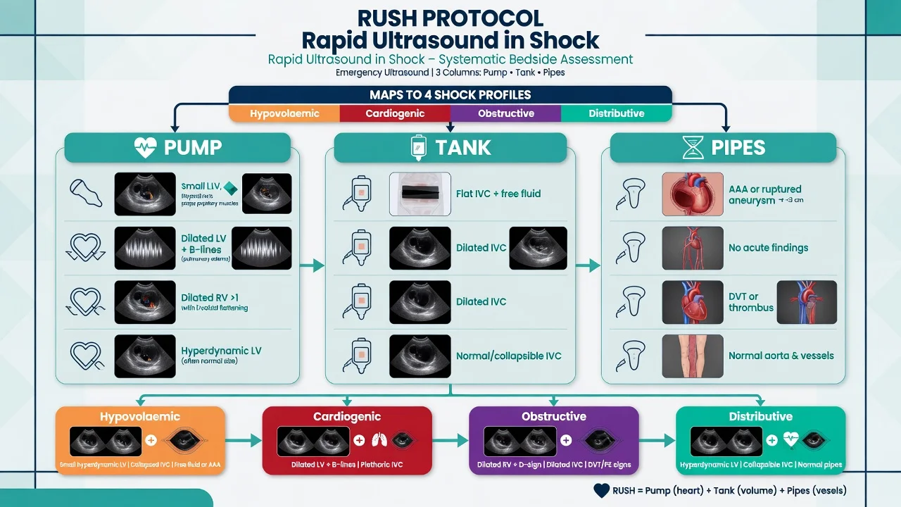
The E-FAST extends the principle to the thorax: a pneumothorax abolishes the normal visceral–parietal pleural sliding and the B-lines, and a haemothorax collects as anechoic fluid above the diaphragm. The inferior vena cava is interrogated for the volume status — a small, collapsing IVC implies hypovolaemia, a plethoric fixed IVC implies right-heart strain or tamponade. [1]
Indications
The FAST is indicated in any patient with significant blunt or penetrating torso trauma in whom free fluid or pericardial blood would change management. The common scenarios are the blunt polytrauma (motor-vehicle crash, fall from height, crush), the penetrating torso injury (stab, gunshot) where intraperitoneal or pericardial breach is suspected, the haemodynamically unstable trauma patient in whom the source of shock must be localised at the bedside, and the pregnant or the anticoagulated trauma patient in whom the threshold to image is lower and the radiation of CT is to be avoided when possible. The FAST is also performed serially — repeated at intervals — in the patient whose initial scan is negative but who remains at risk, because an initially small bleed may accumulate over time. It is part of C (circulation) of the primary survey and is repeated during the secondary survey and as the patient's physiology dictates.[3]
Contraindications — when not to delay for the scan
There is no absolute contraindication to performing a FAST. The contraindication is to delaying a life-saving intervention for the sake of the scan. The patient with an exsanguinating abdominal wound, a peritonitic rigid abdomen, or a clear surgical indication and shock should go to the operating theatre immediately — the FAST is performed on the way, in theatre, or not at all. Equally, the FAST must not interrupt the airway, the breathing or the external-haemorrhage control of the primary survey: it is woven into the circulation assessment once A and B are secured. A technically limited scan (the morbidly obese patient, the patient with extensive subcutaneous emphysema, the uncooperative agitated patient) should be documented as indeterminate rather than falsely read as negative. [1]
Differential diagnosis — the alternative modalities and the mimics
The Fellowship candidate must hold two differential questions at once: which alternative to FAST is appropriate for this patient, and what could mimic a false result. The choice of imaging or disposition hinges on the haemodynamic status, and the mimics explain most of the errors that the scan generates. [1]
Positive FAST + unstable
- Theatre immediately — the operating room, not CT
- Activate the massive haemorrhage protocol, give TXA, crossmatch
- Damage-control laparotomy; the pericardial view may flag tamponade
- Do not delay for further imaging
Positive FAST + stable
- CT with intravenous contrast to define the organ injury and grade it
- Reserve theatre for deterioration or a peritonitic abdomen
- Consider the false positive (ascites, fat, full bladder) before over-calling
- Admit for observation and serial examination
Negative FAST + unstable
- Do not accept as excluding injury — the bleeding may be pelvic-only, retroperitoneal, thoracic or from a long bone
- Repeat the FAST; add the E-FAST chest views and the pelvic X-ray
- If not stabilising with resuscitation, theatre or angioembolisation
- Treat the patient, not the scan
Negative FAST + stable
- CT to exclude solid organ, hollow viscus and retroperitoneal injury
- Admit, observe, serial examination and a serial FAST at intervals
- A negative single scan is reassuring but never definitive
- Watch the elderly, the anticoagulated and the intoxicated closely
The false-positive mimics deserve their own list, because they explain the small but real error rate of an otherwise highly specific test. Ascites in the patient with chronic liver disease, peritoneal dialysate, and the physiological free fluid of a ruptured ovarian cyst or ovulation all read as anechoic fluid. Intraperitoneal fat in the obese patient can mimic fluid at Morrison. A full bladder distends the pelvis and displaces fluid, and can itself be misread as pathology; decompress it before reading the pelvic view. Fluid-filled bowel and perivesical fat are occasional traps. The discipline is to correlate the scan with the clinical picture and to repeat or escalate with CT when the result does not fit. [1]
The false-negative causes are the more dangerous group, because a falsely reassuring negative scan may delay surgery. Pelvic-only bleeding from a pelvic fracture can be confined below the peritoneal reflection and escape the abdominal views. Retroperitoneal bleeding — a renal pedicle injury, a contained pelvic or psoas haematoma — is invisible to intraperitoneal ultrasound. Hollow viscus injury leaks gas more than fluid early on and is notoriously missed. An early scan (within minutes of injury) may precede enough fluid accumulation. Subcutaneous emphysema scatters the ultrasound beam and obliterates the chest views. Morbid obesity degrades every window. The unifying rule is that a negative FAST is a snapshot in time, never a clearance. [1]
Relevant anatomy and the four views
The FAST interrogates four anatomical regions, each chosen because free fluid collects there first in the supine patient. [1]
The four FAST views — HIPE
HIPE
Right upper quadrant — the interface of the liver and the right kidney; the most sensitive single window; ≥200 mL usually visible
The pericardial view through the subxiphoid window; flags haemopericardium and tamponade
Suprapubic transverse and longitudinal; the most dependent pelvic space; decompress the bladder first
Left upper quadrant — the interface of the spleen and the left kidney; harder window, move laterally and posteriorly
The Morrison pouch (the hepatorenal recess) sits between the inferior surface of the right liver lobe and the anterior capsule of the right kidney; free fluid appears as a black stripe in this potential space. The splenorenal recess lies between the spleen and the left kidney, and is the harder window because the stomach gas and the rib cage intervene — the probe is moved posteriorly and the patient is often rolled slightly right. The pelvic view images the bladder (a well-defined anechoic structure) in transverse and longitudinal planes; the pouch of Douglas or rectovesical space lies behind the bladder, where dependent fluid pools. The pericardial view is taken subxiphoid, angled up towards the right shoulder beneath the costal margin, using the liver as an acoustic window; the pericardium is the bright echogenic line, and free fluid appears as an anechoic stripe between the heart and the pericardium. [1]
The E-FAST extension
The extended examination adds the chest and the IVC. The bilateral anterior chest is examined in the second to fourth intercostal spaces at the midclavicular line (right) and midaxillary line (bilaterally) for pneumothorax. The normal pleural interface shows lung sliding — the to-and-fro shimmer of the visceral pleura against the parietal pleura with respiration — and, on M-mode, the seashore sign (a grainy sandy pattern below the pleural line with horizontal wave-like lines above). A pneumothorax abolishes sliding and converts M-mode to the barcode or stratosphere sign (parallel horizontal lines throughout). The lung point — the boundary where sliding lung meets non-sliding pneumothorax — is the most specific sonographic sign of pneumothorax (specificity approaching 100 per cent), though it is insensitive because it is only visible when the pneumothorax is partial and the probe happens to sit on the margin. The chest views also detect a haemothorax as an anechoic collection above the diaphragm. [1]
The inferior vena cava is imaged in the subxiphoid long-axis view a few centimetres from the right atrium. A small (under 1.5 centimetres) IVC that collapses by more than half with a sniff implies a low volume state and hypovolaemia; a plethoric, fixed IVC (over 2.5 centimetres, no collapse) implies right-heart strain, tamponade, or tension pneumothorax — the obstructive causes of shock. The IVC gives a dynamic, repeatable read on volume responsiveness that complements the static vital signs. [1]
Equipment and the machine setup
The FAST is performed with a curvilinear probe (the low-frequency 2 to 5 megahertz abdominal transducer) for the abdominal and pelvic views; a phased-array probe is an acceptable alternative and is preferred for the pericardial and chest views because it fits the intercostal and subxiphoid spaces. The depth is set to 15 to 18 centimetres initially in the adult so that the target organ fills the screen, then adjusted to bring the region of interest to the mid-screen. The gain is set so that the anechoic fluid (the blood) is genuinely black and the parenchyma is mid-grey — too much gain fills free fluid with artefactual echoes and masks small collections. The abdominal preset is selected; the machine is set to two-dimensional mode (B-mode) for the FAST, with M-mode added for the lung sliding assessment. A warm gel and a clean probe cover reduce patient discomfort and infection risk. The probe marker convention is fixed (typically the marker points to the patient's head for longitudinal views and to the patient's right for transverse views) so the images are reproducible. [1]
Patient preparation
The patient is examined supine, fully exposed, as part of the primary survey. The probe is placed directly on the skin with coupling gel, and the four windows are swept to find the optimal image. The scan is integrated into the resuscitation — it is performed by one team member while the airway, the breathing, the access and the haemorrhage control proceed in parallel, and it is interrupted and resumed as the patient demands. Consent is implicit in the emergency setting; the scan is non-invasive and adds no radiation, and the team proceeds under the doctrine of necessity. The bladder is decompressed with a urinary catheter before the pelvic view if a false positive is to be avoided, and the chest leads and the lines are repositioned so they do not obscure the windows. [1]
Stepwise technique — the structured scan
The scan is run to a fixed sequence so that no window is missed under the pressure of the resuscitation. [1]
The FAST and E-FAST, window by window, in order
- Right upper quadrant (Morrison) — place the curvilinear probe in the mid to posterior axillary line at the eighth to eleventh intercostal space, marker to the head. Angle between the ribs to find the liver–kidney interface. A positive scan shows a black anechoic stripe in the recess.
- Left upper quadrant (splenorenal) — move to the left posterior axillary line at the same level, marker to the head. Roll the patient slightly right if stomach gas obscures the view. Identify the spleen and the left kidney; a black stripe in the recess is positive.
- Subxiphoid pericardial — place the probe just below the xiphisternum, marker to the patient's right, angled up towards the right shoulder under the costal margin, using the liver as the window. Identify the pericardium as a bright line around the beating heart; a black stripe between the heart and the pericardium is haemopericardium.
- Pelvis (suprapubic) — place the probe transversely (marker to the patient's right) then longitudinally (marker to the head) two centimetres above the symphysis pubis. Identify the bladder, then the space behind it (pouch of Douglas / rectovesical). A black collection is positive. Decompress the bladder first if a catheter is in place.
- E-FAST right anterior chest — switch to M-mode at the second intercostal space, midclavicular line. Confirm lung sliding and the seashore sign. Absence of sliding and the barcode sign suggest pneumothorax; sweep to seek the lung point.
- E-FAST left anterior chest — repeat on the left side.
- E-FAST chest for haemothorax — return to the upper quadrants and image above the diaphragm; an anechoic collection is a haemothorax.
- E-FAST inferior vena cava — subxiphoid, two-dimensional, a few centimetres from the right atrium. Assess the diameter and the inspiratory collapse for the volume status.
- Document — record a positive or a negative result for each window, the indeterminate windows, and the time. Repeat the scan if the physiology changes or at defined intervals. [1]
The two structural errors are to omit a window because it looked hard, and to declare a single indeterminate view a negative scan. Each window is either positive, negative or indeterminate, and the indeterminate is honestly labelled so it does not falsely reassure. [1]
Drug doses — trauma analgesia and haemorrhage control
The FAST does not itself require any drug, but it is performed within the resuscitation that does. The Fellowship candidate must state the doses for the trauma analgesia and the haemorrhage-control agents that surround the scan. [1]
The drugs that surround the FAST
Morphine (5 milligrams intravenously, titrated in 1 to 2 milligram increments every five minutes to the pain and the sedation) is the standard opioid analgesia for the trauma patient; it blunts the catecholamine surge and the pain without the rapid offset of fentanyl. Its penalties are the hypotension (it is a vasodilator, poorly tolerated in the hypovolaemic) and the respiratory depression, so it is titrated against the conscious level and the blood pressure. Fentanyl (1 microgram per kilogram intravenously, in 25 to 50 microgram aliquots) is the alternative when the offset must be rapid or the blood pressure is precarious, because it preserves the haemodynamics better than morphine. Tranexamic acid (1 gram intravenously over ten minutes within three hours of injury, followed by 1 gram over eight hours) is the haemorrhage-control agent demonstrated by the CRASH-2 trial to reduce all-cause mortality in bleeding trauma; the dose is given early, ideally in the prehospital or the emergency department phase, and is not withheld for the FAST. The massive haemorrhage protocol delivers blood products in a 1:1:1 ratio of packed red cells to fresh frozen plasma to platelets, with the avoidance of crystalloid to prevent the dilutional coagulopathy — and a positive FAST is one of the triggers that activates it. [1]
Complications — the procedural and the interpretive
The FAST is non-invasive and carries no procedural complications beyond the theoretical probe pressure on a tender wound. The real complications are interpretive — the wrong disposition driven by a misread scan. The false positive sends a stable patient with ascites or a cyst to an unnecessary operation, or wastes a CT; the false negative withholds the operation from the patient with pelvic-only or retroperitoneal bleeding who needs it. The indeterminate scan that is reported as negative is the classic trap. The pericardial view may be falsely negative in the patient with a pre-existing pericardial effusion or a pneumopericardium, and the lung views are uninterpretable through subcutaneous emphysema. Each of these is prevented by honest reporting (positive, negative or indeterminate), by correlation with the physiology, and by repeating the scan.[2][3]
Pitfalls and practical tips
The pitfalls are the inverse of the discipline. Accepting a single negative scan as clearance — the patient with an evolving bleed who is discharged or sent to a ward without serial assessment. Missing the retroperitoneum — the renal or pelvic bleed that produces shock with a clean abdominal FAST. Over-reading a full bladder as pelvic fluid — always decompress first. Failing to image the pericardium in the penetrating chest injury — the haemopericardium that progressed to tamponade while the team scanned the abdomen. Setting the gain too high — the artefact that fills a small collection with echoes and hides it. Declaring a window negative because it was hard — the splenorenal view that is skipped for the rib gas and the hidden splenic laceration. Forgetting the E-FAST — the pneumothorax and the haemothorax that the chest views would have found, and the tension pneumothorax that the IVC (plethoric, fixed) would have flagged. The practical tips are the opposite: scan all four windows plus the chest every time, label indeterminate honestly, decompress the bladder before the pelvic view, correlate with the physiology, repeat the scan at intervals and whenever the patient changes, and treat the patient — not the scan. [1]
Post-procedure care and disposition
The disposition hinges on the haemodynamic status and the FAST result together.[1][3] The positive FAST with haemodynamic instability is the strongest indication for immediate laparotomy — the patient is taken to the operating theatre, the massive haemorrhage protocol is running, the TXA is given, and the damage-control laparotomy (control the bleeding, control the contamination, leave the abdomen open, correct the lethal triad, plan the return) is performed. The positive FAST with stability is sent to CT to define and grade the injury, with a low threshold for theatre if the patient deteriorates. The negative FAST with instability is not a clearance — the team searches for the pelvic, retroperitoneal, thoracic and long-bone sources, repeats the scan, images the pelvis, and proceeds to theatre or angiography if the patient does not stabilise. The negative FAST with stability is admitted for observation, serial examination and a serial FAST; CT is performed if the mechanism, the examination or the laboratory picture (a falling haemoglobin, a rising lactate) warrants it. A serial FAST (repeated at 30 minutes, or at any change in physiology) raises the sensitivity as the fluid accumulates, and is the answer to the early negative scan in the patient who remains at risk.
Special populations
The pregnant trauma patient is scanned with the gravid uterus displacing the viscera and the relative hypervolaemia masking the shock until late; the team tilts the uterus left, checks the β-hCG and the Rh status, and remembers that the FAST windows may be distorted — the pericardial and the chest views remain reliable. The paediatric patient is examined with the same views (a smaller probe and a shallower depth), with the recognition that physiological reserve hides the shock and that non-operative management of solid organ injury is the default, so the FAST is a trigger for CT rather than theatre in most stable children. The elderly patient carries the comorbidity, the anticoagulation and the diminished reserve that lower the threshold to scan, to repeat and to CT; the anticoagulated patient bleeds into spaces FAST cannot see, and the negative scan is trusted the least. The obese patient is the technically limited scan, honestly labelled indeterminate where the windows fail. The intoxicated or head-injured patient cannot localise pain, so the FAST and the CT carry the diagnostic load that the history and the examination cannot. [1]
Evidence and regional guidelines
The diagnostic accuracy of the FAST is established in the Rational Clinical Examination systematic review of Nishijima and colleagues, which reported a sensitivity of around 60 to 70 per cent and a specificity of 95 to 98 per cent for intra-abdominal injury on the initial scan, rising with serial examinations; a negative likelihood ratio low enough to support observation but not to exclude injury.[1] The Cochrane reviews of Stengel and colleagues evaluated point-of-care ultrasonography against a reference standard of CT, laparotomy or clinical follow-up in blunt and penetrating trauma, and confirmed that the FAST is most useful as a rule-in test in the unstable patient — its sensitivity is too low to screen out injury in the stable patient.[2][3] The contemporary framework is embedded in the Advanced Trauma Life Support (ATLS) course, the American College of Emergency Physicians / American Institute of Ultrasound in Medicine consensus, and the regional trauma guidelines of Australia, New Zealand, the United Kingdom and Europe, all of which place the FAST in the primary survey as a repeatable, clinician-performed disposition tool, and warn against accepting a single negative scan as clearance.
ANZ practice note. The FAST and E-FAST are core skills of the emergency medicine and trauma training programmes, performed by the treating clinician as part of the primary survey, and reinforced by the ATLS and the Early Management of Severe Trauma (EMST) courses. The positive FAST in the hypotensive patient mandates the operating theatre; the negative scan is repeated at intervals or escalated to CT. The massive transfusion protocol, the tranexamic acid 1 gram loading dose and the 1:1:1 ratio are the standard ANZ resuscitation bundle that surrounds the scan. [1]
SAQs
SAQ — Blunt polytrauma with positive FAST
10 minutes · 10 marks
A 34-year-old man is brought to your major trauma bay after a high-speed motor vehicle crash. He is intoxicated, GCS 13, BP 78/50 mmHg, HR 132 bpm. The primary survey is in progress; airway is patent with cervical spine protection, breath sounds are equal, two large-bore IVs are placed. During the circulation assessment you perform a FAST. The right upper quadrant view shows an anechoic stripe in the Morrison pouch and the pericardial view is negative.
SAQ — Penetrating chest injury and the E-FAST
10 minutes · 10 marks
A 26-year-old man presents after a single stab wound to the left chest, just above the nipple. He is agitated, BP 84/60 mmHg, HR 128 bpm, SpO2 90 per cent on high-flow oxygen, with distended neck veins and reduced breath sounds on the left. You perform an E-FAST during the primary survey. The pericardial view is negative. The left anterior chest view shows absent lung sliding with a barcode sign on M-mode; a lung point is identified laterally. The right upper quadrant view shows a small anechoic stripe.
Exam pearls
- Positive FAST plus hypotension equals the operating theatre, not the CT scanner — the single highest-yield sentence in the topic.
- A negative FAST never excludes injury — the sensitivity is 60 to 70 per cent on a single scan; the retroperitoneum, the hollow viscus and the pelvic-only bleed are invisible.
- The specificity is around 95 per cent — a positive scan in the right context is a reliable trigger to operate.
- The lung point is the most specific sign of pneumothorax (near 100 per cent) but it is insensitive — its absence does not exclude a pneumothorax.
- The seashore sign is normal; the barcode (stratosphere) sign is pneumothorax on M-mode.
- A full bladder produces a false positive at the pelvic view — decompress before you read the pouch of Douglas.
- The Morrison pouch detects around 200 millilitres — the most sensitive single window.
- Repeat the FAST at intervals and whenever the physiology changes; the serial scan raises the sensitivity as the fluid accumulates.
- The IVC is the volume-status adjunct — a small collapsing IVC is hypovolaemia; a plethoric fixed IVC is obstruction (tamponade, tension pneumothorax, massive pulmonary embolism).
- Tranexamic acid 1 gram IV over ten minutes within three hours, then 1 gram over eight hours; morphine 5 milligrams IV titrated; the 1:1:1 ratio for the massive haemorrhage protocol. [1]
Red flags
[1]References
- [1]Nishijima DK, Simel DL, Wisner DH, Holmes JF. Does this adult patient have a blunt intra-abdominal injury? JAMA, 2012.PMID 22496266
- [2]Stengel D, Czebulla C, Achatz G, Ottersbach C, Hildebrand F, Destecke J, Bouillon B. Point-of-care ultrasonography for diagnosing thoracoabdominal injuries in patients with blunt trauma Cochrane Database Syst Rev, 2018.PMID 30548249
- [3]Stengel D, Leisterer J, Ferrada P, Ekkernkamp A, Mutze S, Hoenning A. Emergency ultrasound-based algorithms for diagnosing blunt abdominal trauma Cochrane Database Syst Rev, 2015.PMID 26368505