EM · Acute kidney injury
Acute kidney injury
Also known as AKI · Acute renal failure · Pre-renal uraemia · Acute tubular necrosis
Acute kidney injury (AKI) is an abrupt fall in glomerular filtration producing a rise in creatinine and/or a fall in urine output, classified by KDIGO (creatinine rise of at least 26.5 micromol per litre in 48 hours, or at least 1.5 times baseline within seven days, or urine output under 0.5 mL per kilogram per hour for six hours). Split by site into pre-renal (reduced perfusion, intact tubules), intrinsic (acute tubular necrosis is the emergency exemplar) and post-renal (obstruction). The bedside discriminator is the BUN-to-creatinine ratio over 20 to 1 and a fractional excretion of sodium under 1 per cent for pre-renal disease, against a ratio under 15 and FENa over 2 per cent for intrinsic disease. Management is to treat the cause, give a fluid challenge, stop nephrotoxins, and manage the complications — hyperkalaemia with calcium chloride 10 mL of 10 per cent IV first, then insulin-dextrose and salbutamol, and renal replacement therapy for the AEIOU indications. ACEM-primary, globally tagged.
On this page & tools
Your progress
Saved locally on this device.
5 MCQs with explanations
Target exams
Red flags
Acute kidney injury (AKI) is an abrupt, usually reversible fall in glomerular filtration, manifest as a rise in creatinine or a fall in urine output. It is common, lethal when severe, and — critically for the emergency candidate — the window where intervention reverses the lesion is narrow and measured in hours. The Fellowship examiner tests three things: classification (KDIGO), the pre-renal versus intrinsic versus post-renal split and its urine-chemistry discriminators (BUN-to-creatinine ratio and FENa), and the resuscitation-plus-complication management (treat the cause, fluid challenge, avoid nephrotoxins, control hyperkalaemia, escalate to dialysis under AEIOU). The candidate who reaches for frusemide to "make urine" before securing perfusion fails the station and harms the kidney.[1][2]
Definition and classification
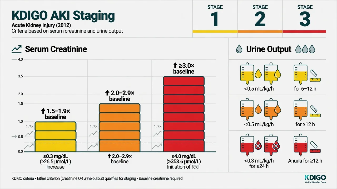
AKI is defined by the KDIGO 2012 criteria, which the candidate must reproduce verbatim. AKI is present when any one of the following holds: a rise in serum creatinine of at least 26.5 micromol per litre (0.3 mg per decilitre) within 48 hours; a rise in creatinine to at least 1.5 times the baseline value, known or presumed to have occurred within the prior seven days; or a urine volume of under 0.5 mL per kilogram per hour for six hours.[1][2]
Severity is then staged 1 to 3, and the stage predicts mortality and guides the threshold for renal replacement therapy. [1]
KDIGO staging of AKI
KDIGO unified the earlier RIFLE (Risk, Injury, Failure, Loss, End-stage) and AKIN (Acute Kidney Injury Network) schemes; the creatinine and urine-output thresholds overlap, and the examiner may use any of the three names. The change in nomenclature from "acute renal failure" to "acute kidney injury" was deliberate — it captures the spectrum from a mild, reversible rise in creatinine through to established tubular necrosis, and it underscores that the mild injury is itself dangerous and worth treating. [1]
Epidemiology and risk
AKI complicates roughly one in five adult hospital admissions and over half of intensive-care admissions; the multinational AKI-EPI study found AKI in approximately 57 per cent of ICU patients, with stage 2 to 3 disease in roughly a third.[3][4] Mortality tracks severity: stage 3 AKI in the critically ill carries a mortality approaching 50 per cent, and even stage 1 independently increases in-hospital death. Survivors carry a markedly elevated risk of progressing to chronic kidney disease. Risk is concentrated in the elderly, in patients with pre-existing CKD, diabetes, heart failure or sepsis, and after major surgery, hypovolaemia, or exposure to nephrotoxins (non-steroidal anti-inflammatories, ACE-inhibitors and angiotensin-receptor blockers, iodinated contrast, aminoglycosides, vancomycin, tenofovir). The commonest single emergency scenario is the volume-deplete septic patient on an ACE-inhibitor who has taken a NSAID and then had a contrast CT.
Pathophysiology — pre-renal, intrinsic, post-renal
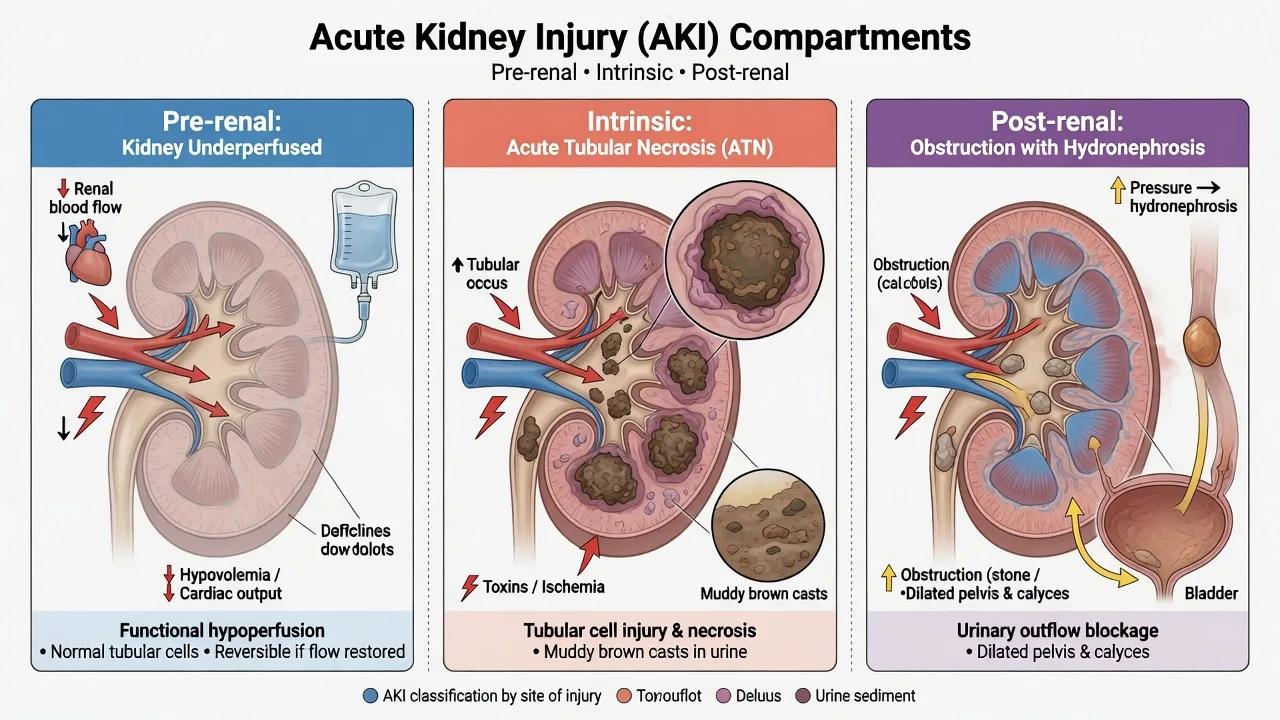
AKI is classified by site into three compartments, and the site decides both the urine chemistry and the treatment. [1]
Pre-renal (reversible)
- Reduced renal perfusion with intact tubules — hypovolaemia, hypotension, sepsis, heart failure, hepatorenal
- Tubule avidly reabsorbs sodium and water: urine sodium under 20, FENa under 1 per cent, BUN:Cr over 20 to 1
- Urine concentrated and bland; muddy casts absent
- Reverses with restoration of perfusion — fluid challenge treats it directly
Intrinsic (ATN exemplar)
- Ischaemic or nephrotoxic tubular injury — prolonged pre-renal, contrast, aminoglycosides, rhabdomyolysis, pigment
- Tubule loses sodium-handling: urine sodium over 40, FENa over 2 per cent, BUN:Cr under 15 to 1
- Muddy brown granular casts on microscopy
- Does NOT reverse with a fluid bolus — remove the insult, support perfusion, await recovery over days to weeks
Post-renal (obstruction)
- Obstruction anywhere from calyx to urethra — stones, BPH, cervical/prostate cancer, retroperitoneal fibrosis, blocked catheter
- Requires obstruction to be bilateral (or single kidney) to raise creatinine
- Bladder palpable/distended; prostate or pelvic mass on exam; hydronephrosis on ultrasound
- Reverses with drainage — catheter, nephrostomy, or stent; do not miss it
The unifying cellular mechanism is tubuloglomerular feedback: a fall in renal blood flow or a direct tubular toxin injures the proximal tubular cell, which releases mediators that constrict the afferent arteriole and collapse the glomerular filtration pressure. In pure pre-renal disease the tubule is structurally intact and simply underperfused, so it conserves sodium; once injury passes a threshold the tubule loses its sodium-pump integrity and becomes ATN — the point at which a fluid challenge no longer reverses the lesion.[1]
Clinical presentation
AKI is usually clinically silent and is discovered on the biochemistry or the urine-output chart, not at the bedside. When symptoms exist they reflect the cause (hypovolaemia, sepsis, obstruction), the uraemic state (nausea, anorexia, confusion, pruritus, pericarditic chest pain), or the complications (dyspnoea from fluid overload, weakness from hyperkalaemia, seizures from severe acidosis). The history interrogates fluid loss (vomiting, diarrhoea, haemorrhage, burns), sepsis source, recent surgery, drug list (NSAIDs, ACE-inhibitors, contrast, antibiotics), and urinary symptoms suggesting obstruction. The single most useful examination finding is the volume status: dry mucosae, low jugular venous pressure and a postural drop point to pre-renal disease and predict a response to fluid, whereas a raised jugular venous pressure, basal crackles and oedema argue against further boluses. [1]
Differential diagnosis
The differential is not a list of diseases but the mechanistic split above, plus the AKI-versus-CKD distinction. AKI on a background of CKD ("acute-on-chronic") is the rule rather than the exception in the elderly renal patient — small kidneys on ultrasound, an anaemia out of proportion to the acute event, and a known baseline creatinine all flag chronicity. The distinguishing features: [1]
AKI (any cause)
- Days of onset; normal-sized kidneys on ultrasound
- Sudden oliguria; sharp creatinine rise over hours to days
- Pre-renal chemistry (FENa under 1 per cent) or ATN chemistry (over 2 per cent)
- Often reversible once the insult is removed
CKD (acute-on-chronic)
- Months of onset; small echogenic kidneys (under 9 cm) on ultrasound
- Insidious; anaemia, hyperphosphataemia, renal bone disease
- Baseline creatinine available in the record; intercurrent trigger for the acute rise
- Treat the acute-on-chronic component; the CKD remains
ATN vs pre-renal
- Pre-renal: FENa under 1 per cent, urine Na under 20, BUN:Cr over 20, urine osmolality over 500
- ATN: FENa over 2 per cent, urine Na over 40, BUN:Cr under 15, urine osmolality under 350
- FENa is invalidated by a recent diuretic — use FEUrea (under 35 per cent supports pre-renal)
- A fluid challenge that fails to improve urine output has converted pre-renal toward ATN
Bedside assessment
Assess and resuscitate in parallel. Secure airway and breathing, attach cardiac monitoring (hyperkalaemia is the lethal complication), and establish intravenous access. Establish the volume status from mucous membranes, jugular venous pressure, capillary refill, lung bases and peripheral oedema, and look for the cause: fever and a source for sepsis, a palpable bladder and an enlarged prostate or pelvic mass for obstruction, a drug chart laden with nephrotoxins, and stigmata of chronic liver or heart failure. Examine for the complications — a pericardial rub (uraemic pericarditis), asterixis (uraemic encephalopathy), and fluid overload. [1]
Send blood for urea and electrolytes with a venous gas (to confirm the potassium and the acid-base immediately), full blood count, creatinine kinase for rhabdomyolysis, calcium, magnesium, phosphate, albumin, coagulation, and a beta-human chorionic gonadotropin in women. Send urine for dipstick, microscopy (casts) and a spot sodium and creatinine for FENa. A bladder scan or in-out catheter excludes retention at the bedside. [1]
Investigations and the discriminators
The venous blood gas delivers the potassium and pH within minutes and sets the urgency. The serum creatinine confirms AKI against a baseline (use the lowest known value in the prior three months); remember creatinine lags the injury and under-represents severity in the small, malnourished patient. Urinalysis narrows the mechanism: bland concentrated urine suits pre-renal disease; muddy brown granular casts and renal tubular epithelial cells on microscopy are the signature of ATN; haematuria and proteinuria with red-cell casts suggest glomerulonephritis; eosinophils and white-cell casts suggest acute interstitial nephritis. [1]
Two urine-chemistry ratios distinguish pre-renal from intrinsic disease and must be reproduced. [1]
[1]Muddy brown granular casts
- The signature of acute tubular necrosis (ATN)
- Pigmented, muddy-brown granular casts plus renal tubular epithelial cells
- Ischaemic or nephrotoxic injury — prolonged pre-renal, contrast, aminoglycosides, rhabdomyolysis
- Does NOT reverse with a bolus — supportive care while the tubule regenerates over 1 to 3 weeks
Red blood cell casts
- Dysmorphic RBCs and RBC casts — the glomerular bleeding signature
- Acute glomerulonephritis (post-streptococcal, IgA, RPGN), anti-GBM, lupus nephritis
- Accompanied by haematuria, proteinuria, hypertension and oedema (nephritic syndrome)
- Urgent renal referral, serology (ANCA, anti-GBM, complement, ANA) and biopsy; immunosuppression may be life-saving
White blood cell casts
- WBC casts and eosinophiluria — the interstitial signature
- Acute interstitial nephritis (AIN) — classically after a beta-lactam (penicillin, cephalosporin), NSAID, or proton-pump inhibitor
- Fever, rash, eosinophilia and a rising creatinine days to weeks after drug exposure form the classic tetrad
- Stop the offending drug; corticosteroids for severe or persistent disease
Bland urine
- Concentrated, clear, cast-free urine with high osmolality
- Pre-renal physiology — the intact tubule concentrates the urine and conserves sodium
- Resolves with restoration of perfusion
- If the urine turns muddy after a failed fluid challenge, pre-renal has converted to ATN
The fractional excretion of urea (FEUrea) is the diuretic-resistant cousin of FENa and the discriminator the candidate must reach for whenever the patient has received frusemide. Urea is reabsorbed passively and partly independently of the sodium-pump that frusemide blocks, so it retains discriminating power after a loop diuretic. Carvounis' landmark Kidney International study showed FEUrea under 35 per cent correctly identified pre-renal azotemia in the diuretic-treated patient where FENa failed.[13] FEUrea = (urine urea × plasma creatinine) ÷ (plasma urea × urine creatinine) × 100.
Urine microscopy and chemistry — the mechanism signatures
Renal tract ultrasound is mandatory in any AKI without a clear pre-renal cause to exclude obstruction (hydronephrosis) and to gauge kidney size and cortical thickness (small kidneys support CKD). A chest radiograph assesses fluid overload, and an electrocardiogram screens for hyperkalaemic change. [1]
Immediate management and resuscitation
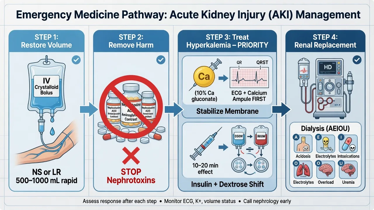
The principles are treat the cause, restore perfusion, stop nephrotoxins, and manage the complications in parallel. Do not reach for frusemide to generate urine before the patient is resuscitated — it converts a hypovolaemic pre-renal state into established ATN. [1]
Treat the cause at the source: control haemorrhage, give antibiotics and source control for sepsis within the hour, drain obstruction with a catheter or nephrostomy, and stop the offending drug. Stop every nephrotoxin on presentation — non-steroidal anti-inflammatories, ACE-inhibitors and angiotensin-receptor blockers, iodinated contrast unless essential, aminoglycosides (switch to a renal-safe regimen), and potassium-sparing agents. Withhold metformin in any AKI (lactic-acidosis risk). [1]
Give a fluid challenge to the volume-deplete patient — 250 to 500 mL of balanced crystalloid (Hartmann's or Plasma-Lyte) over 15 to 30 minutes, and reassess the response by urine output, jugular venous pressure and lung bases before repeating. Normal saline in large volumes causes hyperchloraemic acidosis and worsens renal perfusion, so prefer a balanced solution. The fluid challenge is diagnostic as well as therapeutic: a pre-renal kidney responds within hours; failure to respond means established intrinsic disease or ongoing obstruction, and further blind boluses only cause pulmonary oedema.[6]
Resuscitation doses in AKI
The ED AKI workup in the first 60 minutes
0 to 10 minutes — recognise and protect
Attach cardiac monitoring (hyperkalaemia is the killer), secure two large-bore cannulae, and take a venous gas for the potassium, pH and creatinine immediately. Assess the volume status from the mucous membranes, jugular venous pressure, capillary refill and lung bases — this single finding decides whether the next move is a bolus or a diuretic.
10 to 20 minutes — the bloods and the urine
Draw the full biochemistry with a baseline creatinine from the record, the full blood count, the creatine kinase (rhabdomyolysis), the calcium, phosphate, magnesium, albumin and coagulation; a beta-hCG in women. Send urine for dipstick, microscopy (the casts decide the mechanism) and a spot sodium and creatinine for the FENa. If a loop diuretic was given, order the FEUrea.
20 to 30 minutes — exclude the obstruction at the bedside
Bladder-scan every oliguric or anuric patient, and pass a catheter for retention. A renal tract ultrasound excludes hydronephrosis and gauges the kidney size (small kidneys flag chronicity). Post-renal AKI is the most reversible form and the most embarrassing to miss.
30 to 40 minutes — treat the cause and stop the nephrotoxins
Stop every nephrotoxin on the chart (NSAIDs, ACE-inhibitor, ARB, iodinated contrast, aminoglycoside, metformin). Treat the sepsis with antibiotics and source control within the hour, control the haemorrhage, and address the hepatorenal or the cardiorenal state. The cause is the treatment.
40 to 60 minutes — the fluid challenge and the complication sweep
Give a 250 to 500 mL balanced-crystalloid bolus to the volume-deplete patient and reassess the urine output, JVP and lung bases before repeating. Treat the hyperkalaemia up the ladder (calcium first if there is ECG change, then insulin-dextrose, then salbutamol). Correct the metabolic acidosis and escalate to RRT under AEIOU if conservative measures fail.
Balanced crystalloid (Hartmann, Plasma-Lyte)
- The SMART and SALT-ED trials showed a lower rate of MAKE-30 (major adverse kidney events) with balanced crystalloid than saline
- Avoids the hyperchloraemic metabolic acidosis of large-volume saline that worsens renal vasoconstriction
- The default for the AKI fluid challenge and ongoing matched replacement
- Slightly more expensive; no other downside
Normal (0.9 per cent) saline
- Hyperchloraemic when given in volume; the strong-ion-difference acidosis constricts the renal vasculature and may worsen the AKI
- Reserve for the hyponatraemic or the profoundly hypochloraemic patient, and for the initial bolus where it is cheap and available
- Do not use for sustained or large-volume resuscitation in AKI
- Equivo cal to balanced once >2 L given
Colloid (albumin, gelatin, starch)
- No outcome advantage over crystalloid in AKI; hydroxyethyl starches increase the risk of renal injury and the need for RRT and are contraindicated
- Albumin may be used for the cirrhotic with hepatorenal physiology alongside terlipressin
- Crystalloid is the default; colloid does not "stay in the vessel" long enough to justify the risk in AKI
Definitive management and the escalation ladder
Once resuscitated and the cause addressed, definitive management is supportive while the tubule recovers: maintain perfusion with balanced fluids matched to output, correct electrolyte and acid-base disturbances, treat infection, and nutritionally support. Loop diuretics (frusemide 40 to 80 mg IV) have no role in treating AKI itself — they do not shorten the course or reduce mortality — but may relieve symptomatic fluid overload in the polyuric or responsive patient, and never substitute for dialysis when dialysis is indicated. [1]
Hyperkalaemia is the lethal complication and is treated up the antidote ladder (the candidate must know the order): calcium chloride 10 mL of 10 per cent IV first to stabilise the cardiac membrane whenever there is any ECG change (peaked T, widened QRS, sine wave); then insulin 10 units of soluble insulin with 50 mL of 50 per cent dextrose IV to shift potassium into cells, lowering it by roughly 1 mmol per litre over 15 to 30 minutes; add salbutamol 10 to 20 mg nebulised as a synergistic beta-2 shifter; and remove potassium with gastrointestinal cation-exchange resins or, definitively, haemodialysis.[1]
Renal replacement therapy is indicated when conservative measures fail. The indications are encoded by AEIOU: [1]
Indications for dialysis in AKI
AEIOU
Severe metabolic acidosis (pH under 7.1) refractory to bicarbonate
Refractory hyperkalaemia unresponsive to the shift ladder, or other uncontrolled electrolyte disturbance
Dialysable toxin — lithium, salicylate, methanol, ethylene glycol, metformin
Volume overload refractory to diuretics, with pulmonary oedema
Symptomatic uraemia — pericarditis, encephalopathy, or coagulopathy
The modality — intermittent haemodialysis, continuous renal replacement therapy (CVVHDF), or sustained low-efficiency dialysis — is chosen with the renal and intensive-care teams by haemodynamic stability; continuous therapy suits the shocked patient. Timing of initiation is nuanced: dialyse for an indication, not for a creatinine number alone.[5]
Renal replacement therapy timing — the evidence
The single most examined AKI controversy of the last decade is when to start RRT in the critically ill patient without an absolute indication. Three large multicentre randomised trials frame the modern answer, and the candidate must hold them in the same hand. ELAIN suggested a benefit to early initiation; AKIKI and the much larger STARRT-AKI found no benefit and a signal toward harm (more RRT dependence) with the early strategy. The synthesis: initiate RRT for an indication (AEIOU), not for a creatinine number, and tolerate a period of observation with conservative support in the patient with severe AKI but no absolute indication. [1]
AKIKI — early vs delayed
- Gaudry, NEJM 2016 — 620 critically ill patients with KDIGO stage 3 AKI needing vasopressors or mechanical ventilation
- Early (immediate RRT) vs delayed (RRT only if an absolute indication developed): no difference in 60-day mortality (49 vs 50 per cent)
- RRT was ultimately avoided in half of the delayed group — supporting the "wait for an indication" strategy
- No increase in mortality from waiting
STARRT-AKI — accelerated vs standard
- Wald, NEJM 2020 — 3019 critically ill patients with KDIGO stage 2 to 3 AKI, the largest trial of RRT timing
- Accelerated (RRT within 12 h of eligibility) vs standard (RRT for an indication or persistent AKI): no mortality difference at 90 days
- Adverse events (chiefly catheter-related infection and bleeding) and RRT dependence at 90 days were HIGHER in the accelerated group
- Established the modern standard: do not start RRT early in the absence of an indication
ELAIN — early vs delayed
- Zarbock, JAMA 2016 — 231 surgical ICU patients with KDIGO stage 2 AKI and plasma neutrophil gelatinase-associated lipocalin over 150
- Early (within 8 h of stage 2) vs delayed (within 12 h of stage 3): 90-day mortality reduced (24 vs 39 per cent)
- Smaller, single-centre, selected (post-cardiac-surgery) population; the benefit has not been reproduced in AKIKI or STARRT-AKI
- The outlier that has not changed the cautious consensus
AKIKI-2 — very-delayed vs delayed
- Gaudry, Lancet 2021 — 278 patients with severe AKI, no absolute indication, randomised at 72 h to immediate vs further-delayed RRT
- No difference in the composite of 60-day death or RRT dependence; the trial was stopped early
- Adds nuance: even among those started late, a brief further watch is reasonable, but equipoise remains
AKIKI — early vs delayed RRT in stage 3 AKI (Gaudry, NEJM 2016)
New England Journal of Medicine
PMID 27181456
Key finding
A multicentre randomised trial of 620 critically ill patients with KDIGO stage 3 AKI needing vasopressors or mechanical ventilation, comparing the early strategy (RRT within 6 hours of randomisation) against the delayed strategy (RRT only if an absolute indication — refractory hyperkalaemia, acidosis, overload, or uraemia — or persistent AKI after 6 days). The 60-day mortality was identical at 49 versus 50 per cent, and RRT was avoided entirely in roughly half of the delayed group.
Practice change
Initiating RRT early in severe AKI without an absolute indication does not improve survival and exposes the patient to the dialysis catheter and circuit. The modern standard is to wait for an AEIOU indication. AKIKI underpins the conservative strategy.
STARRT-AKI — accelerated vs standard RRT timing (Wald, NEJM 2020)
New England Journal of Medicine
PMID 32668114
Key finding
The largest trial of RRT timing — 3019 critically ill patients with KDIGO stage 2 to 3 AKI, randomised to the accelerated strategy (RRT within 12 hours of eligibility) versus the standard strategy (RRT initiated for an absolute indication or persistent severe AKI). The 90-day mortality was unchanged (43.9 versus 43.7 per cent), but adverse events — predominantly dialysis-catheter-related bloodstream infection and bleeding — and RRT dependence at 90 days were significantly higher in the accelerated group.
Practice change
The definitive trial against the early-RRT reflex: starting dialysis early in severe AKI without an indication increases the catheter and circuit harm without any survival gain, and leaves more patients dialysis-dependent at three months. Dialyse for an AEIOU indication, not a creatinine trajectory.
ELAIN — early vs delayed RRT (Zarbock, JAMA 2016)
JAMA
PMID 27209269
Key finding
A single-centre randomised trial of 231 predominantly post-cardiac-surgical ICU patients with KDIGO stage 2 AKI and a plasma neutrophil gelatinase-associated lipocalin over 150, randomised to early RRT (within 8 hours of stage 2) versus delayed RRT (within 12 hours of stage 3). The 90-day mortality fell from 39 to 24 per cent in favour of the early arm, and the early group had fewer complications and shorter stays.
Practice change
The positive outlier among the timing trials. The benefit has not been reproduced in the larger AKIKI and STARRT-AKI trials, and the single-centre, post-surgical, biomarker-selected population limits the generalisability. The trial is cited for completeness but does not overturn the indication-based consensus.
AKIKI-2 — very-delayed vs delayed RRT (Gaudry, Lancet 2021)
Lancet
PMID 33812488
Key finding
A multicentre randomised trial of 278 patients with severe AKI, no absolute RRT indication at 72 hours, randomised to immediate RRT versus a further-delayed strategy. No difference in the composite outcome of 60-day death or RRT dependence; the trial was stopped early for futility.
Practice change
Among patients already selected for a conservative strategy, a brief further watch remains reasonable — but the equipoise is genuine, and most units initiate RRT by 72 hours of severe oliguric AKI even without an absolute indication.
Subtypes and special scenarios
Contrast-associated AKI follows iodinated contrast in the at-risk patient (CKD, diabetes, hypovolaemia); prevention is volume expansion with isotonic fluid and avoidance of nephrotoxins, not prophylactic N-acetylcysteine (the evidence is equivocal). Rhabdomyolysis-induced AKI (creatine kinase in the thousands, pigment casts, myoglobin) demands aggressive isotonic fluid expansion to drive a urine output of 1 to 2 mL per kilogram per hour; alkaline diuresis is now debated. Acute interstitial nephritis (eosinophiluria, rash, fever, after a beta-lactam, NSAID or proton-pump inhibitor) requires drug cessation and, for severe disease, corticosteroids. Hepatorenal syndrome in cirrhosis is a pre-renal physiology unresponsive to fluid, treated with albumin and terlipressin. Glomerulonephritis (red-cell casts, hypertension, oedema) demands urgent renal referral, serology and biopsy. [1]
Contrast-associated AKI and the PRESERVE verdict
The earlier enthusiasm for prophylactic N-acetylcysteine and sodium bicarbonate infusions before iodinated contrast was overturned by the PRESERVE trial: in 5177 high-risk patients undergoing angiography, neither intravenous sodium bicarbonate nor acetylcysteine reduced the risk of contrast-associated AKI, dialysis, or death compared with isotonic saline and placebo. The prevention of contrast-associated AKI is now isotonic volume expansion and the avoidance of other nephrotoxins — bicarbonate and N-acetylcysteine add nothing.[14]
PRESERVE — bicarbonate and N-acetylcysteine for contrast-associated AKI (Weisbord, NEJM 2018)
New England Journal of Medicine
PMID 29130810
Key finding
A 2-by-2 factorial randomised trial of 5177 patients with CKD undergoing angiography, comparing intravenous sodium bicarbonate versus isotonic saline, and N-acetylcysteine versus placebo. Neither bicarbonate nor N-acetylcysteine reduced the composite of death, need for dialysis, or persistent kidney impairment at 90 days, nor the rate of contrast-associated AKI.
Practice change
The definitive negative trial: abandon routine sodium bicarbonate and N-acetylcysteine prophylaxis. Isotonic saline volume expansion and holding other nephrotoxins are the only evidence-based prevention for contrast-associated AKI.
SMART — balanced crystalloids versus saline in critically ill adults (Semler, NEJM 2018)
New England Journal of Medicine
PMID 29485925
Key finding
A pragmatic crossover cluster-randomised trial of 15 802 critically ill adults in five ICUs, comparing balanced crystalloids (Hartmann or Plasma-Lyte) against 0.9 per cent saline. Balanced crystalloids reduced the composite outcome of MAKE-30 (death, new RRT, or persistent renal dysfunction) from 15.4 to 14.3 per cent, driven mainly by fewer new RRT starts.
Practice change
Balanced crystalloid is the preferred resuscitation fluid in the critically ill patient with or at risk of AKI — the saline-induced hyperchloraemic acidosis is a real and avoidable renal injury. Use Hartmann or Plasma-Lyte for the AKI fluid challenge.
SALT-ED — balanced crystalloids versus saline in noncritically ill adults (Self, NEJM 2018)
New England Journal of Medicine
PMID 29485926
Key finding
A single-centre randomised trial of 13 347 noncritically ill adults seen in the emergency department and admitted to a ward, comparing balanced crystalloids against saline. Balanced crystalloids reduced the MAKE-30 outcome from 5.6 to 4.7 per cent, again driven by fewer new RRT events.
Practice change
The benefit of balanced crystalloid extends to the ED and the ward patient, not only the ICU. The fluid choice for the AKI resuscitation in the emergency department is balanced crystalloid.
Aetiology memory aids by compartment
Pre-renal (perfusion failure)
- Hypovolaemia — haemorrhage, vomiting, diarrhoea, burns, pancreatitis, third-spacing
- Heart failure and cardiogenic shock — the underperfused failing kidney
- Sepsis — the #1 cause of AKI in the ICU; vasodilatory hypoperfusion plus direct tubular injury
- Hepatorenal syndrome — pre-renal physiology unresponsive to fluid in cirrhosis
- NSAIDs constrict the afferent; ACE-inhibitors dilate the efferent — both drop GFR
Intrinsic (parenchymal)
- ATN from ischaemia or nephrotoxins (contrast, aminoglycosides, rhabdomyolysis, pigment) — the emergency exemplar
- AIN from drugs — penicillins and cephalosporins, NSAIDs, proton-pump inhibitors; the classic fever-rash-eosinophilia tetrad
- Acute glomerulonephritis — post-streptococcal, IgA nephropathy, RPGN, anti-GBM, lupus nephritis; RBC casts
- Vascular — renal vein thrombosis, cholesterol emboli, malignant hypertension, TTP-HUS, scleroderma renal crisis
- Intra-renal: the muddy-brown-cast compartment
Post-renal (obstruction)
- Lower tract — benign prostatic hyperplasia (BPH), prostate cancer, blocked urinary catheter, urethral stricture
- Upper tract — bilateral ureteric stones, single-kidney ureteric stone, retroperitoneal fibrosis, pelvic malignancy (cervical, colorectal, bladder)
- Must be bilateral (or single kidney) to raise the creatinine
- Reversible with drainage — catheter, nephrostomy, or ureteric stent; hydronephrosis on ultrasound
Complications and pitfalls
The complications are fluid, electrolyte and acid-base: hyperkalaemia (the killer), metabolic acidosis (a high anion gap from retained acids), fluid overload and pulmonary oedema, uraemic pericarditis and encephalopathy, anaemia from blunted erythropoietin, infection (the leading cause of death), and bleeding from platelet dysfunction. The pitfalls are the mirror of the management: reaching for frusemide before resuscitating; continuing ACE-inhibitors or NSAIDs in an evolving AKI; missing a drainable obstruction; trusting a FENa taken after a diuretic; over-resuscitating into pulmonary oedema; and delaying dialysis when the hyperkalaemic ladder has failed. [1]
Prognosis and disposition
Outcome tracks severity, cause, and the speed of correction. Pre-renal and post-renal AKI recover fully when the insult is removed promptly; ATN recovers over one to three weeks but carries a mortality that rises with stage. Disposition: stage 1 AKI with a reversible cause may be managed on a ward with daily biochemistry; stage 2 to 3 disease, hyperkalaemia, fluid overload, or any dialysis indication is admitted to a high-dependency or intensive-care bed with continuous monitoring. Survivors need follow-up creatinine, as even recovered AKI raises the long-term risk of chronic kidney disease.[3][4]
Special populations
The elderly have less renal reserve and accumulate nephrotoxins; a small creatinine rise in an 80-year-old is a large loss of filtration. Pre-existing CKD is the single strongest risk factor and is usually the substrate for acute-on-chronic injury. Sepsis is the commonest ICU cause and combines vasodilatory pre-renal hypoperfusion with direct tubular injury. Post-operative patients (especially cardiac and major abdominal surgery) develop AKI from hypoperfusion and third-spacing. Cirrhosis brings hepatorenal physiology. The diabetic on metformin needs the drug withheld and lactic acidosis considered when AKI coexists. [1]
Evidence and regional guidelines
The contemporary framework is the KDIGO 2012 Clinical Practice Guideline for Acute Kidney Injury, summarised operationally by Kellum and Lameire, which codifies the diagnostic criteria, staging, the work-up, and the management principles reproduced above, and is endorsed across ANZ, UK and US practice.[7] The definitive reviews of classification, pathophysiology and management are the Ronco–Bellomo–Kellum Lancet seminar and the Lameire–Vanholder Lancet review; the epidemiology is captured by the world-incidence meta-analysis and the AKI-EPI multinational ICU study.[1][2][3][4] The modern answer to when to start RRT is settled by the three large timing trials — AKIKI and the much larger STARRT-AKI showed no benefit and a harm signal with the early strategy, while the single-centre ELAIN remains the unreproduced positive outlier, and AKIKI-2 added nuance to the delayed arm — together establishing the indication-based consensus.[8][9][10][15] The fluid choice is settled by the SMART and SALT-ED trials in favour of balanced crystalloid over saline for the AKI resuscitation, and the contrast-associated AKI prophylaxis is settled by PRESERVE in favour of isotonic saline alone (no bicarbonate, no N-acetylcysteine).[11][12][14] The timing of dialysis more broadly is addressed in the Macedo–Mehta review, and the harm of fluid overload is detailed in the O'Connor–Prowle review.[5][6] The fractional excretion of urea as the diuretic-resistant discriminator was validated by Carvounis.[13]
ANZ practice note. AKI is managed under KDIGO via hospital renal and intensive-care protocols. The emergency role is to diagnose and stage by KDIGO, classify pre-renal versus intrinsic versus post-renal using FENa and the urine sediment, give a balanced-crystalloid fluid challenge (250 to 500 mL) and reassess, stop every nephrotoxin, and treat hyperkalaemia up the ladder — calcium chloride 10 mL of 10 per cent IV first when there is ECG change, then insulin 10 units with 50 mL of 50 per cent dextrose and salbutamol 10 to 20 mg nebulised. Dialysis is organised under the AEIOU indications in consultation with nephrology, with continuous renal replacement therapy for the haemodynamically unstable patient. Metformin is withheld in any AKI. [1]
Exam pearls
- KDIGO verbatim — at least 26.5 micromol per litre creatinine rise in 48 hours, OR at least 1.5 times baseline in seven days, OR urine output under 0.5 mL per kilogram per hour for six hours. Stage 3 includes the initiation of renal replacement therapy.
- Pre-renal vs ATN urine chemistry — FENa under 1 per cent, urine sodium under 20, BUN:Cr over 20 to 1, urine osmolality over 500 is pre-renal; the reverse is ATN. Diuretics invalidate FENa — switch to FEUrea under 35 per cent.
- Muddy brown granular casts equal ATN — the single best microscopy discriminator for intrinsic tubular injury.
- First drug in hyperkalaemic AKI with ECG change is calcium — chloride 10 mL of 10 per cent IV; it stabilises the membrane but does not lower potassium, so always pair it with insulin-dextrose.
- Frusemide does not treat AKI — it relieves overload and does not shorten recovery; never substitute it for resuscitation or dialysis.
- AEIOU — Acidosis, Electrolytes, Intoxication, Overload, Uraemia — the dialysis indications.
- RBC casts equal glomerulonephritis, WBC casts plus eosinophils equal acute interstitial nephritis — pair the cast to the mechanism; AIN follows penicillins, NSAIDs and PPIs.
- Sepsis is the commonest cause of AKI in the ICU — it combines vasodilatory pre-renal hypoperfusion with direct tubular injury, and is the master diagnosis to exclude.
- NSAIDs constrict the afferent arteriole, ACE-inhibitors dilate the efferent — both drop the glomerular filtration pressure and tip the borderline kidney into AKI; stop both on presentation.
- FEUrea under 35 per cent is the diuretic-resistant discriminator — Carvounis showed it identifies pre-renal azotemia where FENa fails after frusemide.
- Post-renal AKI needs bilateral obstruction (or a single kidney) to raise creatinine — BPH, prostate or cervical cancer, bilateral stones, retroperitoneal fibrosis; drain it and it recovers.
- Creatinine lags the injury — a normal creatinine in the small, malnourished or early patient does not exclude a large loss of filtration; the urine output and the trend are the frontline.
- Normal saline in large volume causes hyperchloraemic acidosis — the SMART and SALT-ED trials made balanced crystalloid (Hartmann, Plasma-Lyte) the default for the AKI fluid challenge.
- PRESERVE killed the bicarbonate-and-N-acetylcysteine reflex — isotonic saline volume expansion and holding nephrotoxins are the only proven prevention for contrast-associated AKI.
- AKIKI and STARRT-AKI settled the RRT-timing question — start dialysis for an AEIOU indication, not for a creatinine number; the early strategy adds catheter harm and RRT dependence without a survival gain. ELAIN is the unreproduced positive outlier.
- Withhold metformin in any AKI — the renally-cleared drug accumulates and precipitates lactic acidosis; check the lactate and the pH in the metformin-treated patient.
- Hydroxyethyl starch is contraindicated in AKI — colloids add no outcome benefit over crystalloid and the starches increase the renal injury and the need for RRT.
- A single normal creatinine never rules out AKI in the elderly — the 80-year-old loses a large filtration fraction for a small creatinine rise; stage by KDIGO and trend the urine output. [1]
Model answer — oliguric AKI with hyperkalaemia and ECG change
Exam practice
SAQ — Septic AKI with life-threatening hyperkalaemia and ECG change
10 minutes · 10 marks
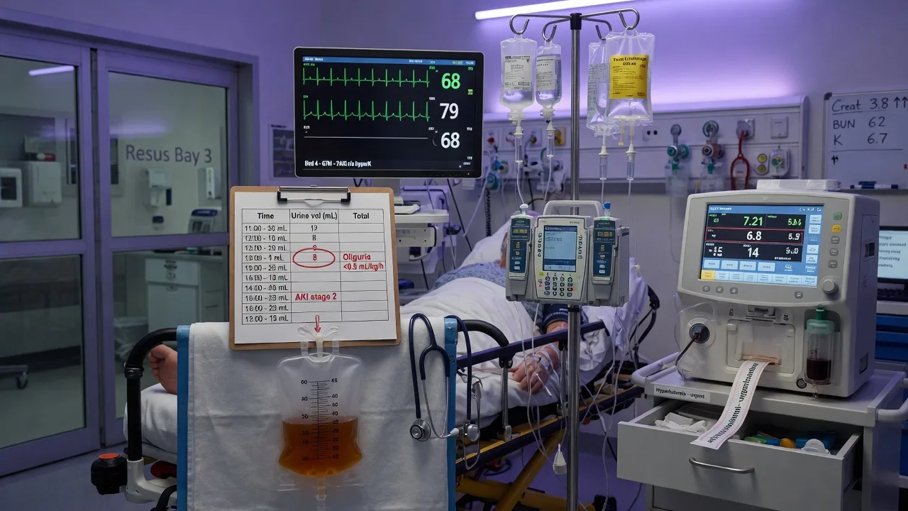
A 68-year-old man with type 2 diabetes, hypertension on ramipril 10 mg daily, and benign prostatic hypertrophy presents with three days of dysuria, fevers and rigors, and 24 hours of oliguria (urine output 10 mL per hour). He has been taking ibuprofen 400 mg three times daily for flank pain. On arrival he is confused (GCS 14), temperature 38.8 degrees C, HR 128, BP 84/52 (MAP 62), RR 26, SpO2 94 per cent on room air. Urine dipstick shows nitrites, large leucocytes and blood. Venous gas: pH 7.24, bicarbonate 14, potassium 7.1 mmol/L, lactate 4.2, creatinine 320 (baseline 95 three months ago). ECG shows widened QRS at 132 ms with peaked T waves and a rate of 128.
SAQ — Pre-renal versus intrinsic AKI: the diuretic-confounded urine chemistry
10 minutes · 10 marks
A 72-year-old woman is admitted with a three-day history of vomiting and diarrhoea from gastroenteritis. She takes frusemide 40 mg daily for heart failure with reduced ejection fraction, ramipril 10 mg daily, and spironolactone 25 mg daily. On arrival she is clinically dry: HR 108, BP 96/60 with a postural drop, JVP not visible, dry mucosae, and no peripheral oedema. Creatinine 280 micromol per litre (baseline 110 four months ago), urea 24 mmol per litre, potassium 3.4, sodium 132. Spot urine sodium 65 mmol per litre, urine creatinine 8 mmol per litre — FENa 2.4 per cent. Urine microscopy shows no muddy casts, no red-cell or white-cell casts; the sediment is bland.
Red flags
[1]References
- [1]Ronco C, Bellomo R, Kellum JA. Acute kidney injury Lancet, 2019.PMID 31777389
- [2]Lameire N, Van Biesen W, Vanholder R. Acute renal failure Lancet, 2005.PMID 15680458
- [3]Susantitaphong P, Cruz DN, Cerda J, et al. World incidence of AKI: a meta-analysis Clin J Am Soc Nephrol, 2013.PMID 23744003
- [4]Hoste EA, Bagshaw SM, Bellomo R, et al. Epidemiology of acute kidney injury in critically ill patients: the multinational AKI-EPI study Intensive Care Med, 2015.PMID 26162677
- [5]Macedo E, Mehta RL. Timing of dialysis initiation in acute kidney injury and acute-on-chronic renal failure Semin Dial, 2013.PMID 24016050
- [6]O'Connor ME, Prowle JR. Fluid Overload Crit Care Clin, 2015.PMID 26410146
- [7]Kellum JA, Lameire N; KDIGO AKI Guideline Work Group. Diagnosis, evaluation, and management of acute kidney injury: a KDIGO summary (Part 1) Crit Care, 2013.PMID 23394211
- [8]Gaudry S, Hajage D, Schortgen F, et al. Initiation Strategies for Renal-Replacement Therapy in the Intensive Care Unit N Engl J Med, 2016.PMID 27181456
- [9]The STARRT-AKI Investigators; Wald R, Adhikari NKJ, et al. Timing of Initiation of Renal-Replacement Therapy in Acute Kidney Injury N Engl J Med, 2020.PMID 32668114
- [10]Zarbock A, Kellum JA, Schmidt C, et al. Effect of Early vs Delayed Initiation of Renal Replacement Therapy on Mortality in Critically Ill Patients With Acute Kidney Injury: The ELAIN Randomized Clinical Trial JAMA, 2016.PMID 27209269
- [11]Semler MW, Self WH, Wanderer JP, et al.; SMART Investigators and the Pragmatic Critical Care Research Group. Balanced Crystalloids versus Saline in Critically Ill Adults N Engl J Med, 2018.PMID 29485925
- [12]Self WH, Semler MW, Wanderer JP, et al.; SALT-ED Investigators. Balanced Crystalloids versus Saline in Noncritically Ill Adults N Engl J Med, 2018.PMID 29485926
- [13]Carvounis CP, Nisar S, Guro-Razuman S. Significance of the fractional excretion of urea in the differential diagnosis of acute renal failure Kidney Int, 2002.PMID 12427149
- [14]Weisbord SD, Gallagher M, Jneid H, et al.; PRESERVE Trial Group. Outcomes after Angiography with Sodium Bicarbonate and Acetylcysteine N Engl J Med, 2018.PMID 29130810
- [15]Gaudry S, Hajage D, Martin-Lefevre L, et al. Comparison of two delayed strategies for renal replacement therapy initiation for severe acute kidney injury (AKIKI 2): a multicentre, open-label, randomised, controlled trial Lancet, 2021.PMID 33812488