EM · Vertigo and dizziness
Vertigo and dizziness
Also known as Acute vestibular syndrome · Peripheral vertigo · Central vertigo · BPPV · Vestibular neuritis
Vertigo and dizziness — the central-versus-peripheral distinction as the load-bearing decision, the HINTS examination (Head Impulse, Nystagmus, Test of Skew) for stroke in the acute vestibular syndrome, the Bárány diagnostic criteria and the Epley manoeuvre for BPPV, vestibular neuritis and labyrinthitis, Meniere disease, the differential (posterior-circulation TIA and stroke, vestibular migraine, cardiac syncope), and the drug doses (prochlorperazine 5 mg, corticosteroids). ACEM-primary, globally tagged.
On this page & tools
Your progress
Saved locally on this device.
2 MCQs with explanations
Target exams
Red flags
Vertigo and dizziness account for up to 5 per cent of emergency department attendances and are deceptively heterogeneous: at one end sits a self-limiting bppv that a single bedside manoeuvre cures, at the other a posterior-circulation stroke whose only feature is vertigo. The Fellowship task is not to classify every dizzy patient but to make one decision reliably — is this a central (potentially dangerous) or a peripheral (usually benign) vestibular problem? — because that decision determines whether the patient is discharged to vestibular rehabilitation or admitted to a stroke pathway. The single most reliable tool for that decision in the acute vestibular syndrome is the HINTS examination, which in the hands of a trained clinician is more sensitive for stroke than early MRI.[1][2]

Definition and classification
Dizziness is the patient's word and is deliberately broad; vertigo is the physician's term for the illusion of motion (spinning, swaying, tilting) of either the self or the surroundings. The standard teaching framework, the Drachman quartet, divides dizziness into vertigo (a vestibular problem), presyncope (cardiovascular, near-faint), disequilibrium (a gait/balance disorder, often neurological), and light-headedness (psychiatric, hyperventilation). The first job at the bedside is to decide which quartet the patient is in, because each has a different dangerous differential: presyncopal dizziness is worked up for a cardiac cause, while true vertigo is worked up for a central-versus-peripheral cause. [1]
Vertigo itself is classified by its temporal pattern into the acute vestibular syndrome (AVS) — continuous vertigo lasting days to weeks with nausea, vomiting and gait instability, typically vestibular neuritis, labyrinthitis or a posterior-circulation stroke — the triggered episodic pattern, where vertigo is provoked by a specific movement or position (BPPV, orthostatic hypotension) — and the spontaneous episodic pattern (Meniere disease, vestibular migraine, TIA). The pattern plus the HINTS examination localises the lesion: the AVS is the high-stakes presentation, because its differential includes stroke. [1]
Vertigo (vestibular)
- The illusion of motion — spinning, swaying or tilting of the self or the surroundings
- Peripheral causes: BPPV, vestibular neuritis, labyrinthitis, Meniere disease; central causes: stroke/TIA, MS, tumour, vestibular migraine
- Nystagmus on examination; nausea and vomiting; either triggered by position or continuous
- Work up the central-versus-peripheral question — the HINTS examination in the acute vestibular syndrome
Presyncope (cardiovascular)
- A near-faint — lightheaded, greying or tunnel vision, a feeling of being about to pass out
- Cardiac: arrhythmia, orthostatic hypotension, aortic stenosis, carotid sinus hypersensitivity, vasovagal
- No nystagmus and no true vertigo; reproduced on standing; an abnormal ECG or positive orthostatics
- Work up as a syncope — ECG, orthostatic blood pressure, the San Francisco Syncope Rule
Disequilibrium (gait/balance)
- A sense of imbalance or unsteadiness, worse on standing or walking — "like being on a boat"
- Neurological: cerebellar disease, sensory ataxia (B12 deficiency, peripheral neuropathy), parkinsonism, normal-pressure hydrocephalus
- No vertigo and no presyncope; an objectively abnormal gait on examination
- Neurological work-up — gait, Romberg test, proprioception, neuroimaging
Lightheadedness (non-specific)
- A vague, floating, hard-to-describe sensation; the patient cannot pin it down
- Anxiety, panic, hyperventilation; medication; a multisensory deficit in the elderly
- A normal examination; reproduced by voluntary hyperventilation; no nystagmus
- A diagnosis of exclusion — exclude the other three quartets first
Pathophysiology — the vestibular apparatus and the VOR
The inner ear houses the vestibular end organ — three semicircular canals (horizontal, anterior, posterior) sensing angular acceleration, and two otolith organs (utricle and saccule) sensing linear acceleration and gravity. Each canal drives the vestibulo-ocular reflex (VOR), the reflex that keeps the visual world stable during head movement: when the head turns, the canals signal the brainstem to drive the eyes equally and oppositely, holding gaze steady. The head impulse test is a direct bedside test of the horizontal canal's VOR: a rapid turn of the head to the side of a deafferented canal produces no opposing eye movement, so the eyes slip off target and a corrective catch-up saccade snaps them back — a positive (abnormal) test. A normal head impulse test (no catch-up saccade) in AVS therefore means the peripheral horizontal canal and its nerve are intact, which points the lesion centrally. [1]
The common peripheral mechanisms are: canalithiasis in BPPV — loose otoconia displaced from the utricle settle into a semicircular canal (the posterior in roughly 85 per cent) and, on head movement, drag the endolymph and produce a false signal of rotation; viral inflammation of the superior vestibular nerve in vestibular neuritis; endolymphatic hydrops — a distension of the endolymphatic space — in Meniere disease; and acute bacterial or viral labyrinthitis when both the vestibular and the auditory apparatus are inflamed together. Central lesions (AICA, PICA, or vertebral artery territory stroke, or a cerebellar/brainstem tumour) interrupt vestibular processing in the brainstem and cerebellum but spare the peripheral VOR — which is exactly why a normal head impulse test is a red flag. [1]
Clinical presentation — the four disorders an examiner rewards
BPPV presents with brief (under one minute) episodes of spinning vertigo provoked by a specific change in head position — rolling over in bed, looking up to a high shelf, bending forward — with no vertigo between attacks and no hearing loss. Vestibular neuritis presents with the acute vestibular syndrome: continuous severe vertigo, nausea and vomiting, gait unsteadiness and a strong preference to lie still, building over hours and peaking within a day, without hearing loss. Labyrinthitis is vestibular neuritis plus acute sensorineural hearing loss and tinnitus on the affected side. Meniere disease presents with the characteristic triad of episodic vertigo (each attack lasting 20 minutes to 12 hours), fluctuating low- to medium-frequency sensorineural hearing loss, and fluctuating aural symptoms (fullness, tinnitus, hearing) in the affected ear.[6] Atypical features that should always trigger a search for a central cause are: vertigo with new headache or neck pain (vertebral dissection), vertigo with diplopia, dysarthria, dysphagia or dysmetria (brainstem/cerebellar signs), vertigo in a patient with vascular risk factors, and any vertical or downbeat nystagmus.
The HINTS examination — the central-versus-peripheral discriminator
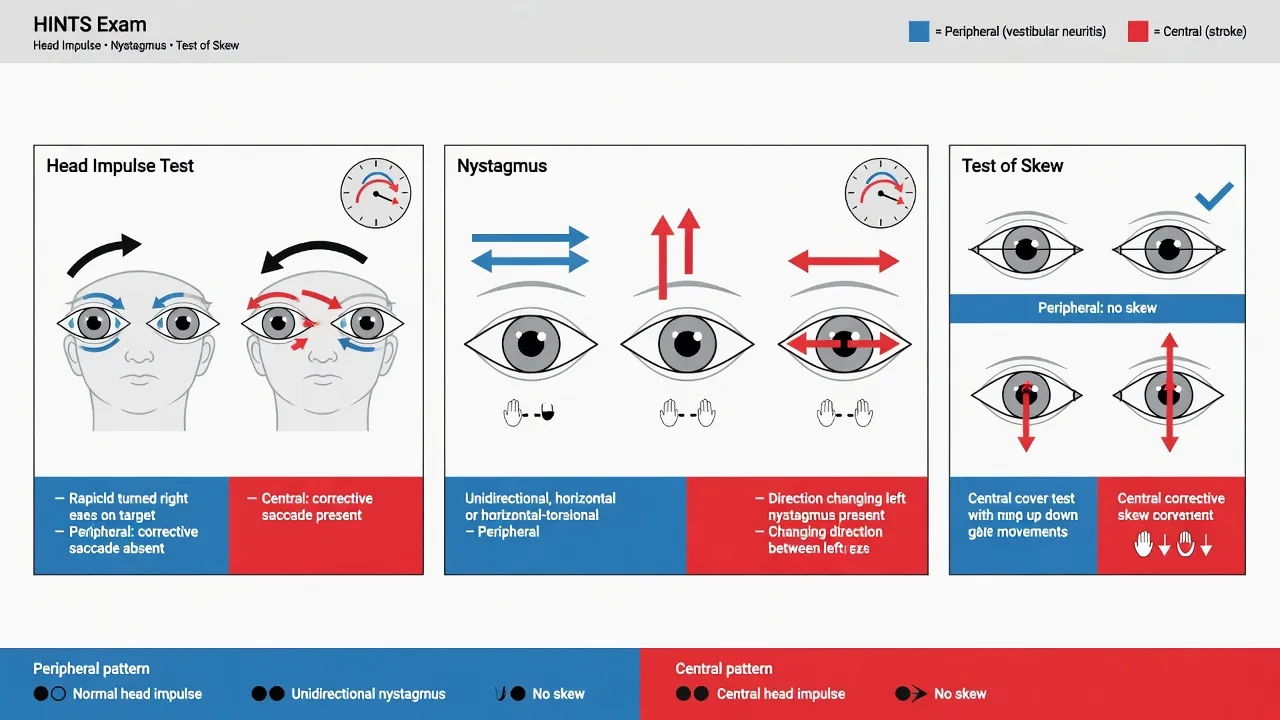
In the AVS the HINTS examination is the discriminator, and reproducing its three components and their interpretation is a Fellowship-examined core. The mnemonic is H-I-N-T-S, with any one of the three central findings being central.[1][2]
HINTS — three bedside signs, ANY one being central means central
HINTS
A POSITIVE (abnormal) head impulse test with a catch-up saccade = peripheral; a NORMAL head impulse = central red flag
Direction-fixed horizontal nystagmus beating away from the affected ear = peripheral; direction-changing (gaze-evoked) or vertical/downbeat = central
A vertical correction on alternate-cover test (skew deviation) = central; absent = peripheral
The rule: ANY one of the three being central is central — HINTS is more sensitive than early MRI within 48 hours (Kattah 2009)
In Kattah's 101-patient AVS cohort, the HINTS examination was 100 per cent sensitive and 96 per cent specific for stroke — more sensitive than early MRI diffusion-weighted imaging, which returned a false negative in 12 per cent of strokes within the first 48 hours.[1] The 2011 systematic review and the 2023 meta-analysis confirmed that a central HINTS pattern is highly specific for stroke and that, unlike imaging, its accuracy holds at the bedside on day one.[2][3]
HINTS and imaging thresholds
HINTS to diagnose stroke in the acute vestibular syndrome — Kattah 2009 (Stroke)
Stroke
PMID 19762709
Key finding
A prospective cohort of 101 patients with the acute vestibular syndrome (continuous vertigo with nystagmus, nausea and gait instability) assessed by the three-step bedside HINTS examination and by early MRI diffusion-weighted imaging. HINTS was 100 per cent sensitive and 96 per cent specific for stroke — and it outperformed early MRI DWI, which returned a false negative in roughly 12 per cent of posterior-circulation strokes within the first 48 hours.
Practice change
In the acute vestibular syndrome, a central HINTS pattern is stroke until proven otherwise, and a normal early MRI does NOT exclude it. Trust the bedside examination over the early scan; repeat the MRI at 48 to 72 hours if the clinical suspicion persists.
Peripheral nystagmus
- Horizontal or horizontal-torsional — never purely vertical
- Direction-FIXED: beats in one direction only (away from the affected ear), in primary and lateral gaze
- Intensifies when gaze is directed toward the fast phase (Alexander law)
- Spontaneous in the acute vestibular syndrome (neuritis), or positional and fatigable in BPPV
Central nystagmus
- Purely vertical (downbeat or upbeat), purely torsional, or horizontal — any plane is possible
- Direction-CHANGING (gaze-evoked): beats left in left-gaze, right in right-gaze
- Downbeat nystagmus that intensifies on lateral gaze or convergence is pathognomonic for a craniocervical junction lesion (Arnold–Chiari, cerebellar degeneration)
- May be positional and NON-fatigable (a positional vertigo that does not fatigue is central until proven otherwise)
HINTS nystagmus rule — read the eyes in three planes
HVD
Beats one way only, away from affected ear, intensified by gaze toward the fast phase (Alexander law) — peripheral vestibular neuritis
Any purely vertical nystagmus is central — a downbeat nystagmus worsened by lateral gaze suggests a craniocervical junction lesion
Beats left in left-gaze and right in right-gaze — pathognomonic of a central (brainstem/cerebellar) gaze-holding failure
The ED approach to the dizzy patient
The ED approach to the dizzy patient — the load-bearing decision
Sort the symptom — which quartet?
If it is vertigo — define the temporal pattern
In the AVS — perform the HINTS examination
Examine the ears and the hearing
Look for the brainstem and cerebellar signs
ECG and a finger-prick glucose — the two mimics
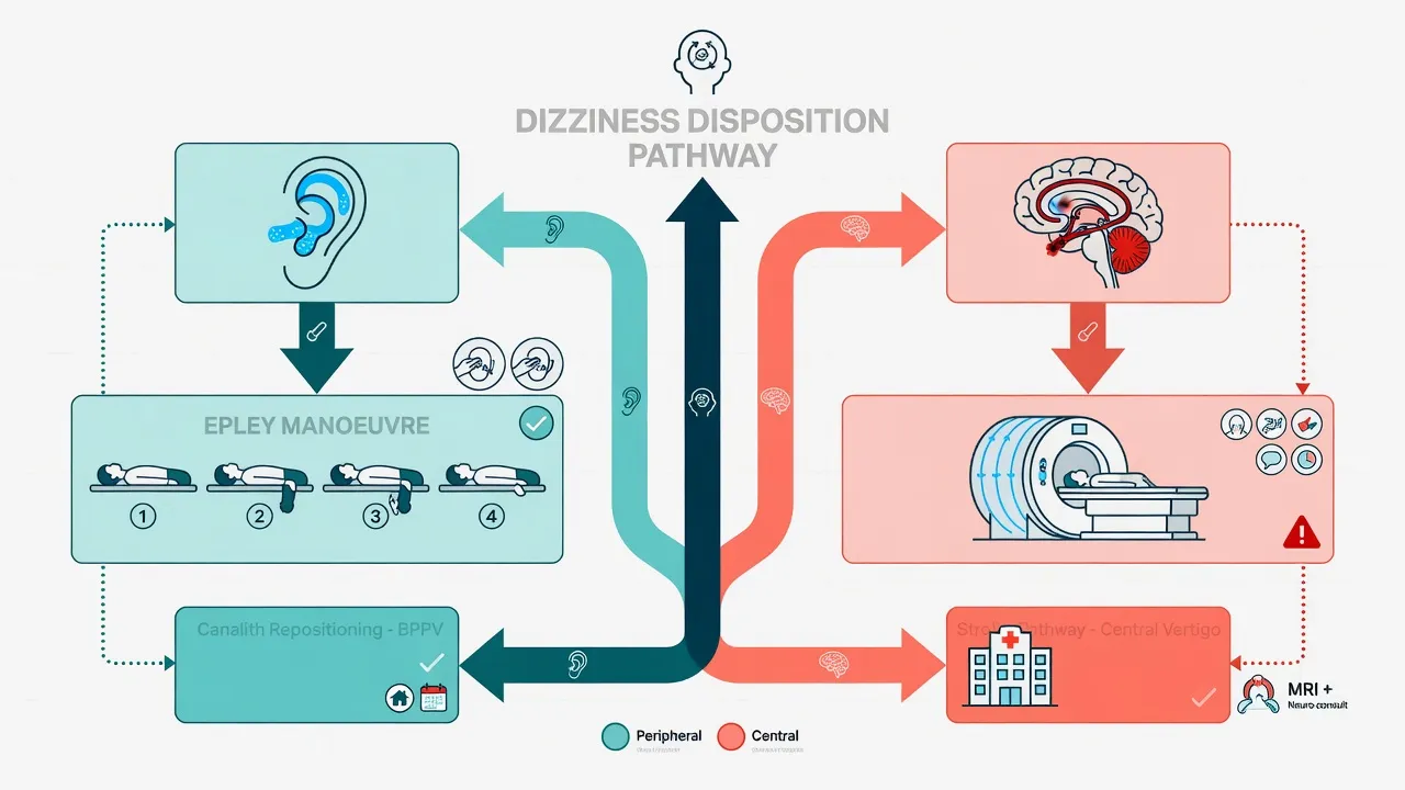
Disposition — the stroke pathway or the vestibular pathway
Differential diagnosis — distinguishing the dangerous from the benign
The differential of acute vertigo is the differential the examiner tests relentlessly, and the can't-miss member is the posterior-circulation stroke that mimics a peripheral vestibulopathy. The discriminating moves are the temporal pattern, the HINTS examination, the hearing, and the vascular risk profile. [1]
Vestibular neuritis (peripheral)
- Acute vestibular syndrome: continuous vertigo hours–days, vomiting, gait unsteady but can walk
- PERIPHERAL HINTS: abnormal (positive) head impulse, direction-fixed nystagmus, no skew
- Hearing preserved; no headache; normal CT
- Symptomatic ± short steroid course; outpatient with vestibular rehabilitation
Posterior-circulation stroke (central)
- Acute vestibular syndrome: identical presentation to neuritis — vertigo, vomiting, ataxia
- CENTRAL HINTS: normal head impulse, direction-changing or vertical/downbeat nystagmus, skew deviation
- Any brainstem sign (diplopia, dysarthria, dysphagia, dysmetria); higher vascular risk; true ataxia (cannot stand)
- Stroke pathway: MRI DWI, admit; CT may be normal — do not discharge on CT
BPPV (peripheral)
- Brief (under 1 min) positional vertigo, no symptoms between attacks, no hearing loss
- Positive Dix–Hallpike reproduces vertigo with torsional upbeating nystagmus after a brief latency
- HINTS not in play (not an AVS); normal gait between attacks
- Epley canalith repositioning manoeuvre at the bedside cures most in one session
Meniere disease (peripheral)
- Spontaneous episodic vertigo 20 min–12 h, fluctuating low-freq SNHL, aural fullness/tinnitus
- Recurrent attacks over months–years; progressive hearing loss
- Diagnosed by Bárány 2015 criteria (definite/probable)
- Betahistine, low-salt diet, diuretic; intratympanic steroids for refractory
Vestibular migraine (central/functional)
- Episodic vertigo 5 min–72 h with migraine features (photo/phonophobia, aura, headache)
- Normal hearing; normal interictal exam; personal or family migraine history
- Diagnosis of exclusion (Bárány/IHS criteria); no brainstem sign during attack
- Migraine prophylaxis; vestibular rehabilitation; avoid vestibular suppressants long-term
Cardiac presyncope (mimic)
- Patient SAYS "dizzy" but describes near-faint, lightheadedness — not true vertigo
- Exertional, postural, with palpitation or chest pain; abnormal ECG; family history of sudden death
- No nystagmus, no true vertigo, no vestibular pattern
- Risk-stratify as syncope — ECG, orthostatics, admit high-risk (San Francisco Syncope Rule)
Two mimics are particularly dangerous because the history is misleading. Cardiac presyncope frequently presents as "dizziness" — the patient uses the word the ED also uses for vertigo, but the symptom is a near-faint with greying vision, not a spin. Failing to take the symptom apart converts a high-risk cardiac presentation into a benign vertigo discharge. Posterior-circulation TIA may produce brief vertigo that mimics BPPV in timing — the give-away is the absence of a positional trigger, the presence of any brainstem sign, and vascular risk factors. Vertebrobasilar TIA causing isolated vertigo is rarer than once thought and is a diagnosis of exclusion after a clean HINTS, but the threat it carries (impending basilar occlusion) means any patient with high vascular risk and recurrent brief vertigo is imaged. [1]
[1]The central causes in depth — the dangerous differentials
The central causes of vertigo are best remembered by the three vascular territories of the posterior circulation plus the three structural mimics. The vascular strokes are PICA (lateral medullary / Wallenberg), AICA (lateral pontine), and the vertebral/basilar perforator and superior cerebellar artery territories; the structural mimics are multiple sclerosis, the cerebellopontine-angle tumour, and vestibular migraine. Each has a signature that the bedside examination can separate.[13]
PICA — lateral medullary (Wallenberg)
- Vertigo + ipsilateral Horner + ipsilateral ataxia + contralateral body pain/temperature loss (crossed sensory)
- Ipsilateral facial sensory loss (spinal trigeminal tract); dysphagia and hoarseness (nucleus ambiguus)
- HINTS: normal head impulse, direction-changing nystagmus, skew — classic central pattern
- A vertebral artery dissection or atherothrombosis; an urgent CTA/MRA of the posterior circulation
AICA — lateral pontine
- Vertigo + a SUDDEN SENSORINEURAL HEARING LOSS (AICA supplies the labyrinth via the internal auditory artery) — the hearing loss is the discriminator from PICA
- Ipsilateral facial palsy (CN VII), Horner, facial sensory loss, and a contralateral body sensory loss
- HINTS: normal head impulse or an abnormal one (AICA can infarct the labyrinth itself), so the hearing loss is the red flag
- A basilar-artery branch occlusion; managed as an acute stroke — consider thrombolysis/thrombectomy
Basilar / SCA — cerebellar
- Vertigo + cerebellar dysmetria and a true gait ataxia; may evolve to coma with bilateral pontine signs (locked-in)
- Any "dizziness" with fluctuating or bilateral brainstem signs is a basilar occlusion — a time-critical emergency
- Often a normal head impulse with direction-changing nystagmus; the threat is progression, not the initial exam
- A basilar thrombosis → immediate CTA and thrombectomy-capable-centre referral; do NOT await repeat imaging if signs progress
Multiple sclerosis
- A young patient (20–40 years) with a plaque in the vestibular nucleus, the eighth-nerve entry zone, or the cerebellar peduncle
- Vertigo may be the first presentation of MS, or part of a relapse; look for previous optic neuritis, transverse myelitis, Lhermitte sign
- HINTS: typically central (normal head impulse, direction-changing nystagmus, or skew); internuclear ophthalmoplegia on lateral gaze is highly suggestive
- An MRI brain and cervical spine with contrast; neurology referral; the vertigo itself is treated supportively
Cerebellopontine-angle tumour
- A vestibular schwannoma — a progressive, asymmetric hearing loss, tinnitus, and an imbalance (NOT an acute vertigo, which is the trap)
- A fifth-nerve (corneal reflex loss) and a seventh-nerve involvement develop late; papilloedema only with large lesions
- An absent corneal reflex on the affected side; an abnormal head impulse on the affected side (peripheral deafferentation)
- An MRI internal auditory meatus with gadolinium; an elective ENT/neurosurgery referral; the ED role is to exclude stroke and refer
Vestibular migraine
- The COMMONEST central cause of recurrent vertigo — episodic vertigo of 5 min to 72 h with migraine features (Bárány/IHS criteria)
- Photophobia, phonophobia, visual aura, or a migraine-type headache during the vertigo; a personal or family migraine history
- A normal interictal examination and a normal head impulse; no brainstem sign during the attack
- A migraine prophylaxis (propranolol, topiramate, amitriptyline, flunarizine) plus vestibular rehabilitation; AVOID prolonged vestibular suppressants
Small strokes causing severe vertigo — false-negative MRIs — Saber Tehrani 2014 (Neurology)
Neurology
PMID 24920847
Key finding
A study of posterior-circulation strokes presenting as the acute vestibular syndrome, characterising the frequency and mechanism of small strokes that mimic a peripheral vestibulopathy. Roughly 80 per cent of the missed strokes were non-lacunar (large-vessel or cardioembolic) rather than lacunar, and a substantial fraction had a false-negative early MRI — reinforcing that stroke in AVS is frequently missed by early imaging and that the HINTS examination outperforms the early scan.
Practice change
The 'stroke that looks like vestibular neuritis' is usually a non-lacunar, large-vessel or cardioembolic event — exactly the strokes that benefit from acute intervention. A normal early MRI in the right clinical context does not close the case; repeat the MRI at 48 to 72 hours and trust a central HINTS pattern over the scan.
Diagnosing stroke in acute vertigo — the HINTS family and the 'eye ECG' — Newman-Toker 2015 (Semin Neurol)
Seminars in Neurology
PMID 26444396
Key finding
A synthesis of the bedside eye-movement tests (HINTS plus its extensions — HINTS-plus, the STANDING algorithm) that distinguish central from peripheral causes in the acute vestibular syndrome, framing the eye-movement examination as the 'eye ECG' — a fast, cheap, bedside, and more-sensitive-than-MRI tool for the time-critical stroke diagnosis.
Practice change
The eye-movement examination in AVS is to the posterior circulation what the ECG is to the anterior circulation: a mandatory first-line test that, in trained hands, outperforms early imaging. ED clinicians should treat the HINTS examination as a core competency, not a specialist skill.
Peripheral causes (inner ear or nerve)
- BPPV — brief positional vertigo (under 1 min), a positive Dix–Hallpike, an Epley-responsive posterior canal
- Vestibular neuritis — the acute vestibular syndrome with a PERIPHERAL HINTS pattern and no hearing loss; typically post-viral
- Labyrinthitis — vestibular neuritis plus a sudden sensorineural hearing loss ± tinnitus
- Meniere disease — an episodic vertigo of 20 min to 12 h plus a fluctuating low-frequency sensorineural hearing loss and aural fullness
- The rarer: Ramsay Hunt syndrome (varicella zoster), otosyphilis, a perilymph fistula, vestibular paroxysmia, aminoglycoside vestibulotoxicity
Central causes (brainstem or cerebellum)
- Posterior-circulation stroke or TIA — the PICA, AICA, vertebral or basilar territory; the killer mimic
- Multiple sclerosis — a demyelinating plaque in the vestibular nucleus or the entry zone of the eighth nerve
- A cerebellopontine-angle tumour — a vestibular schwannoma, usually a progressive imbalance rather than an acute vertigo
- Vestibular migraine — an episodic vertigo of 5 min to 72 h with migraine features (the commonest central cause)
- A vertebral artery dissection — a posterior neck or occipital pain plus vertigo and ataxia; the younger patient
Investigations — HINTS first, then MRI
The investigations run in parallel with the bedside examination. The finger-prick glucose excludes hypoglycaemia (a vertigo and ataxia mimic). The ECG is mandatory — atrial fibrillation drives a cardioembolic posterior-circulation stroke, and the patient who says "dizzy" may have a primary cardiac rhythm. Bloods include full blood count, electrolytes and a coagulation screen; these are baseline rather than diagnostic. [1]
The pivotal imaging question is central versus peripheral, and the answer rests on HINTS, not on CT. Non-contrast CT of the brain is insensitive for posterior-fossa stroke — the dense petrous temporal bones produce beam-hardening artefact that obscures the brainstem and cerebellum, and early ischaemia is missed. A normal CT in AVS therefore excludes neither stroke nor a cerebellar haemorrhage reliably. MRI with diffusion-weighted imaging is the gold standard, but even MRI DWI returns false-negative in roughly 12 to 20 per cent of posterior-circulation strokes within the first 48 hours, which is the entire rationale for trusting the HINTS examination over early imaging.[1] The practical rule: a patient with a central HINTS pattern is managed as a stroke regardless of a normal CT or an early normal MRI, and is admitted to the stroke pathway. Audiometry, when available, characterises the hearing loss in Meniere disease and labyrinthitis; caloric testing and formal vestibular function tests are outpatient evaluations, not ED tests.
Immediate management — symptomatic control and the disposition split
Resuscitation is rarely required for vertigo itself, but vomiting and dehydration often are: establish intravenous access, give fluids (for example 1 litre of normal saline over an hour with antiemetic cover), and treat the nausea. The antiemetic of choice in the acute phase is prochlorperazine 5 mg orally or buccally (repeat if needed), or 12.5 mg intramuscularly for a vomiting patient, or ondansetron 4 mg intravenously as an alternative. The critical principle is that vestibular suppressants are a short-course symptomatic tool — three days at most in vestibular neuritis — because prolonged use impairs central vestibular compensation and prolongs recovery. [1]
[1]Definitive management — the four disorders
BPPV (posterior canal). The diagnosis is made at the bedside by the Dix–Hallpike manoeuvre — sitting the patient up, turning the head 45 degrees to one side, then rapidly lying them back with the head extended 20 degrees over the end of the bed; a positive test reproduces the vertigo after a brief latency (1 to 5 seconds) with a characteristic torsional upbeating nystagmus fatiguing within a minute.[7] Treatment is the Epley canalith repositioning manoeuvre, which rotates the head through a sequence of positions that walk the loose otoconia out of the posterior canal and back into the utricle. The Cochrane review confirms the Epley is significantly more effective than sham or no treatment for resolving symptoms and converting a positive Dix–Hallpike to negative.[4] Horizontal-canal BPPV (the second commonest) is diagnosed by the supine roll test and treated with the Barbecue (Lempert) or Gufoni manoeuvre.
BPPV at the bedside — from the Dix–Hallpike to the Epley
Confirm the diagnosis — the Dix–Hallpike
Identify the canal
Posterior canal — the Epley manoeuvre
Horizontal canal — the Barbecue (Lempert) or the Gufoni
Re-test and give the post-manoeuvre advice
The Epley (canalith repositioning) manoeuvre for BPPV — Hilton and Pinder, Cochrane 2014
Cochrane Database of Systematic Reviews
PMID 25485940
Key finding
A systematic review of the randomised and quasi-randomised trials of the Epley canalith repositioning manoeuvre versus a sham or no treatment for the posterior-canal BPPV. The pooled data showed the Epley was significantly more effective than a sham or no treatment for both a symptom resolution and a conversion of a positive Dix–Hallpike to negative, with no serious adverse effects.
Practice change
The Epley manoeuvre is the first-line treatment for the posterior-canal BPPV and should be performed at the bedside by the emergency clinician at the time of the positive Dix–Hallpike — not deferred to a clinic appointment.
Clinical Practice Guideline: BPPV (Update) — Bhattacharyya 2017, AAO-HNS (Executive Summary)
Otolaryngology–Head and Neck Surgery
PMID 28248609
Key finding
The American Academy of Otolaryngology–Head and Neck Surgery update of the evidence-based guideline for BPPV. The guideline affirms the Dix–Hallpike as the diagnostic standard for posterior-canal BPPV, the supine roll test for horizontal-canal BPPV, and the canalith repositioning (Epley) manoeuvre as first-line therapy; it explicitly recommends AGAINST routinely prescribed vestibular suppressants for BPPV, and recommends against imaging or vestibular testing in the uncomplicated case.
Practice change
BPPV is a clinical, bedside diagnosis-and-treat: a Dix–Hallpike then an Epley at the same visit. Do NOT prescribe prochlorperazine or other suppressants as routine BPPV therapy — they do not treat the canalithiasis and they add sedation, falls risk and delayed referral. Refer only the atypical, recurrent, or multi-canal case.
Canal-specific BPPV — match the manoeuvre to the canal
Posterior canal (85%) — Dix–Hallpike → Epley
Horizontal canal (10–15%) — supine roll test → Barbecue / Gufoni
Anterior canal (rare, 1–2%) — Dix–Hallpike → deep head-hanging / Yacovino
Atypical or non-fatiguing positional vertigo — STOP and reassess
Vestibular neuritis. Symptom control is with a short course of prochlorperazine or an alternative antiemetic (three days, then stop). A tapering course of oral corticosteroids — for example prednisolone 1 mg per kilogram (maximum 60 mg) daily for three to five days then taper over two to three weeks, started within 72 hours of onset — modestly improves long-term caloric recovery, although the modern meta-analyses show inconsistent benefit on symptomatic outcomes and the practice is guideline-variable.[5] Antivirals alone are not effective. Early vestibular rehabilitation (graded head-movement exercises) accelerates central compensation and is the most evidence-based long-term intervention.
[1]Corticosteroids in vestibular neuritis — the updated meta-analysis, Bogdanova 2022
Acta Neurologica Scandinavica
PMID 36029039
Key finding
An updated meta-analysis of the randomised trials of corticosteroids versus a placebo or no treatment in vestibular neuritis. The corticosteroids modestly improved the long-term caloric recovery (a peripheral vestibular-function measure) when started within 72 hours of onset, but the benefit on the symptomatic and patient-centred outcomes was inconsistent across the trials.
Practice change
A short tapering course of an oral corticosteroid (for example the prednisolone 1 mg per kilogram, maximum 60 mg, daily for 3 to 5 days then a taper over 2 to 3 weeks) is a reasonable, guideline-variable option in vestibular neuritis if started within 72 hours — but the vestibular rehabilitation is the more consistently evidence-based intervention. Antivirals alone are ineffective.
Methylprednisolone, valacyclovir, or the combination for vestibular neuritis — Strupp 2004 (NEJM)
New England Journal of Medicine
PMID 15269315
Key finding
A randomised, double-blind, multicentre trial of 141 patients with acute vestibular neuritis, comparing oral methylprednisolone (a taper starting 100 mg daily), valacyclovir, the combination, and placebo, begun within three days of onset. Methylprednisolone alone significantly improved peripheral vestibular function (caloric testing) at 12 months; valacyclovir alone did NOT, and the combination added no benefit over the corticosteroid alone.
Practice change
The case for a corticosteroid in vestibular neuritis rests on this trial: an oral methylprednisolone taper begun early improves objective peripheral recovery. The viral-cause hypothesis (and therefore the antiviral arm) did not pan out — do not add valacyclovir or aciclovir. The benefit on patient-centred symptoms is modest, which is why later meta-analyses call the practice guideline-variable, but the corticosteroid remains a reasonable, evidence-supported option within 72 hours of onset.
Prochlorperazine (D2 antagonist)
- The first-line ED vestibular suppressant/antiemetic; 5 mg orally or buccally, or 12.5 mg intramuscularly
- Effective for acute nausea and vertigo; buccal route avoids the vomiting patient swallowing a tablet
- SHORT-COURSE ONLY (≤3 days): extrapyramidal side effects (akathisia, acute dystonia, parkinsonism) and it impairs central compensation
- Avoid in Parkinson disease and in pregnancy (first trimester); elderly are prone to confusion and falls
Ondansetron (5-HT3 antagonist)
- A useful alternative antiemetic, 4 mg intravenously; less effective on the vertigo itself, more on the nausea
- Favoured when a D2 antagonist is contraindicated (Parkinson disease, young female, elderly fall risk)
- QT prolongation at higher or combined doses; an ECG consideration in the cardiac patient
- Also short-course; the same compensation principle applies — do not prescribe long-term
Diazepam / benzodiazepines
- Sedating, anxiolytic, and a mild vestibular depressant — sometimes used for severe acute vertigo (2–5 mg orally)
- Markedly impairs central compensation and adds sedation, falls, and dependence with repeated dosing
- Reserve for a single, severe, first-night dose if at all; NEVER as a repeat prescription for vestibular neuritis
- Avoid in the elderly (falls, delirium) and in patients on opioids (respiratory depression)
Cinnarizine / flunarizine (Ca-channel)
- A calcium-channel antagonist with anti-histamine activity; used in some regions for vestibular suppression and in Meniere/migraine prophylaxis
- Sedation, weight gain, and (rarely) drug-induced parkinsonism with flunarizine limit long-term use
- A reasonable short-course alternative where prochlorperazine is not tolerated
- Again: short-course in the acute phase; the chronic role is migraine prophylaxis, not vestibular suppression
Betahistine (H1 agonist / H3 antagonist)
- A histamine analogue used for Meniere disease, NOT for acute vestibular neuritis; 16 mg three times daily (regional dosing)
- Mechanism: improves cochlear microcirculation and reduces endolymphatic hydrops; evidence is mixed
- Not subsidised on the PBS in Australia (used selectively); generally well tolerated
- A maintenance therapy for Meniere, not an acute vestibular suppressant — do not confuse the roles
Vestibular suppressant doses and limits
Labyrinthitis. Managed as vestibular neuritis with attention to the hearing loss: urgent audiology, and consider corticosteroids (systemic, and in some pathways intratympanic) for the sudden sensorineural hearing loss, which has its own time window. [1]
[1]Meniere disease. Acute attacks are managed with bed rest, antiemetics and a short vestibular suppressant. Maintenance therapy is betahistine (a histamine analogue that improves cochlear microcirculation), a low-salt diet, and a diuretic (for example hydrochlorothiazide 25 to 50 mg daily or acetazolamide 250 mg twice daily) to reduce endolymphatic hydrops. Refractory disease is treated with intratympanic corticosteroids or gentamicin, and surgery (endolymphatic sac decompression, vestibular nerve section) in the small minority with refractory disabling vertigo. [1]
The functional and chronic vestibular disorders
Not every dizzy patient has an acute peripheral or central lesion. Two chronic, episodic-then-persistent disorders are common, frequently mislabelled as "psychogenic", and missed in the ED: vestibular migraine (the commonest central cause of recurrent vertigo) and persistent postural-perceptual dizziness (PPPD), the new consensus term for the chronic functional dizziness formerly called chronic subjective dizziness or phobic postural vertigo. Both are diagnoses of inclusion (against defined Bárány criteria) made after the dangerous and acute peripheral causes are excluded, and both are managed primarily WITHOUT vestibular suppressants. [1]
Diagnostic criteria for persistent postural-perceptual dizziness (PPPD) — Staab 2017, Bárány Society consensus
Journal of Vestibular Research
PMID 29036855
Key finding
The international consensus diagnostic criteria for PPPD, produced by the Bárány Society Committee for the Classification of Vestibular Disorders. PPPD is defined as three or more months of non-rotational dizziness or unsteadiness, present on most days, that is provoked by upright posture, active or passive motion, and exposure to moving or complex visual stimuli, WITHOUT an alternative explanation and usually triggered by an acute vestibular, medical, or psychological event.
Practice change
PPPD is the commonest cause of chronic dizziness in the neurology clinic, and it usually begins with an acute vestibular event (a neuritis, a BPPV, or even a panic attack) that the brain fails to compensate for. Recognise it after the acute work-up is negative: the patient is dizzy on standing and in visually busy environments (supermarkets, scrolling screens), with a normal neurological and vestibular examination. Treat with serotonin-reuptake-inhibitors / serotonin-noradrenaline-reuptake-inhibitors and vestibular rehabilitation — NOT with vestibular suppressants.
Vestibular migraine
- Episodic VERTIGO (rotational or non-rotational) 5 min to 72 h, ≥5 episodes, with migraine features
- A personal or family migraine history; photophobia, phonophobia, or visual aura during attacks
- A normal interictal examination and a normal head impulse; no brainstem sign
- Migraine prophylaxis (propranolol, topiramate, amitriptyline, flunarizine) + vestibular rehabilitation
PPPD
- Chronic (≥3 months) non-rotational dizziness/unsteadiness on most days, NO vertigo
- Provoked by upright posture, motion, and visually complex environments; normal exam and normal HIT
- Often triggered by a prior acute vestibular event (neuritis, BPPV) that "did not resolve"
- SSRI/SNRI + vestibular rehabilitation + CBT; AVOID vestibular suppressants
Meniere disease
- Episodic VERTIGO 20 min to 12 h with AUDITORY symptoms (fluctuating low-frequency SNHL, aural fullness, tinnitus)
- Recurrent attacks over months to years with progressive hearing loss; defined by Bárány 2015 criteria
- An abnormal audiogram between attacks is the key discriminator from vestibular migraine and PPPD
- Betahistine, low-salt diet, diuretic; intratympanic steroids/gentamicin for refractory
Complications and pitfalls
The complications of mismanaged vertigo are, in order of danger, the missed posterior-circulation stroke (the leading cause of vertigo-related litigation and mortality), prolonged vestibular suppressant use impairing central compensation, recurrence of BPPV (which can be re-treated), and the progression of sudden hearing loss in labyrinthitis when corticosteroids are withheld. The classic pitfalls at the bedside are: trusting a normal non-contrast CT in a patient with central HINTS; failing to perform the head impulse test in any AVS patient (and therefore defaulting to a neuritis diagnosis); prescribing long-term prochlorperazine or diazepam for vestibular neuritis; treating recurrent brief vertigo in a vascular patient as BPPV without excluding vertebrobasilar TIA; and forgetting the ECG in the elderly dizzy patient. [1]
The three peripheral manoeuvres and their canal
Prognosis and disposition
Peripheral vestibular disorders do well with the right intervention: BPPV resolves in the great majority after one or two Epley manoeuvres; vestibular neuritis improves over weeks with vestibular rehabilitation (residual symptoms reflect delayed compensation, often from prolonged vestibular suppressants). Central HINTS carries the prognosis of its underlying stroke and is admitted. The disposition rules are: any central HINTS, any brainstem sign, true gait ataxia, severe headache, or a first attack of Meniere with new hearing loss — admit for imaging and specialist review; a clear peripheral pattern with normal gait — discharge with vestibular rehabilitation, a short antiemetic/steroid course, and a written safety-net for new neurological symptoms. [1]
Special populations
The elderly dizzy patient has a broader and more dangerous differential — posterior-circulation stroke, orthostatic hypotension from polypharmacy, carotid sinus hypersensitivity, cardiac arrhythmia — and the threshold for imaging, ECG monitoring and admission is lower. Polypharmacy (antihypertensives, sedatives, anticonvulsants) is a leading cause of dizziness in this group and is reversible. The anticoagulated patient with vertigo carries a higher risk of posterior-fossa haemorrhage (cerebellar haematoma presenting with vertigo and ataxia) and of cardioembolic stroke, and warrants early imaging even with a clean HINTS. Pregnancy is usually managed without vestibular suppressants (prochlorperazine is generally avoided in the first trimester); positional manoeuvres for BPPV are safe. [1]
Evidence and regional guidelines
The diagnostic evidence base is dominated by the HINTS examination. Kattah and colleagues (2009) showed that the three-step bedside HINTS examination was 100 per cent sensitive and 96 per cent specific for stroke in AVS, and more sensitive than early MRI DWI within 48 hours.[1] Tarnutzer's 2011 systematic review confirmed the high specificity of central ocular motor findings at the bedside,[2] and the 2023 meta-analysis extended this to show that diagnostic accuracy depends on clinician training and stroke location, but remains high in expert hands.[3] The Cochrane review of the Epley manoeuvre (Hilton and Pinder, 2014) confirms its efficacy for posterior-canal BPPV.[4] Corticosteroid meta-analysis for vestibular neuritis (Bogdanova, 2022) shows modest and inconsistent benefit.[5] The Bárány Society International Classification of Vestibular Disorders provides the operational diagnostic criteria for both Meniere disease (Lopez-Escamez, 2015)[6] and BPPV (von Brevern, 2015).[7]
ANZ practice note. Australian and New Zealand practice follows the Stroke Foundation Clinical Guidelines and the local state-wide stroke service for the central-HINTS patient (MRI DWI, stroke-unit admission, consideration of posterior-circulation thrombectomy where a basilar or vertebral large-vessel occlusion is found). Prochlorperazine 5 mg buccal or oral is the standard ED antiemetic for acute vestibular neuritis; oral prednisolone for vestibular neuritis is variable by centre. The Epley manoeuvre is performed at the bedside by the emergency clinician for confirmed posterior-canal BPPV. Betahistine is not subsidised on the PBS for Meniere disease in Australia and is used selectively. [1]
SAQ — Central versus peripheral vertigo — the HINTS examination
10 minutes · 10 marks
A 72-year-old man with hypertension presents with 6 hours of continuous vertigo, nausea and vomiting. He has spontaneous horizontal-torsional nystagmus, gait instability (he can stand but sways), and reports the room is spinning to his left. There are no focal neurological signs on a brief examination.
SAQ — Recurrent positional vertigo — benign paroxysmal positional vertigo
10 minutes · 10 marks
A 44-year-old woman presents with a 1-week history of brief (under 30 seconds) episodes of severe vertigo when rolling over in bed, looking up to a high shelf, or bending to put on shoes. Between episodes she is well. Neurological examination is normal.
Exam pearls
- Central HINTS = NORMAL head impulse + direction-changing (or vertical/downbeat) nystagmus + skew deviation — ANY one central sign is central. The candidate who states this rule wins the vertigo SAQ.
- HINTS is more sensitive than early MRI within 48 hours of AVS onset — a normal head impulse test in a vertiginous patient is a red flag, not reassurance.
- BPPV = brief positional vertigo reproduced by Dix–Hallpike → Epley manoeuvre. The nystagmus is torsional and upbeating on the dependent side, with a brief latency and fatigability.
- Vestibular neuritis = continuous vertigo + PERIPHERAL HINTS (abnormal head impulse) + no hearing loss. Labyrinthitis = neuritis + sudden SNHL.
- Meniere triad = episodic vertigo 20 min–12 h + fluctuating low-frequency SNHL + aural fullness/tinnitus (Bárány 2015).
- Prochlorperazine and other vestibular suppressants are short-course (≤3 days) — prolonged use delays central compensation.
- Never trust a normal non-contrast CT in a suspected posterior-circulation stroke — CT is insensitive in the posterior fossa; pursue MRI or admit.
- Nystagmus reading — direction-fixed horizontal is peripheral; direction-changing (gaze-evoked) or vertical/downbeat is central. Read the eyes in primary gaze then ±30 degrees lateral; never interpret end-point nystagmus.
- A purely vertical or downbeat nystagmus is central — full stop. A downbeat nystagmus worsened by lateral gaze or convergence points to a craniocervical junction lesion (Arnold–Chiari, cerebellar degeneration).
- The AICA stroke produces vertigo + ipsilateral sudden SNHL (AICA supplies the labyrinth); PICA (Wallenberg) spares the hearing. A hearing loss with a normal HIT and direction-changing nystagmus is AICA stroke, not labyrinthitis.
- Horizontal-canal BPPV (10–15%) = horizontal nystagmus on the supine roll test → Barbecue (Lempert) or Gufoni manoeuvre — NOT the Epley. The Epley only treats the posterior canal.
- A positional vertigo that is NON-fatiguing, persistent, or has downbeat nystagmus is a central positional lesion, NOT BPPV — do not reposition, image.
- Vestibular migraine = ≥5 episodes of vertigo 5 min–72 h + migraine history + ≥1 migraine feature per attack (Bárány/IHS, Lempert 2012); headache is NOT required during every attack.
- PPPD = ≥3 months of non-rotational dizziness provoked by posture/motion/visual stimuli after an acute vestibular event; treat with SSRI/SNRI + vestibular rehab, NOT suppressants.
- The corneal reflex (CN V) is the forgotten bedside sign — an absent reflex with a progressive unilateral hearing loss and imbalance is a vestibular schwannoma until MRI proves otherwise.
- Methylprednisolone (Strupp 2004, NEJM) improves objective recovery in vestibular neuritis; valacyclovir does NOT — do not add antivirals.
- A sudden SNHL (≥30 dB over three frequencies in 72 h) gets high-dose oral prednisolone within 7–14 days — this is an ENT emergency, not an outpatient referral.
- The eye-movement examination in AVS is the "eye ECG" — a mandatory first-line test that, in trained hands, outperforms early MRI (Newman-Toker 2015).
- A bilateral abnormal head impulse test with oscillopsia (the world jumping when walking) is a bilateral vestibulopathy — think aminoglycoside vestibulotoxicity. [1]
Red flags
[1]References
- [1]Kattah JC, Talkad AV, Wang DZ, Hsieh YH, Newman-Toker DE. HINTS to diagnose stroke in the acute vestibular syndrome: three-step bedside oculomotor examination more sensitive than early MRI diffusion-weighted imaging Stroke, 2009.PMID 19762709
- [2]Tarnutzer AA, Berkowitz AL, Robinson KA, Hsieh YH, Newman-Toker DE. Does my dizzy patient have a stroke? A systematic review of bedside diagnosis in acute vestibular syndrome CMAJ, 2011.PMID 21576300
- [3]Tarnutzer AA, Gold D, Wang Z, et al. Impact of Clinician Training Background and Stroke Location on Bedside Diagnostic Test Accuracy in the Acute Vestibular Syndrome - A Meta-Analysis Ann Neurol, 2023.PMID 37038843
- [4]Hilton MP, Pinder DK. The Epley (canalith repositioning) manoeuvre for benign paroxysmal positional vertigo Cochrane Database Syst Rev, 2014.PMID 25485940
- [5]Bogdanova A, Karim H, Yoo S, et al. Corticosteroids in patients with vestibular neuritis: An updated meta-analysis Acta Neurol Scand, 2022.PMID 36029039
- [6]Lopez-Escamez JA, Carey J, Chung WH, et al. Diagnostic criteria for Menière's disease J Vestib Res, 2015.PMID 25882471
- [7]von Brevern M, Bertholon P, Brandt T, et al. Benign paroxysmal positional vertigo: Diagnostic criteria J Vestib Res, 2015.PMID 26756126
- [8]Strupp M, Zingler VC, Arbusow V, et al. Methylprednisolone, valacyclovir, or the combination for vestibular neuritis N Engl J Med, 2004.PMID 15269315
- [9]Bhattacharyya N, Gubbels SP, Schwartz SR, et al. Clinical Practice Guideline: Benign Paroxysmal Positional Vertigo (Update) Otolaryngol Head Neck Surg, 2017.PMID 28248609
- [10]Staab JP, Eckhardt-Henn A, Horii A, et al. Diagnostic criteria for persistent postural-perceptual dizziness (PPPD): Consensus document of the committee for the Classification of Vestibular Disorders of the Bárány Society J Vestib Res, 2017.PMID 29036855
- [11]Lempert T, Olesen J, Furman J, et al. [Vestibular migraine: diagnostic criteria: consensus document of the Bárány Society and the International Headache Society] Nervenarzt, 2013.PMID 23532572
- [12]Popkirov S, Stone J, Holle-Lee D. Treatment of Persistent Postural-Perceptual Dizziness (PPPD) and Related Disorders Curr Treat Options Neurol, 2018.PMID 30315375
- [13]Newman-Toker DE, Curthoys IS, Halmagyi GM. Diagnosing Stroke in Acute Vertigo: The HINTS Family of Eye Movement Tests and the Future of the Eye ECG Semin Neurol, 2015.PMID 26444396
- [14]Saber Tehrani AS, Kattah JC, Mantokoudis G, et al. Small strokes causing severe vertigo: frequency of false-negative MRIs and nonlacunar mechanisms Neurology, 2014.PMID 24920847