Paeds · haematology-oncology-and-transfusion
Venous thromboembolism, pulmonary embolism and thrombophilia in children
Also known as Venous thromboembolism · Deep vein thrombosis · Pulmonary embolism · CVAD-associated thrombosis · Catheter-related thrombosis · Hereditary thrombophilia · Paediatric thrombosis
Fellowship guide to venous thromboembolism, pulmonary embolism and thrombophilia in children: the bimodal epidemiology peaking in neonates and adolescents, the predominance of provoked and central-venous-catheter-associated thrombosis, the Virchow triad pathophysiology modified by developmental haemostasis, the clinical picture of leg swelling and catheter dysfunction, the diagnosis with compression ultrasound and D-dimer, the stepwise anticoagulation with low molecular weight heparin and oral rivaroxaban built on the CHEST and ASH guidelines and the EINSTEIN-Jr trials, the selective approach to hereditary thrombophilia testing, and the care of neonates, critically ill children and the transitioning adolescent.
On this page & tools
Your progress
Saved locally on this device.
Practise this topic
Target exams
Red flags
Life stages
Care settings
Clinical exam formats
Board mappings
Overview & Definition
Picture the fourteen-year-old girl admitted after spinal fusion surgery for scoliosis, five days postoperative, now breathless and complaining of right-sided chest pain when she takes a deep breath in, her oxygen saturations dipping to 92 percent. That girl has a pulmonary embolism, and the deep vein thrombosis that seeded it has been quietly forming in her immobilised leg. The recent surgery, the immobility and her combined oral contraceptive pill are the three provoking factors stacked together, and the breathlessness and pleuritic pain are the clinical signature of an embolus in a pulmonary artery. [3] [11]
Venous thromboembolism in children is the formation of a pathological clot within the venous system, encompassing deep vein thrombosis (a clot in a deep vein, most often a leg or a central vein), pulmonary embolism (a clot that has embolised to the pulmonary arterial tree), cerebral sinus venous thrombosis, and renal and portal vein thrombosis. The defining feature of paediatric venous thromboembolism, in contrast to the adult disease, is that it is overwhelmingly provoked and secondary. Over 90 percent of paediatric cases have an identifiable trigger, and the single largest category is thrombosis associated with a central venous access device, which accounts for around half of all events. [3] [2]
The disease is also defined by its developmental context. Children are not small adults when it comes to clotting: the neonatal haemostatic system is physiologically immature, with reduced levels of the vitamin K dependent factors and anticoagulant proteins, and this developmental haemostasis shapes both the risk and the response to anticoagulants. Neonates and infants metabolise heparin faster than older children and need higher weight-based doses, which is why dose tables are age-stratified from birth. [7] [1]
Classification
Paediatric venous thromboembolism is classified along two axes that together drive every management decision: the anatomic site of the clot, and whether the event is provoked or unprovoked. The site tells you which imaging to obtain and what the immediate danger is; the provoked or unprovoked status tells you the duration of anticoagulation and whether to chase an underlying cause. [2] [11]
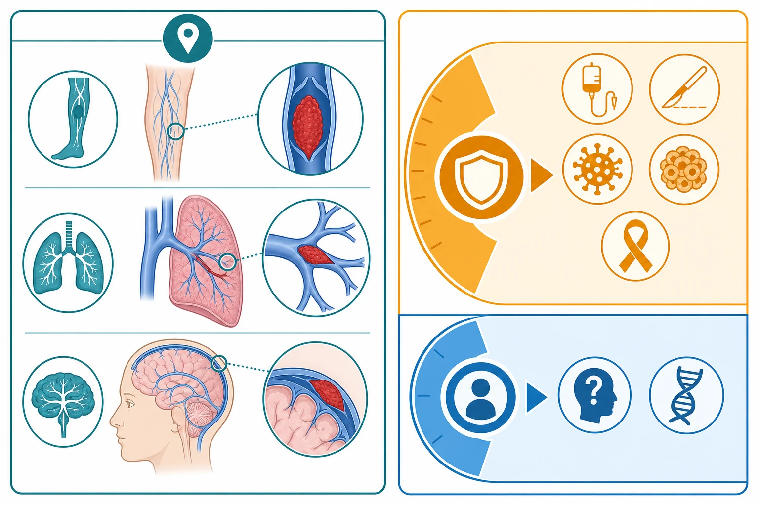
By anatomic site, the commonest is deep vein thrombosis of a lower limb or of a central vein around a catheter tip. Pulmonary embolism is a deep vein thrombus that has broken off and lodged in a pulmonary artery branch. Cerebral sinus venous thrombosis presents with headache, seizures or raised intracranial pressure. Renal vein thrombosis classically affects the neonate, presenting with a flank mass and haematuria, and portal vein thrombosis is a recognised complication of umbilical catheter placement and of neonatal sepsis. [11] [1]
Provoked versus unprovoked venous thromboembolism
Recurrent
second event
The provoked or unprovoked axis is the one that changes management. A provoked thrombosis has an identified trigger, and once that trigger resolves the anticoagulation is time-limited. An unprovoked thrombosis has no trigger, which raises the suspicion of an underlying hereditary or acquired thrombophilia and prompts a more careful evaluation. Recurrent thrombosis, a second event after a completed course, extends the duration of anticoagulation and strongly prompts thrombophilia testing. [2] [9]
A third classification, increasingly used in modern trials, is catheter-related versus non-catheter-related disease, because the two behave differently. Catheter-related thrombosis is often asymptomatic, discovered when a line stops working or a routine surveillance scan finds a clot, and its management may include line removal. Non-catheter-related thrombosis is more often symptomatic and behaves like the adult disease. The EINSTEIN-Jr CVC-VTE subgroup addressed catheter-related disease specifically. [6] [12]
Epidemiology & Risk Factors
Venous thromboembolism in children has risen dramatically over the past two decades. Raffini and colleagues showed that the rate of venous thromboembolism in United States children's hospitals rose from 34 to 58 cases per 10,000 admissions between 2001 and 2007, a 70 percent increase driven by better survival of critically ill children, more invasive procedures, wider use of central venous catheters, and improved imaging and recognition. Despite this rise, paediatric venous thromboembolism remains far rarer than the adult disease, which reaches 1 to 2 per 1000 adults per year. [3]
The incidence is bimodal, with two clear peaks. The first peak is in neonates and infants, reflecting the physiologic immaturity of the haemostatic system, the frequency of central lines, and the complications of critical neonatal care. The second peak is in adolescents, driven by oestrogen exposure, obesity, and the emergence of adult-style risk factors. Between these peaks, in the school-age child, thrombosis is uncommon and almost always provoked by a central line, critical illness or malignancy. [3] [7]
| Risk factor | Why it matters | Typical scenario |
|---|
The risk factors cluster into the three arms of Virchow's triad: venous stasis (immobility, critical illness, paralysed limb, central line), endothelial injury (catheter tip, surgery, infection, previous thrombosis), and hypercoagulability (malignancy, nephrotic syndrome, sepsis, hereditary thrombophilia, oestrogen). A child with several of these stacked together, such as a teenager on the oral contraceptive pill who has spinal surgery, is at substantially elevated risk and may warrant thromboprophylaxis. [7] [11]
Pathophysiology
Thrombus formation in a vein follows Virchow's triad, the three forces that disturb normal venous blood flow and tip the balance of haemostasis toward clotting. The first is venous stasis, the slowing or stagnation of blood in a vein, which occurs with immobility, a paralysed limb, the low-flow state of critical illness, and the disturbed flow around a central venous catheter tip. The second is endothelial injury, damage to the vein's inner lining, which exposes tissue factor and subendothelial collagen and is caused by the physical presence of a catheter, by surgery, by infection, and by the chemical injury of irritant infusions. The third is hypercoagulability, an altered blood composition that favours clotting, seen in malignancy, sepsis, nephrotic syndrome, oestrogen use, and the inherited thrombophilias. [11] [7]
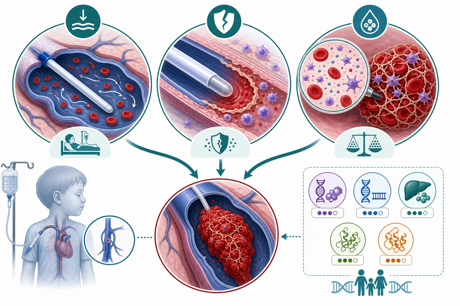
The central venous access device deserves special attention because it is the dominant paediatric trigger, and its mechanism combines all three arms of the triad. The catheter physically occupies the vein lumen and disturbs blood flow (stasis), its tip and the mechanical trauma of insertion injure the endothelium (injury), and the child who needs the line is usually already hypercoagulable from critical illness, malignancy or congenital heart disease (hypercoagulability). This convergence explains why a central line is such a powerful provoking factor and why catheter-related thrombosis is the leading category of paediatric disease. [6] [12]
In children, the third arm of the triad is modified by developmental haemostasis. The neonatal haemostatic system has physiologically low levels of the vitamin K dependent procoagulants (factors II, VII, IX and X) and of the anticoagulant proteins (antithrombin, protein C and protein S), which roughly balances out to protect the neonate from both bleeding and clotting. After the first six months, these levels rise toward adult values. This developmental physiology means that a neonate can be both genuinely deficient in anticoagulant proteins (mimicking thrombophilia on a laboratory panel) and at real risk of thrombosis when a central line is placed, and it explains why laboratory reference ranges are age-specific. [7] [8]
[7] [8]The inherited thrombophilias tip the hypercoagulability arm. Factor V Leiden, the commonest hereditary thrombophilia in people of European ancestry, makes factor V resistant to cleavage by activated protein C and is inherited autosomal dominant. The prothrombin gene mutation G20210A raises prothrombin levels. Antithrombin deficiency, protein C deficiency and protein S deficiency are rarer but stronger risk factors. The practical point, examined heavily, is that these conditions are common in the healthy population and carry a low positive predictive value for a first thrombosis, which is why testing is selective and never universal. [8] [9]
Clinical Presentation
The clinical presentation follows the site of the clot, and the key skill is to think of thrombosis in a child with risk factors, because the signs are easy to dismiss in a child who is already unwell. The single most common presentation in children is the asymptomatic or minimally symptomatic catheter-related thrombosis found on a surveillance scan or when a central line stops drawing back or infusing. [11] [2]
A symptomatic deep vein thrombosis of a limb presents with swelling, warmth, pain or tenderness, and a colour change ranging from erythema to a pale or purple limb. The swelling is often unilateral and the child may hold the limb still or limp. The classic adult signs of calf tenderness and a palpable cord are less reliable in a young child, and the presentation may be subtle, so a high index of suspicion in any child with a central line, critical illness or recent surgery is essential. [1] [11]
Pulmonary embolism presents with sudden dyspnoea, pleuritic chest pain, haemoptysis, tachycardia, hypoxia, or unexplained deterioration in a child with risk factors. The diagnosis is frequently missed in children because it is rare and because the symptoms overlap with much commoner conditions such as chest infection, asthma or anxiety. Any child with a known or suspected deep vein thrombosis who becomes breathless or hypoxic has a pulmonary embolism until proven otherwise, and any adolescent with sudden pleuritic chest pain after surgery or on the oral contraceptive pill warrants consideration of pulmonary embolism. [11] [3]
Limb DVT
swollen leg
- Swelling, warmth, pain, unilateral
- Limp or held limb
- Central line or post-surgical
- Confirm with compression ultrasound
Pulmonary embolism
- Sudden dyspnoea, pleuritic pain
- Tachycardia, hypoxia, haemoptysis
- Often missed in children
- Confirm with CT pulmonary angiography
Cerebral sinus thrombosis
- Headache, seizures, raised pressure
- Neonate or dehydration
- Focal neurological signs
- Confirm with MR or CT venography
Catheter-related (asymptomatic)
- Line will not draw or flush
- Found on surveillance imaging
- Largest single category
- Manage with line removal plus anticoagulation
Cerebral sinus venous thrombosis presents with headache, vomiting, seizures, a depressed conscious state, or focal neurological signs, and in the neonate with apnoea, irritability or seizures. Renal vein thrombosis in the neonate presents with a firm flank mass, haematuria and hypertension, often in an infant of a diabetic mother or after umbilical catheterisation. Portal vein thrombosis after umbilical line placement may be silent in the neonate and present years later with portal hypertension and cavernous transformation. [1] [11]
Differential Diagnosis
The differential diagnosis depends on the presentation, and the task is to distinguish true venous thromboembolism from its common mimics while keeping the provoking context in mind. For the swollen limb, the mimics are cellulitis, a ruptured Baker cyst, a haematoma, a fracture, and the swelling of a lymphatic or vascular malformation. For chest pain and breathlessness, the mimics are pneumonia, pleurisy, asthma, musculoskeletal chest pain, pneumothorax and anxiety. [11]
The decisive step is imaging, but the clinical context sharpens the pre-test probability. A unilateral swollen leg in a child with a central venous catheter, recent surgery, critical illness or malignancy has a high pre-test probability of deep vein thrombosis and warrants compression ultrasound. A symmetrically swollen leg in an otherwise well child is more likely a vascular or lymphatic malformation. Pleuritic chest pain with fever and crackles points toward pneumonia, while pleuritic pain with sudden dyspnoea and hypoxia in a child with risk factors points toward pulmonary embolism. [1] [11]
The hereditary thrombophilias enter the differential when a thrombosis is unprovoked, recurrent, or occurs in an unusual site such as the cerebral sinus or the mesenteric veins, or when there is a strong family history. In these settings the differential includes factor V Leiden, the prothrombin gene mutation, and deficiencies of antithrombin, protein C and protein S, as well as the acquired antiphospholipid syndrome. The distinction matters for counselling and sometimes for the duration of anticoagulation, but it rarely changes the acute management. [8] [9]
A final and important mimic is the artefactual abnormality. An indwelling central line can produce a flow artefact on ultrasound that looks like a clot, and a poor venous Doppler signal in a moving or crying child can give a false positive. A genuinely positive compression ultrasound, where the vein does not compress under probe pressure, is the diagnostic standard, and equivocal studies should be repeated or confirmed with a second modality. [11] [1]
Clinical & Bedside Assessment
Assessment begins with recognising the provoking context, because in children the context often precedes and explains the clot. Ask about the presence of a central venous catheter, a recent admission to the paediatric intensive care unit, surgery within the past three months, malignancy, congenital heart disease, infection, nephrotic syndrome, and oestrogen use including the combined oral contraceptive pill. Ask about a family history of thrombosis, particularly first-degree relatives with clots before the age of 45, and about previous thrombosis in the child. [7] [11]
Examine the affected limb for swelling (measure and compare both limbs at matched points), warmth, tenderness, colour change, and the state of any central line site. Look for the signs of pulmonary embolism: tachypnoea, tachycardia, hypoxia, pleural rub, and a raised jugular venous pressure. In the neonate, look for the firm flank mass of renal vein thrombosis, the irritability and seizures of cerebral sinus thrombosis, and the discoloured or swollen limb of a catheter-related thrombosis. Examine the abdomen for hepatosplenomegaly and ascites that may point to portal or hepatic vein involvement. [11] [1]
[11] [3]Assess the child's overall clinical state, haemodynamic stability, renal function (which governs the safety of low molecular weight heparin), platelet count (to exclude heparin-induced thrombocytopenia, which is rare in children), and the planned duration of any central venous access. Document the timing of symptom onset and of any central line insertion, because these govern both the imaging choice and the decision about line removal. Involve the paediatric haematology team early, because paediatric thrombosis management is specialised and benefits from a coordinated multidisciplinary approach. [2] [7]
Investigations
The investigation strategy serves three purposes: to confirm the clot, to define its extent, and to identify provoking factors and any underlying thrombophilia. The first-line test for a suspected limb deep vein thrombosis is compression ultrasonography, performed by a sonographer experienced in children. The diagnostic criterion is non-compressibility of the vein under probe pressure; a Doppler flow assessment alone is not sufficient. A negative ultrasound with a high clinical suspicion should be repeated within a week, because an early clot may not yet be visible. [1] [11]
The paediatric thrombosis investigation profile
For suspected pulmonary embolism, the imaging of choice in a stable child is CT pulmonary angiography, which directly visualises the clot in the pulmonary arteries. A chest X-ray is usually normal or non-specific but helps exclude pneumonia. A D-dimer is a useful adjunct: a clearly normal D-dimer with a low clinical probability supports exclusion, but in hospitalised children the D-dimer is frequently elevated from the underlying illness and cannot rule out thrombosis on its own. An echocardiogram assesses right ventricular strain in a large pulmonary embolism and guides the decision about thrombolysis. [11] [1]
For cerebral sinus venous thrombosis, magnetic resonance or CT venography visualises the occluded sinus. For renal vein thrombosis, Doppler ultrasound shows an enlarged kidney with altered flow. Once the clot is confirmed, baseline bloods guide anticoagulation: a full blood count, coagulation screen, renal function and liver function. The anti-factor Xa level is the monitoring test for low molecular weight heparin, drawn around four hours after a subcutaneous dose, with a therapeutic target of 0.5 to 1.0 IU per mL for twice-daily dosing. [7] [1]
Enoxaparin (low molecular weight heparin)
Dose
Neonates under two months: 1.5 mg per kg subcutaneously every 12 hours; Children over two months: 1 mg per kg subcutaneously every 12 hours; Monitor with anti-factor Xa level at 4 hours post-dose; target 0.5 to 1.0 IU per mL for twice-daily dosing; Adjust the dose in increments of 25 percent and recheck until steady and therapeutic
Thrombophilia testing is selective, not routine, and is guided by the clinical context rather than applied to every child. Testing is reasonable when the thrombosis is unprovoked, recurrent, in an unusual site, or when there is a strong family history. It is generally deferred until the acute phase has passed, because acute thrombosis and inflammation transiently alter protein C, protein S and antithrombin levels. The panel includes factor V Leiden and prothrombin gene mutation (genetic, unaffected by the acute phase), antithrombin, protein C and protein S activity, lupus anticoagulant, and anticardiolipin and beta-2 glycoprotein I antibodies. [8] [9]
Management — Resuscitation
The resuscitation priority in paediatric venous thromboembolism is to recognise and stabilise the life-threatening presentation, which is a large pulmonary embolism causing haemodynamic compromise. A child with hypotension, severe hypoxia, signs of right heart failure, or cardiac arrest from a massive pulmonary embolism needs oxygen, circulatory support, and urgent involvement of the paediatric intensive care and haematology teams, with systemic thrombolysis considered if the child is unstable and the diagnosis is confirmed or highly likely. [11] [2]
Resuscitation of a suspected large pulmonary embolism in a child
Recognise: sudden dyspnoea, hypoxia, tachycardia, hypotension, syncope or cardiac arrest in a child with risk factors
Stabilise: high-flow oxygen, circulatory support, escalate to the paediatric intensive care team
Confirm urgently: CT pulmonary angiography if the child is stable enough, or bedside echocardiography for right ventricular strain if unstable
Anticoagulate: start therapeutic low molecular weight heparin once the diagnosis is confirmed or strongly suspected
Consider thrombolysis: systemic alteplase or catheter-directed thrombolysis for a life-threatening massive pulmonary embolism with shock
For the haemodynamically stable child with confirmed deep vein thrombosis or pulmonary embolism, the immediate management is to start therapeutic anticoagulation with low molecular weight heparin. Enoxaparin 1 mg per kg subcutaneously every 12 hours for a child over two months, or 1.5 mg per kg for a neonate, is the standard initial dose, with an anti-factor Xa level checked after the third or fourth dose and the dose titrated to a target of 0.5 to 1.0 IU per mL. Analgesia, elevation of a swollen limb, and mobilisation as tolerated are the supportive measures. [7] [1]
General supportive measures apply alongside anticoagulation: avoid intramuscular injections, ensure adequate hydration, treat the underlying provoking factor such as infection or sepsis, and remove a central venous catheter if it is no longer needed and can be removed safely. A child with a central line that is the source of the thrombosis usually has the line removed after three to five days of anticoagulation, once the clot has stabilised, to reduce the risk of embolism at removal. [6] [11]
Management — Definitive & Stepwise
The definitive management of paediatric venous thromboembolism is therapeutic anticoagulation for a defined duration, chosen to prevent clot extension and embolisation while minimising bleeding. The modern landscape offers three main agents: low molecular weight heparin, the vitamin K antagonist warfarin, and the direct oral anticoagulant rivaroxaban. The choice is governed by the child's age, the site and provoking status of the clot, renal and hepatic function, the ability to swallow tablets, and local availability. [2] [4]
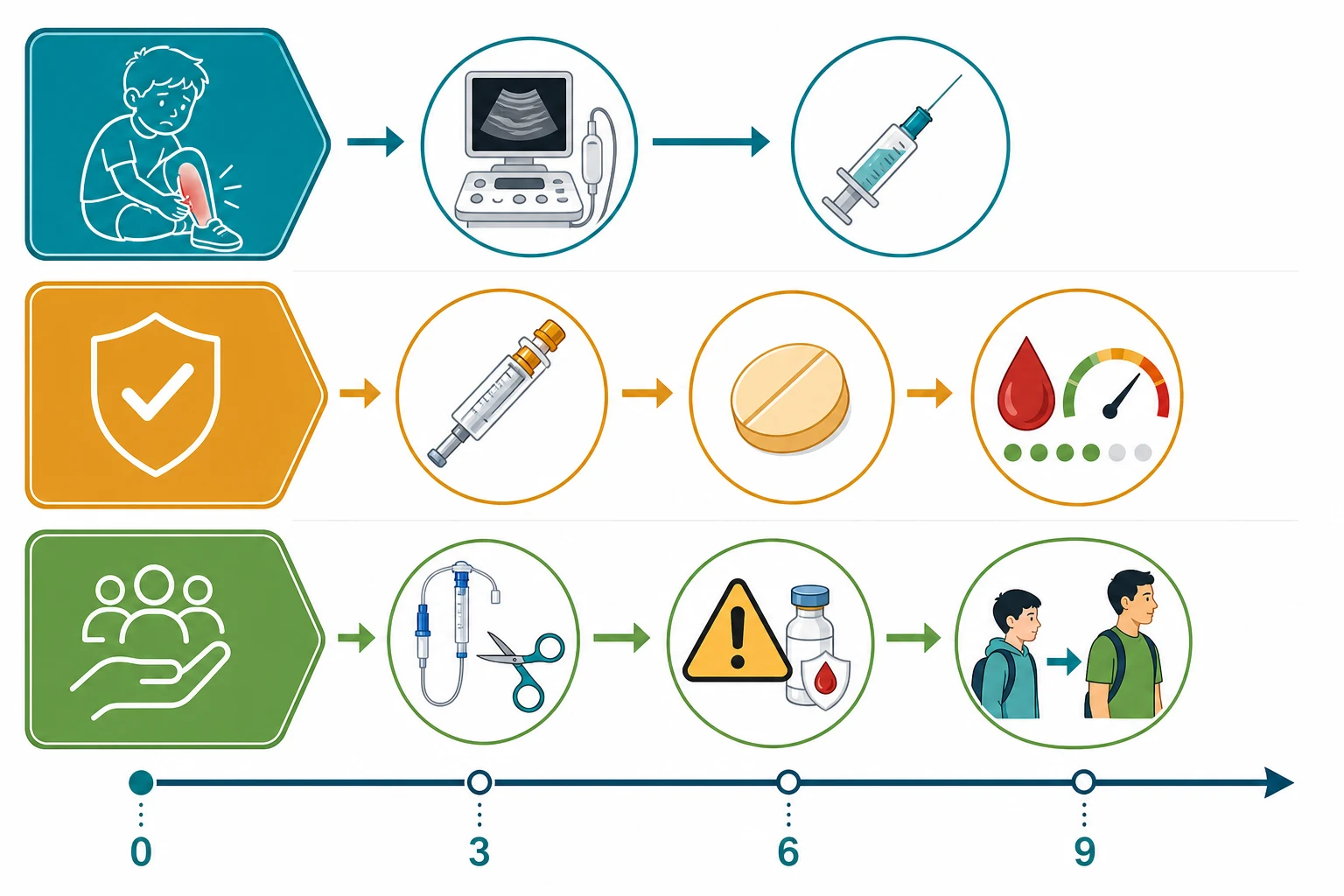
Low molecular weight heparin remains the workhorse of paediatric anticoagulation and is recommended by both the CHEST and ASH guidelines as a preferred agent. It is given subcutaneously, has a predictable dose response, does not require routine coagulation monitoring beyond the anti-factor Xa level, and is reversible with protamine. Its drawbacks are the need for twice-dightly injections, the discomfort for the child and family, and the rare risk of heparin-induced thrombocytopenia, which is far less common in children than in adults. Enoxaparin or dalteparin are the commonly used agents. [1] [7]
The EINSTEIN-Jr programme (Monagle 2019, Male 2020, Thom 2020)
Multicentre phase 2 and phase 3 trials of bodyweight-adjusted oral rivaroxaban versus standard anticoagulants (low molecular weight heparin or warfarin) in children with venous thromboembolism, including a catheter-related subgroup
Key finding
Bodyweight-adjusted rivaroxaban achieved comparable or better thrombus resolution and symptom improvement with a similar or lower bleeding rate, including in catheter-related disease, establishing rivaroxaban as a licensed oral option for children.
Practice change
Rivaroxaban is now a first-line oral alternative to low molecular weight heparin for children, avoiding injections for many families, and it is specifically evaluated for catheter-related thrombosis.
The direct oral anticoagulants have transformed paediatric care. Rivaroxaban, a direct factor Xa inhibitor, is now licensed for children based on the EINSTEIN-Jr programme. The phase 2 studies established a bodyweight-adjusted dosing schedule that achieves adult-equivalent drug exposure, and the phase 3 trial showed that rivaroxaban compared favourably with standard anticoagulants for recurrent thrombosis and bleeding. The catheter-related subgroup confirmed efficacy and safety in central venous catheter thrombosis specifically. Rivaroxaban is given as a tablet or, for younger children, an oral suspension, and avoids the need for injections and monitoring, which is a major quality-of-life advantage. [4] [5] [6]
Warfarin, the vitamin K antagonist, is now a third-line option for most children because it requires frequent international normalised ratio monitoring, interacts with many foods and medications, and has a slow onset, but it remains useful for children with mechanical heart valves or antiphospholipid syndrome, and in settings where the direct oral anticoagulants are unavailable. The duration of anticoagulation follows the provoked or unprovoked status: a provoked thrombosis with a reversible trigger such as a central line is treated for 6 weeks to 3 months; an unprovoked thrombosis for 3 to 6 months; and a recurrent thrombosis or a strong thrombophilia for 6 to 12 months or longer. [2] [1]
Specific Subtypes & Scenarios
Central venous access device-associated thrombosis is the single most common scenario in paediatric practice and deserves its own management logic. The thrombosis is often asymptomatic, discovered when a line will not draw or flush, or found on a surveillance ultrasound. Symptomatic catheter-related thrombosis is treated with therapeutic anticoagulation for 6 weeks to 3 months, and the central line is removed once it is no longer needed and the clot has stabilised. The EINSTEIN-Jr CVC-VTE subgroup specifically addressed this population and confirmed the safety of anticoagulation including rivaroxaban. Routine pharmacologic prophylaxis to prevent catheter-related thrombosis is not recommended, as a Cochrane review found no clear benefit. [6] [12]
Pulmonary embolism in the adolescent is a rising scenario and a frequent exam subject. The adolescent girl on the combined oral contraceptive pill who develops sudden pleuritic chest pain and breathlessness, sometimes after surgery or a long journey, presents like an adult. The diagnosis is confirmed with CT pulmonary angiography, and management is therapeutic anticoagulation for 3 to 6 months, with consideration of thrombophilia testing for an unprovoked event and advice to avoid oestrogen-containing contraception in future. [3] [11]
Neonatal thrombosis is the third key scenario and includes renal vein thrombosis (the firm flank mass and haematuria, often in an infant of a diabetic mother), cerebral sinus venous thrombosis (seizures and apnoea), and catheter-related thrombosis from umbilical and central lines. The neonatal haemostatic system and the higher heparin clearance demand age-specific dosing: enoxaparin 1.5 mg per kg twice daily. Supportive care, treatment of the underlying cause, and removal of an unnecessary catheter are the principles, and anticoagulation is used selectively because the bleeding risk is higher in the neonate. [1] [7]
CLOTS
Complications & Pitfalls
The complications of paediatric venous thromboembolism divide into those of the disease and those of its treatment. The dominant disease complication is the post-thrombotic syndrome, a chronic venous insufficiency that follows damage to the deep vein valves, presenting with limb swelling, pain, heaviness, skin discolouration and occasionally ulceration. It affects a substantial minority of children after a limb deep vein thrombosis, develops over months to years, and is the main long-term morbidity. Recurrent thrombosis and fatal pulmonary embolism are the acute dangers. [1] [11]
The treatment complications are primarily bleeding and heparin-induced thrombocytopenia. Major bleeding with low molecular weight heparin occurs in a small percentage of treated children and is higher in the critically ill and in infants, and protamine partially reverses the effect. Heparin-induced thrombocytopenia, a paradoxical prothrombotic antibody reaction, is far rarer in children than in adults but must be considered when the platelet count falls between 5 and 14 days after starting heparin, and it mandates switching to a non-heparin anticoagulant. [7] [1]
The avoidable pitfalls are well defined. The first is failing to think of thrombosis in a child because the disease is rare, so a swollen leg in a child with a central line is misattributed to cellulitis and the diagnosis is delayed. The second is over-testing for hereditary thrombophilia in a child with a clearly provoked catheter-related thrombosis, generating anxiety and labels without changing management. The third is using adult heparin doses in a neonate, which are subtherapeutic because of the higher neonatal clearance. The fourth is prescribing oestrogen-containing contraception to a girl with a personal or strong family history of thrombosis without screening. [8] [9]
The interaction of the inherited thrombophilias with contraception is a classic exam point. A girl heterozygous for factor V Leiden who takes the combined oral contraceptive pill faces a substantially increased relative risk of venous thromboembolism, and a personal or strong family history should prompt thrombophilia screening and a preference for progestogen-only or non-hormonal contraception. This counselling is increasingly part of the adolescent medicine encounter and the transition clinic. [8] [9]
Prognosis & Disposition
The prognosis of paediatric venous thromboembolism is generally favourable for survival but carries a significant burden of recurrence and post-thrombotic syndrome. Mortality directly attributable to the thrombosis is low in modern practice, though the conditions that provoked it (critical illness, malignancy) carry their own mortality. Recurrence occurs in around 10 percent of children over the years following a first event, and is higher in those with a persistent provoking factor, an unprovoked event, or a strong thrombophilia. [1] [2]
The events that worsen the prognosis are a persistent or unremovable provoking factor, an underlying strong thrombophilia (especially antithrombin deficiency or antiphospholipid syndrome), and adherence problems with long-term injected anticoagulation. The post-thrombotic syndrome develops insidiously and is the main source of long-term disability, which is why adequate initial treatment and follow-up imaging matter. [11] [2]
Disposition is through a paediatric haematology service that coordinates the diagnostic workup, the choice and duration of anticoagulation, the monitoring (anti-factor Xa levels for heparin, international normalised ratio for warfarin), and the transition to adult care for the adolescent. The child and family learn injection technique, bleeding precautions, the warning signs of recurrence and pulmonary embolism, and the importance of adherence. A clear written action plan, a medic alert device, and coordination with the primary care team and the school complete the disposition. [7] [11]
The disparity between high-income and resource-limited settings is real. Access to paediatric haematology expertise, to anti-factor Xa monitoring, and to the direct oral anticoagulants varies widely, and in many settings injected low molecular weight heparin remains the only option, with the burden of twice-daily injections falling on the family. The move toward licensed oral direct anticoagulants for children is narrowing this gap, and access programmes are extending their reach. [4] [2]
Special Populations
The neonate is the population where developmental haemostasis and the frequency of central lines converge to produce the highest risk. Neonatal thrombosis is dominated by catheter-related events, renal vein thrombosis, and cerebral sinus venous thrombosis, and the management uses age-specific enoxaparin dosing of 1.5 mg per kg twice daily. The neonatal haemostatic system also means that low anticoagulant protein levels on a panel may be developmental rather than hereditary, so thrombophilia results are interpreted with caution and confirmed by repeat or genetic testing later in infancy. [1] [7]
The critically ill child in the paediatric intensive care unit is the population where thrombosis is commonest and hardest to detect, because immobility, multiple central lines, sepsis and surgery stack the risk factors, and the signs are obscured by sedation and the underlying illness. A high index of suspicion, surveillance imaging in the highest-risk groups, and early involvement of the haematology team govern the approach, and thromboprophylaxis is considered selectively in the highest-risk older adolescents. [3] [2]
The adolescent girl and the transitioning young person deserve attention for the oestrogen-related risks and the adherence challenges. The combined oral contraceptive pill substantially raises the risk of thrombosis, particularly with an underlying thrombophilia, so contraception counselling is part of every adolescent encounter where thrombosis is in the history or family. Transition to adult care is structured and deliberate, handing over the anticoagulation regimen, the thrombophilia status, the contraception advice, and the psychosocial needs, because the transition period is a high-risk time for disengagement and recurrence. [8] [9]
[8] [9]Evidence, Guidelines & Regional Differences
The cornerstone guidance is the American College of Chest Physicians ninth edition antithrombotic guideline for neonates and children, which set the framework for risk stratification, diagnosis, and the use of low molecular weight heparin, and the 2018 American Society of Hematology guideline for the treatment of paediatric venous thromboembolism, which reinforced low molecular weight heparin as a preferred agent and gave conditional recommendations on duration and thrombophilia testing. These two guidelines frame practice worldwide and are the references examiners expect. [1] [2]
The landmark modern evidence is the EINSTEIN-Jr programme. The phase 2 studies established the bodyweight-adjusted rivaroxaban dosing that achieves adult-equivalent exposure in children, the phase 3 randomised trial compared rivaroxaban with standard anticoagulants and showed comparable safety and efficacy, and the catheter-related subgroup addressed the dominant paediatric category directly. These trials established rivaroxaban as a licensed oral alternative to injected low molecular weight heparin, transforming the quality of life for many families. [4] [5] [6]
The frontier of paediatric thrombosis is moving toward simpler, oral therapy. Additional direct oral anticoagulants, including dabigatran and apixaban, are being evaluated in children, and extended-interval low molecular weight heparin preparations and antidote strategies are improving safety. The broader challenge is equitable access: many children worldwide still rely on injected heparin and lack the monitoring and expertise to use the oral agents safely. [4] [2]
Exam Pearls
Paediatric venous thromboembolism is provoked and secondary in over 90 percent of cases, and the central venous access device is the single largest provoking factor, accounting for around half of all events. The incidence is bimodal, peaking in neonates and in adolescents, and it has risen dramatically with better survival of critically ill children and wider use of central lines. Think of thrombosis in any child with a central line, critical illness, recent surgery, malignancy or oestrogen use who has a swollen limb, chest pain and breathlessness, or an unexplained deterioration. [3] [11]
The treatment backbone is low molecular weight heparin: enoxaparin 1.5 mg per kg twice daily for a neonate and 1 mg per kg twice daily for a child over two months, monitored by the anti-factor Xa level with a target of 0.5 to 1.0 IU per mL for twice-daily dosing. The neonatal dose is 50 percent higher because neonates clear heparin faster. Bodyweight-adjusted oral rivaroxaban is now licensed for children after the EINSTEIN-Jr trials and avoids injections for many families. Duration follows the provoked or unprovoked status: 6 weeks to 3 months for a provoked catheter-related clot, 3 to 6 months for an unprovoked event. [1] [7] [4]
Thrombophilia testing is selective, never universal, and is reserved for an unprovoked, recurrent or unusual-site thrombosis or a strong family history. The common abnormalities such as factor V Leiden have a low positive predictive value, and over-testing labels healthy children and families. Test outside the acute phase, because protein C, protein S and antithrombin levels are transiently altered by active thrombosis and inflammation, and interpret neonatal results with caution because of developmental haemostasis. [8] [9]
The high-yield one-liners an examiner rewards: the central venous catheter is the commonest provoking factor; the anti-factor Xa target for twice-daily low molecular weight heparin is 0.5 to 1.0 IU per mL; a girl with factor V Leiden should avoid the combined oral contraceptive pill; the post-thrombotic syndrome is the main long-term morbidity of a limb deep vein thrombosis; and pulmonary embolism is increasingly recognised and frequently missed in children. [11] [8] [6]
References
- [1]Monagle P, Chan AKC, Goldenberg NA, et al. Antithrombotic therapy in neonates and children: Antithrombotic Therapy and Prevention of Thrombosis, 9th ed: American College of Chest Physicians Evidence-Based Clinical Practice Guidelines. Chest, 2012.PMID 22315277
- [2]Monagle P, Cuello CA, Augustine C, et al. American Society of Hematology 2018 Guidelines for management of venous thromboembolism: treatment of pediatric venous thromboembolism. Blood Adv, 2018.PMID 30482766
- [3]Raffini L, Huang YS, Witmer C, et al. Dramatic increase in venous thromboembolism in children's hospitals in the United States from 2001 to 2007. Pediatrics, 2009.PMID 19736261
- [4]Male C, Lensing AWA, Palumbo JS, et al. Rivaroxaban compared with standard anticoagulants for the treatment of acute venous thromboembolism in children: a randomised, controlled, phase 3 trial. Lancet Haematol, 2020.PMID 31699660
- [5]Monagle P, Lensing AWA, Thelen K, et al. Bodyweight-adjusted rivaroxaban for children with venous thromboembolism (EINSTEIN-Jr): results from three multicentre, single-arm, phase 2 studies. Lancet Haematol, 2019.PMID 31420317
- [6]Thom K, Lensing AWA, Nurmeev I, et al. Safety and efficacy of anticoagulant therapy in pediatric catheter-related venous thrombosis (EINSTEIN-Jr CVC-VTE). Blood Adv, 2020.PMID 33002131
- [7]Law C, Raffini L A guide to the use of anticoagulant drugs in children. Paediatr Drugs, 2015.PMID 25711916
- [8]van Ommen CH, Nowak-Göttl U Inherited Thrombophilia in Pediatric Venous Thromboembolic Disease: Why and Who to Test. Front Pediatr, 2017.PMID 28352625
- [9]Heleen van Ommen C, Middeldorp S Thrombophilia in childhood: to test or not to test. Semin Thromb Hemost, 2011.PMID 22187402
- [10]Mahajerin A, Obasaju P, Eckert G, et al. Thrombophilia testing in children: a 7 year experience. Pediatr Blood Cancer, 2014.PMID 24249220
- [11]Monagle P Diagnosis and management of deep venous thrombosis and pulmonary embolism in neonates and children. Semin Thromb Hemost, 2012.PMID 23034828
- [12]Pelland-Marcotte MC, Amiri N, Avila ML, et al. Low molecular weight heparin for prevention of central venous catheter-related thrombosis in children. Cochrane Database Syst Rev, 2020.PMID 32557627