Rheumatology · Rheumatology
Sjogren's Syndrome
Also known as sicca syndrome · Sicca syndrome · autoimmune epithelitis · Sjogren's disease · Mikulicz disease
Sjogren's syndrome is a chronic systemic autoimmune epithelitis characterised by focal lymphocytic infiltration of exocrine glands leading to sicca syndrome (dry eyes plus dry mouth), with variable extraglandular involvement. It is the second most common autoimmune rheumatic disease after rheumatoid arthritis, with a 9 to 1 female preponderance and peak onset at age 40 to 60. Anti-Ro/SSA is the key serological marker. Primary Sjogren's is classified by the ACR/EULAR 2016 weighted score of at least 4. Management is symptomatic first (artificial tears, saliva substitutes, secretagogues such as pilocarpine 5 mg four times daily), with immunosuppression reserved for organ-threatening disease. Lifelong surveillance for B-cell (MALT) lymphoma is essential.
On this page & tools
Your progress
Saved locally on this device.
Exam tags
Red flags
Overview & Definition
Sjogren's syndrome is a chronic, systemic autoimmune disease in which lymphocytes infiltrate and progressively destroy the exocrine glands — principally the lacrimal and major salivary glands — producing the sicca syndrome of xerophthalmia (dry eyes) and xerostomia (dry mouth). Because the glandular epithelium itself is the autoimmune target and actively participates in the inflammatory response, the disease is increasingly and more accurately termed autoimmune epithelitis. The process is not confined to the glands: extraglandular (systemic) involvement of skin, joints, lungs, kidneys, nerves and the haematological system is variable but clinically important, and the disease carries a characteristic predisposition to B-cell non-Hodgkin lymphoma.[1]
The syndrome sits within the family of systemic autoimmune rheumatic diseases, alongside rheumatoid arthritis, systemic lupus erythematosus and systemic sclerosis, with which it shares autoantibodies, genetic susceptibility and many clinical features. Two operational categories are recognised: primary Sjogren's syndrome (standalone, without another defined connective-tissue disease) and secondary Sjogren's syndrome (sicca features occurring in the setting of another autoimmune disease, most often rheumatoid arthritis). The distinction matters for prognosis, classification criteria, lymphoma risk and the focus of therapy.[1]
Classification & Definition — Primary versus Secondary
The classification of Sjogren's into primary and secondary forms is the first practical question at the bedside and at every examination. [1]
Primary Sjogren's
- Standalone autoimmune disease — no other defined connective-tissue disease coexists
- Classified by the ACR/EULAR 2016 weighted score of at least 4 (with applicable exclusions)
- Higher anti-Ro/SSA positivity (60 to 95 percent depending on assay) and a higher lymphoma risk
- Extraglandular disease more prominent and florid (vasculitis, renal, lung, neuropathy)
- Older 2002 AECG and 1993/1996 European criteria superseded; 2016 criteria are data-driven
Secondary Sjogren's
- Sicca features occur WITH another defined connective-tissue disease
- Most commonly rheumatoid arthritis, then systemic lupus erythematosus, systemic sclerosis, mixed connective-tissue disease
- No validated classification criteria — diagnose sicca on objective testing (Schirmer, biopsy, anti-Ro)
- Lymphoma risk lower than primary form but still elevated versus general population
- Management directed at the underlying disease PLUS sicca measures; secondary SS is not separately classified
The currently accepted international standard for primary Sjogren's syndrome is the 2016 American College of Rheumatology/European League Against Rheumatism (ACR/EULAR) classification criteria, which superseded the older European Economic Community (1993/1996, Vitali), American-European Consensus Group (AECG, 2002) and ACR (2012) criteria. The 2016 criteria were derived by international consensus and data-driven analysis of three large cohorts, deliberately combining the strongest objective items (biopsy and serology, each worth three points) with three objective secretory tests (each worth one point).[2]
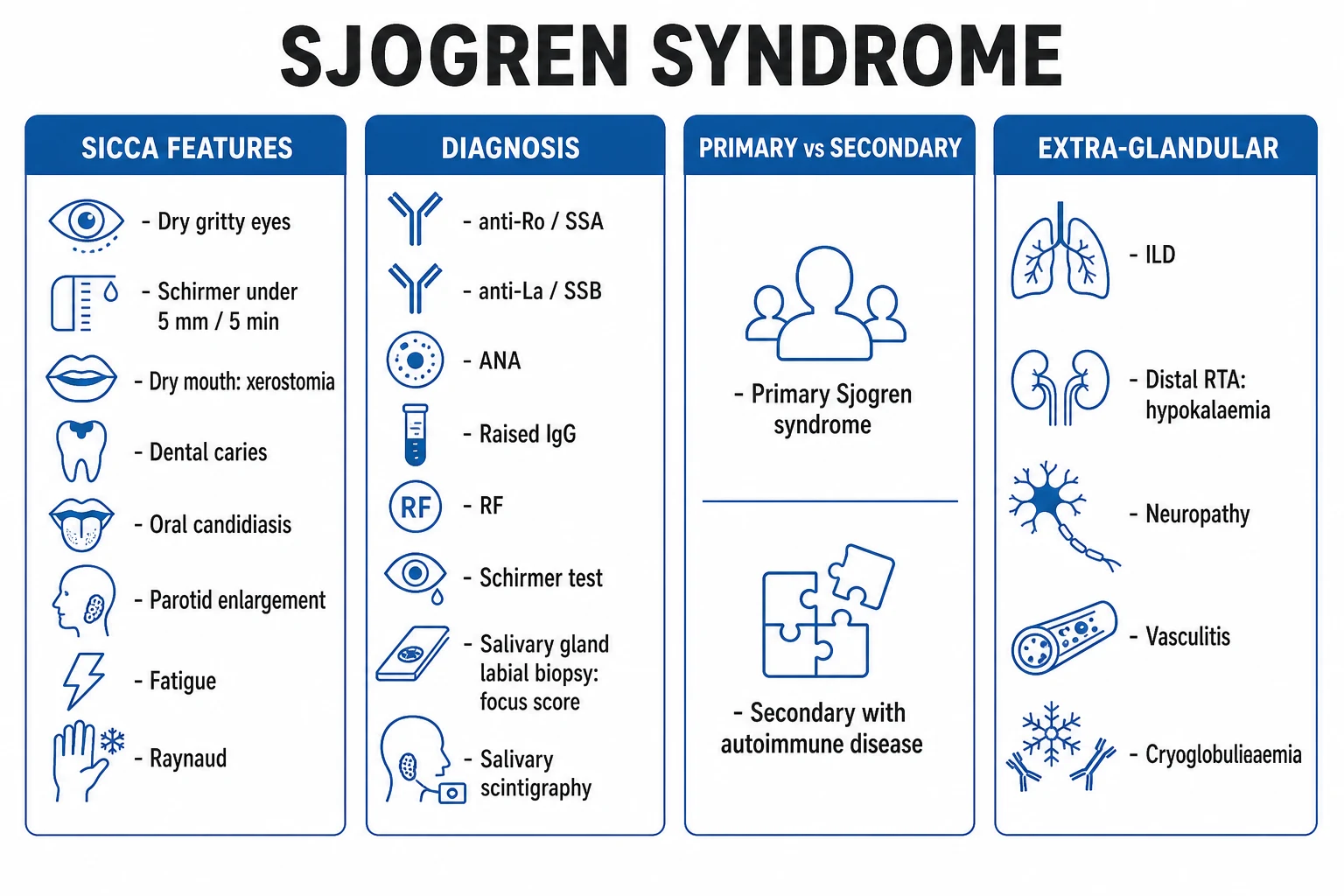
The ACR/EULAR 2016 criteria are the international standard and are used by all major rheumatology societies for primary Sjogren's. No validated criteria exist for secondary Sjogren's; diagnosis rests on objective sicca testing in a patient already diagnosed with another connective-tissue disease.[2]
Epidemiology & Risk Factors
Sjogren's syndrome is the second most common systemic autoimmune rheumatic disease after rheumatoid arthritis, with an estimated prevalence of 0.1 to 0.6 percent of the general population. The female-to-male ratio is approximately 9 to 1, and the typical age of onset is 40 to 60 years, peaking around the menopause — a pattern implicating oestrogen both as a permissive factor and, by its decline, as a trigger of disease expression. Men who develop the disease are often under-diagnosed, with a delayed workup for sicca.[1][12]
Sjogren's syndrome — headline epidemiology
Recognised risk factors and associations include female sex and oestrogen exposure, genetic susceptibility (HLA-DRB1*03, HLA-DR2 and HLA-DRw52, plus the type I interferon pathway polymorphisms IRF5 and STAT4), a family history of autoimmunity, and viral triggers — Epstein-Barr virus, coxsackievirus, and (importantly as an exclusion) hepatitis C virus. Secondary Sjogren's complicates roughly 20 to 30 percent of rheumatoid arthritis and a clinically significant fraction of systemic lupus erythematosus and systemic sclerosis cohorts. A north-south epidemiological gradient has been observed, with southern European cohorts exhibiting more systemic (extraglandular) involvement at presentation.[12]
Although overwhelmingly a disease of middle-aged women, Sjogren's can occur in men (often under-recognised), in the elderly, and rarely in children — in whom recurrent parotitis, dental caries and ANA positivity are often the presenting clues, rather than sicca. [1]
Pathophysiology
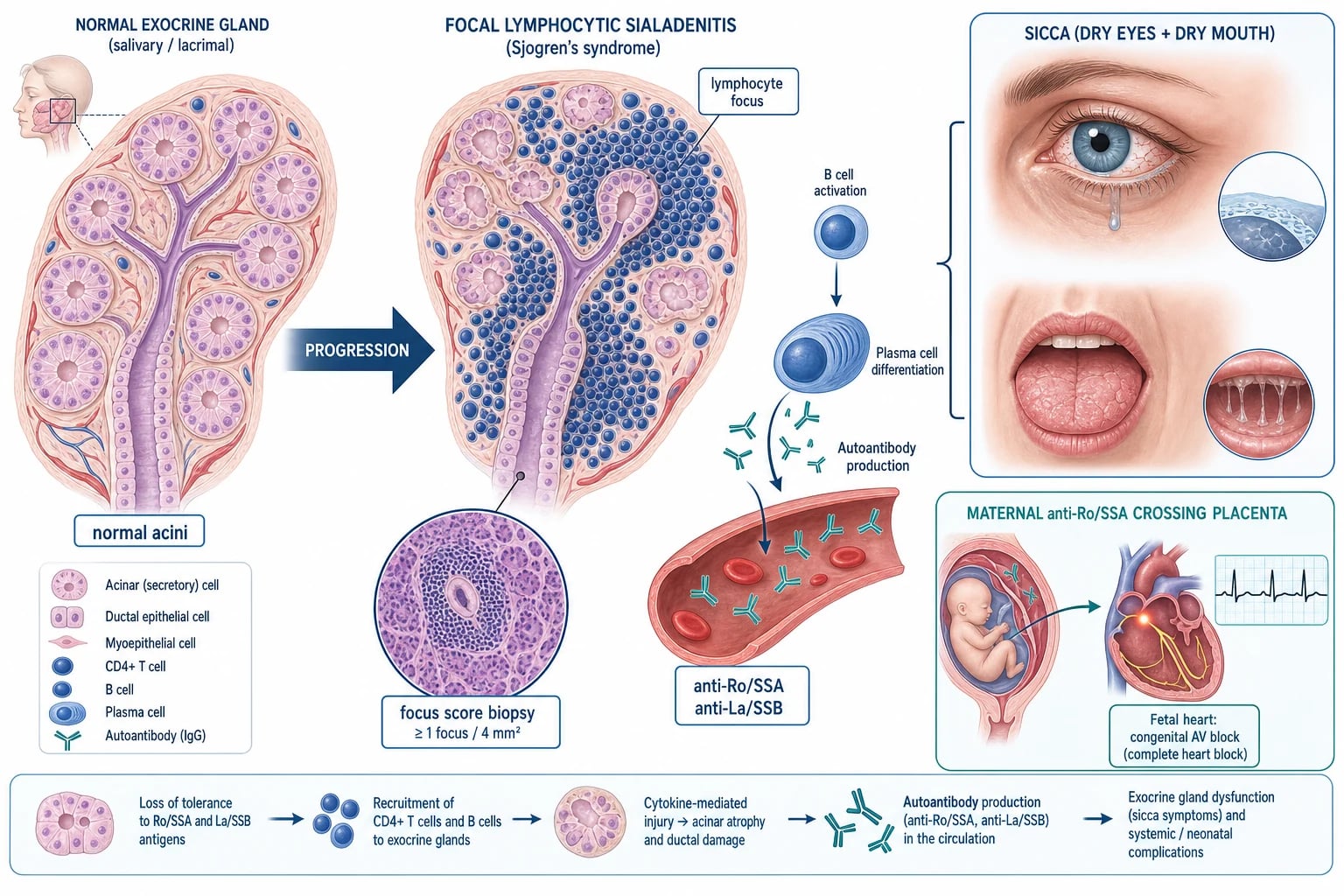
Sjogren's syndrome is best understood as an epithelium-centred autoimmune process in which the glandular epithelial cell is both the target and an active participant. The defining lesion is focal lymphocytic sialadenitis — dense periductal aggregates of CD4-positive T-helper cells, B cells and plasma cells surrounding the salivary (and, by parallel process, the lacrimal) ducts, with progressive acinar destruction and ductal narrowing. Function fails because the surviving acini cannot secrete and the narrowed ducts cannot deliver saliva or tears.[1]
The histological hallmark — and a gold-standard diagnostic test — is the focus score on minor (labial) salivary gland biopsy. A focus is defined as a cluster of at least 50 mononuclear cells per 4 mm squared of glandular tissue; a focus score of at least 1 (one or more foci per 4 mm squared, with non-focal periductal lymphocytes ignored) is diagnostic-grade for the ACR/EULAR 2016 criteria and earns three weighted points. Ectopic germinal-centre-like lymphoid organisation within the glands is seen in a subset of patients and predicts subsequent lymphoma development.[9]
Two intertwined immune axes drive the injury: [1]
The anti-Ro/SSA antibodies (against Ro52/TRIM21 and Ro60 ribonucleoproteins) and anti-La/SSB antibodies (against the La/SSB ribonucleoprotein) are the serological signatures of this B-cell activation. They are not directly gland-toxic but define the disease serologically, contribute to immune-complex formation, and — of critical importance in pregnancy — cross the placenta to cause neonatal lupus and congenital heart block. Hypergammaglobulinaemia, rheumatoid factor positivity (even without rheumatoid arthritis) and cryoglobulinaemia all reflect this B-cell overdrive.[1]
Clinical Presentation
The clinical picture combines glandular (sicca) features — the universal core of the disease — with a variable but important extraglandular (systemic) component. Presentation may be sicca-dominated or, less commonly, dominated by an extraglandular manifestation (vasculitic purpura, distal renal tubular acidosis, peripheral neuropathy, recurrent dental caries or parotid swelling) that brings the patient to attention before sicca is volunteered.[1]
Glandular (sicca) features
- Xerostomia (dry mouth): difficulty swallowing dry foods (the patient sips water to swallow — the "cracker sign"), difficulty speaking for long periods, altered taste, a furrowed, fissured tongue, rapid and accelerated dental caries (often the earliest clue), recurrent oral candidiasis, and sialadenitis. Unstimulated whole saliva flow is reduced.
- Xerophthalmia (keratoconjunctivitis sicca): a gritty, sandy or foreign-body sensation, burning, photophobia, paradoxical tearing in response to irritation, and the well-known inability to produce tears while crying emotionally. Untreated, keratitis, corneal abrasion and, rarely, corneal perforation may supervene.
- Major salivary gland enlargement: bilateral, recurrent or persistent parotid and submandibular enlargement occurs in roughly 30 to 50 percent of patients over the disease course. Persistent, unilateral, hard or rapidly growing parotid enlargement is a red flag for lymphoma. [1]
Extraglandular (systemic) features
Constitutional & musculoskeletal
- Profound **fatigue** — frequently the most disabling symptom and a major driver of quality-of-life loss
- Low-grade fever, non-erosive symmetrical small-joint **arthralgia or arthritis**
- **Raynaud phenomenon** in roughly 20 to 40 percent; myalgia and fibromyalgia overlap
Cutaneous & vascular
- **Palpable purpura** on the lower legs — leucocytoclastic or cryoglobulinaemic vasculitis (a lymphoma predictor)
- Annular erythema, xerosis; sometimes urticarial vasculitis
- Raynaud phenomenon; rarely digital ulcers
Respiratory
- Dry cough from tracheobronchial sicca; **interstitial lung disease** (NSIP pattern most common)
- **Lymphocytic interstitial pneumonia (LIP)** — relatively characteristic of Sjogren's
- Bronchiectasis from sicca of the airways; pleuritis; rare lymphoma
Renal
- **Distal (type 1) renal tubular acidosis** — hypokalaemia (occasionally periodic paralysis), nephrocalcinosis, osteomalacia
- Tubulointerstitial nephritis; rarely glomerulonephritis (often membranous or cryoglobulinaemic)
Neurological
- Predominantly **sensory (small-fibre) peripheral neuropathy** and painful neuropathy
- **Trigeminal neuropathy**; less commonly sensorimotor neuropathy or mononeuritis multiplex (vasculitic)
- Rare **CNS** involvement — transverse myelitis, optic neuritis; consider **neuromyelitis optica spectrum** overlap (anti-aquaporin-4)
Hepatic & haematological
- Overlap with **primary biliary cholangitis** (anti-mitochondrial antibody), autoimmune hepatitis
- **Lymphadenopathy**, splenomegaly, cytopenias (anaemia, leukopenia, lymphopenia)
- **Polyclonal hypergammaglobulinaemia**, cryoglobulinaemia; monoclonal gammopathy may herald lymphoma
Atypical presentations
Examiners deliberately probe the corners. Sjogren's may present atypically as: [1]
- Extraglandular presentation with mild or delayed sicca — e.g. a young woman with palpable purpura, distal RTA, or a sensory neuropathy, in whom sicca emerges only on direct questioning.
- Recurrent dental caries or parotid swelling as the first clue in an otherwise asymptomatic patient.
- Hypokalaemic periodic paralysis from undiagnosed distal RTA.
- Older men misattributed to "ageing" or drug side-effect.
- Children with recurrent parotitis and ANA positivity rather than sicca. [1]
Neurological Sjogren's — pattern recognition
Neurological involvement is among the most challenging manifestations to diagnose and is frequently tested. Three patterns recur. Sensory (small-fibre) peripheral neuropathy — burning, painful distal extremities with normal nerve conduction studies and reduced intra-epidermal nerve fibre density on skin biopsy — is the commonest and often the most disabling. Trigeminal sensory neuropathy (often unilateral, numbness rather than pain) is relatively characteristic of Sjogren's. Sensorimotor neuropathy or mononeuritis multiplex signals vasculitic disease and warrants urgent immunosuppression. Central nervous system involvement is rarer and controversial; reported forms include transverse myelitis, optic neuritis and a multiple-sclerosis-like illness. Any patient with Sjogren's who develops optic neuritis or a longitudinally extensive spinal-cord lesion should be tested for anti-aquaporin-4 (NMO-IgG), because Sjogren's overlaps with neuromyelitis optica spectrum disorder and the management differs fundamentally.[1]
Differential Diagnosis
Sicca symptoms are common and nonspecific; the differential is wide and must be worked through systematically. At least three distinguishing features should be considered for each. [1]
Drug-induced sicca
- Anticholinergics, tricyclic antidepressants, antihistamines, opioids, diuretics, antipsychotics, alpha-blockers
- Reversible on withdrawal or dose reduction; no objective inflammation
- Negative anti-Ro/La; normal labial biopsy
Dehydration / age-related
- Elderly with reduced fluid intake, mouth-breathing, reduced glandular reserve with age
- No autoantibodies or extraglandular features; corrects with hydration
- Normal objective secretory testing once hydration restored
Head/neck radiotherapy
- Clear history of prior radiation for head/neck cancer or lymphoma — an **exclusion criterion** for primary SS
- Acute and chronic gland destruction; other radiation stigmata
- Anti-Ro/La negative
Hepatitis C
- Causes sicca and mixed cryoglobulinaemia; **exclusion criterion** for primary SS — always screen
- Transaminitis, positive HCV RNA, mixed cryoglobulins, low C4
- Labial biopsy may show lymphocytic sialadenitis but with different serology
HIV (diffuse infiltrative lymphocytosis syndrome)
- Causes sicca and parotid enlargement; **exclusion criterion** for primary SS — always screen
- CD8 lymphocytosis, positive HIV serology; affects men more often
- Anti-Ro/La typically negative; biopsy shows CD8 predominant infiltrate
IgG4-related disease
- Painless salivary/lacrimal enlargement (Mikulicz syndrome), pancreatitis, retroperitoneal fibrosis; older men
- Elevated serum IgG4; **storiform fibrosis, obliterative phlebitis, IgG4-positive plasma cells** on histology
- Dramatic steroid response — **exclusion criterion** for primary SS
Sarcoidosis
- May cause sicca and parotid enlargement (Heerfordt syndrome); **exclusion criterion**
- Non-caseating granulomas on biopsy; hilar lymphadenopathy; raised ACE
- Hypercalcaemia; anti-Ro/La negative
GVHD / amyloidosis
- Graft-versus-host disease after haematopoietic stem cell transplant; amyloid infiltration
- respective transplant history or amyloid biopsy (Congo red)
- Both **exclusion criteria** for primary SS
Parotid differential — mumps, sialolithiasis, lymphoma
- **Mumps**: acute, painful, viral prodrome, unilateral or bilateral parotitis
- **Sialolithiasis**: unilateral, meal-time-related swelling, duct stone on ultrasound
- **Parotid lymphoma/malignancy**: persistent, hard, unilateral, nerve palsy — refer
The dry-eye differential also includes meibomian gland dysfunction and chronic blepharitis, and the fatigue must be distinguished from depression, hypothyroidism (a co-existent autoimmune association), iron deficiency, sleep apnoea and fibromyalgia overlap — all of which are common in this demographic and frequently co-exist. [1]
Clinical & Bedside Assessment
A focused, structured assessment at the bedside combines history, oral and ocular examination, and a search for extraglandular disease and lymphoma red flags. [1]
History — ask about: dryness onset, progression and diurnal pattern; medication review (anticholinergics); photosensitivity, Raynaud, joint pain and swelling; dental history (rapid caries, recurrent extractions); recurrent oral thrush; parotid swelling (intermittent or persistent, unilateral or bilateral); palpable purpura, neuropathic symptoms, dry cough; miscarriage or offspring with congenital heart block (anti-Ro); and family history of autoimmunity. Quantify fatigue and dryness using the ESSPRI (EULAR Sjogren's Syndrome Patient Reported Index) — a 0 to 10 visual analogue scale across dryness, fatigue and limb pain.[4]
Bedside sicca examination — inspect the mouth for a dry, furrowed or fissured tongue, atrophic mucosa, candidiasis, caries and dental work; palpate the parotid and submandibular glands (size, tenderness, symmetry, consistency — hard, fixed or progressively enlarging glands are concerning); examine the eyes for conjunctival injection, corneal staining and a reduced tear meniscus. Demonstrate Schirmer's test: hook a standard filter paper strip over the lower eyelid margin (lateral third) for 5 minutes with the eyes gently closed; at most 5 mm of wetting in at least one eye supports keratoconjunctivitis sicca.[1]
Extraglandular examination — palpate for palpable purpura on the lower limbs, assess for Raynaud and annular lesions, examine joints for non-erosive synovitis, listen for crackles of interstitial lung disease, examine the abdomen for hepatosplenomegaly (PBC overlap or lymphoma), and perform a screening neurological examination for sensory neuropathy and trigeminal involvement. Check for lymphadenopathy in all cervical, axillary and inguinal regions. [1]
Lymphoma red flags — actively look for: new or persistent parotid enlargement, lymphadenopathy or splenomegaly; unexplained weight loss or night sweats; and a serological profile of low complement C4, cryoglobulins, palpable purpura, monoclonal gammopathy, or a falling IgM or rheumatoid factor (the loss of immune complexes can herald transition to lymphoma). [1]
Investigations
No single test establishes primary Sjogren's syndrome. The diagnosis rests on the ACR/EULAR 2016 weighted score, which integrates objective histology, serology and secretory function. The total score must be at least 4 (out of a possible 9), with applicable exclusions excluded, to classify primary Sjogren's.[2]
The ACR/EULAR 2016 weighted score — reproduced verbatim
The criteria apply to any patient with at least one symptom of ocular or oral dryness (or suspicion of SS from the ESSDAI questionnaire). The score is: [1]
- Labial salivary gland biopsy with focal lymphocytic sialadenitis and focus score at least 1 (at least 50 mononuclear cells per 4 mm squared) — 3 points
- Anti-SSA/Ro positive (anti-Ro/SSA, including anti-Ro52 and anti-Ro60) — 3 points
- Ocular staining score at least 5 (van Bijsterveld or SICCA/Oxford scheme) — 1 point
- Schirmer's test at most 5 mm in 5 minutes (in at least one eye) — 1 point
- Unstimulated whole saliva flow at most 0.1 mL per minute (equivalent to at most 1.5 mL in 15 minutes) — 1 point [1]
A total score of at least 4 classifies primary Sjogren's syndrome, provided no exclusion applies.[2]
Exclusion criteria (ACR/EULAR 2016, verbatim)
Because of overlapping clinical features or interference with the criteria tests, the following exclude a classification of primary Sjogren's syndrome: [1]
- History of head and neck radiation treatment
- Active hepatitis C (HCV) infection
- AIDS (HIV)
- Sarcoidosis
- Amyloidosis
- Graft-versus-host disease (GVHD)
- IgG4-related disease (IgG4-RD) [1]
Note that the 2016 criteria do not list rheumatoid arthritis or SLE as exclusions — the criteria are designed for the classification of primary Sjogren's syndrome, so a patient with another defined connective-tissue disease is simply not classified as primary SS by these criteria; their sicca is labelled secondary. [1]
Autoantibody profile
- Anti-Ro/SSA: the key antibody in primary Sjogren's, positive in roughly 60 to 95 percent of primary cases (the most sensitive and most useful serology; 3 criteria points). Anti-Ro52 (TRIM21) and anti-Ro60 may be reported separately.[1]
- Anti-La/SSB: positive in approximately 40 to 60 percent; more specific but less sensitive, and rarely positive without anti-Ro. Not part of the weighted score but supports the diagnosis.
- Antinuclear antibody (ANA): frequently positive, often with a speckled pattern.
- Rheumatoid factor (RF): positive in roughly 40 to 60 percent, even in the absence of rheumatoid arthritis — a marker of B-cell hyperactivity.
- Anti-mitochondrial antibody (AMA): screen for primary biliary cholangitis overlap when liver enzymes are cholestatic.
- Cryoglobulins, complement (C3, C4): cryoglobulinaemia and low C4 predict systemic vasculitis and lymphoma.
General laboratory tests
- Full blood count: anaemia of chronic disease, leukopenia, lymphopenia, occasionally eosinophilia.
- ESR and CRP: a characteristic discordance — ESR is often markedly elevated (driven by hypergammaglobulinaemia) while CRP remains normal unless there is infection or active vasculitis. This pattern frequently confuses activity assessment and is a high-yield examinable point.
- Immunoglobulins: polyclonal hypergammaglobulinaemia is typical; a falling IgG or IgM, or a monoclonal band, prompts lymphoma evaluation.
- Liver function, renal function, urinalysis: screen for hepatic overlap, distal RTA (urine pH inappropriately high, metabolic acidosis) and tubular proteinuria.
- Hepatitis B and C, HIV: always screen — these are exclusion criteria for primary SS. [1]
Histology — labial (minor) salivary gland biopsy
A 3 to 5 mm incision on the lower inner lip yields 3 to 5 minor salivary glands. Focal lymphocytic sialadenitis with a focus score of at least 1 is the histological gold standard (3 criteria points). The presence of ectopic germinal-centre-like structures is an independent predictor of subsequent lymphoma.[9] Biopsy is reserved for cases where anti-Ro is negative or where systemic disease demands confirmation — it is not required when anti-Ro is positive and the score is already 4 or more.
Objective secretory tests
- Schirmer's test: at most 5 mm in 5 minutes (1 point).
- Ocular staining score: van Bijsterveld (rose Bengal) at least 4, or SICCA/Oxford scheme at least 5 (1 point).
- Unstimulated whole saliva flow: at most 0.1 mL/min, equivalent to at most 1.5 mL in 15 minutes (1 point).
- Salivary gland scintigraphy and sialography: older tests showing reduced uptake/excretion (scintigraphy) or punctate sialectasis and ductal changes (sialography) — useful where the criteria tests are equivocal but not part of the 2016 weighted score.
- Major salivary gland ultrasound: an emerging tool with growing evidence. The characteristic findings are parenchymal inhomogeneity, multiple roundish hypoechoic areas, hyperechoic bands and, in advanced disease, cystic changes — scored on the Hocevar 0 to 48 scale across the four major glands. A normal ultrasound lowers the probability of primary Sjogren's, and ultrasound is now being used in some centres as a less invasive alternative when biopsy is declined; it is not part of the 2016 weighted score but is a candidate for future criteria revisions.[2]
Investigations for extraglandular disease
- Pulmonary function tests and high-resolution CT for interstitial lung disease.
- Nerve conduction studies and skin biopsy (small-fibre neuropathy) for neurological involvement.
- Renal function, arterial blood gas, urine pH for distal (type 1) renal tubular acidosis — a high urine pH with metabolic acidosis and hypokalaemia is diagnostic.
- CT chest, abdomen and pelvis or PET-CT when lymphoma is suspected. [1]
Activity and damage measurement (EULAR tools)
The EULAR Sjogren's Syndrome Disease Activity Index (ESSDAI) and Patient Reported Index (ESSPRI) are the validated instruments for measuring systemic activity and patient-reported burden respectively, and are used in trials and longitudinal care.[4]
Management — Resuscitation & Symptom Control
Sjogren's syndrome is rarely a medical emergency, but several organ-threatening or acute presentations demand immediate action: cryoglobulinaemic vasculitis with mononeuritis or glomerulonephritis, severe hypokalaemia of distal RTA (occasionally with periodic paralysis), acute interstitial lung disease, and suspected lymphoma. The principles below cover the immediate symptom-control and safety-net measures that apply to every patient.[3]
Foundation measures for all patients: [1]
- Meticulous oral hygiene: regular dental review (3 to 6 monthly), topical fluoride (5000 ppm toothpaste), chlorhexidine or alcohol-free mouthwash, sugar-free diet, smoking cessation.
- Avoid anticholinergic drugs (tricyclic antidepressants, antihistamines, opioids, diuretics, antispasmodics) — review and switch where possible.
- Humidification of the bedroom, saline nasal sprays, and vaginal lubricants for mucosal dryness.
- Vaccinate (pneumococcal, influenza, COVID-19, hepatitis B); screen for and treat latent TB and hepatitis B/C before any biologic. [1]
Acute parotitis or suppurative sialadenitis is treated with warm compresses, gland massage, sialogogues, hydration, and antistaphylococcal antibiotics (for example flucloxacillin 500 mg four times daily orally for 7 to 10 days, or amoxicillin-clavulanate). Recurrent or unilateral obstructive symptoms warrant imaging for duct stone or stricture. [1]
Before escalating immunosuppression — always distinguish a disease flare from infection (fever, raised CRP, focal signs); investigate and treat infection first when unclear, because B-cell depletion (rituximab) or cyclophosphamide given during active infection can be catastrophic.[3]
Management — Definitive & Stepwise
Management is staged: symptom control first, then systemic therapy for organ-threatening disease. The EULAR 2020 recommendations and the supporting systematic review underpin this ladder.[3][11] A central, frequently-tested point is that no disease-modifying drug has been conclusively shown to improve sicca symptoms; secretagogues and topical measures remain the backbone for glandular hypofunction.[11]
Step 1 — Topical ocular and oral therapy (first line for sicca)
- Preservative-free artificial tears as required (e.g. carmellose 0.5 to 1 percent, hyaluronic acid 0.1 to 0.4 percent, hydroxypropyl cellulose inserts).
- Lubricating ointment (e.g. simple eye ointment, paraffin-based) at night.
- Punctal plugs or cautery for moderate-to-severe disease (after confirming the ocular surface is quiescent).
- Treat co-existent blepharitis and meibomian gland dysfunction (lid hygiene, warm compresses, oral doxycycline 50 to 100 mg once daily if rosacea-like).
- Saliva substitutes (sprays, gels, lozenges) and frequent sips of water.
- Sugar-free chewing gum or lozenges to stimulate residual flow.
- Topical fluoride (5000 ppm toothpaste) and chlorhexidine mouthwash to prevent caries.
- Trial topical ciclosporin 0.1 percent or pilocarpine oral rinse in refractory oral dryness.
Step 2 — Secretagogues (muscarinic M3 agonists) for inadequate topical response
When topical measures are inadequate, oral secretagogues stimulate residual salivary and lacrimal secretion via the muscarinic M3 receptor.[11]
Secretagogues — the dose essentials
Step 3 — Musculoskeletal and constitutional disease
- NSAIDs (e.g. naproxen 500 mg twice daily, ibuprofen 400 to 600 mg three times daily) for arthralgia and mild arthritis.
- Hydroxychloroquine 200 to 400 mg daily (typically 200 mg twice daily, maximum 5 mg/kg/day) for fatigue, arthralgia and cutaneous disease, with annual retinal screening. The JOQUER trial did not show benefit on the primary sicca endpoint, but hydroxychloroquine is widely used and reasonable for musculoskeletal and skin disease.[5]
- Methotrexate 7.5 to 15 mg once weekly (with folic acid 5 mg weekly) or leflunomide 10 to 20 mg daily as steroid-sparing agents for persistent arthritis.
- A short course of low-dose oral prednisolone (5 to 10 mg daily, tapered over 2 to 4 weeks) for flares is acceptable; minimise long-term use.
Step 4 — Immunosuppression for organ-threatening extraglandular disease
Severe vasculitis / CNS / cryoglobulinaemia
- **Cyclophosphamide** 0.5 to 1 g/m squared intravenous monthly (or 1 to 2 mg/kg/day orally) with glucocorticoid
- High-dose **prednisolone** 0.5 to 1 mg/kg/day (or pulse methylprednisolone 500 mg to 1 g daily for 3 days) for induction
- **Plasmapheresis** for severe cryoglobulinaemic vasculitis with neuropathy, ulcers or renal involvement
- Rituximab for refractory or relapsing disease
ILD / renal / haematological
- **Mycophenolate mofetil** 2 g/day or **azathioprine** 1 to 2 mg/kg/day as steroid-sparing maintenance
- Azathioprine is the preferred immunosuppressant in **pregnancy** (and pre-conception)
- Rituximab for refractory systemic disease
Biologic therapy
- **Rituximab** (anti-CD20) 1 g IV at weeks 0 and 2, or 375 mg/m squared weekly for 4 weeks
- Used for severe, refractory systemic disease; sicca trials (TEARS) showed mixed benefit on systemic — not primary sicca — endpoints
- **Belimumab** (anti-BAFF, BELISS) shows signals of benefit; further trials ongoing
The place of rituximab is nuanced. The TEARS randomised trial showed some improvement in fatigue and certain objective measures but did not meet its primary sicca endpoint; rituximab is therefore reserved for refractory systemic disease (vasculitis, cryoglobulinaemia, arthritis, severe parotid enlargement, neurological involvement) rather than for sicca itself.[6] Belimumab (anti-BAFF) showed signals of benefit in the BELISS open-label trial, particularly in early active disease.[7]
Step 5 — Organ-specific therapy
- Distal (type 1) renal tubular acidosis: oral sodium bicarbonate 1 to 2 g four times daily (or potassium citrate 10 mEq three times daily) titrated to serum bicarbonate at least 22 mmol/L, with potassium replacement (potassium chloride) to correct hypokalaemia; prevents nephrocalcinosis, osteomalacia and hypokalaemic complications.
- Lymphoma: urgent haemato-oncology referral. Most low-grade MALT lymphomas of the salivary glands are indolent and may be observed or treated with localised rituximab or radiotherapy; high-grade transformation is treated with standard lymphoma protocols (R-CHOP).[8]
Specific Subtypes & Scenarios
Primary versus secondary Sjogren's
In secondary Sjogren's, the underlying connective-tissue disease drives therapy: rheumatoid arthritis (methotrexate or a biologic such as a TNF inhibitor or rituximab); systemic lupus erythematosus (hydroxychloroquine with immunosuppression for organ-threatening disease); systemic sclerosis (vasodilators, immunosuppression for ILD). Sicca measures are layered on top.[3]
Sjogren's-associated B-cell lymphoma
The hallmark malignancy is an extranodal marginal-zone (MALT) B-cell lymphoma of the major salivary glands (parotid most common), with a lifetime incidence of approximately 5 to 10 percent and a roughly 16-fold increased relative risk of non-Hodgkin lymphoma versus the general population.[8] The clinical and serological predictors that should prompt urgent haematology referral are:
Lymphoma in Sjogren's — predictors to suspect (SICCA)
SICCA
persistent major salivary gland enlargement — the single most important clinical predictor
a falling IgM or rheumatoid factor (loss of immune complexes) heralds lymphoma
hypocomplementaemia — particularly low C4 — is a key adverse prognostic marker
cryoglobulinaemia and palpable purpura raise lymphoma risk
new lymphadenopathy or splenomegaly — refer to haematology for biopsy
Ectopic germinal-centre formation on labial biopsy and a monoclonal gammopathy are additional risk markers.[9]
Sjogren's with renal involvement
Distal (type 1) renal tubular acidosis is the classic renal lesion — causing hypokalaemia (occasionally periodic paralysis), nephrocalcinosis and osteomalacia. Less commonly, tubulointerstitial nephritis and (especially in cryoglobulinaemic disease) membranous or membranoproliferative glomerulonephritis occur. Diagnosis of distal RTA rests on a metabolic acidosis with an inappropriately high urine pH (above 5.5) and a positive urinary anion gap; treatment is oral bicarbonate or citrate and potassium replacement.[1]
Sjogren's with vasculitis
Cryoglobulinaemic or leucocytoclastic vasculitis presents as palpable purpura on the lower limbs, leg ulcers, mononeuritis multiplex or glomerulonephritis. Management combines immunosuppression (glucocorticoids with cyclophosphamide or rituximab) and, for severe cryoglobulinaemia, plasmapheresis.[3]
Sjogren's in pregnancy
Anti-Ro/SSA antibodies cross the placenta, conferring a roughly 1 to 2 percent risk of congenital heart block in a first pregnancy; the recurrence risk rises to 15 to 20 percent after one affected child. Counsel and risk-stratify; continue hydroxychloroquine (the maternal use of hydroxychloroquine is associated with a reduced risk of recurrent cardiac manifestations of neonatal lupus); monitor with serial fetal echocardiography from 16 to 26 weeks; if first- or second-degree heart block is detected, consider dexamethasone 4 mg daily (avoid fluorinated steroids unless myocarditis is present). Neonatal lupus rash and cytopenias are transient; third-degree (complete) heart block is irreversible and may require pacing.[10]
Paediatric Sjogren's
In children, recurrent parotitis (often mislabelled as recurrent mumps), dental caries and ANA positivity may precede sicca by years; the same classification criteria apply but sicca may be under-reported. Consider primary Sjogren's in a child with parotid swelling, ANA/anti-Ro positivity and unexplained fatigue. [1]
Complications & Pitfalls
The major complications of Sjogren's syndrome span glandular failure, systemic organ damage, and the ever-present risk of lymphoma. [1]
Major complications — frequency and consequence
Classic pitfalls — the recurring ways Sjogren's is missed or mismanaged: [1]
- Attributing sicca to age or drugs and not investigating; especially in men and the elderly.
- Not screening for HCV and HIV — both are exclusion criteria, and a missed hepatitis C misdirects therapy.
- Failing to perform a labial biopsy when anti-Ro is negative — without the biopsy score, classification is impossible.
- Missing hypokalaemia of distal RTA — a young woman with fatigue and periodic paralysis may have undiagnosed Sjogren's.
- Attributing fatigue to depression or fibromyalgia while overlooking lymphoma, anaemia, hypothyroid overlap or active systemic disease.
- Giving unchecked pilocarpine to a patient with asthma or narrow-angle glaucoma — risks bronchospasm or acute angle-closure crisis.
- Long-term glucocorticoids without bone protection, glycaemic monitoring, infection vigilance and PJP prophylaxis for high doses.
- Forgetting vaccination and live-vaccine avoidance on immunosuppression/biologics. [1]
Exam application bank (NEET-PG / INICET)
One-line answer
Sjogren's syndrome is a chronic systemic autoimmune epithelitis characterised by focal lymphocytic infiltration of exocrine glands leading to sicca syndrome (dry eyes plus dry mouth), with variable extraglandular involvement. It is the second most common autoimmune rheumatic disease after rheumatoid arthritis, with a 9 to 1 female preponderance and peak onset at age 40 to 60. Anti-Ro/SSA is the key serological marker. Primary Sjogren's is classified by the ACR/EULAR 2016 weighted score of at least 4. Management is symptomatic first (artificial tears, saliva substitutes, secretagogues such as pilocarpine 5 mg four times daily), with immunosuppression reserved for organ-threatening disease. Lifelong surveillance for B-cell (MALT) lymphoma is essential. [1]
Worked stems (answer without another resource)
Stem 1 — Classic presentation. Map symptoms to mechanism; name the first investigation and first treatment step with dose/route if drug therapy is standard. [1]
Stem 2 — Unstable / complicated. List red flags that force immediate resuscitation, theatre, ICU, antidote, or reperfusion — and what you do in the first 15 minutes. [1]
Stem 3 — Atypical group. Elderly, pregnancy, child, or immunocompromised: how presentation and thresholds change. [1]
Stem 4 — Differential trap. Name the three closest mimics and one discriminator for each. [1]
Stem 5 — Disposition. Who goes home with safety-netting, who is admitted, who needs HDU/ICU/theatre, and what follow-up is mandatory. [1]
Rapid viva checklist
- Definition + classification
- Pathophysiology chain
- Bedside signs / criteria
- Score with exact components (if any)
- Emergency bundle
- Definitive therapy with doses
- Complications of disease and of treatment
- Special populations
- Guideline/trial name if classic
- Three exam traps
Coverage self-check
If you cannot answer any stem above from this page alone, re-read the matching section — the page is intended to be self-sufficient for final-prof and NEET-PG/INICET questions on Sjogren's Syndrome.
Prognosis & Disposition
Primary Sjogren's has a variable but generally favourable course: many patients have stable, mild disease for decades, and overall mortality is near-normal in unselected cohorts. Excess mortality is concentrated in those with systemic complications or lymphoma; the leading causes of excess death are lymphoma (the single largest contributor), systemic vasculitis and interstitial lung disease.[1]
The adverse prognostic markers — the "high-risk profile" — are low complement C4, cryoglobulinaemia, palpable purpura, persistent salivary gland enlargement, splenomegaly, lymphadenopathy, monoclonal gammopathy and a falling IgM/rheumatoid factor. These predict lymphoma and systemic activity, and should lower the threshold for investigation and referral.[8][9]
Disposition — most patients are managed jointly between rheumatology and primary care, with multidisciplinary referral as needed: ophthalmology (corneal disease, punctal plugs), dentistry/maxillofacial (caries prevention, biopsy, salivary ultrasound), haematology (suspected lymphoma), nephrology (renal tubular acidosis, glomerulonephritis), respiratory (interstitial lung disease), neurology (neuropathy, myelitis) and obstetrics/maternal-fetal medicine (anti-Ro-positive pregnancy). Fatigue is frequently the most disabling symptom for quality of life, even when objective disease is mild — patient education, exercise, cognitive-behavioural approaches and self-management are central and should not be neglected.[4]
Monitoring and follow-up — patients are reviewed at least annually, more frequently if systemic disease is active or immunosuppression is in use. Each visit should include: a focused history for new sicca, fatigue and systemic symptoms; an oral and dental review (caries, candidiasis, parotid enlargement); an eye assessment (corneal staining, tear break-up time); a search for lymphoma red flags (new parotid enlargement, lymphadenopathy, splenomegaly, weight loss); and baseline bloods — full blood count, ESR and CRP, renal and liver function, immunoglobulins, complement (C3, C4) and cryoglobulins where indicated. A falling IgG/IgM or a monoclonal band, persistently low C4, or new glandular enlargement lowers the threshold for imaging (CT or PET-CT) and haematology referral. Use the ESSDAI to track systemic activity across its 12 domains and the ESSPRI to capture patient-reported burden; a rising ESSDAI or sustained high ESSPRI prompts escalation. Vaccination status (pneumococcal, influenza, COVID-19, hepatitis B, zoster where appropriate) should be confirmed and updated before any B-cell-depleting therapy.[4]
Special Populations
Pregnancy (anti-Ro/SSA positive)
Screen anti-Ro/SSA at booking; counsel regarding the 1 to 2 percent risk of congenital heart block and 15 to 20 percent recurrence after one affected child. Continue hydroxychloroquine through pregnancy and breastfeeding (associated with reduced recurrence of cardiac neonatal lupus). Perform serial fetal echocardiography 16 to 26 weeks; if heart block is detected, consider dexamethasone. Avoid mycophenolate, methotrexate, cyclophosphamide and leflunomide; azathioprine is the preferred immunosuppressant if needed.[10]
Men
Under-diagnosed; sicca burden may be lower and the workup delayed. The same criteria, serology, biopsy and management apply. A higher index of suspicion is required for a man with unexplained sicca, parotid swelling or distal RTA. [1]
Children
Recurrent parotitis, dental caries and ANA positivity may precede sicca by years. Consider primary Sjogren's in a child with parotid swelling and ANA/anti-Ro positivity; classification uses the same criteria, with age-appropriate dose adjustments for secretagogues and immunosuppression. [1]
Elderly
Sicca is common and partly age-related; apply criteria carefully to avoid over-diagnosis. Weigh secretagogue risks — urinary retention (especially in men with prostatism), narrow-angle glaucoma, bradycardia and falls — and review the medication list meticulously for anticholinergic burden. [1]
Co-existing autoimmune disease
Screen for and treat organ-specific autoimmunity: autoimmune thyroiditis, primary biliary cholangitis (anti-mitochondrial antibody), autoimmune hepatitis, coeliac disease, pernicious anaemia and type 1 diabetes. The combination is common and influences prognosis and therapy. [1]
Patients on immunosuppression or biologics
Screen for and treat latent TB (interferon-gamma release assay or chest X-ray), hepatitis B and C, and HIV before rituximab or other biologics. Avoid live vaccines on immunosuppression. Monitor for infection, cytopenias and hypogammaglobulinaemia (especially after repeated rituximab), and ensure vaccination status is up to date before B-cell depletion (rituximab blunts vaccine responses for 6 to 12 months). [1]
Evidence, Guidelines & Regional Differences
The evidence base in Sjogren's syndrome is anchored by a handful of landmark classification, outcome and therapeutic studies. [1]
The Shiboski 2016/2017 paper (data-driven merge of three international cohorts) is the current international standard for classifying primary Sjogren's: a weighted score of at least 4, with biopsy or anti-Ro each worth 3 points and three objective secretory tests each worth 1 point. It superseded the older European (1993/1996), AECG (2002) and ACR (2012) criteria.[2]
Seror and colleagues (2015) validated the EULAR Sjogren's Syndrome Disease Activity Index (ESSDAI) across 12 systemic domains and the Patient Reported Index (ESSPRI) (dryness, fatigue, limb pain on a 0 to 10 scale). These are the standard outcome measures for trials and longitudinal care.[4]
JOQUER (hydroxychloroquine, Gottenberg 2014) showed no significant benefit on the primary sicca endpoint.[5] TEARS (rituximab, Meijer 2010) showed signals on fatigue and some objective measures but did not meet its primary sicca endpoint, limiting rituximab to systemic use.[6] BELISS (belimumab, Mariette 2015) showed signals of benefit in early active disease.[7]
Izmirly (2020) reported that maternal hydroxychloroquine use is associated with a reduced risk of recurrent anti-Ro/SSA-associated cardiac manifestations of neonatal lupus, supporting routine continuation in anti-Ro-positive pregnancy.[10]
ACR/EULAR 2016 classification and EULAR 2020 management recommendations are the international standard, used by all major rheumatology societies (ACR, EULAR, British Society for Rheumatology, Pan-American League of Associations for Rheumatology).[2][3]
Exam Pearls
- Sjogren's = autoimmune epithelitis of exocrine glands — the glandular epithelium is the target, not an innocent bystander.
- Anti-Ro/SSA is the key antibody — most sensitive, 3 criteria points, and the cause of neonatal lupus and congenital heart block in pregnancy. Anti-La/SSB is more specific but less sensitive and rarely positive without anti-Ro.
- Focus score (at least 50 mononuclear cells per 4 mm squared) on labial biopsy — the histological gold standard; focus score at least 1 earns 3 criteria points.
- Schirmer's test: at most 5 mm of wetting in 5 minutes supports keratoconjunctivitis sicca (1 point).
- ACR/EULAR 2016: weighted score at least 4 (biopsy or anti-Ro = 3 points each; ocular staining, Schirmer, saliva flow = 1 point each); excludes head/neck radiation, HCV, HIV, sarcoidosis, amyloidosis, GVHD, IgG4-RD.
- Secretagogues = pilocarpine (5 mg QID) and cevimeline (30 mg TDS) — muscarinic M3 agonists; contraindicated in asthma and narrow-angle glaucoma.
- ESR high but CRP normal is typical — driven by polyclonal hypergammaglobulinaemia; a discordance examiners love.
- LYMPHOMA (MALT, salivary gland) is the hallmark malignancy — 5 to 10 percent lifetime, 16-fold relative risk. Predictors: persistent parotid enlargement, low C4, cryoglobulins, palpable purpura, falling IgM/RF.
- Distal (type 1) renal tubular acidosis — hypokalaemia (occasionally periodic paralysis), nephrocalcinosis, osteomalacia; treat with bicarbonate and potassium.
- Anti-Ro in pregnancy — congenital heart block (1 to 2 percent, recurrence 15 to 20 percent); hydroxychloroquine reduces recurrence; fetal echo 16 to 26 weeks.
- HCV and HIV are exclusion criteria — always screen before classifying primary SS.
- No disease-modifying drug convincingly improves sicca — secretagogues and topical measures remain the backbone (JOQUER negative; TEARS sicca-endpoint negative).
- Fatigue is often the most disabling symptom for quality of life; do not dismiss it as psychological.
- EULAR tools: ESSDAI (clinician-assessed systemic activity, 12 domains), ESSPRI (patient-reported dryness, fatigue, limb pain). [1]
References
- [1]Mariette X, Criswell LA. Primary Sjögren's Syndrome N Engl J Med, 2018.PMID 29514034
- [2]Shiboski CH, Shiboski SC, Seror R, Criswell LA, et al. 2016 American College of Rheumatology/European League Against Rheumatism Classification Criteria for Primary Sjögren's Syndrome: A Consensus and Data-Driven Methodology Involving Three International Patient Cohorts Arthritis Rheumatol, 2017.PMID 27785888
- [3]Ramos-Casals M, Brito-Zeron P, Bombardieri S, et al. EULAR recommendations for the management of Sjögren's syndrome with topical and systemic therapies Ann Rheum Dis, 2020.PMID 31672775
- [4]Seror R, Theander E, Brun JG, et al. Validation of EULAR primary Sjögren's syndrome disease activity (ESSDAI) and patient indexes (ESSPRI) Ann Rheum Dis, 2015.PMID 24442883
- [5]Gottenberg JE, Ravaud P, Puechal X, et al. Effects of hydroxychloroquine on symptomatic improvement in primary Sjögren syndrome: the JOQUER randomized clinical trial JAMA, 2014.PMID 25027140
- [6]Meijer JM, Meiners PM, Vissink A, et al. Effectiveness of rituximab treatment in primary Sjögren's syndrome: a randomized, double-blind, placebo-controlled trial Arthritis Rheum, 2010.PMID 20131246
- [7]Mariette X, Seror R, Quartuccio L, et al. Efficacy and safety of belimumab in primary Sjögren's syndrome: results of the BELISS open-label phase II study Ann Rheum Dis, 2015.PMID 24347569
- [8]Nocturne G, Boudaoud S, Ly TVs, et al. Sjögren Syndrome-associated lymphomas: an update on pathogenesis and management Br J Haematol, 2015.PMID 25316606
- [9]Theander E, Vasaitis L, Baecklund E, et al. Lymphoid organisation in labial salivary gland biopsies is a possible predictor for the development of malignant lymphoma in primary Sjögren's syndrome Ann Rheum Dis, 2011.PMID 21715359
- [10]Izmirly PM, Costedoat-Chalumeau N, Pisoni CN, et al. Hydroxychloroquine to Prevent Recurrent Congenital Heart Block in Fetuses of Anti-SSA/Ro-Positive Mothers J Am Coll Cardiol, 2020.PMID 32674792
- [11]Brito-Zeron P, Retamozo S, Seror R, et al. Efficacy and safety of topical and systemic medications: a systematic literature review informing the EULAR recommendations for the management of Sjögren's syndrome RMD Open, 2019.PMID 31749986
- [12]Brito-Zeron P, Acar-Denizli N, Ng WF, et al. Epidemiological profile and north-south gradient driving baseline systemic involvement of primary Sjögren's syndrome Rheumatology (Oxford), 2020.PMID 31873754